
Evaluation of Selected Bacteria and Yeast for Probiotic Potential in Poultry Production

 and
and
Abstract
:1. Introduction
2. Materials and Methods
2.1. Microbes and Culture Conditions
2.2. In Vitro Tests for Survival of Select Microbes in the Gastrointestinal Tract Environment
2.2.1. Acid Tolerance
2.2.2. Bile Tolerance
2.3. Assessment of the Ability of Probiotic Microbials to Adhere to Intestinal Epithelial Tissue Microbial Culture Preparation
2.4. In Vitro Adhesion Assay
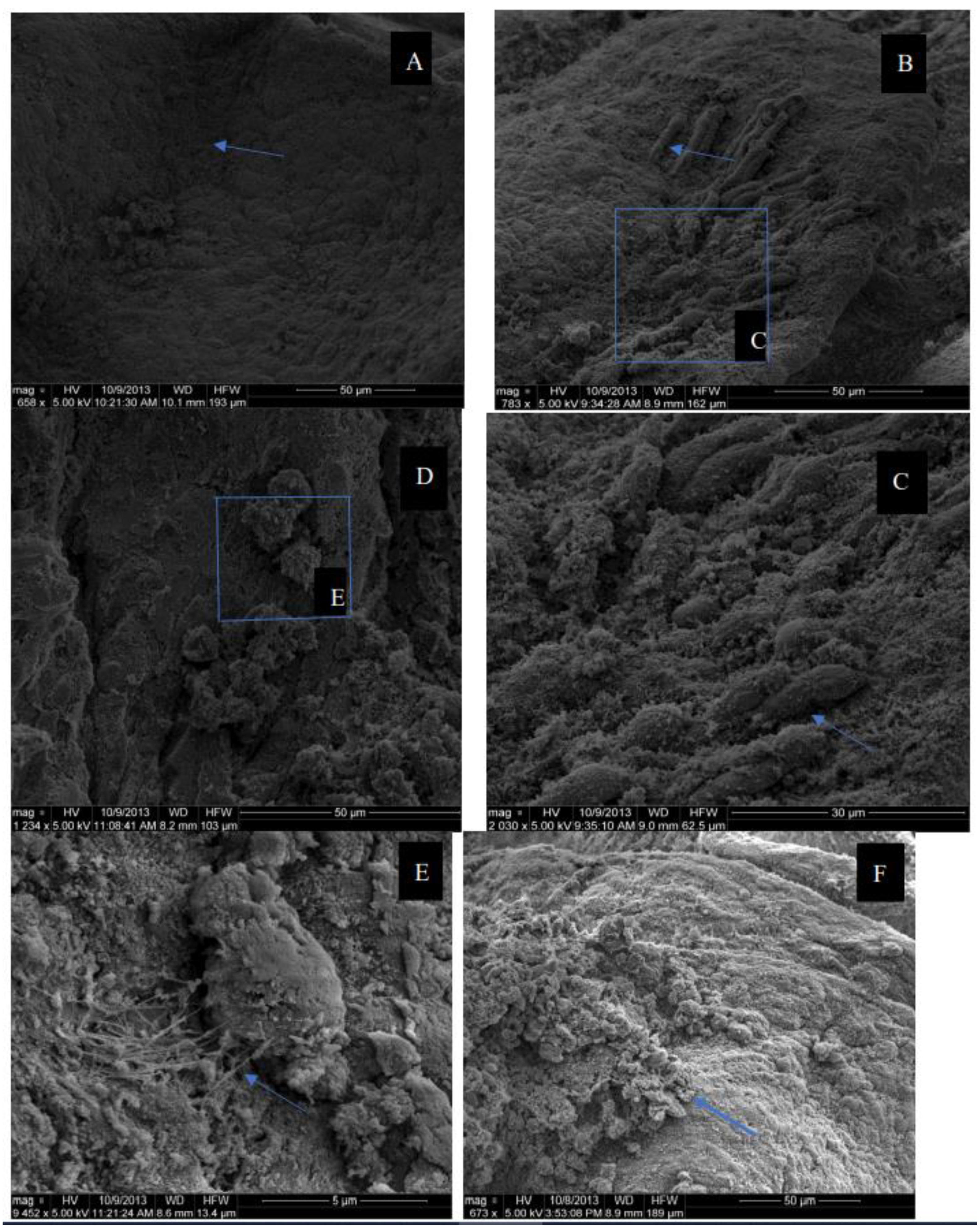
2.5. Sample Preparation for SEM Evaluation
2.6. Agar Spot Test of Probiotic Microorganisms to Competitively Exclude Pathogenic Bacteria in the Chicken Gastrointestinal Tract
2.7. Statistical Analysis
3. Results and Discussion
4. Conclusions
Author Contributions
Funding
Conflicts of Interest
Abbreviations
| GIT | Gastrointestinal tract |
| CFU | Colony-Forming Units |
| IM | Intestinal Microflora |
| TYP | Tryptone, Peptone, Yeast Extract |
| DMEM | Dulbecco’s Modified Eagle’s Medium |
References
- FAO/WHO. Health and Nutritional Properties of Probiotics in Food Including Powder Milk with Live Lactic Acid Bacteria; Report of a Joint FAO/WHO Expert Consultation on Evaluation of Health and Nutritional Properties of Probiotics in Food Including Powder Milk with Live Lactic Acid Bacteria; FAO/WHO: American Córdoba Park Hotel, Córdoba, Argentina, 2001; pp. 1–34. [Google Scholar]
- Ricke, S.C. Potential of fructooligosaccharide prebiotics in alternative and nonconventional poultry production systems. Pout. Sci. 2015, 94, 1411–1418. [Google Scholar] [CrossRef] [PubMed]
- Park, S.H.; Hanning, I.; Perrota, A.; Bench, B.J.; Alm, E.; Ricke, S.C. Modifying the gastrointestinal ecology in alternatively raised poultry and the potential for molecular and metabolomic assessment. Poult. Sci. 2013, 92, 546–561. [Google Scholar] [CrossRef] [PubMed]
- Nahashon, S.N.; Nakaue, H.S.; Mirosh, L.W. Nutrient retention and production parameters of Single Comb White Leghorn layers fed diets with varying crude protein levels and supplemented with direct-fed microbials. Anim. Feed Sci. 1996, 61, 17–26. [Google Scholar] [CrossRef]
- Gadde, U.; Kim, W.H.; Oh, S.T.; Lillehoj, H.S. Alternatives to antibiotics for maximizing growth performance and feed efficiency in poultry: A review. Anim. Health Res. Rev. 2017, 18, 26–45. [Google Scholar] [CrossRef] [PubMed]
- Fuller, R. Probiotics in man and animals. J. Appl. Bacteriol. 1989, 66, 365–378. [Google Scholar] [PubMed]
- Schrezenmeir, J.; de Vrese, M. Probiotics, prebiotics and synbiotics-approaching a definition. Am. J. Clin. Nutr. 2001, 73, 361S. [Google Scholar] [CrossRef] [Green Version]
- Mead, G.C. Prospects for competitive exclusion treatment to control Salmonellas and other foodborne pathogens in poultry. Vet. J. 2000, 159, 111–123. [Google Scholar] [CrossRef]
- Bhogoju, S.; Nahashon, S.; Wang, X.; Darris, C.; Kilonzo-Nthenge, A. A comparative analysis of microbial profile of Guinea fowl and chicken using metagenomic approach. PLoS ONE 2018, 13, e0191029. [Google Scholar] [CrossRef] [Green Version]
- Santos, F.B.O.; Sheldon, B.W.; Santos, A.A., Jr.; Ferket, P.R.; Lee, M.D.; Petroso, A.; Smith, D. Determination of ileum microbial diversity of broilers fed triticale- or corn-based Diets and colonized by salmonella. J. Appl. Poult. Res. 2007, 16, 563–573. [Google Scholar] [CrossRef]
- Shi, H.N.; Walker, A. Bacterial colonization and the development of intestinal defenses. Can. J. Gastroenterol. 2004, 18, 493–500. [Google Scholar] [CrossRef] [Green Version]
- Goran, M. Probiotics in Foods Not Containing Milk or Milk Constituents, with Special Reference to Lactobacillus plantarum 299v. Am. J. Clin. Nutr. 2001, 73, 380S–385S. [Google Scholar]
- Allan Walker, W. Mechanisms of Action of Probiotics. Clin. Infect. Dis. 2008, 46, S87–S91. [Google Scholar] [CrossRef]
- Awad, W.A.; Ghareeb, K.; Abdel-Raheem, S.; Böhm, J. Effects of dietary inclusion of probiotic and symbiotic on growth performance, organ weights, and intestinal histomorphology of broiler chickens. Poult. Sci. 2009, 88, 49–56. [Google Scholar] [CrossRef] [PubMed]
- Chichlowski, M.; Croom, W.J.; Edens, F.W.; McBride, B.W.; Qiu, R.; Chiang, C.C.; Daniel, L.R.; Havenstein, G.B.; Koci, M.D. Microarchitecture and spatial relationship between bacteria and ileal, cecal, and colonic epithelium in chicks fed a direct-fed microbial, primalac, and salinomycin. Poult. Sci. 2007, 86, 1121–1132. [Google Scholar] [CrossRef] [PubMed]
- Samanya, M.; Yamauchi, K.E. Histological alterations of intestinal villi in Chickens fed dried Bacillus subtilis var. natto. Comp. Biochem. Physiol. A Mol. Integr. Physiol. 2002, 133, 95–104. [Google Scholar] [CrossRef]
- Nyamambi, B.; Ndlovu, L.R.; Naik, Y.S.; Kock, N.D. Intestinal growth and function of broiler chicks fed sorghum-based diets differing in condensed tannin levels. S. Afr. J. Anim. Sci. 2007, 37, 202–214. [Google Scholar] [CrossRef] [Green Version]
- Amat, C.; Planas, M.J.; Moreto, M. Kinetics of hexose uptake by the small and large intestine of the chicken. Am. J. Physiol. 1996, 271, R1085–R1089. [Google Scholar] [CrossRef]
- Samli, H.E.; Senkoylu, N.; Koc, F.; Kanter, M.; Agma, A. Effects of Enterococcus faecium and dried whey on broiler performance, gut histomorphology and microbiota. Arch. Anim. Nutr. 2007, 61, 42–49. [Google Scholar] [CrossRef]
- Pluske, J.R.; Tompson, M.J.; Atwood, C.S.; Bird, P.H.; Williams, I.H.; Hartmann, P.E. Maintenance of villus height and crypt depth, and enhancement of disaccharide digestion and monosaccharide absorption, in piglets fed on cow’s whole milk after weaning. Br. J. Nutr. 1996, 76, 409–422. [Google Scholar] [CrossRef] [Green Version]
- Todd, R.K.; Kellen, M.J. Selection and design of probiotics. Int. J. Food Microbiol. 1999, 50, 45–57. [Google Scholar]
- Perez, P.F.; Minnaard, Y.; Disalvo, E.A.; De Antoni, G.L. Surface properties of Bifidobacterial strains of human origin. Appl. Environ. Microbiol. 1998, 64, 21–26. [Google Scholar] [CrossRef] [PubMed] [Green Version]
- Jin, L.Z.; Ho, Y.W.; Abdullah, N.; Jalaludin, S. Acid and bile tolerance of Lactobacillus isolated from chicken intestine. Lett. Appl. Microbiol. 1998, 27, 183–185. [Google Scholar] [CrossRef]
- Vernazza, C.L.; Gibson, G.R.; Rastall, R.A. Carbohydrate preference, acid tolerance and bile tolerance in five strains of Bifidobacterium. J. Appl. Microbiol. 2006, 100, 846–853. [Google Scholar] [CrossRef] [PubMed]
- Santini, C.; Baffoni, L.; Gaggia, F.; Granata, M.; Gasbarri, R.; di Gioia, D.; Biavati, B. Characterization of probiotic strains: An application as feed additives in poultry against Campylobacter jejuni. Int. J. Food. Microbiol. 2010, 141, S98–S108. [Google Scholar] [CrossRef] [PubMed]
- Kizerwetter-Swida, M.; Binek, M. Selection of potential probiotic Latobacillus strains towards their inhibitory activity against poultry enteropathogenic bacteria. Pol. J. Microbiol. 2005, 54, 287–294. [Google Scholar]
- SAS Institute Inc. SAS User’s Guide; Statistics; SAS Institute Inc.: Cary, NC, USA, 2002. [Google Scholar]
- Biavati, B.; Vescovo, M.; Torriani, S.; Bottazzi, V. Bifidobacteria: History, ecology, physiology and applications. Ann. Microbiol. 2000, 50, 117–131. [Google Scholar]
- Van de Casteele, S.; Vanheuverzwijn, T.; Ruyssen, T.; van Assche, P.; Swings, J.; Huys, G. Evaluation of culture media for selective enumeration of probiotic strains of lactobacilli and bifidobacteria in combination with yogurt or cheese starters. Int. Dairy J. 2006, 16, 1470–1476. [Google Scholar] [CrossRef]
- Giraud, E.; Lelong, B.; Raimbault, M. Influence of pH and initial lactate concentration on the growth of Lactobacillus plantarum. Appl. Microbiol. Biotechnol. 1991, 36, 96–99. [Google Scholar] [CrossRef]
- Graff, S.; Chaumeil, J.C.; Boy, P.; Lai-Kuen, R.; Charrueau, C. Formulations for protecting the probiotic Saccharomyces boulardii from degradation in acidic condition. Biol. Pharm. Bull. 2008, 31, 266–272. [Google Scholar] [CrossRef] [Green Version]
- Sanhueza, E.; Paredes-Osses, E.; González, C.L.; García, A. Effect of pH in the survival of Lactobacillus salivarius strain UCO_979C wild type and the pH acid acclimated variant. Electron. J. Biotechnol. 2015, 18, 343–346. [Google Scholar] [CrossRef] [Green Version]
- An, H.; Douillard, F.P.; Wang, G.; Zhai, Z.; Yang, J.; Song, S.; Cui, J.; Ren, F.; Luo, Y.; Zhang, B.; et al. Integrated Transcriptomic and Proteomic Analysis of the Bile Stress Response in a Centenarian-originated Probiotic Bifidobacterium longum BBMN68. Mol. Cell. Proteom. 2015, 13, 2558–2572. [Google Scholar] [CrossRef] [PubMed] [Green Version]
- Begley, M.; Hill, C.; Gahan, C.G. Bile salt hydrolase activity in probiotics. Appl. Environ. Microbiol. 2006, 72, 1729–1738. [Google Scholar] [CrossRef] [PubMed] [Green Version]
- Carabeo, R.A.; Scott, S.G.; Hackstadt, T. Chlamydia trachomatis induces remodeling of actin cytoskeleton during attachment and entry in HeLa cells. Infect. Immun. 2002, 70, 3793–3803. [Google Scholar] [CrossRef] [PubMed] [Green Version]
- O’sullivan, M.G.; Thorton, G.; O’sullivan, G.C.; Collins, J.K. Probiotic bacteria: Myth or reality? Trends Food Sci. Technol. 1992, 3, 309–314. [Google Scholar] [CrossRef]
- Hoepelman, A.I.M.; Tuomanen, E.I. Consequences of microbial attachment: Directing host cell functions with adhesins. Infect. Immun. 1992, 60, 1729–1733. [Google Scholar] [CrossRef] [Green Version]
- Fuller, R. Probiotics in human medicine. Gut 1991, 32, 439–442. [Google Scholar] [CrossRef] [Green Version]
- Asha, D.G. Antagonistic potential of Lactobacillus spp. against enteropathogenic bacteria; purification and characterization of their bacteriocins. Adv. J. Food Sci. Technol. 2012, 4, 265–269. [Google Scholar]
- Slačanac, V.; Hardi, J.; Čuržik, D.; Pavlović, H.; Lučan, M.; Vlainić, M. Inhibition of the in vitro growth of Salmonella enteritidis D by goat and cow milk fermented with probiotic bacteria Bifidobacterium longum Bb-46. Czech J. Food Sci. 2007, 25, 351–358. [Google Scholar] [CrossRef] [Green Version]
- Im, E.; Pothoulakis, C. Recent advances in Saccharomyces boulardii research. Gastroenterol. Clin. Biol. 2010, 34, S62–S70. [Google Scholar] [CrossRef]

| Genus | Species | Growth Conditions | Media |
|---|---|---|---|
| Salmonella | enteriditis, pullorum, typhimurium | Aerobic, 18–24 h at 37 °C | Tryptic Soy Broth (TSB)/Tryptic Soy Agar (TSA) |
| Campylobacter | jejuni, coli, lari | Microaerophilic, 48–72 h at 42 °C | Bolton Selective Enrichment Broth w/supplement and 5% horse blood customized agar * |
| Enterococcus | Faecalis, faecium, | Aerobic, 20–24 h at 35 °C | Tryptic Soy Broth (TSB)/Tryptic Soy Agar (TSA) |
| Escherichia coli | E. coli 0157:H7, E. coli 01: K1:H7, ATCC 25922 | Aerobic, 18–24 h at 37 °C | Tryptic Soy Broth (TSB)/Tryptic Soy Agar (TSA) |
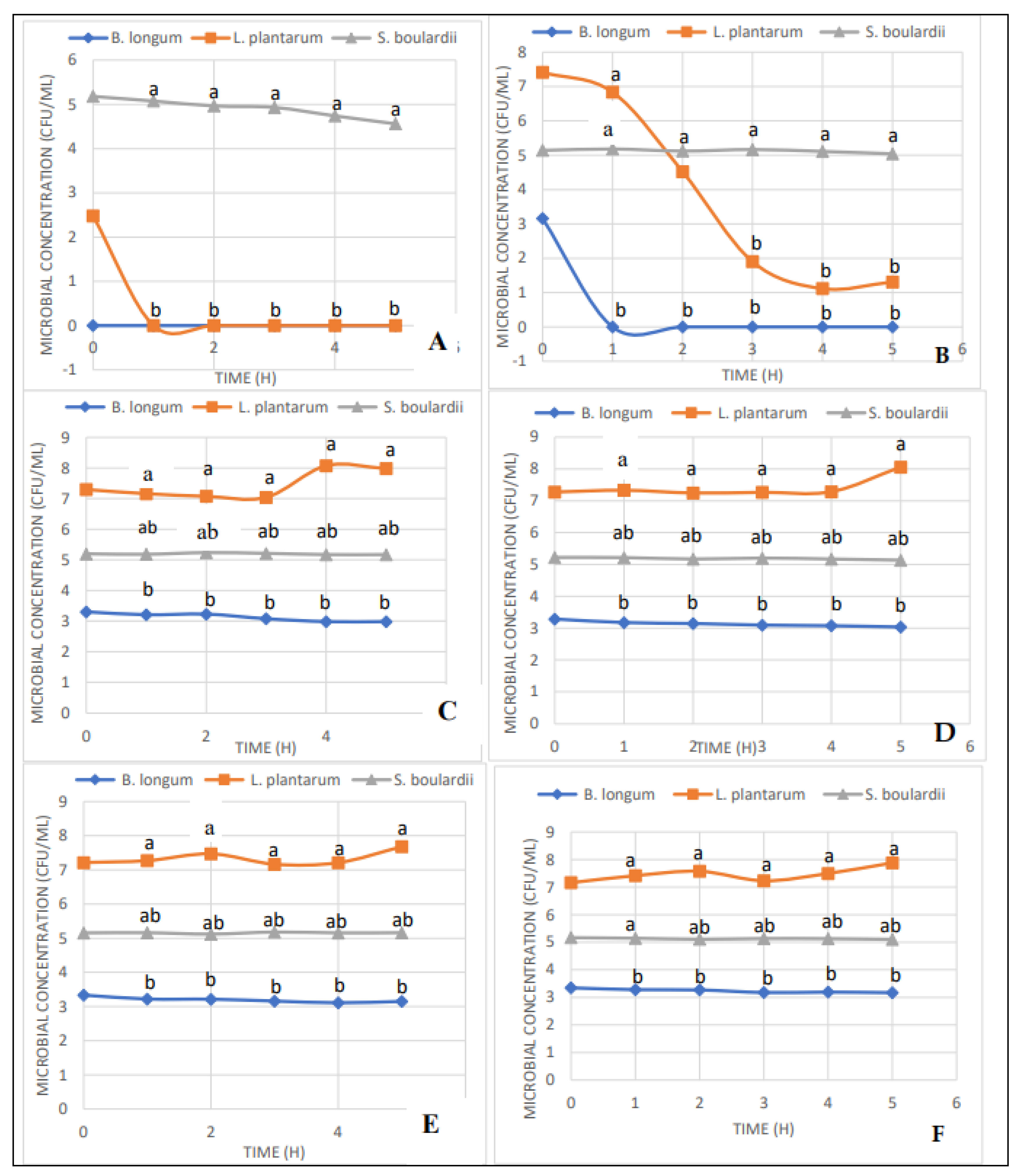
| Bile Concentration (%) | 3 | 2 | 1.5 | 1 | 0 | |
|---|---|---|---|---|---|---|
| Probiotic | Time (h) | Concentration (CFU/mL) | ||||
| B. Longum | 1 | 1.64 bz | 1.585 byz | 1.425 bxy | 1.3 bx | 4.89 az |
| 2 | 1.605 bz | 1.46 byz | 1.27 by | 1.19 bx | 4.92 az | |
| 3 | 1.625 bz | 1.41 bz | 1.285 by | 1.065 bx | 5.265 ay | |
| 4 | 1.81 byz | 1.52 byz | 1.17 by | 1.015 bx | 5.66 axy | |
| 5 | 2.065 bxy | 1.71 by | 1.355 bxy | 1.085 bx | 6.16 awx | |
| 6 | 2.345 bx | 2.09 bx | 1.82 bx | 1.43 bx | 6.675 aw | |
| L. Plantarum | 1 | 7.285 az | 7.315 az | 7.385 az | 6.61 bz | 7.1 abz |
| 2 | 7.395 ayz | 7.08 az | 6.96 az | 6.82 bz | 7.455 az | |
| 3 | 7.72 abcxy | 7.445 bcyz | 6.955 bcz | 7.245 cy | 8.165 ay | |
| 4 | 7.9 bx | 7.785 bxy | 7.35 bz | 7.7 bx | 8.705 ay | |
| 5 | 8.295 bw | 8.205 bcx | 8.025 cy | 7.975 cwx | 9.92 ax | |
| 6 | 8.35 b cw | 8.38 bcx | 8.655 bx | 8.14 cw | 10.15 ax | |
| S. boulardii | 1 | 3.765 ax | 3.72 axy | 3.57 axyz | 3.61 ay | 3.82 az |
| 2 | 3.815 abx | 3.59 by | 3.82 abwz | 3.72 abxy | 3.93 az | |
| 3 | 3.775 bcx | 3.785 bxy | 3.51 cy | 3.955 bxy | 4.33 ay | |
| 4 | 3.74 bx | 3.69 bxy | 3.81 bwx | 3.885 bxy | 4.545 axy | |
| 5 | 3.7 cx | 3.95 bx | 3.885 bcw | 4.065 bx | 4.72 awx | |
| 6 | 3.77 bx | 3.965 bx | 4.055 bw | 4.09 bx | 4.83 aw | |
| Probiotics | |||
|---|---|---|---|
| Enteropathogens | L. plantarum | B. longum | S. boulardii |
| Zone of Inhibition (mm) * | |||
| E. coli O157:H7 | 34.0 ay | 21.9 bx | 0 cy |
| E. coli 25922 | 29.5 ay | 14.8 by | 0 cy |
| E. coli 11775 | 30.0 ay | 14.6 by | 0 cy |
| S. pullorum | 43.6 ax | 22.8 bx | 0 cy |
| S. enteritidis | 31.3 ay | 13.6 by | 0 cy |
| S. typhimurum | 31.8 ay | 16.0 by | 0 cy |
| E. faecium | 24.0 az | 4.5 bz | 0 by |
| E. faecalis | 33.0 ay | 24.1 bx | 0 cy |
| C. coli | 17.8 az | 16.3 by | 0 cy |
| C. lari | 30.0 ay | 20.0 bx | 12.0 cx |
| C. jejuni | 22.0 az | 15.8 by | 12.0 cx |
Publisher’s Note: MDPI stays neutral with regard to jurisdictional claims in published maps and institutional affiliations. |
© 2022 by the authors. Licensee MDPI, Basel, Switzerland. This article is an open access article distributed under the terms and conditions of the Creative Commons Attribution (CC BY) license (https://creativecommons.org/licenses/by/4.0/).
Share and Cite
Dixon, B.; Kilonzo-Nthenge, A.; Nzomo, M.; Bhogoju, S.; Nahashon, S. Evaluation of Selected Bacteria and Yeast for Probiotic Potential in Poultry Production. Microorganisms 2022, 10, 676. https://doi.org/10.3390/microorganisms10040676
Dixon B, Kilonzo-Nthenge A, Nzomo M, Bhogoju S, Nahashon S. Evaluation of Selected Bacteria and Yeast for Probiotic Potential in Poultry Production. Microorganisms. 2022; 10(4):676. https://doi.org/10.3390/microorganisms10040676
Chicago/Turabian StyleDixon, Beverly, Agnes Kilonzo-Nthenge, Maureen Nzomo, Sarayu Bhogoju, and Samuel Nahashon. 2022. "Evaluation of Selected Bacteria and Yeast for Probiotic Potential in Poultry Production" Microorganisms 10, no. 4: 676. https://doi.org/10.3390/microorganisms10040676
APA StyleDixon, B., Kilonzo-Nthenge, A., Nzomo, M., Bhogoju, S., & Nahashon, S. (2022). Evaluation of Selected Bacteria and Yeast for Probiotic Potential in Poultry Production. Microorganisms, 10(4), 676. https://doi.org/10.3390/microorganisms10040676