Article

Multi-Functional Potential of Lactic Acid Bacteria Strains and Antimicrobial Effects in Minimally Processed Pomegranate (Punica granatum L. cv Jolly Red) Arils

1
Laboratory of Protein Engineering and Bioactive Molecules (LR11ES24), INSAT (Institut National des Sciences Appliquées et de Technologie Centre Urbain Nord), University of Carthage, Tunis BP 676-1080, Tunisia
2
Department of Agriculture, Food and Environment (Di3A), University of Catania, Via S. Sofia 100, 95123 Catania, Italy
3
CREA (Consiglio per la Ricerca in Agricoltura e l’analisi dell’Economia Agraria), Centro di Ricerca Olivicoltura, Frutticoltura e Agrumicoltura, Corso Savoia 190, 95024 Acireale, Italy
4
ProBioEtna SRL, Spin-off of University of Catania, Via S. Sofia 100, 95123 Catania, Italy
5
CERNUT (Interdepartmental Research Centre in Nutraceuticals and Health Products), University of Catania, Via le A. Doria 6, 95125 Catania, Italy
*
Author to whom correspondence should be addressed.
These authors contributed equally to this work.
Microorganisms 2022, 10(10), 1876; https://doi.org/10.3390/microorganisms10101876
Submission received: 24 August 2022 / Revised: 9 September 2022 / Accepted: 14 September 2022 / Published: 20 September 2022

Abstract

:
This study aimed to evaluate the antimicrobial activity of both cells, and cell-free supernatants (CFS) of 7 selected lactic acid bacteria (LAB) strains belonging to Limosilactobacillus fermentum (4 strains), Lacticaseibacillus paracasei (1 strain), Lacticaseibacillus rhamnosus (1 strain), and Enterococcus faecium (1 strain) species, against Listeria monocytogenes, Escherichia coli, Salmonella Typhimurium, Pseudomonas aeruginosa and Staphylococcus aureus, by both the agar-well diffusion and co-culture methods. In addition, probiotic and safety traits were also detected. Great variability was detected on antimicrobial effects, whereas all tested strains were found sensitive to most of the tested antibiotics, and without any DNase, gelatinase, or hemolytic activity. Moreover, strains showed excellent survival in acidic conditions and exhibited tolerance to pepsin and bile salts. Based on the in vitro results, the CFSs of two selected L. fermentum strains were applied, in a mixed solution, as bio-preservative into minimally processed pomegranate arils, inoculated with a cocktail of L. monocytogenes and E. coli. Samples, packaged in an ordinary atmosphere, were analyzed during refrigerated storage, for up to 12 days, for physicochemical (as weight loss, texture, color, pH, total soluble solids and organic acid content) and for microbiological traits. Results revealed the effectiveness of CFS, up to 12 days, in reducing weight loss and microbial growth, without any significant effect on texture, total soluble solid content and color, found comparable to the acid citric treatment, highlighting the multi-functional potential of selected probiotic strains.

1. Introduction

The global ready-to-eat fresh produce market has rapidly grown in recent years due to changes in consumer food attitudes, and a wide variety of products is available in the market with functional foods positively considered by consumers for their beneficial effects and nutritional traits [1]. Nevertheless, fresh and minimally processed vegetables represent a favorable matrix for both spoilage and pathogenic microorganisms, which cause economic losses and human diseases, such as acute enteritis with fever, bloody diarrhea and pseudo appendicitis [2]. Indeed, ready-to-eat fruits are extremely perishable due to the occurrence of physiological and biochemical changes, with significant degradation of sensorial traits, such as browning and modification of texture and flavor. Several studies reported the presence of pathogenic species, including Listeria monocytogenes, Salmonella enterica, Escherichia coli, Yersinia enterocolitica, Aeromonas hydrophila and Staphylococcus aureus both on fresh produce and minimally processed products [3,4,5,6]. In a recent survey, upon 10,070 samples of fresh fruit and vegetables collected in retail stores, the most isolated microorganisms were E. coli, Salmonella enterica and E. coli O157: H7 [7]. In the last 30 years, different strategies, mainly based on the addition of chemical substances (such as sodium lactate, sodium diacetate, trisodium phosphate, sodium hypochlorite, peroxyacetic acid and salicylic acid) have been applied to avoid both spoilage and foodborne pathogen growth [6,8]. Alternative strategies, based on physical treatments, such as electrolyzed water, high-intensity ultrasound, UV light, pulsed light, ionizing radiation, cold gas plasma and the use of phages have been developed [9,10]. However, none of these strategies is effective in avoiding microbial growth, being unable to completely eradicate microbial presence [11,12].
Pomegranate (Punica granatum L.) is a fruit typically growing in the Northern and Southern hemispheres, from September to February, and from March to May. Pomegranate fruits are sources of bioactive compounds with high nutritional value and health-promoting effects [13,14]. Although pomegranates are usually sold as whole fruit, recently the consumer’s demand for ready-to-eat pomegranate arils has increased. The main hurdle for the marketing of the arils is the short shelf life which is not always consistent with the needs of food retailers [15,16]. Moreover, consumers require healthier food without chemical additives and industries continuously propose novel bio-additive compounds. The exploitation of natural bio-preservatives, or addition of lactic acid bacteria (LAB) and/or their metabolites with antagonistic effects against the deleterious microorganisms are continuously proposed [17,18], and successfully applied for controlling the growth of other microorganisms and/or for enhancing the nutritional values [19]. In this scenario, the proposal of applying post-biotics has emerged with the advantage of a lower interaction with the food matrix and greater ease of use [20,21,22]. Postbiotics are defined as the preparation of inanimate microorganisms and/or their components that confers a health benefit on the host [23,24]. Post-biotics include any compounds released or produced through the metabolic activity of microorganisms, such as exopolysaccharides, enzymes, cell wall fragments, short chain fatty acids (SCFAs) or bacterial lysates. The major benefits of post-biotics are their inherent stability in industrial processes and storage, and intellectual property protection (as no live microorganisms can be isolated from post-biotic). Several studies have succeeded in identifying LAB metabolites which mainly include organic acids (acetic, formic, and propionic, among others), fatty acids, hydrogen peroxide (H2O2), proteinaceous compounds, as bacteriocins (nisin, reutericyclin, pediocin, lacticin, reuterin and sakacin), and some volatiles such as diacetyl, and/or ethyl alcohol [25,26,27]. However, the amount of the metabolites has generally been found in concentrations much lower than their minimal inhibitory concentrations (MICs), indicating that the antimicrobial metabolites might act in synergy [28].
The aim of the present study was to evaluate the antibacterial effects, safety and functional traits of a panel of LAB strains. Moreover, for the first time, the cell-free supernatant (CFS) of the two most promising strains were sprayed, as a mixed solution, on minimally processed pomegranate arils to evaluate its effects on a mixed culture of L. monocytogenes and E. coli, during refrigerated storage up to 12 days.

2. Materials and Methods

2.1. Bacterial Strains and Culture Conditions

The LAB used in the present study included strains previously isolated from traditional fermented dairy-products collected in Beja, Ben Arous, Zaghouan and Tunis [29]. LAB strains were genetically identified by 16S rRNA gene sequencing as four strains of Limosilactobacillus fermentum LBF4, LBF5, LBF15 and LBF17 (strains accession number OM165034, OM165035, OM165036, OM165038, respectively), Lacticaseibacillus paracasei LBF19 (strain accession number OM171261), Lacticaseibacillus rhamnosus LBF16 (strain accession number OM165037), and Enterococcus faecium LBF20 (strain accession number OM165039). All strains belong to the Dairy Science Department, Faculty of Agriculture, Kafr El-Sheikh University, Egypt.
LAB strains were grown in Man Rogosa Sharpe (MRS) and M17 (Liofilchem srl, Roseto degli Abruzzi (TE), Italy) broth, incubated at 37 °C and stored at −80 °C in the same broth media, supplemented with 20% (w/v) of glycerol. In order to determine the phases of the microbial growth, 1 mL of an overnight culture of each LAB strain (OD600 ≈ 0.82) was inoculated into 10 mL of MRS or M17 and incubated at 37 °C. Cell enumeration was determined immediately after inoculation and during incubation time (after 2, 4, 6, 8, 10 and 24 h). Serial dilutions were performed, plated into MRS and the plates were incubated for 24 h at 37 °C. The experiment was performed in triplicate.

2.2. Evaluation of Functional Traits

2.2.1. Tolerance to Acidic Conditions

LAB strains were tested for tolerance at different pH values, as previously reported [29,30]. Acidic tolerance was tested as follows: overnight cultures were centrifuged, washed twice with phosphate-buffered saline (PBS) at pH 7.0, and re-suspended to reach a final density of 108 bacteria/mL in MRS broth acidified, using HCl (1 M), at pH 3.0 and pH 2.0. One mL of each culture was diluted and plated on MRS and M17 agar. The viable bacterial counts were detected at initial time and after 3 h of incubation at 37 °C, and the survival rate was calculated and expressed as survival rate percentage (SR%), based on the initial and the final number of viable cells. The experiment was performed in triplicate.

2.2.2. Bile Salt Tolerance

Bacterial cells from an overnight culture were collected by centrifugation and re-suspended in MRS broth added with 0.5 and 2.0% of bile salts (Oxgall, Sigma-Aldrich, L’lsle D’Abeau Chesnes, France), respectively and anaerobically incubated at 37 °C. MRS broth without bovine bile salts was used as a control. Aliquots were taken at initial time and after 4 h, diluted and plated on MRS agar. The survival rates were determined as:
SR (%) = (N1/N0) × 100
where N1 (Log CFU/mL) is the total viable count of selected strains after 4 h and N0 (Log CFU/mL) the total viable count of selected strains at the initial time. The experiment was performed in duplicate.

2.2.3. Pepsin Tolerance

LAB cultures in MRS broth, incubated at 37 °C for 24 h, were centrifuged (10,000 rpm for 10 min), washed twice with PBS and re-suspended to a final concentration of 108 bacteria/mL in MRS or M17 broth at pH 2.0 and pH 3.0, supplemented with 3 mg/mL of pepsin (Sigma-Aldrich, Germany). The acidification of media was performed using HCl (1 M). The broth cultures were incubated at 37 °C, and aliquots of 100 µL were sampled at the initial time and after 3 h, inoculated into MRS agar and incubated for 48 h at 37 °C. Viable cells were counted for the determination of survival rate (SR%), according to the above-mentioned equation [29]. The experiment was performed in triplicate.

2.2.4. Auto-Aggregation Assay

The auto-aggregation assay was performed as previously described [30]. In detail, overnight cultures were centrifuged at 5000 rpm for 10 min at 4 °C. The pellets were washed twice with PBS 20 mM, pH 6.0 and re-suspended in the same buffer. Equal volumes of each bacterium were vortexed for 10 s and incubated at 30 °C for 24 h, without agitation. The auto-aggregation activity was calculated using the equation:
% Auto-aggregation = 1 − (A1/A0) × 100
where A1 and A0 were the absorbance of LAB cultures after and before incubation. The experiment was performed in triplicate.

2.3. Evaluation of Safety Traits

2.3.1. DNase, Gelatinase and Hemolytic Assays

For detection of DNase, as previously reported by Mokdad and co-workers [31], strains were streaked in duplicate on DNase agar (Sigma, St. Louis, MO, USA) and plates were incubated at 37 °C for 48 h. After flooding, the plates with 1 N HCl, the positive indication of DNase production was visualized as a clear pink zone around the colonies.
The gelatinase activity was investigated using Nutrient agar medium (Liofilchem srl, Roseto degli Abruzzi (TE), Italy) containing 3.0% (w/v) of gelatin (Sigma Aldrich, Germany), as previously reported by Pino and co-workers [30]. All analyses were performed in duplicate.
Hemolytic activity was determined on Columbia agar medium (Biolife, Novi Ligure (AL), Italy), containing 5.0% of sheep blood, by streaking each overnight LAB culture on the surface of the solid medium. The plates were then incubated, under aerobic conditions, at 37 °C for 24–48 h and visually examined for the green zone (α-hemolysis), clear zones (β-positive) or no zone around colonies (γ-negative), as previously reported [32]. Streptococcus pyogenes ATCC 19615 and Streptococcus pneumoniae ATCC 6303 were used as positive controls.

2.3.2. Antibiotic Susceptibility Test

For the seven LAB strains the antibiotic susceptibility was tested using a minimum inhibitory concentration (MIC) assay, as previously described [33]. The LAB strains were considered resistant or sensitive to each tested antibiotic (ampicillin, vancomycin, gentamycin, kanamycin, streptomycin, erythromycin, clindamycin, tetracycline, and chloramphenicol), according to breakpoints proposed by the European Food Safety Authority [34]. LAB strains were incubated at 37 °C for 24 h in the LSM formulation medium, constituted by 90% of IST broth (Iso-Sensi test broth, Oxoid Ltd., UK) supplemented with 10% of MRS broth. Each strain was standardized, by using a 1.0 McFarland standard solution, to reach the final inoculum density of 108 CFU/mL. 100 µL of each standardized culture were used and incubated for 48 h at 37 °C, in the absence (positive control) and in the presence of each antibiotic at the following concentration (mg/L) ranges: ampicillin (1–32), vancomycin (0.5–16), gentamicin (4–128), kanamycin (8–256), streptomycin (8–256), erythromycin (0.125–4), clindamycin (0.125–4), tetracycline (1–32), and chloramphenicol (1–32). An aliquot of 100 μL of antibiotic, diluted into an LSM medium with the composition mentioned by ISO 10932/IDF 223 [33,35], was inoculated with 100 μL of bacterial inoculum in a microplate (Euroclone SpA, Pero, Italy). The MIC was determined as the lowest antibiotic concentration where no visible growth was observed [36]. All analyses were performed in duplicate.

2.4. Antibacterial Activity

2.4.1. Target Strains and Growth Conditions

The antimicrobial activity was evaluated against a panel of five target bacteria, including Listeria monocytogenes ATCC 19114, Escherichia coli ATCC 25922, Salmonella enterica Typhimurium ATCC 14026, and Pseudomonas aeruginosa ATCC 3224 and Staphylococcus aureus ATCC 29213. The target strains were revitalized in Luria–Bertani (LB) medium (Liofilchem, Roseto degli Abruzzi (TE) Italy) at 37 °C.

2.4.2. Antibacterial Activity of LAB Strains (as Cells or CFSs)

Antibacterial activity of LAB strains was tested, by both agar-well diffusion and dilution methods, against the same target bacteria (namely L. monocytogenes ATCC 19114, E. coli ATCC 25922, S. Typhimurium ATCC 14026, P. aeruginosa ATCC 3224, and Staphylococcus aureus ATCC 29213). The turbidity of the microbial suspensions was adjusted to 0.5 McFarland, giving an approximate cell density of 1.5 × 10⁸ CFU/mL [37]. The target cultures were spread on LB agar medium, using a cotton swab, and 50 µL of LAB cultures were injected on wells (6 mm diameter), previously cut into agar plates. Likewise, 50 µL of both crude supernatants, obtained by centrifugation (10,000 rpm for 20 min at 4 °C) of the LAB cultures, grown overnight at 37 °C in MRS broth, and deacidified supernatant (adjusted to pH 6.5 with 1 N of NaOH to rule out acidic inhibition), filtered through a 0.22 µm membrane filter (Millipore, Billerica, MA, USA) were injected on wells. The plates were aerobically incubated at 37 °C for 24 h and the antibacterial activity was evaluated by the measurement of the inhibition diameter around the well (≤ 5 mm, low inhibition; 5–10 mm, medium inhibition, ≥10 mm, high inhibition) [38].
The dilution method was performed using the broth microdilution assay in a 96-well plate, as previously reported [39]. 100 µL of both standardized LAB cultures (108 CFU/mL) and diverse cell density of target bacteria, fixed from 106 to 103 CFU/mL, were inoculated in a mixed media of MRS and BHI (Brain Heart Infusion) broth (Liofilchem srl, Roseto degli Abruzzi (TE), Italy) and co-incubated for 48 h at 37 °C. After incubation, both LAB and target bacteria were enumerated on MRS and BHI agar, respectively. Single cultivation of each microorganism was used as a control. The experiments were performed in triplicate.

2.5. Fruit Processing and Treatments

2.5.1. Vegetal Matrix

Pomegranate fruits (Punica granatum L. cv Jolly Red) harvested in autumn 2021, at the commercial ripening stage, and classified as C4 category [40] were used. The fruits were kindly provided by a local grower located in Bronte (Sicily) on the same day of harvesting. Fruits were transferred to the laboratory of Di3A (at the University of Catania) and immediately treated.

2.5.2. Fruit Treatments

Arils were manually extracted with a knife and washed in water containing sodium hypochlorite solution (0.5% w/v) for 2 min. The washed arils were then collected and placed in sterile containers. Arils that presented blemishes were removed. Afterwards, arils were dried and weighed at portions of 100 g under aseptic conditions, in an ordinary atmosphere, into transparent polystyrene bags PS 6 (code: V00501/OPS) with the following characteristics L: 127 × 115 mm; 1: 45 mm; 0.08 m3.
To evaluate the bio-preservative effect of CFS, fruits were inoculated with a mixed culture of L. monocytogenes ATCC 19114 and E. coli ATCC 8739 (ratio 1:1 at ~104 CFU/100 g) and subsequently sprayed with the CFS obtained from L. fermentum LBF4 (OM165034) and L. fermentum LBF5 (OM165035) cultures. Moreover, in order to compare the antimicrobial effect of CFS to citric acid, samples (100 g) of pomegranate arils inoculated with the mixed culture of L. monocytogenes ATCC 19114 and E. coli ATCC 8739 (ratio 1:1 at ~104 CFU/100 g) were sprayed with a fresh solution of 3% (v/w) of citric acid. Samples without CFS neither citric acid were considered as controls. All samples were packaged, as described above, stored at refrigerated conditions (4 °C) for up to 12 days, and analyzed at the initial time (T0), after 3 (T3), 7 (T7), and after 12 (T12) days of storage. Physicochemical and microbiological analyses were performed at each sampling time.

2.5.3. Physicochemical Analyses

The weight loss of pomegranate aril samples was detected after 3, 7 and 12 days of refrigerated storage. The weight of samples was measured using a precision electronic scale accurate to two decimal places (Gibertini EU-C 2002 RS, Novate Milanese, Italy) with an accuracy of ±0.20 g. Weight loss (WL) was calculated using the equation:
WL = (W0 − W1)/W0 × 100
where W0 is the initial weight (g) and W1 is the final weight (g).
The firmness of pomegranate aril samples was determined using a Universal machine (Zwick/Roell DO-FB0.5 TS model 2002, Genoa, Italy) and referred as the maximum force required for breaking the fruit skin. The firmness was evaluated in fresh fruits and in minimally processed samples, differently treated, at an initial time and after 3, 7 and 12 days of storage at 4 °C. The firmness analyzer was equipped with a 5.8 mm diameter cylinder probe (P6); the set parameters of each test were: pre-test speed 2 mm/s, test speed 0.5 mm/s and distance of 2 mm, post-test speed 4 mm/s and force max 1 N. For each treatment, three replicates of five arils were measured. The maximum force of the peak reached during aril tissue breakage was registered as firmness and expressed in Newton (N).
The colorimetric analyses were performed in fresh fruits and in minimally processed samples, differently treated, at an initial time and after 3, 7 and 12 days of storage at 4 °C. The color coordinates L* (brightness), a* (green-red component), and b* (blue-yellow component) were detected and reported as the average of three transmittance measurements for a single replicate, using a CM-5 spectrophotometer (Minolta, Milan, Italy), with the D65 illuminant, according to the CIELAB scale. Color was assessed according to the Commission Internationale de l’Éclairage (CIE) and expressed as L*, a*, b* color values. The coordinates L*, a* and b* indicate the lightness of the color (L* = 0 and L* = 100 represented black and white, respectively), its position between green and red (negative and positive a* values indicate greenness and redness, respectively) and between blue and yellow (negative and positive b* values point towards blueness and yellowness, respectively).
Once firmness and color were determined, the arils were blended, and the juice was stored at −20 °C for further analyses.
The pH of juice obtained from fresh fruits and from minimally processed samples, differently treated, at an initial time and after 3, 7 and 12 days of storage at 4 °C, was measured using a Mettler DL25 pH meter (Mettler-Toledo International Inc., Columbus, OH, USA).
The total soluble solids (TSS) of juice obtained from fresh fruits and from minimally processed samples, differently treated, at an initial time and after 3, 7 and 12 days of storage at 4 °C, were determined using a refractometer (Atago, RX-5000, Fisher Scientific, Rodano (MI), Italy), and expressed as °Brix.
The organic acid content of juice, obtained from fresh fruits and from minimally processed samples, differently treated, at an initial time and after 3, 7 and 12 days of storage at 4 °C, was determined by injecting the samples filtered through a 0.45 μm PTFE syringe filter (Merck, Frankfurter Str. 250, 64293 Darmstadt, Germany) into the chromatographic HPLC system.
In addition, the organic acid profile of L. fermentum LBF4 (OM165034) plus L. fermentum LBF5 (OM165035) supernatant (CFS) was also determined. The system consisted of a Waters Alliance 2695 HPLC liquid chromatograph, equipped with a Waters 996 photodiode array (PDA) detector set at 210 nm and Waters Empower software (Waters Corporation, Milford, MA, USA). Isocratic elution with 5 mM sulfuric acid was performed on a Rezex ROA Organic Acid H+ column (Phenomenex, Torrence, CA, USA) maintained in an oven at 26 °C. The run time was set at 50 min with a flow rate of 0.6 mL/min. The quantification of the peaks was obtained through calibration curves of pure standard of lactic, citric, acetic, propionic, isobutyric and butyric acids (all purchased from Sigma-Aldrich, Italy) injected at different concentrations. All analyses were performed in triplicate for each sample.

2.6. Microbiological Analyses

The microbiological analyses were performed according to Thakur and Sharma [32], with slight modifications. In detail, 10 g of each pomegranate sample were aseptically collected and homogenized in sterile bags (Whirl-Pak1, Nasco, Fort Atkinson, WI, USA), containing 90 mL of physiological water (0.9% NaCl), using a stomacher 80 lab blender (Seward Ltd., West Sussex, England). After performing serial decimal dilutions, a volume of 0.1 mL was plated onto appropriate microbiological media: EC X-gluc (Biolife srl, Novi Ligure (AL), Italy) for E. coli and Listeria Palcam Agar for L. monocytogenes enumeration and plates were incubated for 48–72 h at 37 °C. The same procedures were performed for control samples. Randomly, in order to confirm species affiliation, additionally basic microbiological tests, such as Gram staining, microscopic observation and catalase tests were carried out on colonies growth on selective media. In addition, total mesophilic microorganisms (TMM) and total psychrotrophic microorganisms (TPM) were counted on plate count agar (PCA) and incubated at 32 °C for 24 h and at 4 °C for 5 days, respectively. All media were purchased from Liofilchem srl (Roseto degli Abruzzi (TE), Italy). Results were expressed as a mean of log10 CFU/g ± standard deviation (SD) of two replications.

2.7. Statistical Analyses

All data were expressed as mean ± standard deviation (SD). Statistical significance was determined using an analysis of variance (ANOVA). The values were considered as significantly different when p ≤ 0.05. The SPSS software (version 21.0, IBM Statistics, Armonk, NY, USA) was used for data processing. Statistical analysis of the obtained results was performed using one-way analysis of variance (ANOVA), and Tukey’s HSD post-hoc test for means separation at a significance level of p ≤ 0.05. To evaluate the relationships among the different physical, chemical and microbiological parameters of minimally processed arils, differently treated, data were subjected to a one-tailed Pearson’s correlation.

3. Results

3.1. Growth Rate of LAB Strains

The tested LAB proliferated immediately after inoculation into MRS and M17 broth media. No significant differences in growth rate among LAB strains were observed; they showed a short lag phase (about 2 h) and an exponential phase that lasted up to 8 h. L. fermentum LBF15 showed the highest biomass yield, reaching a density around 7.4 Log CFU/mL (data not shown).

3.2. Functional Traits Assessment

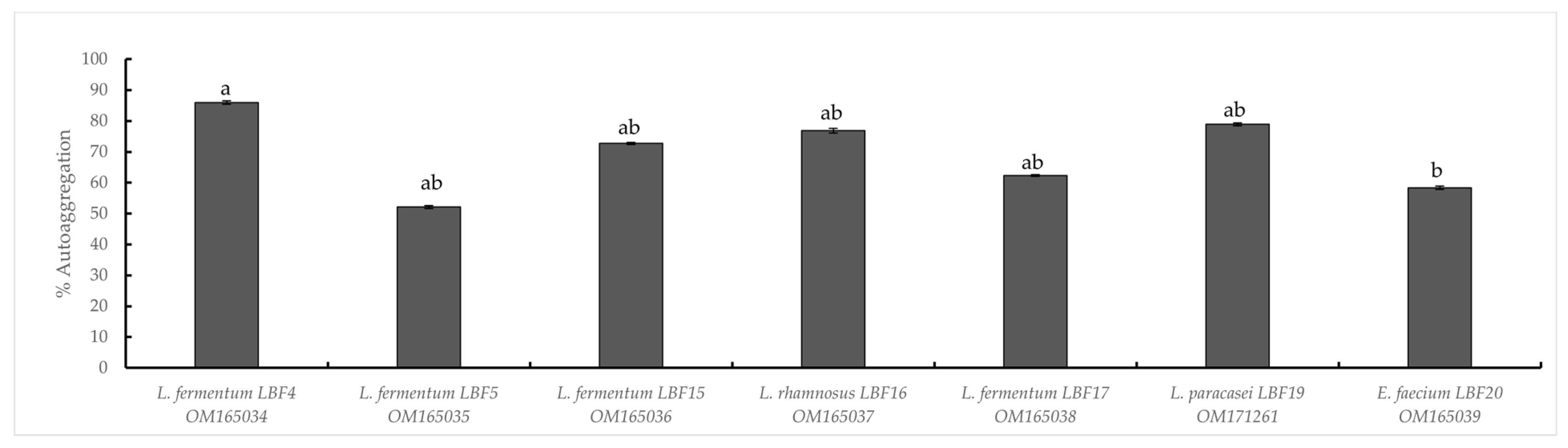
Acidic, Bile Salt, Pepsin Tolerance and Auto-Aggregation

Regarding acidic tolerance, starting from an initial number of viable cells (control cells) ranging from 6.80 to 6.47 Log CFU/mL, a survival rates ≥ 90% were observed at pH 3.0. The survival rate at pH 2 was slightly lower, showing values between 85.74 and 96.67%. Significant differences were observed among LAB strains (p ≤ 0.01), except for the ability to survive at pH 3.0 (Table 1).
Results of bile salt tolerance, shown in Table 2, revealed that after 3 h of incubation, LAB strains highlighted significant differences (p ≤ 0.01), exhibiting high viability with a survival rate always above 90%, at 0.5% (w/v) of bile salt concentration, and between 87.79 and 96.82% at 1.0% (w/v) of bile salt presence. Lower survival rates were observed for the highest tested bile salt concentration (2%), except for LBF15 and LBF5 strains which showed values close to 92.89 and 89.47%, respectively (Table 2).
Data on pepsin tolerance (Table 3) revealed no significant differences (p > 0.05) in the ability to survive at both tested pH values. All strains showed a good resistance at both pH 3.0 and pH 2.0, when 3 mg/mL of pepsin were added, reaching a survival rate always higher than 90% (Table 3).
As shown in Figure 1, all tested strains exhibited auto-aggregation activity, ranging from 60% to 85%. L. fermentum LBF4 showed the highest auto-aggregating value, as 85% after 24 h of incubation at 30 °C (Figure 1).

3.3. Safety Evaluation

DNase, Gelatinase, Hemolytic Activity and Antibiotic Resistance

None of the tested LAB strains showed the ability to produce DNase and gelatinase, or to exert hemolytic activity, fulfilling such safety requirements. Results on antibiotic susceptibility, evaluated by MIC method, are shown in Table 4. In details, L. fermentum LBF4, L. fermentum LBF5 and L. rhamnosus LBF16 strains were sensitive to all tested antibiotics, while L. paracasei LBF19 showed resistance to streptomycin and chloramphenicol. The remaining strains, L. fermentum LBF15 and LBF17 were resistant to ampicillin, kanamycin and erythromycin and, as expected, E. faecium LBF 20 showed resistance towards ampicillin, vancomycin, streptomycin, tetracycline and chloramphenicol (Table 4).

3.4. Antibacterial Activity of Lab Strains

Results of antibacterial activity showed that, overall, both the cells and the CFSs obtained from L. fermentum LBF4, L. fermentum LBF5, and L. rhamnosus LBF16 displayed the highest inhibition against L. monocytogenes ATCC 19114, E. coli ATCC 8739, and S. Typhimurium ATCC 14026 (Supplementary Table S1). After all, L. fermentum LBF5 exhibited the highest inhibition effect also against P. aeruginosa ATCC 3224. Although with different alone sizes, L. fermentum LBF4 and L. rhamnosus LBF16 showed the highest inhibition effect against S. aureus ATCC 29213. A similar inhibitory effect against all target bacteria was observed for E. faecium LBF20.
No activity was detected for the neutralized CFSs against any tested target bacteria, highlighting that the antagonistic activity is attributable to the acidic content (mainly lactic, citric and acetic acids).
Regarding the inhibition activity against the target bacteria in a liquid medium, the results were independent on the bacterial species and highlighted no influence caused by co-cultivation with the pathogenic bacteria, remaining after 24 h of incubation, always constant (Figure 2, panels from a to g). Overall, for all LAB strains a complete inhibition of target bacteria at 103 CFU/mL concentration was observed (data not shown). Among the tested strains, the L. fermentum LBF4 and L. fermentum LBF5 exhibited the strongest inhibitory effect against E. coli ATCC 8739 and L. monocytogenes ATCC 19114, at both 105 and 104 CFU/mL concentrations, with an inhibition rate higher than 80% (Figure 2, panels a and b). Similar behaviour was observed for L. rhamnosus LBF16 and L. paracasei LBF19 strains, while for E. faecium LBF20, an inhibition rate slight lower against E. coli ATCC 8739 at 104 and 105 concentrations was observed (Figure 2, panel d, f, and g). Finally, L. paracasei LBF19 and E. faecium LBF20 showed an inhibition rate around 60% against L. monocytogenes ATCC 19114 at both tested concentrations (Figure 2, panels f and g). After all, strains showed an inhibition rate lower than 60% against the remaining tested target bacteria at any concentrations (Figure 2, panels from a to g).

3.5. Physicochemical Analyses of Minimally Processed Pomegranate Arils Differently Treated during Storage at Refrigerated Conditions

During storage, the weight of the pomegranate arils constantly decreased (Figure 3), mainly starting from the third day. Nevertheless, the weight loss did not exceed the 1% value until the third day of storage in samples treated with CFS or citric acid. For control samples and for untreated contaminated samples, the initial weight loss exceeded 2% and 3%, respectively (Figure 3). Increasing of weight loss was detected without any significant difference (p > 0.05) within the untreated (inoculated and control) samples, reaching 14% and 15.6%, respectively, on the twelfth day of storage. In contrast, in treated samples (with the addition of citric acid or CFS), the weight loss reached values of 8.7% and 9.6%, respectively after 12 days of storage. Indeed, as expected, in samples inoculated with pathogens and without any treatments, the weight loss was positively related with both E. coli and L. monocytogenes cell densities (R = 0.847 and R = 0.805, respectively), whereas in samples inoculated with pathogens and treated with CFS a negative correlation (R= −0.779) was found for L. monocytogenes.
Regarding physicochemical traits, the fresh pomegranate fruits revealed a firmness (N) value of 10.18 ± 1.39 (Table 5), which decreased in all processed samples after 3 days of storage (Table 5), reaching final values, scored between 6.38 and 7.48, at the end of storage time, with any significant difference among samples. A different behaviour was observed after 7 days in samples without any treatments (control and samples inoculated with pathogens), which showed values of firmness of 9.15 and 9.05, respectively, higher than values detected in treated samples, with citric acid (7.72) or with CFS (7.08) (Table 5).
The color coordinates of fresh fruits and pomegranate aril differently treated, detected at the initial time and during storage, are reported in Table 5. The effects of treatments and storage time were not significant for lightness (L*) values (p > 0.05) after 3 and 7 days. Similarly, the analysis of variance showed that no significant effect among samples on all colorimetric coordinates, used as a color stability indicator, was observed after 7 days of storage. For all samples, the redness values increased after 7 days and decreased after 12 days of storage, resulting markedly higher than that found in fresh fruits. On the contrary, for samples inoculated with pathogens, the values increased during the whole storage period. Regarding the b* index, although the values detected in control samples decreased during the storage, both treated and contaminated samples showed an increase after 7 days of storage, followed by a decrease at the end of storage (Table 5).
Chemical data obtained from fruits, CFS and minimally processed arils, during storage time, are reported in Table 6. Focusing on CFS, results showed the presence of lactic (6788.38 mg/L), citric (3023.48 mg/L), acetic (2878.54 mg/L), and propionic (31.63 mg/L) acids. The same organic acids were detected in samples differently treated at different storage times. Moreover, the presence of butyric acid revealed only in CFS at 1476.23 mg/L, was never detected in any samples, neither in aril samples inoculated with pathogens and treated with CFS.
Overall, the chemical results showed a significant effect of treatments during the storage period (p ≤ 0.05). Focusing on pH, the values of all tested samples decreased during storage (Table 6), whereas no significant difference was observed for TSS value until the seventh day of storage. Indeed, on the twelfth day of storage, the TSS values decreased, reaching the lowest values in samples inoculated with pathogens (14.7 mg/L).
Regarding the acid content, the storage time showed a significant effect on citric and lactic acids for all treatments (p ≤ 0.05), significantly decreasing in all samples mainly at the twelfth day of storage. Conversely, no significant difference was detected for propionic acid content in any samples, both at initial and final storage times, while after 7 days of storage the lowest values were observed (Table 6). After all, the presence of acetic acid, revealed only in samples inoculated with pathogens starting from the third day of storage, increased in all samples at the seventh day of storage, showing significant differences at the twelfth day, when the highest values were reached in samples inoculated with pathogens and treated with CFS or citric acid.

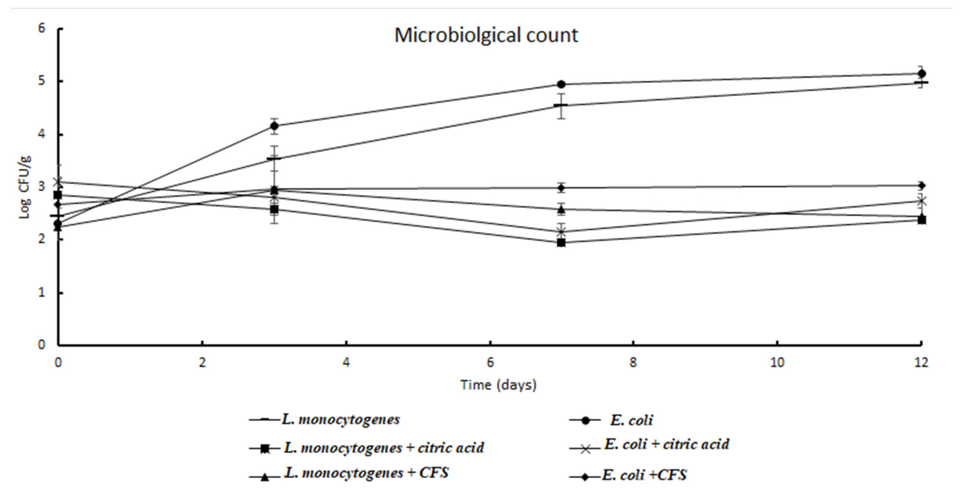
3.6. Microbiological Analyses of Minimally Processed Pomegranate Arils Differently Treated during Storage at Refrigerated Conditions

Results of microbiological analyses performed on minimally processed pomegranate arils are shown in Figure 4 and Figure 5. In control samples (Figure 4), the initial total mesophilic microorganisms (TMM) and the total psychrotrophic microorganisms (TPM) densities were found as 0.9 ± 0.1 Log CFU/g and 1.3 ± 0.12 Log CFU/g, respectively, indicating satisfactory microbiological traits. During storage, the TMM and TPM counts increased, reaching values of 4.19 and 3.73 Log CFU/g, respectively, after 12 days of refrigerated conditions.
Focusing on L. monocytogenes, the mean initial density in pomegranate aril samples (2.5 Log CFU/g) increased during the storage time. In particular, in control samples, presumptive L. monocytogenes density climbed to 3.54 Log CFU/g after 3 days, to reach the value of 4.98 Log CFU/g, after 12 days of storage, with an increase in 2.53 Log units. In samples inoculated with pathogens, the treatment with citric acid resulted in a decrease in almost 0.5 Log units with a final count, after 12 days, of 2.37 Log CFU/g, with the highest listericidal effect between the third and the seventh day of storage. Regarding the pomegranate aril samples inoculated with pathogens, the treatment with CFS resulted in a slight increase (0.72 Log unit) after 3 days, followed by a weak decrease after 7 and 12 days, when the L. monocytogenes density was found as 2.45 Log CFU/g. A similar trend was observed for E. coli counts. In samples inoculated with pathogens, without any treatment, the presumptive E. coli count jumped from an initial 2.3 to final 5.12 Log CFU/g, with an increase of 2.85 Log units. In samples treated with citric acid, a 0.49 Log unit reduction was detected up to 12 days of refrigerated conditions, whilst in samples treated with CFS a final value of 3.0 Log CFU/g was detected, with an overall increase in 0.33 Log units.

4. Discussion

Due to the change of lifestyles, a relevant increase in the request of fresh-cut horticultural produce has been observed [41,42] and the major challenge facing the fresh-cut industry is to find natural alternatives to the chemicals, in order to prevent browning and microbial growth, which still represent major health concerns and cause economic losses [43,44]. Among the various natural strategies based on the bioprotection [42], the use of post-biotics has emerged. The proposal of applying probiotic metabolites has the advantage, for consumers, to lower the risk of infection and for the food industry, to be easy to use lowering the interactions with the food matrix [20,21,22].
In the present study, seven LAB strains were proposed as potential probiotic bacteria based on the safety evaluation and their ability to survive at acidic conditions, in the presence of bile salts and pepsin [45]. The latest result is in contrast with a previous report [46], where no strain was viable after exposure to pH 2.0, when 3 mg/mL of pepsin was added.
All strains showed high autoaggregation scores (above 60%), as recently reported for lactobacilli and enterococci [39,47].
Overall, the seven strains were sensitive to all tested antibiotics with MIC lower than EFSA breakpoints, except the two L. fermentum strains (LBF15 and LBF17), which were slightly resistance to ampicillin, kanamycin and erythromycin and L. paracasei LBF19 that was resistant to streptomycin and chloramphenicol. Whereas, E. faecium LBF20 showed resistance to ampicillin, vancomycin, streptomycin, tetracycline and chloramphenicol, although Russo and co-workers [33] observed a high incidence of resistance to different antibiotics (namely rifampicin, erythromycin and gentamicin) within the Enterococcus genus.
The tested strains showed no hemolytic or DNase activity, satisfying the two most relevant safety criteria for probiotic selection [47,48,49].
In the present study, bio-protective cultures of selected LAB strains against a broad spectrum of target bacteria, using both agar-well diffusion and co-culture methods, were explored. Overall, both LAB and CFSs showed antagonistic activity against target microorganisms, although the inhibition effect varied among LAB and target strains, in agreement with previous reports [18,50,51]. The most relevant effect was observed against E. coli and L. monocytogenes. Indeed, in co-culture, the two strains (LBF4 and LBF5) showed a significant reduction of E. coli and L. monocytogenes densities, with an inhibition rate higher than 79%. Furthermore, a bactericidal effect was observed when a lower density of target bacteria was used, as previously reported [52].
Results obtained from CFS demonstrated that the inhibition effect was due to the organic acid contents, as previously reviewed by Mani-López and co-workers [53]. The in vitro efficacy of the CFSs of the most promising LAB strains was confirmed on minimal processed pomegranate arils inoculated with a mixed culture of L. monocytogenes and E. coli.
Indeed, the CFSs addition resulted in a microbial density reduction, for E. coli and L. monocytogenes, as counted CFU, comparable to the citric acid addition.
According to our results, the weight loss was highest in samples inoculated with pathogens and lowest in samples inoculated with pathogens and treated with citric acid.
Focusing on texture, the firmness detected in arils was not significantly different among samples (p > 0.05), mainly after 3 and 12 days of storage, indicating that it is not affected exclusively by the percentage of water loss. In fact, the slight differences found at the seventh day of storage could be related to the higher microbial metabolism of both the naturally present microbiota and inoculated pathogens. Similar results were reported when heat-shrinkable films were applied on pomegranate fruits [54,55] or in fruits differently packaged [56]. Moreover, the quality of pomegranate arils is strongly related to their red color, which mainly depends on the cultivar and on the ripeness state. In the present study, no significant effect among treated samples (p > 0.05) was found on colorimetric coordinates. Although the red parameter decreased in all samples, an increase in samples treated with CFS was exhibited, supposing a correlation between the CFS and a greater stabilisation of the anthocyanin component [57,58]; this finding was confirmed for the b* parameter, which showed a decrease in yellow in all samples, except in sample containing CFS.
As expected, during the whole storage a reduction in organic acid was observed, as previously reported [59], although, starting from the seventh day of storage, the acetic acid appearance was detected. Looking at Pearson’s test, a negative correlation was observed in untreated samples inoculated with pathogens, between lactic acid and L. monocytogenes and E. coli. (R= −0.787 and R= −0.814, respectively), highlighting that lactic acid could be used as a source of nourishment. Moreover, in samples inoculated with pathogens and treated with citric acid, the pathogen densities were negatively affected by citric acid content (with an R value of −0.753 and −0.783, for L. monocytogenes and E. coli, respectively) and even more by propionic acid (with an R value of −0.960 and −0.956, for L. monocytogenes and E. coli, respectively). Considering the propionic acid content detected in fresh arils and in CFS, its increased concentration revealed in minimally processed arils, starting from the third day of storage, could be related to the metabolism of anaerobic bacteria.
Focusing on pH values, a slight decrease was detected during the whole storage, explainable by the effect of refrigeration or treatments on fruit respiration rate. Moreover, results confirmed that the CFS treatment slightly reduced the pH of pomegranate aril samples after 3 days of storage, confirming that the direct addition of a post-biotic mixture avoids the synergistic activities between organic acids and other metabolites [20,21,22].
In addition, although several organic acids were found in pomegranate aril juice, citric acid is reported as the most relevant in many varietal accessions, as found by Cirillo and co-workers [60] and by Feng et al. [61], through transcriptomic analyses.
The TSS includes mainly sugars, commonly used as maturation index, that are relevant in affecting the shelf-life of fruits [60,62]. In non-climacteric fruits, like pomegranates, a reduction in TSS content is commonly reported during storage [63]. However, in the present study, the TSS content remained constant, suffering a slight decrease at the end of storage, which can be explained by the observed water loss, as revealed by the negative correlation (R = −0.988) between TSS and percentage of weight loss in control samples. However, different studies reported not significant effects of cold storage on TSS content in different minimally processed fruits [64].

5. Conclusions

Results of the present study showed a great variability on in vitro probiotic, safety and antibacterial activities among the tested LAB strains. For their promising probiotic features, two L. fermentum strains were selected and the bio-preservative effect of their CFS, used in a mixed solution, was proven, for the first time, on pomegranate arils inoculated with L. monocytogenes and E. coli. Results revealed the good vocation of the Jolly Red cultivar to be transformed into minimally processed fruits. Moreover, based on physicochemical and microbiological results, the CFS of selected strains can be proposed as a bio-preservative for pomegranate arils with a clean label.
However, the CFS stability, as well as its shelf-life, needs to be better understood and a specific legislation is required to fix the allowed limit, the method of application/addition and the CFS classification as an ingredient or additive.

Supplementary Materials

The following supporting information can be downloaded at: https://www.mdpi.com/article/10.3390/microorganisms10101876/s1, Table S1: Antibacterial activity of LAB both as cells or CFSs (expressed as mm) against target bacteria by agar-well diffusion method.

Author Contributions

Conceptualization, F.V.R., C.L.R. and F.A.; methodology, L.B.F. and F.V.R.; formal analysis, L.B.F., N.R and P.F.; investigation, L.B.F. and N.R.; resources, C.C.; data curation, F.V.R., P.F. and C.C.; writing—original draft preparation, L.B.F.; writing—review and editing, N.R., C.L.R. and C.C.; visualization, F.A. and C.C.; supervision, C.C. and F.A.; project administration, C.C. and F.A.; funding acquisition, C.C. All authors have read and agreed to the published version of the manuscript.

Funding

The present work was supported by the project “Conservabilità, qualità, sicurezza dei prodotti ortofrutticoli ad alto contenuto di servizio”—POFACS. B64I20000070005.

Informed Consent Statement

Not applicable.

Data Availability Statement

All related data and methods are presented in this paper. Additional inquiries should be addressed to the corresponding author.

Acknowledgments

The present work was partially supported by the Tunisian Ministry of Higher Education and Scientific Research. We thank the company Azienda Agricola La Teleferica Di Elia Longhitano (Bronte, Sicily) for kindly supplying the pomegranate fruits.

Conflicts of Interest

All the authors declare no conflict of interest.

References

  1. De Corato, U. Improving the shelf-life and quality of fresh and minimally-processed fruits and vegetables for a modern food industry: A comprehensive critical review from the traditional technologies into the most promising advancements. Crit. Rev. Food Sci. Nutr. 2020, 60, 940–975. [Google Scholar] [CrossRef] [PubMed]
  2. World Health Organization. Microbiological Hazards in Fresh Leafy Vegetables and Herbs: Meeting Report; World Health Organization: Geneva, Switzerland, 2008; Volume 14. [Google Scholar]
  3. Caggia, C.; Scifò, G.O.; Restuccia, C.; Randazzo, C.L. Growth of acid-adapted Listeria monocytogenes in orange juice and in minimally processed orange slices. Food Control 2009, 20, 59–66. [Google Scholar] [CrossRef]
  4. Randazzo, C.L.; Scifò, G.O.; Tomaselli, F.; Caggia, C. Polyphasic characterization of bacterial community in fresh cut salads. Int. J. Food Microbiol. 2009, 128, 484–490. [Google Scholar] [CrossRef] [PubMed]
  5. Lokerse, R.; Maslowska-Corker, K.; Van de Wardt, L.; Wijtzes, T. Growth capacity of Listeria monocytogenes in ingredients of ready-to-eat salads. Food Control 2016, 60, 338–345. [Google Scholar] [CrossRef]
  6. Manzocco, L.; Da Pieve, S.; Bertolini, A.; Bartolomeoli, I.; Maifreni, M.; Vianello, A.; Nicoli, M.C. Surface decontamination of fresh-cut apple by UV-C light exposure: Effects on structure, colour and sensory properties. Postharvest Biol. Technol. 2011, 61, 165–171. [Google Scholar] [CrossRef]
  7. Zhang, H.; Yamamoto, E.; Murphy, J.; Locas, A. Microbiological safety of ready-to-eat fresh-cut fruits and vegetables sold on the Canadian retail market. Int. J. Food Microbiol. 2020, 335, 108855. [Google Scholar] [CrossRef]
  8. García-Pastor, M.E.; Zapata, P.J.; Castillo, S.; Martínez-Romero, D.; Guillén, F.; Valero, D.; Serrano, M. The effects of salicylic acid and its derivatives on increasing pomegranate fruit quality and bioactive compounds at harvest and during storage. Front. Plant Sci. 2020, 11, 668. [Google Scholar] [CrossRef]
  9. Oliveira, M.; Viñas, I.; Colàs, P.; Anguera, M.; Usall, J.; Abadias, M. Effectiveness of a bacteriophage in reducing Listeria monocytogenes on fresh-cut fruits and fruit juices. Food Microbiol. 2014, 38, 137–142. [Google Scholar] [CrossRef]
  10. Ashtari, M.; Khademi, O.; Soufbaf, M.; Afsharmanesh, H.; Sarcheshmeh, M.A.A. Effect of gamma irradiation on antioxidants, microbiological properties and shelf life of pomegranate arils cv.‘Malas Saveh’. Sci. Hortic. 2019, 244, 365–371. [Google Scholar] [CrossRef]
  11. Ali, A.; Yeoh, W.K.; Forney, C.; Siddiqui, M.W. Advances in postharvest technologies to extend the storage life of minimally processed fruits and vegetables. Crit. Rev. Food Sci. Nutr. 2018, 58, 2632–2649. [Google Scholar] [CrossRef]
  12. Martínez-Hernández, G.B.; Navarro-Rico, J.; Gómez, P.A.; Otón, M.; Artés, F.; Artés-Hernández, F. Combined sustainable sanitising treatments to reduce Escherichia coli and Salmonella Enteritidis growth on fresh-cut kailan-hybrid broccoli. Food Control 2015, 47, 312–317. [Google Scholar] [CrossRef]
  13. Sorrenti, V.; Randazzo, C.L.; Caggia, C.; Ballistreri, G.; Romeo, F.V.; Fabroni, S.; Timpanaro, N.; Raffaele, M.; Vanella, L. Beneficial effects of pomegranate peel extract and probiotics on pre-adipocyte differentiation. Front. Microbiol. 2019, 10, 660. [Google Scholar] [CrossRef] [PubMed]
  14. Laurindo, L.F.; Barbalho, S.M.; Marquess, A.R.; Grecco, A.I.d.S.; Goulart, R.d.A.; Tofano, R.J.; Bishayee, A. Pomegranate (Punica granatum L.) and metabolic syndrome risk factors and outcomes: A systematic review of clinical studies. Nutrients 2022, 14, 1665. [Google Scholar] [CrossRef] [PubMed]
  15. Caleb, O.J.; Opara, U.L.; Witthuhn, C.R. Modified atmosphere packaging of pomegranate fruit and arils: A review. Food Bioprocess Technol. 2012, 5, 15–30. [Google Scholar] [CrossRef]
  16. López-Rubira, V.; Conesa, A.; Allende, A.; Artés, F. Shelf life and overall quality of minimally processed pomegranate arils modified atmosphere packaged and treated with UV-C. Postharvest Biol. Technol. 2005, 37, 174–185. [Google Scholar] [CrossRef]
  17. Randazzo, C.L.; Pitino, I.; Scifò, G.O.; Caggia, C. Biopreservation of minimally processed iceberg lettuces using a bacteriocin produced by Lactococcus lactis wild strain. Food Control 2009, 20, 756–763. [Google Scholar] [CrossRef]
  18. Arrioja-Bretón, D.; Mani-López, E.; Palou, E.; López-Malo, A. Antimicrobial activity and storage stability of cell-free supernatants from lactic acid bacteria and their applications with fresh beef. Food Control 2020, 115, 107286. [Google Scholar] [CrossRef]
  19. Bonaccorso, A.; Russo, N.; Romeo, A.; Carbone, C.; Grimaudo, M.A.; Alvarez-Lorenzo, C.; Randazzo, C.; Musumeci, T.; Caggia, C. Coating Lacticaseibacillus rhamnosus GG in Alginate Systems: An Emerging Strategy Towards Improved Viability in Orange Juice. AAPS PharmSciTech 2021, 22, 1–14. [Google Scholar]
  20. Moradi, M.; Kousheh, S.A.; Almasi, H.; Alizadeh, A.; Guimarães, J.T.; Yılmaz, N.; Lotfi, A. Postbiotics produced by lactic acid bacteria: The next frontier in food safety. Compr. Rev. Food Sci. Food Saf. 2020, 19, 3390–3415. [Google Scholar] [CrossRef]
  21. Scarpellini, E.; Rinninella, E.; Basilico, M.; Colomier, E.; Rasetti, C.; Larussa, T.; Santori, P.; Abenavoli, L. From pre-and probiotics to post-biotics: A narrative review. Int. J. Environ. Res. Public Health 2021, 19, 37. [Google Scholar] [CrossRef]
  22. Vinderola, G.; Sanders, M.E.; Salminen, S. The Concept of Postbiotics. Foods 2022, 11, 1077. [Google Scholar] [CrossRef] [PubMed]
  23. Salminen, S.; Collado, M.C.; Endo, A.; Hill, C.; Lebeer, S.; Quigley, E.M.; Sanders, M.E.; Shamir, R.; Swann, J.R.; Szajewska, H. The International Scientific Association of Probiotics and Prebiotics (ISAPP) consensus statement on the definition and scope of postbiotics. Nat. Rev. Gastroenterol. Hepatol. 2021, 18, 649–667. [Google Scholar] [CrossRef] [PubMed]
  24. Nguyen, N.K.; Dong, N.T.N.; Nguyen, H.T.; Le, P.H. Lactic acid bacteria: Promising supplements for enhancing the biological activities of kombucha. Springerplus 2015, 4, 1–6. [Google Scholar] [CrossRef] [PubMed]
  25. Arthur, J.C.; Gharaibeh, R.Z.; Mühlbauer, M.; Perez-Chanona, E.; Uronis, J.M.; McCafferty, J.; Fodor, A.A.; Jobin, C. Microbial genomic analysis reveals the essential role of inflammation in bacteria-induced colorectal cancer. Nat. Commun. 2014, 5, 1–11. [Google Scholar] [CrossRef]
  26. Zheng, J.; Wittouck, S.; Salvetti, E.; Franz, C.M.; Harris, H.; Mattarelli, P.; O’Toole, P.W.; Pot, B.; Vandamme, P.; Walter, J. A taxonomic note on the genus Lactobacillus: Description of 23 novel genera, emended description of the genus Lactobacillus Beijerinck 1901, and union of Lactobacillaceae and Leuconostocaceae. Int. J. Syst. Evol. Microbiol. 2020, 70, 2782–2858. [Google Scholar] [CrossRef]
  27. Meireles, A.; Giaouris, E.; Simões, M. Alternative disinfection methods to chlorine for use in the fresh-cut industry. Food Res. Int. 2016, 82, 71–85. [Google Scholar] [CrossRef]
  28. Siedler, S.; Balti, R.; Neves, A.R. Bioprotective mechanisms of lactic acid bacteria against fungal spoilage of food. Curr. Opin. Biotechnol. 2019, 56, 138–146. [Google Scholar] [CrossRef]
  29. Baccouri, O.; Ben Farhat, L.; Boukerb, A.M.; Zébré, A.; Zimmermann, K.; Domann, E.; Cambronel, M.; Barreau, M.; Maillot, O.; Rincé, I. Probiotic potential and safety evaluation of Enterococcus faecalis OB14 and OB15, isolated from traditional Tunisian Testouri cheese and Rigouta, using physiological and genomic analysis. Front. Microbiol. 2019, 10, 881. [Google Scholar] [CrossRef]
  30. Pino, A.; Bartolo, E.; Caggia, C.; Cianci, A.; Randazzo, C.L. Detection of vaginal lactobacilli as probiotic candidates. Sci. Rep. 2019, 9, 1–10. [Google Scholar] [CrossRef]
  31. Mokdad, F.H.; Benmechernene, Z.; Benyoucef, A.; Russo, N.; Randazzo, C.L.; Caggia, C.; Kihal, M. Characterization of bioactive Leuconostoc mesenteroides producing bacteriocin strains isolated from camel’s and goat’s Algerian raw milks. Int. J. Sci. Res. 2020, 76, 32–61. [Google Scholar]
  32. Thakur, M.; Sharma, R. Development of probiotic pomegranate beverage and its physico-chemical and microbial characterization. Int. J. Pure App. Biosci. 2017, 5, 35–41. [Google Scholar] [CrossRef]
  33. Russo, N.; Caggia, C.; Pino, A.; Coque, T.M.; Arioli, S.; Randazzo, C.L. Enterococcus spp. in ragusano PDO and pecorino siciliano cheese types: A snapshot of their antibiotic resistance distribution. Food Chem. Toxicol. 2018, 120, 277–286. [Google Scholar] [CrossRef]
  34. Rawat, S. Food Spoilage: Microorganisms and their prevention. Asian J. Plant Sci. Res. 2015, 5, 47–56. [Google Scholar]
  35. Committee, E.S. Guidance on selected default values to be used by the EFSA Scientific Committee, Scientific Panels and Units in the absence of actual measured data. EFSA J. 2012, 10, 2579. [Google Scholar]
  36. Milk, I. Milk Products: Determination of the Minimal Inhibitory Concentration (MIC) of Antibiotics Applicable to Bifidobacteria and Non-enterococcal Lactic Acid Bacteria. ISO Stand. 2010, 10932, 2010. [Google Scholar]
  37. Pundir, R.K.; Rana, S.; Kashyap, N.; Kaur, A. Probiotic potential of lactic acid bacteria isolated from food samples: An in vitro study. J. Appl. Pharm. Sci. 2013, 3, 85. [Google Scholar]
  38. Kumherová, M.; Veselá, K.; Kosová, M.; Mašata, J.; Horáčková, Š.; Šmidrkal, J. Novel potential probiotic Lactobacilli for prevention and treatment of vulvovaginal infections. Probiotics Antimicrob. Proteins 2021, 13, 163–172. [Google Scholar] [CrossRef]
  39. Klimko, A.I.; Cherdyntseva, T.A.; Brioukhanov, A.L.; Netrusov, A.I. In vitro evaluation of probiotic potential of selected lactic acid bacteria strains. Probiotics Antimicrob. Proteins 2020, 12, 1139–1148. [Google Scholar] [CrossRef]
  40. Holland, D.; Hatib, K.; Bar-Ya’akov, I. 2 Pomegranate: Botany, Horticulture, Breeding. Hortic. Rev. 2009, 35, 127–191. [Google Scholar]
  41. Linares-Morales, J.R.; Gutiérrez-Méndez, N.; Rivera-Chavira, B.E.; Pérez-Vega, S.B.; Nevárez-Moorillón, G.V. Biocontrol processes in fruits and fresh produce, the use of lactic acid bacteria as a sustainable option. Front. Sustain. Food Syst. 2018, 2, 50. [Google Scholar] [CrossRef]
  42. Bhatia, K.; Asrey, R. Minimal processing of pomegranates (Punica granatum L.)—A review on processing, quality, and shelf life. J. Food Process. Preserv. 2019, 43, e14281. [Google Scholar] [CrossRef]
  43. Snyder, A.B.; Worobo, R.W. The incidence and impact of microbial spoilage in the production of fruit and vegetable juices as reported by juice manufacturers. Food Control 2018, 85, 144–150. [Google Scholar] [CrossRef]
  44. Atta, O.M.; Manan, S.; Ul-Islam, M.; Ahmed, A.A.Q.; Ullah, M.W.; Yang, G. Development and characterization of plant oil-incorporated carboxymethyl cellulose/bacterial cellulose/glycerol-based antimicrobial edible films for food packaging applications. Adv. Compos. Hybrid Mater. 2022, 5, 973–990. [Google Scholar] [CrossRef]
  45. Salah, R.B.; Trabelsi, I.; Mansour, R.B.; Lassoued, S.; Chouayekh, H.; Bejar, S. A new Lactobacillus plantarum strain, TN8, from the gastro intestinal tract of poultry induces high cytokine production. Anaerobe 2012, 18, 436–444. [Google Scholar] [CrossRef] [PubMed]
  46. Benmechernene, Z.; Chentouf, H.F.; Yahia, B.; Fatima, G.; Quintela-Baluja, M.; Calo-Mata, P.; Barros-Velázquez, J. Technological aptitude and applications of Leuconostoc mesenteroides bioactive strains isolated from Algerian raw camel milk. BioMed Res. Int. 2013, 2013, 418132. [Google Scholar] [CrossRef]
  47. Shazadi, K.; Arshad, N. Evaluation of inhibitory and probiotic properties of lactic acid bacteria isolated from vaginal microflora. Folia Microbiol. 2022, 1–19. [Google Scholar] [CrossRef]
  48. Ganguly, N.; Bhattacharya, S.; Sesikeran, B.; Nair, G.; Ramakrishna, B.; Sachdev, H.; Batish, V.; Kanagasabapathy, A.; Muthuswamy, V.; Kathuria, S. ICMR-DBT guidelines for evaluation of probiotics in food. Indian J. Med. Res. 2011, 134, 22. [Google Scholar]
  49. Sirichokchatchawan, W.; Pupa, P.; Praechansri, P.; Am-In, N.; Tanasupawat, S.; Sonthayanon, P.; Prapasarakul, N. Autochthonous lactic acid bacteria isolated from pig faeces in Thailand show probiotic properties and antibacterial activity against enteric pathogenic bacteria. Microb. Pathog. 2018, 119, 208–215. [Google Scholar] [CrossRef]
  50. da Costa, W.K.A.; de Souza, G.T.; Brandão, L.R.; de Lima, R.C.; Garcia, E.F.; dos Santos Lima, M.; de Souza, E.L.; Saarela, M.; Magnani, M. Exploiting antagonistic activity of fruit-derived Lactobacillus to control pathogenic bacteria in fresh cheese and chicken meat. Food Res. Int. 2018, 108, 172–182. [Google Scholar] [CrossRef]
  51. Giles-Gómez, M.; García, J.G.S.; Matus, V.; Quintana, I.C.; Bolívar, F.; Escalante, A. In vitro and in vivo probiotic assessment of Leuconostoc mesenteroides P45 isolated from pulque, a Mexican traditional alcoholic beverage. SpringerPlus 2016, 5, 1–10. [Google Scholar] [CrossRef]
  52. Shah, N.; Patel, A.; Ambalam, P.; Holst, O.; Ljungh, A.; Prajapati, J. Determination of an antimicrobial activity of Weissella confusa, Lactobacillus fermentum, and Lactobacillus plantarum against clinical pathogenic strains of Escherichia coli and Staphylococcus aureus in co-culture. Ann. Microbiol. 2016, 66, 1137–1143. [Google Scholar] [CrossRef]
  53. Mani-López, E.; Arrioja-Bretón, D.; López-Malo, A. The impacts of antimicrobial and antifungal activity of cell-free supernatants from lactic acid bacteria in vitro and foods. Compr. Rev. Food Sci. Food Saf. 2022, 21, 604–641. [Google Scholar] [CrossRef] [PubMed]
  54. Nanda, S.; Rao, D.S.; Krishnamurthy, S. Effects of shrink film wrapping and storage temperature on the shelf life and quality of pomegranate fruits cv. Ganesh. Postharvest Biol. Technol. 2001, 22, 61–69. [Google Scholar] [CrossRef]
  55. D’Aquino, S.; Palma, A.; Schirra, M.; Continella, A.; Tribulato, E.; La Malfa, S. Influence of film wrapping and fludioxonil application on quality of pomegranate fruit. Postharvest Biol. Technol. 2010, 55, 121–128. [Google Scholar] [CrossRef]
  56. Lufu, R.; Ambaw, A.; Opara, U.L. The Influence of internal packaging (liners) on moisture dynamics and physical and physiological quality of pomegranate fruit during cold storage. Foods 2021, 10, 1388. [Google Scholar] [CrossRef]
  57. Martí, N.; Pérez-Vicente, A.; García-Viguera, C. Influence of storage temperature and ascorbic acid addition on pomegranate juice. J. Sci. Food Agric. 2002, 82, 217–221. [Google Scholar] [CrossRef]
  58. Kapetanakou, A.; Stragkas, I.; Skandamis, P. Developing an antimicrobial packaging of ready-to-eat pomegranate arils based on vapors of brandy or distillery ethanol. Food Res. Int. 2015, 69, 141–150. [Google Scholar] [CrossRef]
  59. Zahran, A.; Hassanein, R.A.; AbdelWahab, A.T. Effect of chitosan on biochemical composition and antioxidant activity of minimally processed ‘Wonderful’pomegranate arils during cold storage. J. Appl. Bot. Food Qual. 2015, 88, 241–248. [Google Scholar]
  60. Cirillo, A.; Magri, A.; Scognamiglio, M.; D’Abrosca, B.; Fiorentino, A.; Petriccione, M.; Di Vaio, C. Evaluation of morphological, qualitative, and metabolomic traits during fruit ripening in pomegranate (Punica granatum L.). Horticulturae 2022, 8, 384. [Google Scholar] [CrossRef]
  61. Feng, T.; Wang, J.; Yang, G.; Yang, J.; Wu, M. Pomegranate-structured ZnMn2O4 microspheres for long cycle life lithium ion anode and elucidation of its conversion mechanism. J. Electrochem. Soc. 2020, 167, 060507. [Google Scholar] [CrossRef]
  62. Ranjbar, A.; Ramezanian, A.; Shekarforoush, S.; Niakousari, M.; Eshghi, S. Antifungal activity of thymol against the main fungi causing pomegranate fruit rot by suppressing the activity of cell wall degrading enzymes. LWT 2022, 161, 113303. [Google Scholar] [CrossRef]
  63. Dong, F.; Wang, X. Effects of carboxymethyl cellulose incorporated with garlic essential oil composite coatings for improving quality of strawberries. Int. J. Biol. Macromol. 2017, 104, 821–826. [Google Scholar] [CrossRef] [PubMed]
  64. Duan, J.; Wu, R.; Strik, B.C.; Zhao, Y. Effect of edible coatings on the quality of fresh blueberries (Duke and Elliott) under commercial storage conditions. Postharvest Biol. Technol. 2011, 59, 71–79. [Google Scholar] [CrossRef]
Figure 1. Autoaggregation of LAB strains (as a percentage). Data are expressed as means ± SD. Different letters indicate significant differences of means (n = 3) based on Tukey’s HSD test at p ≤ 0.05.
Microorganisms 10 01876 g001
Figure 2. Antimicrobial activity (as percentage of growth inhibition) of L. fermentum species (ac,e), L. rhamnosus species(d), L. paracasei species (f), E. faecium (g) species against target bacteria at different concentrations in co-culture.
Microorganisms 10 01876 g002
Figure 3. Weight loss (%) in pomegranate aril samples was differently treated during storage at refrigerated conditions.
Microorganisms 10 01876 g003
Figure 4. Total mesophilic microorganism (TMM) and total psychrotrophic microorganisms (TPM) densities in pomegranate aril samples without any treatment (controls) during storage at refrigerated conditions.
Microorganisms 10 01876 g004
Figure 5. L. monocytogenes and E. coli counts in pomegranate aril samples were differently treated during storage at refrigerated conditions.
Microorganisms 10 01876 g005
Table 1. Tolerance to acidic conditions.
LAB StrainsViable Count (Log CFU/mL)Survival Rate (%)
T0T3SR pH 2SR pH 3
pH 3pH 2pH 3pH 2
L. fermentum LBF46.80 ± 0.01 a4.60 ± 0.22 c6.48 ± 0.084.17 ± 0.28 d90.6595.29
L. fermentum LBF56.47 ± 0.03 c6.60 ± 0.11 a6.34 ± 0.166.38 ± 0.05 a96.6797.99
L. fermentum LBF156.80 ± 0.02 a5.75 ± 0.05 b6.42 ± 0.055.33 ± 0.03 b92.6994.41
L. rhamnosus LBF166.47 ± 0.03 c5.66 ± 0.03 b6.15 ± 0.275.08 ± 0.11 ab89.7595.05
L. fermentum LBF176.60 ± 0.07 b5.51 ± 0.19 b6.37 ± 0.095.07 ± 0.07 ab91.0196.51
L. paracasei LBF196.57 ± 0.01 bc5.79 ± 0.10 b6.25 ± 0.025.38 ± 0.16 b92.9195.13
E. faecium LBF206.52 ± 0.02 bc5.54 ± 0.05 b6.17 ± 0.034.75 ± 0.05 c85.7494.63
Sig.****n.s.**
Data are expressed as means ± SD. Different letters in the same column indicate significant differences based on Tukey’s HSD test. ** Significant at p ≤ 0.01; n.s. not significant.
Table 2. Tolerance to bile salt presence at different concentrations.
LAB StrainsViable Count (Log CFU/mL)Survival Rate (%)
T0T3
0.5%1.0%2.0%0.5%1.0%2.0%SR 0.5%SR 1.0%SR 2.0%
L. fermentum LBF48.72 ± 0.037.43 ± 0.07 a6.27 ± 0.11 b8.56 ± 0.05 a7.17 ± 0.07 a5.52 ± 0.31 ab98.1696.5088.04
L. fermentum LBF58.81 ± 0.016.33 ± 0.31 c5.51 ± 0.04 c8.61 ± 0.18 a6.02 ± 0.10 b4.93 ± 0.00 b97.7395.1089.47
L. fermentum LBF158.93 ± 0.087.15 ± 0.21 ab6.75 ± 0.05 a8.68 ± 0.03 a6.79 ± 0.11 a6.27 ± 0.02 a97.2094.9692.89
L. rhamnosus LBF168.52 ± 0.145.02 ± 0.34 d2.79 ± 0.04 d8.29 ± 0.28 ab4.69 ± 0.33 c2.38 ± 0.31 c97.3093.4385.30
L. fermentum LBF178.14 ± 0.187.54 ± 0.00 a6.66 ± 0.01 a7.91 ± 0.01 b7.30 ± 0.01 a5.51 ± 0.04 ab97.1796.8282.73
L. paracasei LBF198.61 ± 0.076.53 ± 0.07 bc5.74 ± 0.16 c8.33 ± 0.16 ab6.21 ± 0.03 b5.06 ± 0.64 b96.7595.1088.15
E. faecium LBF208.49 ± 0.526.47 ± 0.12 bc6.41 ± 0.10 ab8.10 ± 0.11 ab5.68 ± 0.02 b5.25 ± 0.02 ab95.4187.7981.90
Sig.n.s.**********
Data are expressed as means ± SD. Different letters in the same column indicate significant differences based on Tukey’s HSD test. ** Significant at p ≤ 0.01; n.s. not significant.
Table 3. Tolerance to pepsin at different pH values.
StrainsViable Count (Log CFU/mL)Survival Rate (%)
T0T3SR pH3SR pH2
pH 3pH 2pH 3pH 2
L. fermentum LBF47.22 ± 0.25 a6.83 ± 0.05 a6.97 ± 0.266.38 ± 0.2296.5493.41
L. fermentum LBF56.55 ± 0.01 b6.54 ± 0.05 abc6.39 ± 0.046.23 ± 0.0397.5695.26
L. fermentum LBF156.72 ± 0.03 b6.79 ± 0.11 ab6.42 ± 0.026.29 ± 0.0095.5492.64
L. rhamnosus LBF166.59 ± 0.02 b6.43 ± 0.03 c6.38 ± 0.166.14 ± 0.3396.8195.49
L. fermentum LBF176.56 ± 0.04 b6.60 ± 0.08 abc6.37 ± 0.546.31 ± 0.0497.1095.61
L. paracasei LBF196.56 ± 0.07 b6.60 ± 0.08 abc6.35 ± 0.536.32 ± 0.0296.8095.76
E. faecium LBF206.60 ± 0.11 b6.47 ± 0.12 bc6.38 ± 0.066.20 ± 0.0996.6795.83
Sig.***n.s.n.s.
Data are expressed as means±SD. Different letters in the same column indicate significant differences based on Tukey’s HSD test. ** Significant at p ≤ 0.01; * Significant at p ≤ 0.05; n.s. not significant.
Table 4. Antibiotic susceptibility of LAB strains.
StrainsAntibiotic (µg/mL)
AMP
(≥2 a)
VAN
(nr)
GEN
(≥16 a)
KAN
(≥32 a)
STRE
(≥64 a)
ERY
(≥1 a)
CLI
(≥1 a)
TET
(≥8 a)
CHL
(≥4 a)
L. fermentum LBF42 a0.5 nr16 a16 a32 a1 a0.5 a4 a4 a
L. fermentum LBF52 a16 nr16 a32 a64 a1 a1 a8 a4 a
L. fermentum LBF154 R4 nr8 a128 R8 a2 R1 a4 a1 a
L. rhamnosus LBF164 a32 nr8 a64 a8 a1 a0.5 a1 a4 a
L. fermentum LBF1732 R16 nr4 a256 R4 a4 R0.5 a4 a4 a
L. paracasei LBF19416 nr16 a64 a256 R0.5 a0.5 a416 R
E. faecium LBF2032 R16 R8 a256 a256 R1 a4 a16 R32 R
R: resistant; a: EFSA breakpoints; nr: Not required.
Table 5. Texture and color coordinates of pomegranate aril samples differently treated during storage.
SamplesTime (Days)Firmness (N)L* (D65)a* (D65)b* (D65)
Fresh pomegranate arils T010.18 ± 1.3934.93 ± 1.175.67 ± 1.391.35 ± 0.57
ControlT37.31 ± 1.97 n.s33.09 ± 2.19 n.s.9.91 ± 2.44 a3.86 ± 1.77 a
Pathogens + citric acidT36.75 ± 1.94 n.s.32.59 ± 1.09 n.s.5.97 ± 3.58 ab1.48 ± 1.37 ab
PathogensT37.48 ± 0.56 n.s.32.06 ± 0.96 n.s.6.28 ± 2.55 a1.46 ± 0.93 ab
Pathogens + CFST36.39 ± 0.77 n.s32.68 ± 0.94 n.s.5.17 ± 2.31 b1.13 ± 0.19 b
ControlT79.15 ± 1.56 a34.00 ± 1.19 n.s6.98 ± 2.24 n.s1.54 ± 1.20 n.s.
Pathogens + citric acidT77.72 ± 1.67 ab34.22 ± 1.54 n.s.8.91 ± 3.95 n.s2.63 ± 1.60 n.s.
PathogensT79.05 ± 0.58 a33.62 ± 1.60 n.s.8.56 ± 3.95 n.s.2.22 ± 1.45 n.s.
Pathogens + CFST77,08 ± 0.93 b33.50 ± 1.20 n.s.7.49 ± 1.57 n.s.2.76 ± 1.35 n.s.
ControlT126.94 ± 1.99 n.s.34.97 ± 1.73 a6.38 ± 1.98 ab1.62 ± 1.07 ab
Pathogens + citric acidT126.75 ± 0.84 n.s.31.73 ± 1.09 b5.01 ± 2.03 b0.84 ± 1.07 b
PathogensT127.48 ± 0.56 n.s.31.64 ± 1.20 b6.79 ± 1.37 ab1.76 ± 0.63 ab
Pathogens + CFST126.38 ± 0.77 n.s.32.47 ± 0.69 b8.76 ± 2.65 a2.00 ± 0.53 a
Data are expressed as means ± SD. Different letters in the same column for each sampling time indicate significant differences between the means (n = 3) based on Tukey’s test (p ≤ 0.05). L* (brightness), a* (green-red component), and b* (blue-yellow component). n.s. not significant.
Table 6. Chemical analyses and organic acids detected by HPLC in pomegranate aril samples during storage.
SampleTime (Days)pHTSS
(mg/L)
Citric Acid
(mg/L)
Lactic Acid
(mg/L)
Acetic Acid
(mg/L)
Propionic Acid
(mg/L)
Fresh pomegranate arils T04.42 ± 0.03 19.97 ± 0.062443.47 ± 0.2272.75 ± 3.450.00 ± 0.00 72.77 ± 0.03
CFST04.16 ± 0.05----3023.48 ± 30.436785.38 ± 50.022878.54 ± 67.0831.63 ± 1.3
ControlT34.13 ± 0.01 a16.94 ± 0.02 n.s2458.12 ± 25.32 a2155.86 ± 169.16 a0.00 ± 0.00 b453.05 ± 7.77 n.s.
Pathogens + citric acidT33.95 ± 0.02 ab16.31 ± 0.01 n.s2374.97 ± 0.01 ab1405.00 ± 33.05 b0.00 ± 0.00 b452.76 ± 0.89 n.s.
PathogensT33.90 ± 0.01 b16.41 ± 0.03 n.s2311.49 ± 41.66 b2305.90 ± 33.12 a228.63 ± 20.64 a420.83 ± 23.67 n.s.
Pathogens + CFST34.12 ± 0.04 a16.32 ± 0.02 n.s2425.13 ± 24.64 a1625.35 ± 92.74 ab0.00 ± 0.00 b372.00 ± 3.00 n.s
ControlT73.90 ± 0.03 a16.31 ± 0.01 n.s2317.06 ± 6.46 n.s.1575.06 ± 75.89 n.s175.50 ± 32.10 n.s.434.90 ± 8.29 a
Pathogens + citric acidT73.83 ± 0.01 b16.06 ± 0.07 n.s2307.32 ± 4.66 n.s1541.71 ± 176.52 n.s183.24 ± 8.06 n.s.341.11 ± 9.00 c
PathogensT73.85 ± 0.01 b16.53 ± 0.02 n.s2314.23 ± 18.37n.s.1767.40 ± 105.99 n.s204.33 ± 20.23 n.s.413.45 ± 30.75 ab
Pathogens + CFST73.86 ± 0.02 ab16.26 ± 0.08 n.s2386.38 ± 14.7 n.s.1430.47 ± 203.20 n.s159.13 ± 5.02 n.s.380.83 ± 8.52 bc
ControlT123.77 ± 0.06 a15.09 ± 0.01 b2317.39 ± 13.45 a1327.94 ± 201.57 a568.72 ± 59.13 b434.33 ± 5.51 n.s.
Pathogens + citric acidT123.51 ± 0.02 c14.94 ± 0.12 b2288.66 ± 15.54 ab895.26 ± 43.98 b1072.07 ± 29.41 a407.34 ± 16.70 n.s.
PathogensT123.66 ± 0.01 b 14.75 ± 0.08 c2248.04 ± 12.16 b916.13 ± 23.14 b601.48 ± 72.15 b368.14 ± 26.11 n.s.
Pathogens + CFST123.49 ± 0.02 c 15.33 ± 0.01 a2287.71 ± 6.57 ab1165.06 ± 41.77 a1179.62 ± 8.21 a425.32 ± 20.91 n.s.
Data are expressed as means ± SD. Different letters in the same column for each sampling time indicate significant differences between the means (n = 3) based on Tukey’s test (p ≤ 0.05). n.s not significant.
Publisher’s Note: MDPI stays neutral with regard to jurisdictional claims in published maps and institutional affiliations.

