Article

Type II Restriction-Modification System from Gardnerella vaginalis ATCC 14018

by
Aistė Bulavaitė
,
Indre Dalgediene
,
Vilma Michailoviene
and
Milda Pleckaityte
*
Institute of Biotechnology, Life Sciences Center, Vilnius University, Sauletekio al. 7, LT-10257 Vilnius, Lithuania
*
Author to whom correspondence should be addressed.
Pathogens 2020, 9(9), 703; https://doi.org/10.3390/pathogens9090703
Submission received: 1 July 2020 / Revised: 18 August 2020 / Accepted: 25 August 2020 / Published: 27 August 2020
(This article belongs to the Special Issue Vaginal Bacteria from the Genus Gardnerella)

Abstract

:
Intensive horizontal gene transfer may generate diversity and heterogeneity within the genus Gardnerella. Restriction-modification (R-M) systems and CRISPR-Cas are the principal defense tools against foreign DNA in bacteria. Nearly half of the tested Gardnerella spp. isolates harbored the CRISPR-Cas system. Several putative R-M systems of Gardnerella spp. strains were identified in the REBASE database. However, there was no experimental evidence for restriction endonuclease (REase) activity in the isolates. We showed that G. vaginalis strain ATCC 14018 contains the REase R.Gva14018I, which recognizes GGCC and most probably generates blunt ends on cleavage. Bioinformatics evidence and the activity of recombinant methyltransferase M.Gva14018I in vivo indicate that ATCC 14018 possesses a HaeIII-like R-M system. The truncated R.Gva14018I-4 lacking the C-terminal region was expressed in Escherichia coli and displayed wild-type REase specificity. Polyclonal antibodies against R.Gva14018I-4 detected the wild-type REase in the cell lysate of ATCC 14018. The cofactor requirements for activity and bioinformatics analysis indicated that R.Gva14018I belongs to the PD-(D/E)XK family of REases. The REase-like activity was observed in 5 of 31 tested Gardnerella spp. strains, although none of these matched the DNA digestion pattern of R.Gva14018I.

1. Introduction

Vaginal anaerobic dysbiosis, bacterial vaginosis (BV), affects women’s self-esteem and sex life [1] and is associated with poor reproductive and obstetric sequelae [2,3,4]. An indicator of BV is the presence of ‘clue cells,’ which are desquamated vaginal epithelial cells coated with a biofilm [5,6]. The biofilm matrix is built of Gardnerella spp. and other less numerous bacterial species are embedded within the matrix [6]. Gardnerella spp. are considered as key in the progression of anaerobic dysbiosis because of their predominance in BV and exceptional virulence potential compared to other BV-associated bacteria [5,7,8,9]. However, Gardnerella spp. are often found in high numbers in asymptomatic women with Lactobacillus-dominated vaginal microbiota [10,11,12]. Different clinical phenotypes suggest heterogeneity and diversity within the genus Gardnerella [13,14,15,16]. Research groups using distinct genetic targets clustered Gardnerella isolates into four clades/subgroups with different genomic properties [13,17]. In 2019, the Gardnerella taxonomic description was amended based on comparisons of whole-genome sequences and MALDI-TOF mass spectrometry analysis, resulting in four species (G. vaginalis, G. leopoldii, G. swidsinskii, and G. piotii) and nine ‘genome species’ [18]. It is likely that named Gardnerella species and ‘genome species’ are specifically associated with BV due to differences in their virulence potential. The expression profiles of virulence-associated genes can be affected by interactions among Gardnerella species/subgroups [19] and between Gardnerella and other bacterial species [20,21] and the host.
High variability in gene contents of Gardnerella spp. isolates may originate from horizontal gene transfer realized via the acquisition of mobile genetic elements, prophages, and foreign DNA from the surrounding medium [22,23]. The principal prokaryotic defense tools against foreign invading DNA consist of clustered regularly interspaced short palindromic repeats and their associated Cas proteins (CRISPR-Cas) and restriction-modification system [24,25,26]. The CRISPR-Cas system has been experimentally identified in approximately 50% of analyzed Gardnerella clinical isolates [27]. The spacer contents in the CRISPR arrays suggest that the loci in CRISPR-positive isolates were active [27].
Restriction-modification (R-M) systems are widespread among prokaryotes; an average of two R-M systems are found in the bacterial genome [28]. The abundance of R-M systems varies among microorganisms, from a single system in the majority of Francisella noatunensis and Bacillus anthracis strains, to the large number of systems in Neisseria gonorrhoeae and Helicobacter pylori [29,30,31] (rebase.neb.com/rebase/rebase.html), and the complete lack of systems in Anaplasma, Rickettsia, Coxiella, Borrelia, and Chlamydia [32] (rebase.neb.com/rebase/rebase.html). The R-M system consists of two enzymatic activities that ensure discrimination between self and nonself DNA: restriction endonuclease (REase) and methyltransferase (MTase) [33,34]. Microorganisms with smaller genomes often possess REases that recognize a short 4-bp target sequence. Those with large genomes tend to have enzymes that recognize longer sequences and minimize accidental breaks of the host genome [32]. R-M systems are classified into four types and their respective subtypes on the basis of the mechanism of target recognition, genetic and molecular organization, and cofactor requirements [33]. Several putative R-M systems (type I–III) of Gardnerella spp. strains were identified in the REBASE database [35] (rebase.neb.com/rebase/rebase.html). However, there was no data on REase activity in Gardnerella spp. isolates. Here, we identified and characterized the R-M system encoded in the genome of Gardnerella vaginalis strain ATCC 14018. We produced G. vaginalis–derived recombinant REase R.Gva14018I and characterized its enzymatic activity. A collection of 31 Gardnerella spp. isolates were assessed for the REase activity.

2. Results

2.1. R-M Systems in Gardnerella spp. and Detection of Restriction Endonuclease Activity

Nuclease activity was observed in cell-free sTSB medium (TSB supplemented with 0.15% (w/v) soluble starch and 2% (v/v) horse serum) collected after 24 h of G. vaginalis ATCC 14018 cultivation (Figure 1A). A particular array of DNA fragments was produced by incubating pBR322 with the cell-free culture supernatants and clarified cell lysates from serum-free sBHI (BHI supplemented with 2% (w/v) gelatin, 0.5% yeast extract (w/v), and 0.1% soluble starch (w/v)) culture medium, suggesting a restriction of endonuclease (REase) activity (Figure 1B).
The samples of cell-free culture supernatants withdrawn consecutively during strain cultivation displayed an increasing DNA cleavage activity (Figure S1). The genomic DNA (gDNA) extracted from ATCC 14018 was resistant to cleavage by the respective supernatant, whereas gDNAs from G. vaginalis ATCC 49145 and Gardnerella spp. 114.2 strains [12] were susceptible to cleavage (Figure 2A). The results were consistent with the activity of a bacterial restriction-modification (R-M) system comprised of two enzymes: REase combined with methyltransferase (MTase), which modifies bacterial DNA to protect from the respective REase digestion [34].
The putative R-M systems of three Gardnerella spp. strains are presented in the REBASE database: G. swidsinskii strain GV37 [18,36] has two Type I systems, one Type II system, and one Type IV system; G. vaginalis strain ATCC 49145 has one each of Type I and Type III systems; and G. vaginalis strain ATCC 14018 has one complete Type II system with the indicated GGCC target sequence. No endonuclease activity was detected using a Dam and Dcm methylated substrate in the cell-free culture medium of ATCC 49145 (Figure 1B), GV37, and 35 other Gardnerella isolates of different subgroups obtained from the study [12] (data not shown). The cell lysates of ATCC 49145 (Figure 1B), GV37, and 31 Gardnerella spp. isolates were assayed for endonuclease activity using both Dam and Dcm methylated and non-methylated substrates. Specific arrays of DNA fragments were generated by adding cell lysates of five isolates with presumed REase-like activity (Figure S2). Substrates with both methylation patterns were affected by clarified cell lysates of strains 47.3 and 105.1 (both of clade 1), whereas lysates of strains 63.2, 78.1, and 86.3 (all of clade 2) digested only non-methylated DNA (Figure S2). The distinct patterns of DNA fragments produced by REases of these five strains did not match that digested by the ATCC 14018 enzyme.
The DNA fragments produced by pBR322 digestion of the ATCC 14018 supernatant were cloned into the pJET1.2 vector and subjected to sequencing. The results identified the GGCC sequence as a cleavage target. Incubation of the DNA fragments with blunting enzyme (see subchapter Determination of REase cleavage site) before ligation retained the GGCC motif between the ligated fragments. The blunting enzyme has a proofreading activity assuring the removal of 3′-overhangs and fill-in of 5′-overhangs. The retaining of GGCC strongly suggested the blunt-ended cleavage GG↓CC by the REase. The detected REase target sequence was consistent with that presented in the REBASE database (http://rebase.neb.com/cgi-bin/seqget?Gva14018ORF1109P).
The DNA locus predicted to encode the R-M system of G. vaginalis ATCC 14018 strain was designated gva14018I. The sequences coding for REase and MTase are in opposite orientations (Figure 2B). The gva14018IR locus codes for the putative Type II restriction endonuclease R.Gva14018I protein of 34 kDa. The locus gva14018IM codes for the putative methyltransferase M.Gva14018I protein of 38 kDa. The other 36 Gardnerella spp. isolates and ATCC 49145 strain were negative for gva14018IR and gva14018IM in the gene-specific PCR assays (Figure S3).

2.2. Cloning and Expression of gva14018IM and gva14018IR in E. coli

The amplified gva14018IM gene (GenBank accession no. MN938913) was cloned downstream of the tetracycline resistance gene promoter in plasmid pACYC184. The recombinant plasmid was transformed into E. coli DH10B, ER2566, and LMG194 strains. The recombinant MTase ensured protection against the wild-type REase digestion: gDNAs extracted from the pACYC-Me–transformed E. coli strains were protected from cleavage by the G. vaginalis ATCC 14018 culture supernatant, whereas gDNAs from the non-transformed E. coli strains were susceptible to cleavage (Figure 3A). Transformed strains that displayed a high level of protection were selected for subsequent cloning of REase (Table S1).
The amplified gva14018IR gene was cloned into pJET1.2, pET28a(+), and pBAD vectors, and transformed into E. coli strains that displayed either high-level protection or no protection against cognate REase. DNA sequencing revealed the mutations, which most likely appeared during gene cloning. Each mutation resulted in changes in the amino acid sequence of the R.Gva14018I protein. The mutation-free gva14018IR gene was obtained by cloning the PCR product into the pUC57 vector, with subsequent transformation into the methylation-proficient DH10B-Me strain. Recombinant plasmids were selected that harbored the gva14018IR gene in opposite orientation to the lacZ promoter and the distant β-lactamase promoter. It was expected that reduced transcription of the target gene from the major annotated promoters would result in the mutation-free gene. However, subcloning of gva14018IR into the expression vectors pET28a(+) and pBAD did not yield the mutation-free gene even in the presence of glucose, which represses the activity of the respective promoters. We speculated that the synthesized amount of M.Gva14018I was too low to perform complete methylation in vivo, and an insufficient level of protection against the wild-type REase may promote the in vivo selection of colonies harboring mutated gva14018IR. To increase the amount of MTase, we constructed plasmid pRSFori-Me, which has a higher copy number than pACYC-Me. The gDNAs extracted from DH10B-RSFMe and LMG194-RSFMe were resistant to cleavage by cognate REase (Figure 3B). However, subcloning of gva14018IR into the pBAD expression vector with subsequent transformation in DH10B-RSFMe strain resulted in the occurrence of mutations in gva14018IR.

2.3. Expression of Mutated gva14018IR Genes in E. coli

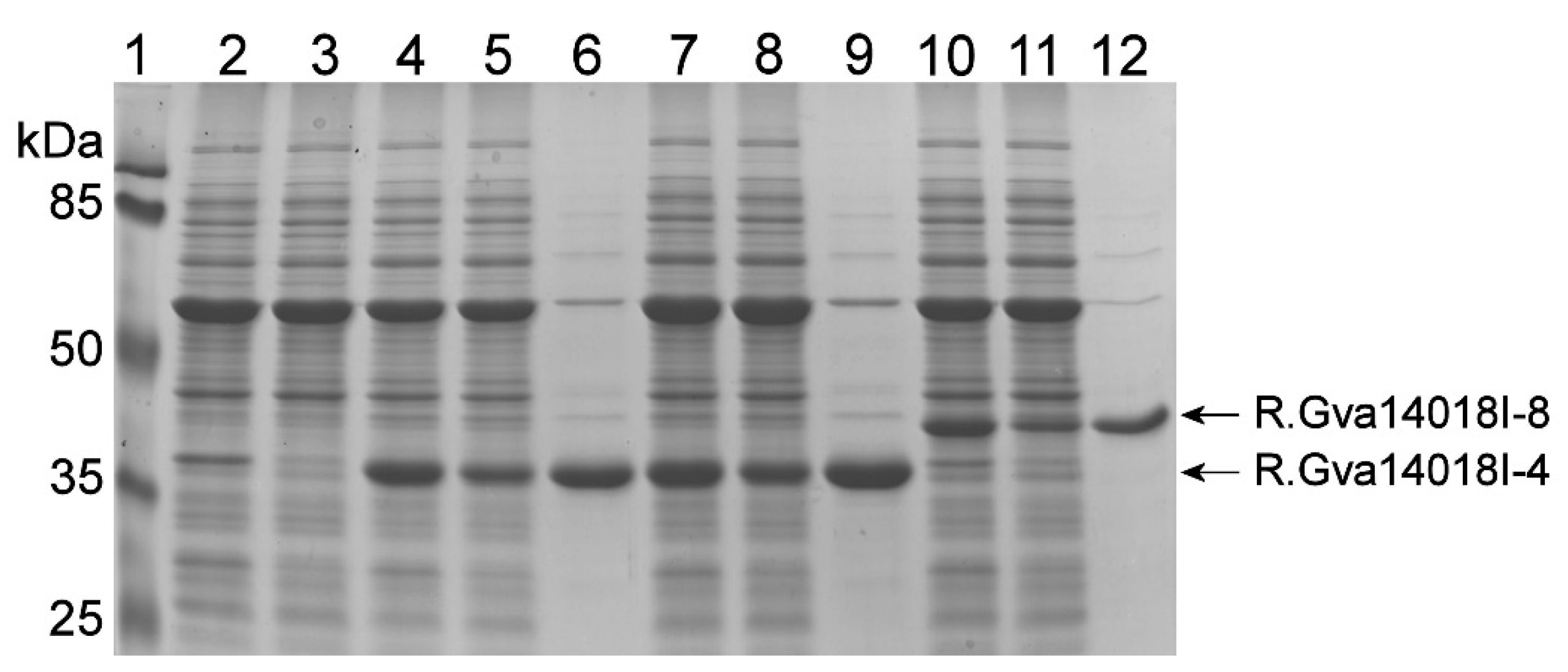
Two mutated gva14018IR genes were selected for further experiments. The first gene variant had one nucleotide deletion, which generated a fragment encoding the truncated protein comprising the first 259 amino acids of the wild-type REase (298 amino acids) followed by WKGEMLYMND from a mutation-generated frameshift. This gene was named gva14018IR-4 (GenBank accession number MN738700). The second gene variant gva14018IR-8 encoded the REase protein containing the W129R amino acid substitution (GenBank accession no. MN938912). Both gene variants were cloned into pET28a(+) and transformed into E. coli strains Tuner (DE3), ER2566, and ArcticExpress (DE3). The E. coli strains selected for transformation did not possess a plasmid harboring the gva14018IM gene. R.Gva14018I-4 and R.Gva14018I-8 were expressed in E. coli as N-terminal His-tagged proteins with apparent molecular weights slightly higher than calculated (33 and 36 kDa, respectively) (Figure 4). E. coli ArcticExpress (DE3) was selected for recombinant protein production because it has low endogenous nuclease activity (Figure 4).
Cell lysates and crude protein preparations exhibited R.Gva14018I-specific activity (Figure 5A). R.Gva14018I-4 displayed higher REase activity than R.Gva14018I-8 and was selected for further experiments. A fraction of soluble R.Gva14018I-4 obtained after the induction of protein synthesis in ArcticExpress (DE3) at 12 °C was subjected to purification via single-step Ni-affinity chromatography (Figure 5).

2.4. Polyclonal Antibodies against Recombinant R.Gva14018I-4 also Detected R.Gva14018I

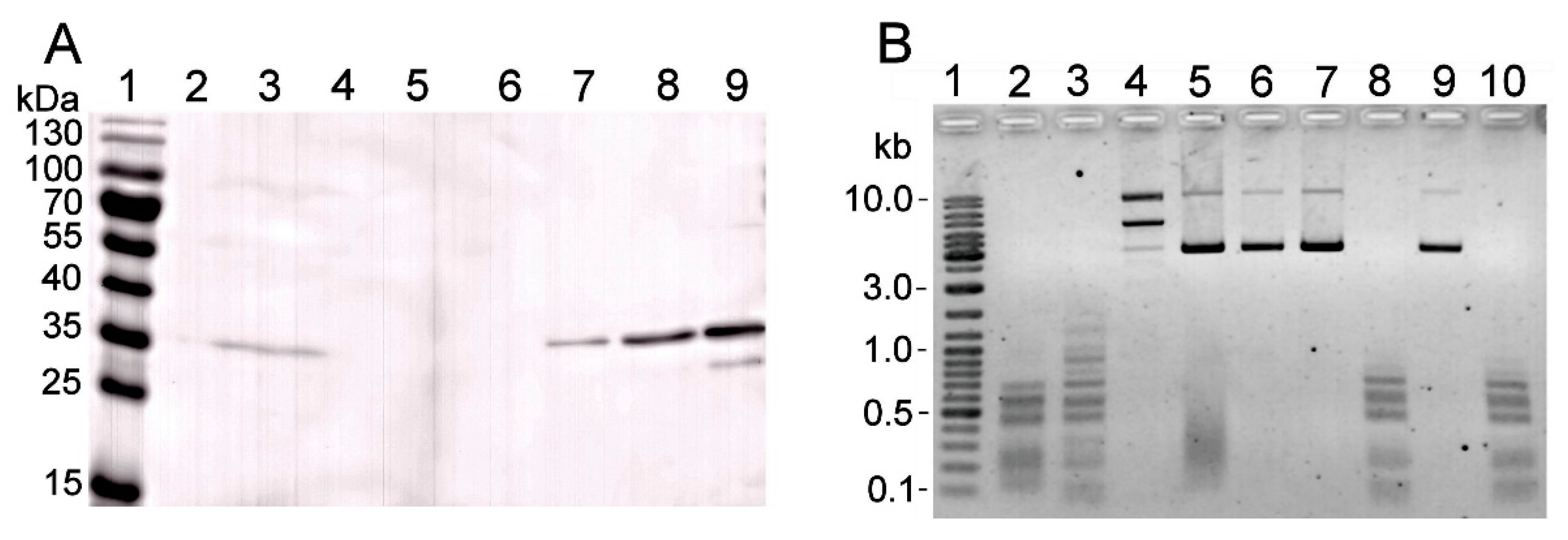
To detect the wild-type REase R.Gva14018I produced by G. vaginalis cells, the polyclonal antibodies (pAbs) were raised by immunizing mice with purified R.Gva14018I-4. The pAb immunoprecipitates of R.Gva14018I-4 were subjected to mass spectrometry analysis. The major peak in the HPLC/ESI-MS chromatogram corresponded to 33,075.20 Da (Figure S4). The detected molecular mass of R.Gva14018I-4 was in good agreement with the mass deduced from its amino acid sequence (33,074.29 Da without the N-terminal methionine; https://web.expasy.org/protparam/). Immunoblot analysis of cell lysates of G. vaginalis strain ATCC 14018 revealed a band in the range of 35–40 kDa using pAbs against R.Gva14018I-4 as the primary antibody (Figure 6A). No bands were visualized via immunoblot analysis of the cell lysates of G. vaginalis strain 49145. REase activity was detected by immunoblot only in the ATCC 14018 cell lysate (Figure 6B).

2.5. Characterization of the R.Gva14018I-4 Protein

The specificities of wild-type and recombinant REases were compared by digesting DNA substrates with ATCC 14018 culture supernatant and purified R.Gva14018I-4 (Figure 5B). Similar arrays of DNA fragments were obtained after the digestion of pUC57 and pBR322 plasmids. Incubation of pBR322 with the supernatant yielded an extra band, which was most likely a product of incomplete DNA cleavage (Figure 5B, lane 6). Both REases digested gDNAs from calf thymus, HCT116 human cell line, and E. coli LMG194, whereas gDNA from the LMG194-Me strain was resistant to cleavage.
To determine the target sequence of R.Gva14018I-4, a single-site PCR substrate was digested, and the DNA fragments were cloned into pJET1.2 plasmid and then sequenced. The results showed that R.Gva14018I-4 cleaved the GGCC sequence. Thus, both the wild-type and recombinant G. vaginalis 14018 REases recognized the same target sequence and were sensitive to the methylation pattern generated by recombinant M.Gva14018I.
The single-site PCR substrate was used to evaluate the effects of pH and salt and divalent cation concentrations on purified R.Gva14018I-4 activity. At neutral pH 7.2, R.Gva14018I-4 displayed maximum activity in the presence of 1–10 mM Mg2+, but the activity significantly decreased at pH 5.0 (Figure 7A). At neutral pH, increasing concentrations of NaCl or KCl from 0–200 mM inhibited REase activity. Divalent metal ions Mg2+ and Mn2+ were preferred cofactors. R.Gva14018I-4 displayed maximum activity in the presence of 1 mM Mg2+, whereas the addition of 0.1 or 10 mM Mg2+ resulted in incomplete substrate digestion. The optimum concentration of Mn2+ in the reaction mixture was 0.1–10 mM. Low REase activity was observed at 1 mM Ni2+ (Figure 7B). The addition of Ca2+ (0.1–10 mM) to the reaction mixture did not induce REase activity. In the presence of 1 mM Mg2+, increasing Ca2+ concentrations inhibited substrate digestion (Figure 7B). Similar effects of pH and salt and divalent cation concentrations on REase activity were observed with pUC57, which has multiple R.Gva14018I-4 recognition sites (Figure S5).

2.6. Bioinformatics Analyses of M.Gva14018I, R.Gva14018I, and R.Gva14018I-4

M.Gva14018I was used as a query sequence for a BLASTp search which revealed multiple bacterial proteins characterized as C-5 cytosine-specific DNA methyltransferases (pfam00145) that are often found with HaeIII family (pfam09556) REases. HaeIII cleaves the double-stranded sequence GGCC and generates blunt ends (UniprotKB entry O68584). Its cognate methyltransferase modifies cytosine to 5-methylcytosine (GGm5CC) on both DNA strands (UniprotKB P20589). The gDNA extracted from DH10B-Me was resistant to cleavage by BsuRI (Figure 3A); BsuRI does not cleave the methylated target GGm5CC sequence [37]. These combined results suggest that M.Gva14018I modifies the first cytosine in the GGCC sequence.
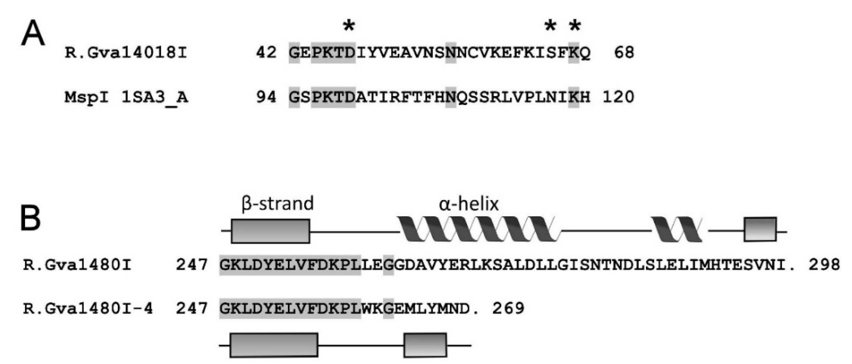
A BLASTp search with the R.Gva14018I sequence found 39% identity and 57% similarity with the restriction endonuclease BspRI (GenBank CBE66553.1), a single protein with a known function among multiple matches. BspRI is an experimentally determined Type IIP REase that cleaves DNA at the sequence GGCC to produce blunt-end fragments [38]. Sequence analysis of R.Gva14018I using the PDEXK server [39] showed a probability of 0.95 that the nuclease belongs to the PD-(D/E)XK superfamily. The HHpred analysis (https://toolkit.tuebingen.mpg.de/tools/hhpred) revealed that the N-terminal region (amino acids 38–104) is similar to the amino acid 90–156 region of MspI. This result suggests that the active site of R.Gva14018I is located between 38–104 amino acids [40]. Amino acids D47, S65, and K67 most likely constitute part of the active site. Amino acid S65 conforms to neither D/E in the PD-(D/E)XK motif, nor N117 in the respective motif of MspI. However, BsuFI displays S254 in the same position [40]. The presence of the first 259 amino acids at the N-terminus presumes that R.Gva14018I-4 contains an intact catalytic center. Secondary structure prediction of wild-type and mutant R.Gva14018I was performed by the PSIPRED server (http://bioinf.cs.ucl.ac.uk/psipred). The truncated R.Gva14018I-4 protein lacked the C-terminal alpha helix (263–277 amino acids) (Figure 8). We speculate that the difference in secondary structure might affect the enzyme conformation and activity. The absence of the C-terminus might result in inefficient DNA cleavage by R.Gva14018-4 compared to that of the wild-type REase. This presumption is supported by the fact that E. coli cells transformed with a plasmid bearing the mutant gene gva14018IR-4 survived even in the absence of cognate MTase, whereas the high toxicity of wild-type R.Gva14018I endonuclease in E. coli impairs its gene cloning.

3. Discussion

In this study, we identified and characterized the R-M system from G. vaginalis strain ATCC 14018. The genes predicted to encode the R-M system were indicated in the REBASE database. The open reading frames encoding the putative REase and MTase were on complementary strands interspaced by 53 bp and oriented toward each other. A putative transcription regulator (GenBank BAQ33762.1) is encoded immediately upstream of the MTase. The REase and MTase are the first characterized R-M enzymes from the genus Gardnerella. G. vaginalis ATCC 14018 possesses the REase R.Gva14018I and its cognate MTase. R.Gva14018I cleaves the GGCC sequence, most probably generating blunt ends. A BLASTp search identified similarity with the characterized REase BspRI, which cleaves the GG↓CC sequence. Experimental evidence indirectly demonstrated that M.Gva14018I modifies the first cytosine residue in the GGCC sequence. Moreover, similarity searches for M.Gva14018I showed many C-5 cytosine-specific DNA MTases, which generate GGm5CC methylation on both DNA strands. G. vaginalis ATCC 14018 possesses a HaeIII-like R-M system, which is widespread among bacterial species [38,40,43]. A HaeIII-like restriction endonuclease activity is an effective tool against foreign DNA because the short cleavage site frequently occurs [43].
The gene encoding MTase was cloned and expressed in E. coli. Recombinant M.Gva14018I was active in vivo. Cloning and expression of R.Gva14018I in E. coli cells was challenging due to toxicity, which is common for REases [38,44]. R.Gva14018I mutants were successfully expressed in E. coli. The truncated R.Gva14018I-4 protein lacking the C-terminal region demonstrated the highest REase activity. The same target specificity was observed for purified R.Gva14018I-4 and wild-type R.Gva14018I from the cell-free lysate of G. vaginalis ATCC 14018. R.Gva14018I-4 requires Mg2+ for its enzymatic activity; Mn2+ can substitute as a cofactor for Mg2+, whereas Ca2+ cannot. The same cofactor requirements are characteristic of Type II REases of the PD-(D/E)XK family, whereas REases of the GIY-YIG and HNH families use various metal cations and Ca2+ as cofactors [45,46]. Our bioinformatics analysis strongly supports the prediction that R.Gva14018I belongs to the PD-(D/E)XK family of REases.
The REase activity was detected in G. vaginalis cell lysate and cell-free growth medium. REase activity in cell-free growth medium is presumably due to cell death and decomposition as the DNA cleavage activity increased over the time course of cultivation. We were unable to purify wild-type R.Gva14018I from cell-free extracts or cell-free growth medium. Polyclonal antibodies against R.Gva14018I-4 detected the wild-type R.Gva14018I REase in the cell lysate of G. vaginalis strain ATCC 14018. The pAbs did not react with the cell lysate of strain ATCC 49145, which did not display REase activity. The apparent molecular mass of the protein recognized by pAbs was slightly larger than the predicted 34 kDa for R.Gva14018I. R.Gva14018I-4 produced a band that was observed at a higher position on the gel than that of its calculated molecular mass, although mass spectrometry of the pAb-immunoprecipitated complexes showed good agreement between calculated and experimental values for R.Gva14018I-4 molecular mass. Immunoprecipitation of wild-type R.Gva14018I from the cell lysate was not successful, possibly due to the high amount of interfering compounds.
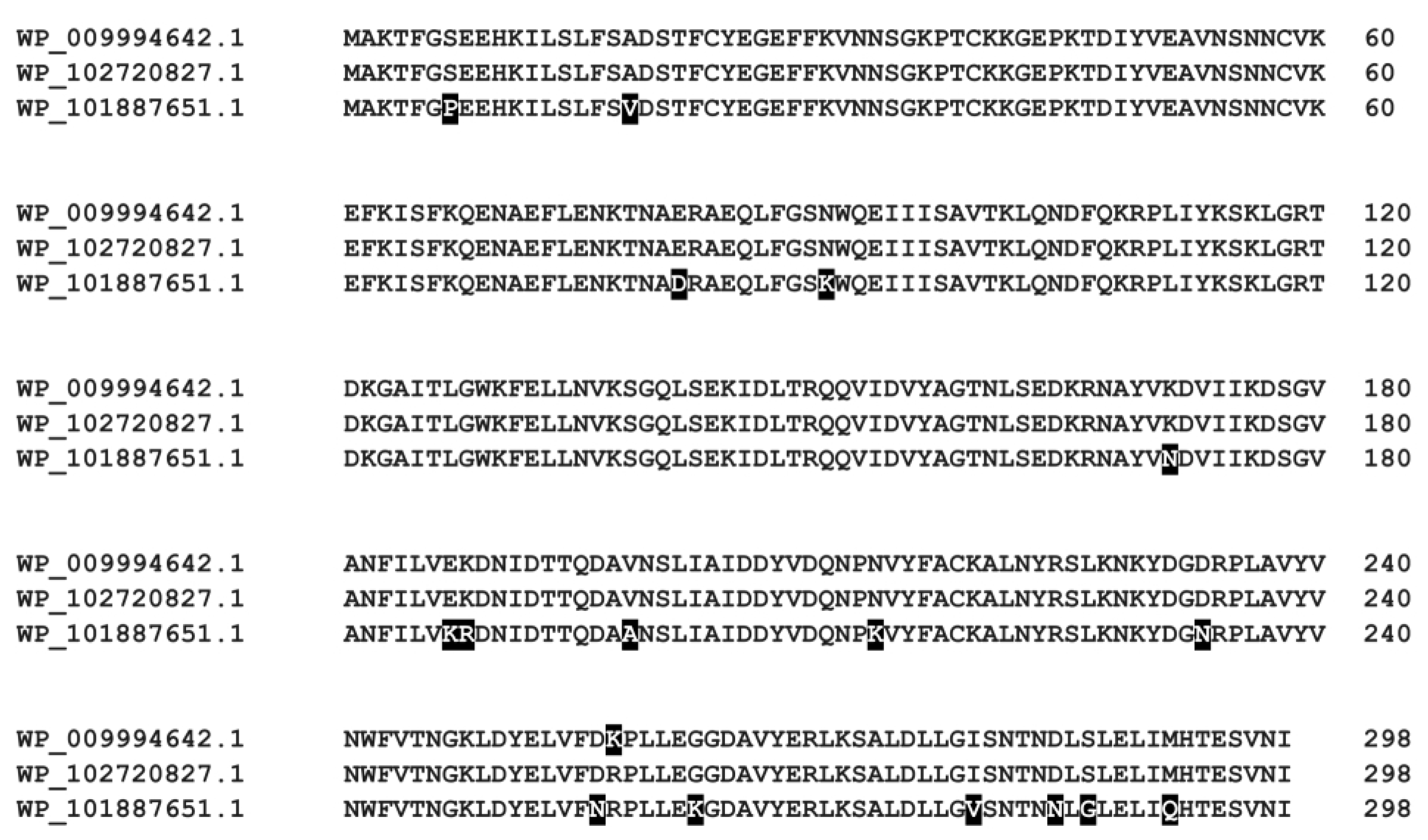
The BLASTp search revealed that several Gardnerella spp. strains encode proteins highly similar to R.Gva14018I (100%, 99%, or 94% sequence identity) (Figure 9). The Gardnerella spp. strains encoding R.Gva14018I homologs also contain cognate methylases that are highly similar to M.Gva14018I (100% or 99% sequence identity). This indicates that these strains possess a full R-M system. In addition to ATCC 14018, G. vaginalis ATCC 14019 and G. vaginalis DSM 4944 possess putative REase that recognize GGCC (rebase.neb.com/rebase/rebase.html). Genome analysis revealed a wide diversity of R-M genes at varying frequencies across 106 Gardnerella spp. [47]. However, G. vaginalis ATCC 49145 and G. swidsinskii GV37 with putative R-M genes (rebase.neb.com/rebase/rebase.html; 47) did not produce restriction endonuclease activity in our hands when grown in vitro.
Gardnerella species were found not to be defined by the presence or absence of particular R-M systems [47], however, these systems are among the barriers to interspecies and intraspecies HGT [25,48]. Horizontal gene transfer is recognized to be a major force shaping the diversity of Gardnerella spp. [13,16,47,49]. More HGT was observed within the same Gardnerella species or closely related species (e.g., between G. piotii and G. vaginalis) than distantly related species/genomospecies, therefore allowing it to maintain species separation in the same vaginal niche [13,47]. G. vaginalis and G. piotii are the most often isolated species from BV-positive women and those species seem to possess a greater virulence potential relative to G. leopoldii/G. swidsinskii [12,15]. G. vaginalis has a larger pangenome and the core genome is more recombinant than G. piotii that suggest more frequent G. vaginalis engagement in HGT [47]. Differences in abundance and target specificity of R-M systems could determine differences in HGT between G. vaginalis and G. piotii. In this study, cell lysates of 31 Gardnerella spp. were tested for the REase activity. Among ten isolates of clade 2 (emended description G. piotii, manuscript in preparation), three isolates produced Dam or Dcm methylation-sensitive REases (Figure S2). Highly similar digestion patterns suggest the same specificity of these REases. Among 16 isolates of clade 1 (emended description G. vaginalis), including ATCC strains, three strains produced enzymes that cut DNA regardless of methylation. Two strains produced REases with similar DNA digestion patterns; however, none of them matched the DNA digestion pattern of R.Gva14018I, and PCR analysis did not reveal the presence of gva14018IM and gva14018IR coding genes in these strains. Our data show that the REase activity was more frequently detected among G. piotii strains than G. vaginalis that may support the potential role of R-M systems as a barrier to HGT in G. piotii. Moreover, Type II REases of distinct specificity were limited to the particular Gardnerella species. Five isolates of clade 4 (emended description G. leopoldii/G.swidsinskii) did not display the REase activity that suggests other barriers and mechanisms of protection against foreign DNA. However, the major limitation to the evaluation of the distribution of Type II REases among species/clades is the small number of strains analyzed in this study. Among limitations is the test for REase activity, which allows the detection of substrate digestion patterns characteristic of Type II REases. Gardnerella spp. isolates may produce rare-cutting restriction endonucleases (e.g., Type I or Type III) that require specific DNA substrates to detect their activity. The gene expression behavior of Gardnerella spp. depends on the mode of growth, the presence of other BV-associated bacteria, and the host [20]. The expression of genes involved in R-M systems might be affected by the same factors that are not accounted for when cultivating in vitro.
In conclusion, we identified and characterized the R-M system of Type II from the G. vaginalis ATCC 14018 strain. R.Gva14018I is a four-base cutter and displays a HaeIII-like restriction endonuclease activity that is widespread in bacteria and archaea. The detected REase activity in G. vaginalis and G. piotii isolates suggest that Type II R-M systems might constitute the inter-species barriers to HGT. However, the particular role of R-M systems in shaping and maintaining HGT barriers remains to be determined.

4. Materials and Methods

4.1. Bacterial Strains and Cultivation Conditions

Gardnerella vaginalis strains ATCC 14018 and 49145 were purchased from the American Type Culture Collection (ATCC). Gardnerella spp. isolates were obtained from previously characterized cultured vaginal samples of women from Lithuania [12]. Frozen bacterial stocks were stored at −80 °C in tryptic soy broth (TSB) (Liofilchem, Roseto degli Abruzzi, Italy) supplemented with 20% (v/v) horse serum (Oxoid, Thermo Fisher Scientific, Waltham, MA, USA) and 15% (v/v) glycerol. All Gardnerella strains were revived on chocolate agar with Vitox (Oxoid) and incubated at 36 °C under 5% CO2 for 48 h. G. vaginalis ATCC 14018, ATCC 49145, Gardnerella spp. strains [12], and GV37 were grown in 7 mL of liquid sTSB medium (TSB supplemented with 0.15% (w/v) soluble starch and 2% (v/v) horse serum) for 24 h, as described previously [15]. Horse serum included into sTSB medium contains nucleases [50]; therefore, unless otherwise indicated, G. vaginalis ATCC 14018, ATCC 49145 strains were cultivated in 7 mL of sBHI medium (brain-heart infusion broth (Liofilchem) supplemented with 2% (w/v) gelatin, 0.5% yeast extract (w/v), and 0.1% soluble starch (w/v)) in tightly closed tubes at 36 °C for 22 h. Typically, after cultivation, the ATCC 14018 strain reached OD600 = 0.6–0.8 and ATCC 49145 reached OD600 = 0.9–1.0. After measurement of the optical density, cells were separated via centrifugation at 13,000× g for 2 min, and the supernatants were filtered through 0.1-µm membrane filters (Ultrafree-MC PVDF, Merck Millipore). To assess the endonuclease activity during growth, Gardnerella spp. strains were cultivated in 30 mL of sBHI. The samples were collected after incubating for 0, 16, 24, and 44 h at 36 °C. The supernatants were filtered and stored frozen.
E. coli strains used in this study are listed in Table S1. LB medium (Thermo Fisher Scientific) was supplemented with the following antibiotics: 100 µg/mL ampicillin for recombinant pJET1.2, pUC57, and pBAD/HisA-based plasmids; 25 µg/mL kanamycin for recombinant pET28a-based plasmids; and 12 or 36 µg/mL chloramphenicol for recombinant pACYC184-based plasmids (Table S2). ArcticExpress (DE3) cultures were incubated overnight in 20 µg/mL gentamicin. Recombinant protein expression in E. coli cells harboring prophage DE3 was induced with 0.1 mM isopropyl β-D-1-thiogalactopyranoside (IPTG). Where indicated, the basal expression of recombinant proteins from pET28a(+) [51] and pBAD/HisA [52] plasmids was repressed by adding 0.5% or 1% (w/v) D(+)-glucose (AppliChem, Merck) to the culture medium.

4.2. Amplification of gva14018IR and gva14018IM Genes

A putative R-M system of G. vaginalis strain ATCC 14018 is indicated in the REBASE database [35]. Enzymes and kits obtained from Thermo Fisher Scientific were used to manipulate DNA according to standard protocols. The gDNA was extracted using the GeneJET Genomic DNA Purification Kit (#K0721) according to the manufacturer’s recommendations. Genes encoding REase (gva14018IR) and MTase (gva14018IM) in Gardnerella spp. strains were detected with primer pairs GVRe5/GVRe3 and GVMe5/GVMe3 (Table 1). PCR was performed with Platinum II Hot Start Taq polymerase in a 15-µL reaction volume. Amplification reactions included initial denaturation at 94 °C for 2 min, 35 amplification cycles consisting of denaturation for 20 s at 94°C, annealing for 20 s at 43 °C (for gva14018IR) or 40 °C (for gva14018IM), and extension for 1 min at 72 °C. For cloning, the genes encoding M.Gva14018I and R.Gva14018I were amplified using Phusion Flash II DNA Polymerase and the primer pairs GVMe5/GVMe3, GVRe5/GVRe3, and GvRe5-NheI/GvRe3-XhoI (Table 1). Purified gDNA from the strain ATCC 14018 was used as a template for PCR.

4.3. Cloning of the gva14018IM Gene

The DNA fragment coding for M.Gva14018I was cloned in the plasmid pJET1.2/blunt and verified using sequencing. The DNA fragment was excised with Eco105I, and then cloned into the plasmid pACYC184 digested with Eco32I. As a result, the gva14018IM gene was inserted downstream of the tetracycline resistance gene promoter. The plasmid was transformed into E. coli DH10B, and methyltransferase-positive clones were grown for 18 h. Protection against R.Gva14018RI and BsuRI cleavage was assessed as described in a subsequent section (MTase activity assay). The plasmid providing the best protection was named pACYC-Me, and the derived strain harboring this plasmid was named DH10B-Me. Accordingly, E. coli strains ER2566 and LMG194 transformed with pACYC-Me were named ER2566-Me and LMG194-Me (Table S1).
The replication origin p15A of the plasmid pACYC-Me was replaced with that of pRSF-Duet1 (Novagen, Merck) to increase the copy number [53] of the gva14018IM-containing plasmid. The replication origin of pRSF-Duet-1 was excised with Bsp68I/XbaI, and subsequently cloned into Bst1107I/XbaI–digested pACYC-Me. The selected recombinant plasmid was named pRSFori-Me. Strains DH10B and LMG194 were transformed with pRSFori-Me and assessed for digestion with the G. vaginalis 14018 culture supernatant. The derived strains were named DH10B-RSFMe and LMG194-RSFMe.

4.4. Cloning the gva14018IR Gene

The DNA fragment amplified with the GvRe5 and GvRe3 primers was cloned into pJET1.2. The resulting plasmid was transformed into strains DH10B and DH10B-Me. Alternatively, the DNA fragment amplified with the GvRe5-NheI and GvRe3-XhoI primers was digested with the respective enzymes and cloned into pET28a(+) and pBAD/HisA. Each resulting plasmid was transformed into DH10B-Me. The thirteen recombinant plasmids from the transformed strains were subjected to sequencing, which revealed the presence of mutations in the gva14018IR gene.
Several strategies were used to select the mutation-free gva14018IR gene. The amplicon generated with GvRe5-NheI and GvRe3-XhoI primers was cloned into the Eco32I-digested pUC57 vector and transformed into DH10B-Me. IPTG was added to the LB agar plates to induce the lacZ promoter. Five recombinant plasmids were confirmed by sequencing to be free of mutations in gva14018IR. The gene was excised with NheI and XhoI, cloned into NheI/SalI-digested pET28a(+), and transformed into E. coli ArcticExpress (DE3). The plating medium contained 0.5% (w/v) glucose. No positive clones were obtained.
The DNA fragment coding for R.Gva14018I was cloned into the NheI/XhoI-digested pBAD plasmid isolated from the DH10B-Me strain. The resulting plasmid was transformed into LMG194-Me, DH10B-Me, and DH10B-RSFMe. The plating medium contained 0–1% (w/v) glucose. Sequencing results confirmed the presence of mutations in the gva14018IR gene.

4.5. Expression of R.Gva14018I Mutants in E. coli

Recombinant plasmids bearing either gva14018IR-4 encoding the truncated protein or gva14018IR-8 encoding the protein with one amino acid substitution were selected for further experiments. Both fragments were excised from pJET1.2 with NheI/SalI and cloned into respectively digested pET28a(+) to generate N-terminal hexahistidine-tagged proteins. The resulting plasmids bearing gva14018IR-4 and gva14018IR-8 were transformed into DH10B. Positive colonies were selected, and the recombinant plasmids were transformed into E. coli Tuner (DE3), ER2566, and ArcticExpress (DE3) strains.

4.6. Small-Scale Synthesis of Recombinant Proteins

The synthesis of R.Gva14018I-4 and R.Gva14018I-8 in E. coli Tuner (DE3), ER2566, and ArcticExpress (DE3) was induced with 0.1 mM IPTG, and then cells were cultivated for 2.5 h at 37 °C, 4 h at 25 °C, or 22–24 h at 12–16 °C, respectively. The cells were harvested and disrupted via sonication (Bandelin Sonopuls HD 3100, Bandelin Electronic, Germany). The soluble fraction was separated, and proteins were purified using the His-Spin Protein Miniprep kit (Zymo Research, Irvine, CA, USA). The insoluble fraction was resuspended in PBS containing 2% SDS and incubated for 10 min at 100 °C. The samples were analyzed by 12% SDS-PAGE under reducing conditions.

4.7. Large-scale Protein Synthesis of Gva14018RI-4 and Protein Purification

E. coli ArcticExpress (DE3) transformed with pET28 bearing gva14018IR-4 was cultivated with shaking at 37 °C for 2 h until cultures reached OD600 ~ 0.4. The flasks were cooled to 4 °C for 1 h. IPTG was added to a final concentration of 0.1 mM, and cultures were incubated at 12 °C for 24 h with shaking (OD600 = 1). The cells were collected by centrifugation at 5000× g for 15 min at 4 °C, washed in PBS followed by centrifugation at 3000× g for 20 min at 4 °C, and kept frozen at −20 °C. A total of 6 g of wet weight biomass was obtained from 3.6 L culture medium. The cell pellet was suspended in lysis buffer (25 mM Tris-HCI pH 7.4, 0.5% Triton X-100, 0.2 M NaCl, 0.1 M imidazole, 10% glycerol, and 2 mM 2-mercaptoethanol) containing 1 mM phenylmethylsulfonyl fluoride and Halt™ Protease Inhibitor Cocktail (Thermo Fisher Scientific). The cells were incubated at 4 °C for 60 min and then disrupted by sonication. The supernatant was clarified by centrifugation at 30,000× g for 25 min. The soluble His-tagged R.Gva14018I-4 protein was purified using Chelating Sepharose Fast Flow with immobilized Ni2+ ions (GE Healthcare Bio-Sciences AB, Sweden). The purified protein was dialyzed overnight at 4 °C against buffer (20 mM HEPES pH 7.5, 150 mM NaCl, 5% glycerol, and 1 mM dithiothreitol), and the purified protein was stored at −80 °C. Protein concentration was determined using UV-visual spectrophotometry (calculated extinction coefficient 40,005 M−1cm−1 at 280 nm) and the standard Bradford method.

4.8. Determination of REase Cleavage Site

Plasmid pBR322 was subjected to digestion with cell-free culture supernatant (OD600 = 0.6) of G. vaginalis strain ATCC 14018. The DNA cleavage products, either untreated or treated with blunting enzyme (CloneJET PCR Cloning kit, Thermo Fisher Scientific), were purified and cloned into pJET1.2 using the CloneJET PCR Cloning kit. The recombinant plasmids were subjected to sequencing. The insert sequences were determined, and the fragment contents were elucidated via alignment with pBR322. The pUC57 plasmid was a template for a single-site PCR substrate generated using primers GV-pUC57-For and GV-pUC57-Rev (Table 1). The obtained DNA fragment of 868 bp was digested with the purified R.Gva14018I-4 protein. The DNA cleavage products were cloned into pJET1.2 and sequenced.

4.9. REase Activity Assays

DNA cleavage assays were performed in 18-µL reactions at 37 °C. The following DNA samples were used as substrates: pBR322, pUC57 (both plasmids isolated from E. coli dam+ dcm+; Thermo Fisher Scientific), gDNAs (from calf thymus, HCT116 human cell line, and E. coli), and a single-site PCR fragment. Assays to detect REase activity in culture supernatants, cell lysates, and crude protein preparations were typically performed in Fast Digest reaction buffer (Thermo Fisher Scientific) containing 0.25 µg pBR322 and 0.1 µg/µL of RNase A. The test sample (2.5 µL) was added to the reaction mixture and incubated for 1–2 h at 37 °C. The reaction was stopped by adding DNA loading dye with SDS (Thermo Fisher Scientific), and the mixture was heated at 65 °C for 10 min before loading on a 1% agarose gel in 1×TAE. The native REase activity in cell-free sTSB medium from Gardnerella spp. strains was determined using buffer containing 50 mM Tris-HCl (pH 7.2 at 37 °C) and buffer containing 50 mM sodium acetate (pH 5.0), both supplemented with 1 mM MgCl2 and 1 mM CaCl2. To compare the specificities of wild-type and recombinant REases, 1 µL of supernatant or 0.7 µg of purified R.Gva14018I-4 was mixed with the substrate (0.25 µg of pUC57 or 0.5 µg of gDNA) in 1× buffer B (10 mM Tris-HCl pH 7.5, 10 mM MgCl2, and 0.1 mg/mL BSA) (Thermo Fisher Scientific) containing 0.1 µg/µL of RNase A. The reaction mixture was incubated at 37 °C for 16 h.
The effects of salt concentration, pH, and divalent metal ions were determined using 25 mM Tris-HCl buffer (pH 7.2 at 37 °C) and 25 mM sodium acetate buffer (pH 5.0) containing 0–200 mM NaCl or KCl, 0–15 mM MgCl2, 0–10 mM MnCl2, CaCl2, or NiCl2. The cleavage reactions were performed by incubating 1 µg of purified R.Gva14018I-4 with 0.25 µg of pUC57 or 0.4 µg of purified R.Gva14018I-4 with 0.2 µg of single-site DNA fragment for 30 min at 37 °C.

4.10. MTase Activity Assay

In vivo DNA methylation by wild-type and recombinant M.Gva14018I was tested by performing the DNA protection assay as follows: 0.5 µg of gDNA was incubated with 1 µL of G. vaginalis 14018 culture supernatant (OD600 = 0.6) or 0.25 µL BsuRI (Fast Digest, Thermo Fisher Scientific) in the presence of 0.1 µg/µL RNase A in 1× FastDigest (Thermo Fisher Scientific) buffer at 37 °C for 16 h.

4.11. Generation of Murine Polyclonal Antibodies Against Recombinant R.Gva14018I-4

Polyclonal antibodies (pAbs) against R.Gva14018I-4 were produced in mice. Animal maintenance and experimental protocols were performed in accordance with FELASA guidelines and Lithuanian and European legislation in the Department of Biological Models (Institute of Biochemistry, Life Sciences Center, Vilnius University). The approval to use mice for immunizations was obtained from the Lithuanian State Food and Veterinary Agency (permission No. G2-117, issued 11 June 2019). Three female 8-week-old BALB/c mice were immunized with 50 µg of recombinant protein R.Gva14018I-4 by subcutaneous injection four times every 28 days. For the first and second immunizations, 50 µg of R.Gva14018I-4 was emulsified with complete (Sigma Aldrich, St. Louis, MO, USA) and incomplete Freund’s adjuvants, respectively. After 94 days, mice were sacrificed, and blood samples were collected. The blood serum was mixed with saturated ammonium sulfate solution to precipitate pAbs.

4.12. Immunoblot Detection of R.Gva14018I

G. vaginalis strains ATCC 14018 and 49145 were grown for 28 h on chocolate agar with Vitox (Oxoid). The cells were harvested, washed with 0.85% NaCl solution, and kept at −20 °C. The biomass was suspended in PBS containing Halt™ Protease Inhibitor Cocktail and then disrupted by sonication. Cell lysates were clarified by centrifugation at 16,100× g for 2 min, and then loaded on a 10% SDS-PAGE gel. After electrophoresis, proteins were transferred to a polyvinylidene difluoride (PVDF) blotting membrane (GE Healthcare Life Science, Freiburg, Germany) under semi-dry conditions. The membrane was blocked with 2% powdered milk (Roth, Germany) in PBS, and then incubated with pAbs against recombinant R.Gva14018I-4. After washing in PBST (PBS supplemented with 0.05% Tween 20), the membrane was incubated with anti-mouse IgG-HRP (Bio-Rad, Hercules, CA, USA) for 1 h. After rinsing, protein bands were developed using 4-chloro-1-naphthol (Sigma Aldrich, St. Louis, MO, USA).

4.13. Immunoprecipitation and Mass Spectrometry Analysis of Recombinant R.Gva14018I-4

R.Gva14018I-4 protein was immunoprecipitated with anti-R.Gva14018I-4 pAbs that were coupled to MagnaBind™ Protein A Beads (Pierce Biotechnology, Thermo Fisher Scientific). The pAbs were incubated with magnetic beads in binding buffer (Tris-HCl pH 8.0) with mixing for 1 h. After several washes with binding buffer, the magnetic beads were incubated with R.Gva14018I-4 protein in PBS with mixing for 2 h. After several washes with PBS, the immunocomplexes of pAbs and R.Gva14018I-4 were eluted from the magnetic beads with 0.1 M glycine (pH 3.0). The mixture was supplemented with DTT to a final concentration of 50 mM. Samples were stored at −20 °C. The immunocomplexes were analyzed on an integrated HPLC (Agilent 1290 Infinity)/ESI-MS (Agilent Q-TOF 6520) system equipped with a Poroshell 300SB-C8 column (2.1 × 75 mm, 5 µm) via elution with a linear gradient of solvent A (1% formic acid in water) and B (1% formic acid in acetonitrile).

4.14. Bioinformatics Analysis

DNA and protein similarity searches were performed using BLAST (https://blast.ncbi.nlm.nih.gov/Blast.cgi). The HHpred server [41] (https://toolkit.tuebingen.mpg.de/tools/hhpred) was used for structural similarity searches. Secondary structure predictions were performed using the PSIPRED server (http://bioinf.cs.ucl.ac.uk/psipred) [42]. The PDEXK recognition server (http://bioinformatics.ibt.lt/pdexk) [39] was implemented to identify nucleases of the PD-(D/E)XK superfamily.

Supplementary Materials

The following are available online at https://www.mdpi.com/2076-0817/9/9/703/s1, Figure S1: Enzymatic activity in the cell-free culture supernatants from G. vaginalis ATCC 14018 at various time points during cultivation, Figure S2: REase activity in the clarified whole cell lysates of Gardnerella spp. strains 47.3, 63.2, 78.1, 86.3, 105.1, G. vaginalis ATCC 49145 and ATCC 14018, Figure S3: Detection of the gva14018IR (A) and gva14018IM (B) genes in Gardnerella spp. isolates by gene-specific PCR with the primer pairs GVRe5/GVRe3 and GVMe5/GVMe3, Figure S4: Molecular mass determination of R.Gva14018I-4 by mass spectrometry, Figure S5: Effects of cofactors, pH and salt concentration on the R.Gva14018I-4 activity, Table S1: Bacterial strains used in this study, Table S2: Plasmids used in this study.

Author Contributions

Conceptualization, M.P., A.B. and I.D.; Methodology, A.B., M.P., I.D. and V.M.; Software, A.B.; Formal analysis, M.P., A.B., V.M. and I.D.; Resources, M.P.; Writing—Original Draft Preparation, A.B., M.P., V.M. and I.D.; Writing—Review & Editing, M.P. and A.B., Visualization, A.B. and M.P.; Supervision, M.P. and A.B.; Project Administration, M.P.; Funding Acquisition, M.P. All authors have read and agreed to the published version of the manuscript.

Funding

This research was funded by the Research Council of Lithuania (http://lmt.lt/en/), grant number S-MIP-17-49.

Acknowledgments

We thank Audrone Ruksenaite for mass spectrometry determination of protein molecular mass. We acknowledge Rolandas Meskys, Giedrius Sasnauskas, Nina Urbeliene, and Migle Janulaitiene for their advice.

Conflicts of Interest

The authors declare no conflict of interest.

References

  1. Bilardi, J.E.; Walker, S.; Temple-Smith, M.; McNair, R.; Mooney-Somers, J.; Bellhouse, C.; Fairley, C.K.; Chen, M.Y.; Bradshaw, C. The burden of bacterial vaginosis: Women’s experience of the physical, emotional, sexual and social impact of living with recurrent bacterial vaginosis. PLoS ONE 2013, 8, e74378. [Google Scholar] [CrossRef] [PubMed] [Green Version]
  2. Leitich, H.; Kiss, H. Asymptomatic bacterial vaginosis and intermediate flora as risk factors for adverse pregnancy outcome. Best Pract. Res. Clin. Obstet. Gynaecol. 2007, 21, 375–390. [Google Scholar] [CrossRef]
  3. Dingens, A.S.; Fairfortune, T.S.; Reed, S.; Mitchell, C. Bacterial vaginosis and adverse outcomes among full-term infants: A cohort study. BMC Pregnancy Childbirth 2016, 16, 278. [Google Scholar] [CrossRef] [Green Version]
  4. Shipitsyna, E.; Khusnutdinova, T.; Budilovskaya, O.; Krysanova, A.; Shalepo, K.; Savicheva, A.; Unemo, M. Bacterial vaginosis-associated vaginal microbiota is an age-independent risk factor for Chlamydia trachomatis, Mycoplasma genitalium and Trichomonas vaginalis infections in low-risk women, St. Petersburg, Russia. Eur. J. Clin. Microbiol. Infect. Dis. 2020, 39, 1221–1230. [Google Scholar] [CrossRef] [PubMed]
  5. Swidsinski, A.; Mendling, W.; Loening-Baucke, V.; Ladhoff, A.; Swidsinski, S.; Hale, L.P.; Lochs, H. Adherent biofilms in bacterial vaginosis. Obstet. Gynecol. 2005, 106, 1013–1023. [Google Scholar] [CrossRef] [PubMed] [Green Version]
  6. Swidsinski, A.; Loening-Baucke, V.; Mendling, W.; Dörffel, Y.; Schilling, J.; Halwani, Z.; Jiang, X.F.; Verstraelen, H.; Swidsinski, S. Infection through structured polymicrobial Gardnerella biofilms (StPM-GB). Histol. Histopathol. 2014, 29, 567–587. [Google Scholar] [CrossRef] [PubMed]
  7. Gelber, S.E.; Aguilar, J.L.; Lewis, K.L.T.; Ratner, A.J. Functional and phylogenetic characterization of vaginolysin, the human-specific cytolysin from Gardnerella vaginalis. J. Bacteriol. 2008, 190, 3896–3903. [Google Scholar] [CrossRef] [Green Version]
  8. Patterson, J.L.; Stull-Lane, A.; Girerd, P.H.; Jefferson, K.K. Analysis of adherence, biofilm formation and cytotoxicity suggests a greater virulence potential of Gardnerella vaginalis relative to other bacterial-vaginosis-associated anaerobes. Microbiology 2010, 156, 392–399. [Google Scholar] [CrossRef] [Green Version]
  9. Robinson, L.S.; Schwebke, J.; Lewis, W.G.; Lewis, A.L. Identification and characterization of NanH2 and NanH3, enzymes responsible for sialidase activity in the vaginal bacterium Gardnerella vaginalis. J. Biol. Chem. 2019, 294, 5230–5245. [Google Scholar] [CrossRef]
  10. Zozaya-Hinchliffe, M.; Lillis, R.; Martin, D.H.; Ferris, M.J. Quantitative PCR assessments of bacterial species in women with and without bacterial vaginosis. J. Clin. Microbiol. 2010, 48, 1812–1819. [Google Scholar] [CrossRef] [Green Version]
  11. Srinivasan, S.; Hoffman, N.G.; Morgan, M.T.; Matsen, F.A.; Fiedler, T.L.; Hall, R.W.; Ross, F.J.; McCoy, C.O.; Bumgarner, R.; Marrazzo, J.M.; et al. Bacterial communities in women with bacterial vaginosis: High resolution phylogenetic analyses reveal relationships of microbiota to clinical criteria. PLoS ONE 2012, 7, e37818. [Google Scholar] [CrossRef] [PubMed]
  12. Janulaitiene, M.; Paliulyte, V.; Grinceviciene, S.; Zakareviciene, J.; Vladisauskiene, A.; Marcinkute, A.; Pleckaityte, M. Prevalence and distribution of Gardnerella vaginalis subgroups in women with and without bacterial vaginosis. BMC Infect. Dis. 2017, 17, 394. [Google Scholar] [CrossRef] [PubMed]
  13. Ahmed, A.; Earl, J.; Retchless, A.; Hillier, S.L.; Rabe, L.K.; Cherpes, T.L.; Powell, E.; Janto, B.; Eutsey, R.; Hiller, N.L.; et al. Comparative genomic analyses of 17 clinical isolates of Gardnerella vaginalis provide evidence of multiple genetically isolated clades consistent with subspeciation into genovars. J. Bacteriol. 2012, 194, 3922–3937. [Google Scholar] [CrossRef] [PubMed] [Green Version]
  14. Hickey, R.J.; Forney, L.J. Gardnerella vaginalis does not always cause bacterial vaginosis. J. Infect. Dis. 2014, 210, 1682–1683. [Google Scholar] [CrossRef] [Green Version]
  15. Janulaitiene, M.; Gegzna, V.; Baranauskiene, L.; Bulavaitė, A.; Simanavicius, M.; Pleckaityte, M. Phenotypic characterization of Gardnerella vaginalis subgroups suggests differences in their virulence potential. PLoS ONE 2018, 13, e0200625. [Google Scholar] [CrossRef]
  16. Castro, J.; Jefferson, K.K.; Cerca, N. Genetic heterogeneity and taxonomic diversity among Gardnerella species. Trends Microbiol. 2020, 28, 202–211. [Google Scholar] [CrossRef] [Green Version]
  17. Schellenberg, J.J.; Paramel Jayaprakash, T.; Withana Gamage, N.; Patterson, M.H.; Vaneechoutte, M.; Hill, J.E. Gardnerella vaginalis subgroups defined by cpn60 sequencing and sialidase activity in isolates from Canada, Belgium and Kenya. PLoS ONE 2016, 11, e0146510. [Google Scholar] [CrossRef] [Green Version]
  18. Vaneechoutte, M.; Guschin, A.; Van Simaey, L.; Gansemans, Y.; Van Nieuwerburgh, F.; Cools, P. Emended description of Gardnerella vaginalis and description of Gardnerella leopoldii sp. nov., Gardnerella piotii sp. nov. and Gardnerella swidsinskii sp. nov., with delineation of 13 genomic species within the genus Gardnerella. Int. J. Syst. Evol. Microbiol. 2019, 69, 679–687. [Google Scholar] [CrossRef]
  19. Khan, S.; Voordouw, M.J.; Hill, J.E. Competition among Gardnerella subgroups from the human vaginal microbiome. Front. Microbiol. 2019, 9, 374. [Google Scholar] [CrossRef] [Green Version]
  20. Castro, J.; Machado, D.; Cerca, N. Unveiling the role of Gardnerella vaginalis in polymicrobial bacterial vaginosis biofilms: The impact of other vaginal pathogens living as neighbors. ISME J. 2019, 13, 1306–1317. [Google Scholar] [CrossRef]
  21. Castro, J.; Rosca, A.S.; Cools, P.; Vaneechoutte, M.; Cerca, N. Gardnerella vaginalis enhances Atopobium vaginae viability in an in vitro model. Front. Microbiol. 2020, 10, 83. [Google Scholar] [CrossRef] [PubMed] [Green Version]
  22. Lawrence, J.G. Gene transfer in bacteria: Speciation without species? Theor. Popul. Biol. 2002, 61, 449–460. [Google Scholar] [CrossRef] [PubMed] [Green Version]
  23. Wiedenbeck, J.; Cohan, F.M. Origins of bacterial diversity through horizontal genetic transfer and adaptation to new ecological niches. FEMS Microbiol. Rev. 2011, 35, 957–976. [Google Scholar] [CrossRef] [PubMed] [Green Version]
  24. Terns, M.P.; Terns, R.M. CRISPR-based adaptive immune systems. Curr. Opin. Microbiol. 2011, 14, 321–327. [Google Scholar] [CrossRef] [PubMed] [Green Version]
  25. Oliveira, P.H.; Touchon, M.; Rocha, E.P.C. Regulation of genetic flux between bacteria by restriction–modification systems. Proc. Natl. Acad. Sci. USA 2016, 113, 5658–5663. [Google Scholar] [CrossRef] [PubMed] [Green Version]
  26. Bernheim, A.; Sorek, R. The pan-immune system of bacteria: Antiviral defence as a community resource. Nat. Rev. Microbiol. 2020, 18. [Google Scholar] [CrossRef]
  27. Pleckaityte, M.; Zilnyte, M.; Zvirbliene, A. Insights into the CRISPR/Cas system of Gardnerella vaginalis. BMC Microbiol. 2012, 12, 301. [Google Scholar] [CrossRef] [Green Version]
  28. Oliveira, P.H.; Touchon, M.; Rocha, E.P.C. The interplay of restriction-modification systems with mobile genetic elements and their prokaryotic hosts. Nucleic Acids Res. 2014, 42, 10618–10631. [Google Scholar] [CrossRef]
  29. Stein, D.C.; Gunn, J.S.; Radlinska, M.; Piekarowicz, A. Restriction and modification systems of Neisseria gonorrhoeae. Gene 1995, 157, 19–22. [Google Scholar] [CrossRef]
  30. Alm, R.A.; Trust, T.J. Analysis of the genetic diversity of Helicobacter pylori: The tale of two genomes. J. Mol. Med. 1999, 77, 834–846. [Google Scholar] [CrossRef]
  31. Ando, T.; Xu, Q.; Torres, M.; Kusugami, K.; Israel, D.A.; Blaser, M.J. Restriction-modification system differences in Helicobacter pylori are a barrier to interstrain plasmid transfer. Mol. Microbiol. 2000, 37, 1052–1065. [Google Scholar] [CrossRef] [PubMed]
  32. Vasu, K.; Nagaraja, V. Diverse functions of restriction-modification systems in addition to cellular defense. Microbiol. Mol. Biol. Rev. 2013, 77, 53–72. [Google Scholar] [CrossRef] [PubMed] [Green Version]
  33. Roberts, R.J.; Belfort, M.; Bestor, T.; Bhagwat, A.S.; Bickle, T.A.; Bitinaite, J.; Blumenthal, R.M.; Degtyarev, S.K.; Dryden, D.T.F.; Dybvig, K.; et al. Survey and summary: A nomenclature for restriction enzymes, DNA methyltransferases, homing endonucleases and their genes. Nucleic Acids Res. 2003, 31, 1805–1812. [Google Scholar] [CrossRef] [PubMed] [Green Version]
  34. Pingoud, A.; Wilson, G.G.; Wende, W. Type II restriction endonucleases—A historical perspective and more. Nucleic Acids Res. 2014, 42, 7489–7527. [Google Scholar] [CrossRef]
  35. Roberts, R.J.; Vincze, T.; Posfai, J.; Macelis, D. REBASE—A database for DNA restriction and modification: Enzymes, genes and genomes. Nucleic Acids Res. 2015, 43, D298–D299. [Google Scholar] [CrossRef]
  36. Tankovic, J.; Timinskas, A.; Janulaitiene, M.; Zilnyte, M.; Baudel, J.-L.; Maury, E.; Zvirbliene, A.; Pleckaityte, M. Gardnerella vaginalis bacteremia associated with severe acute encephalopathy in a young female patient. Anaerobe 2017, 47, 132–134. [Google Scholar] [CrossRef]
  37. Gunthert, U.; Storm, K.; Bald, R. Restriction and modification in Bacillus subtilis. Localization of the methylated nucleotide in the BsuRI recognition sequence. Eur. J. Biochem. 1978, 90, 581–583. [Google Scholar] [CrossRef]
  38. Raskó, T.; Dér, A.; Klement, É.; Ślaska-Kiss, K.; Pósfai, E.; Medzihradszky, K.F.; Marshak, D.R.; Roberts, R.J.; Kiss, A. BspRI restriction endonuclease: Cloning, expression in Escherichia coli and sequential cleavage mechanism. Nucleic Acids Res. 2010, 38, 7155–7166. [Google Scholar] [CrossRef] [Green Version]
  39. Laganeckas, M.; Margelevičius, M.; Venclovas, Č. Identification of new homologs of PD-(D/E)XK nucleases by support vector machines trained on data derived from profile–profile alignments. Nucleic Acids Res. 2011, 39, 1187–1196. [Google Scholar] [CrossRef] [Green Version]
  40. Xu, Q.S.; Roberts, R.J.; Guo, H.-C. Two crystal forms of the restriction enzyme MspI-DNA complex show the same novel structure. Protein Sci. 2005, 14, 2590–2600. [Google Scholar] [CrossRef] [Green Version]
  41. Zimmermann, L.; Stephens, A.; Nam, S.-Z.; Rau, D.; Kübler, J.; Lozajic, M.; Gabler, F.; Söding, J.; Lupas, A.N.; Alva, V.A. Completely reimplemented MPI bioinformatics toolkit with a new HHpred server at its core. J. Mol. Biol. 2018, 430, 2237–2243. [Google Scholar] [CrossRef] [PubMed]
  42. Buchan, D.W.A.; Minneci, F.; Nugent, T.C.O.; Bryson, K.; Jones, D.T. Scalable web services for the PSIPRED Protein Analysis Workbench. Nucleic Acids Res. 2013, 41, W349–W357. [Google Scholar] [CrossRef] [PubMed]
  43. Chung, D.-H.; Huddleston, J.R.; Farkas, J.; Westpheling, J. Identification and characterization of CbeI, a novel thermostable restriction enzyme from Caldicellulosiruptor bescii DSM 6725 and a member of a new subfamily of HaeIII-like enzymes. J. Ind. Microbiol. Biotechnol. 2011, 38, 1867–1877. [Google Scholar] [CrossRef] [Green Version]
  44. Skowron, P.M.; Vitkute, J.; Ramanauskaite, D.; Mitkaite, G.; Jezewska-Frackowiak, J.; Zebrowska, J.; Zylicz-Stachula, A.; Lubys, A. Three-stage biochemical selection: Cloning of prototype class IIS/IIC/IIG restriction endonuclease-methyltransferase TsoI from the thermophile Thermus scotoductus. BMC Mol. Biol. 2013, 14, 17. [Google Scholar] [CrossRef] [PubMed] [Green Version]
  45. Gasiunas, G.; Sasnauskas, G.; Tamulaitis, G.; Urbanke, C.; Razaniene, D.; Siksnys, V. Tetrameric restriction enzymes: Expansion to the GIY-YIG nuclease family. Nucleic Acids Res. 2007, 36, 938–949. [Google Scholar] [CrossRef] [Green Version]
  46. Chan, S.-H.; Opitz, L.; Higgins, L.; O’Loane, D.; Xu, S. Cofactor requirement of HpyAV restriction endonuclease. PLoS ONE 2010, 5, e9071. [Google Scholar] [CrossRef] [Green Version]
  47. Bohr, L.L.; Mortimer, T.D.; Pepperell, C.S. Lateral gene transfer shaper diversity of Gardnerella spp. Front. Cell. Infect. Microbiol. 2020, 10, 293. [Google Scholar] [CrossRef]
  48. Budroni, S.; Siena, E.; Hotopp, J.C.D.; Seib, K.L.; Serruto, D.; Nofroni, C.; Comanducci, M.; Riley, D.R.; Daugherty, S.C.; Angiuoli, S.V.; et al. Neisseria meningitidis is structured in clades associated with restriction modification systems that modulate homologous recombination. Proc. Natl. Acad. Sci. USA 2011, 108, 4494–4499. [Google Scholar] [CrossRef] [Green Version]
  49. Yeoman, C.J.; Yildirim, S.; Thomas, S.M.; Durkin, A.S.; Torralba, M.; Sutton, G.; Buhay, C.J.; Ding, Y.; Dugan-Rocha, S.P.; Muzny, D.M.; et al. Comparative genomics of Gardnerella vaginalis strains reveal substantial differences in metabolic and virulence potential. PLoS ONE 2010, 5, e12411. [Google Scholar] [CrossRef] [Green Version]
  50. Herriot, R.M.; Connolly, J.H.; Gupta, S. Blood nucleases and infectious viral nucleic acids. Nature 1961, 189, 817–820. [Google Scholar] [CrossRef]
  51. Novy, R.; Morris, B. Use of glucose to control basal expression in the pET system. InNovations 2001, 13, 8–10. [Google Scholar]
  52. Guzman, L.M.; Belin, D.; Carson, M.J.; Beckwith, J. Tight regulation, modulation, and high-level expression by vectors containing the arabinose PBAD promoter. J. Bacteriol. 1995, 177, 4121–4130. [Google Scholar] [CrossRef] [PubMed] [Green Version]
  53. Tolia, N.H.; Joshua-Tor, L. Strategies for protein coexpression in Escherichia coli. Nat. Methods 2006, 3, 55–64. [Google Scholar] [CrossRef] [PubMed]
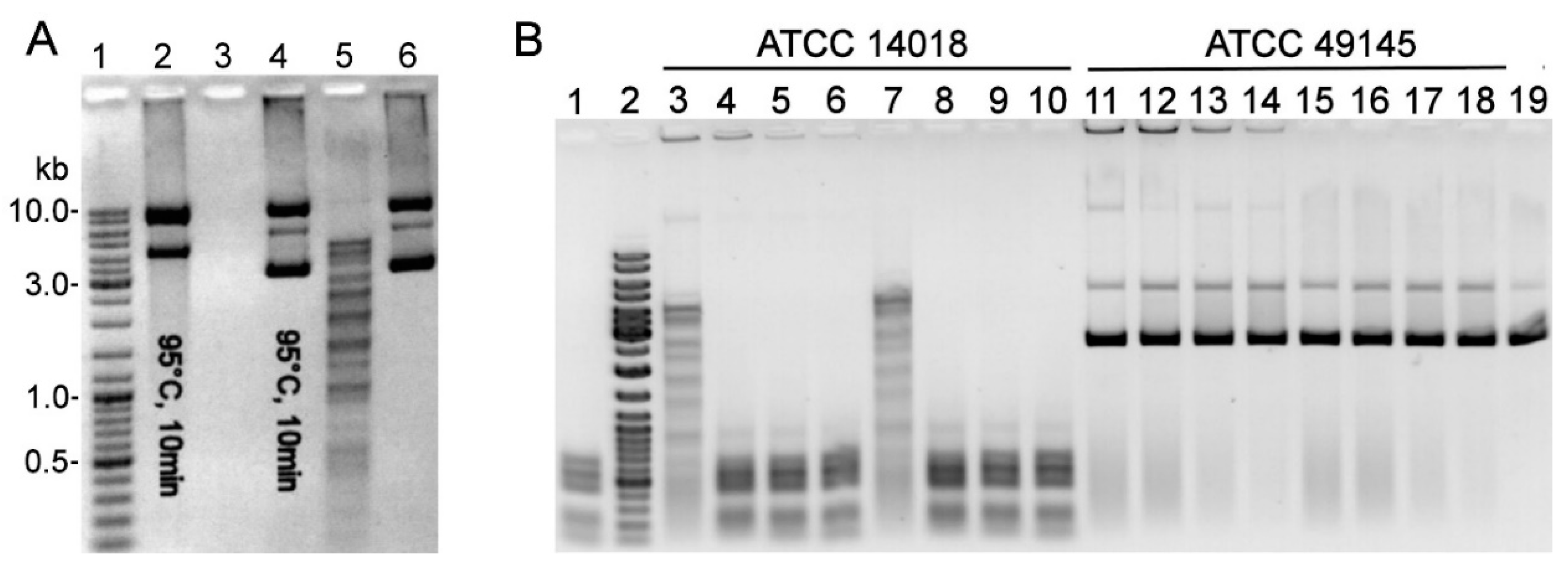
Figure 1. Detection of enzymatic activity. (A) Enzymatic activity in the cell-free sTSB culture medium (optical density (OD600 ) reached 0.65) of G. vaginalis ATCC 14018; 2 µL of culture medium (lanes 4, 5) or 1 unit of DNaseI (lanes 2, 3) was incubated with pBR322 for 45 min at 37 °C. Lanes 2, 4: the reaction mixture was preincubated at 95 °C for 10 min; lane 6: untreated pBR322; lane 1: DNA size standard Gene Ruler™ Ladder Mix (Thermo Fisher Scientific). (B) Enzymatic activity in the cell lysates of G. vaginalis ATCC 14018 and ATCC 49145 strains. Various amounts of the PBS-diluted sonicated (lanes 3–6, 11–14) and centrifuged (lanes 7–10, 15–18) cell lysates were added to the pBR322-containing reaction mixture and incubated for 10 min at 20 °C (lanes 3, 7, 11, 15) or 2 h at 37 °C (lanes 4–6, 8–10, 12–14, 16–18). Lane 1: pBR322 treated with the cell-free G. vaginalis ATCC 14018 culture supernatant (OD600 = 0.6); lane 2: DNA size standards Gene Ruler™ Ladder Mix; lane 19: untreated pBR322.
Pathogens 09 00703 g001
Figure 2. (A) The susceptibility of gDNAs to cleavage by endonuclease in Gardnerella spp. cell-free culture supernatants. gDNAs from G. vaginalis ATCC 14018 and ATCC 49145, Gardnerella spp. 114.2, and Bifidobacterium bifidum were incubated with culture supernatants from strains ATCC 14018 (lanes 3, 8, 13, 17) and 49145 (lanes 4, 9) as described in Methods. Lane 1: Gene Ruler™ Ladder Mix; lanes 2, 7, 12, 16: respective gDNA treated with fresh sBHI medium (negative control); lanes 5, 10, 14, 18: untreated respective gDNA; lanes 6, 11, 15, 19: respective gDNAs treated with the S. pneumoniae culture supernatant. (B) The chromosome position of the DNA locus coding for putative R-M system proteins in G. vaginalis ATCC 14018 (http://tools.neb.com/genomes/view.php?enzname=Gva14018ORF1109P). GAVG_1107 conserved hypothetical protein (1,469,182–1,469,529 nt); GAVG_1108 conserved hypothetical protein (Gva14018ORF1109P, 1,469,855–1,470,751 nt); GAVG_1109 DNA methyltransferase (M.Gva14018ORF1109P, 1,470,803–1,471,804 nt); and GAVG_1110 putative transcriptional regulator (1,471,801–1,472,019 nt).
Pathogens 09 00703 g002
Figure 3. Susceptibility of E. coli gDNA to cleavage by REase in G. vaginalis ATCC 14018 culture supernatant. (A) gDNAs extracted from E. coli DH10B (lanes 2–4) and three individual DH10B-Me colonies (lanes 5–13) were incubated with fresh sBHI medium (negative control) (lanes 2, 5, 8, 11), G. vaginalis ATCC 14018 culture (OD600 = 0.6) supernatant (lanes 3, 6, 9, 12), and BsuRI (lanes 4, 7, 10, 13). (B) gDNAs extracted from E. coli DH10B (lanes 2–3), three individual DH10B-RSFMe colonies (lanes 4–9), LMG194 (lanes 10–11), and three individual LMG194-RSFMe colonies (lanes 12–17) were incubated with fresh sBHI medium (negative control) (lanes 2, 4, 6, 8, 10, 12, 14, 16) and G. vaginalis ATCC 14018 culture (OD600 = 0.6) supernatant (lanes 3, 5, 7, 9, 11, 13, 15, 17). Lanes 18, 19: untreated pBR322 and pBR322 incubated with ATCC 14018 culture supernatant, respectively. (A, B) Lane 1: Gene Ruler™ Ladder Mix.
Pathogens 09 00703 g003
Figure 4. SDS-PAGE analysis of R.Gva14018I mutants produced in E. coli ArcticExpress (DE3). R.Gva14018I-4–expressing cell lysates (lanes 4, 7), clarified lysates (lanes 5, 8), and captured His-tagged R.Gva14018I-4 from an IMAC column (Zymo Research) (lanes 6, 9). R.Gva14018I-8–expressing cell lysates (lane 10), clarified lysates (lane 11), and captured His-tagged R.Gva14018I-8 (lane 12). As controls, lysates (lane 2) and clarified lysates (lane 3) of E. coli cells transformed with pET28(+) vector were loaded on the gel. Lane 1: prestained Protein Molecular Weight Marker (Thermo Fisher Scientific). Arrows indicate the migration positions of R.Gva14018I mutants.
Pathogens 09 00703 g004
Figure 5. REase activity of R.Gva14018I mutants. (A) pBR322 incubated with E. coli ArcticExpress (DE3) expressing R.Gva14018I-4: cell lysates (lanes 3, 4), clarified lysates (lanes 8, 9), and crude protein R.Gva14018I-4 preparations (lane 15). pBR322 incubated with R.Gva14018I-8-expressing cell lysates (lanes 5, 6), clarified lysates (lanes 10, 11), and crude protein R.Gva14018I-8 preparations (lane 16). Lanes 2, 7: pBR322 treated with cell lysate and clarified lysate of E. coli cells transformed with pET28(+) vector, respectively. Lanes 12, 13: pBR322 treated with undiluted (OD600 = 0.6) and a 10-fold diluted G. vaginalis ATCC 14018 cell-free supernatant, respectively. Lane 14: untreated pBR322. (B) REase activity of the purified recombinant R.Gva14018I-4 and wild-type REase in cell-free supernatant of ATCC 14018. The substrates pUC57, pBR322, gDNAs from calf thymus, human cell line HCT116, E. coli LMG194, and E. coli LMG194-Me were incubated with the supernatant (lanes 3, 6, 9, 12, 15, 18) or R.Gva14018I-4 (lanes 4, 7, 10, 13, 16, 19). Lanes 2, 5, 8, 11, 14, 17: untreated respective DNA substrate. (A, B) Lane 1: Gene Ruler™ Ladder Mix.
Pathogens 09 00703 g005
Figure 6. Identification of wild-type and recombinant REases via immunoblot (A) and REase activity towards the DNA substrate (B). (A) Immunoblot with purified R.Gva14018I-4 pAbs. Lane 3: cell lysate of ATCC 14018 (approx. 0.3 mg of the total protein); lane 5: cell lysate of ATCC 49145 (approx. 0.4 mg of the total protein); lanes 7–9: 0.2, 0.5, and 1 µg of recombinant R.Gva14018I-4. Lanes 2, 4, 6: empty lanes; lane 1: PageRuler Prestained Protein Ladder (Thermo Fisher Scientific #26616). (B) Cell lysates subjected to immunoblot analysis (A) were tested for REase activity via a DNA agarose gel electrophoresis. pBR322 was incubated with 10-fold serially diluted cell lysates of strain ATCC 14018 (lanes 2–4) and ATCC 49145 (lanes 5–7), purified R.Gva14018I-4 (lane 8), PBS (lane 9), and cell-free culture supernatant of ATCC 14018 (lane 10). Lane 1: Gene Ruler™ Ladder Mix.
Pathogens 09 00703 g006
Figure 7. Effects of cofactors, pH, and salt concentrations on R.Gva14018I-4 activity. (A) Single-site PCR substrate was digested with purified R.Gva14018I-4 in the presence of increasing amounts of Mg2+ (0, 0.1, 1, 10 mM) at pH 7.2 (lanes 2–5) and pH 5.0 (lanes 6–9). The reaction mixture containing 1 mM Mg2+ at pH 7.2 (lane 11) was supplemented with 50, 100, 150, and 200 mM NaCl or KCl to test the effects of salt concentration (lanes 12–19). Lanes 1, 20: untreated substrate. (B) Single-site PCR substrate cleavage at pH 7.2 in the presence of increasing amounts (0.1, 1, 10 mM) of Mg2+ (lanes 4–6), Mn2+ (lanes 7–9), Ni2+ (lanes 10–12), and Ca2+ (lanes 13–15). R.Gva14018I-4 was incubated with DNA in reactions supplemented with 1 mM Mg2+ and increasing amounts (0, 0.1, 1, 10 mM) of Ca2+ (lanes 16–19). Lanes 2, 20: untreated substrate; lane 3: no divalent metal ions added. Lane 10 (A) and lane 1 (B): Gene Ruler™ Ladder Mix.
Pathogens 09 00703 g007
Figure 8. Bioinformatics analysis of R.Gva1408I sequence motifs. (A) Sequence alignment between the active site of MspI [40] and the putative active-site region of R.Gva1408I. Identical residues are shaded in gray. R.Gva1408I sequence similarity with MspI (PDB ID 1SA3 chain A) was determined using HHpred [41]. The conserved amino acids D47, S65, and K67 of the predicted PD-(D/E)XK active site of R.Gva1408I are marked with asterisks [39,40]. (B) Sequence alignment of the C-terminus of wild-type R.Gva14018I and truncated R.Gva14018I-4. Identical residues are shaded in gray. PSIPRED [42] was used to predict the secondary structure elements of the molecules.
Pathogens 09 00703 g008
Figure 9. Amino acid sequence alignment for R.Gva14018I and its homologs detected in Gardnerella spp. strains. BLASTp (http://blast.ncbi.nlm.nih.gov/) was used for sequence similarity comparisons of R.Gva14018I to NCBI non-redundant protein sequences. The alignment was performed using ClustalW (https://npsa-prabi.ibcp.fr/cgi-bin/npsa_automat.pl?page=npsa_clustalw.html). Nonidentical residues are depicted on a black background. WP_009994642.1 (100% identity) also comprises hypothetical proteins of Gardnerella strains ATCC 14019, DSM 4944, GH021, and NCTC10287. WP_102720827.1 (99% identity) indicates hypothetical protein of strain UMB0768. WP_101887651.1 (94% identity) comprises hypothetical proteins of strains DNF01149, UMB0032A, and UMB0032B.
Pathogens 09 00703 g009
Table 1. Oligonucleotides used in this study.
Primer NamePrimer Sequence 5′→3′T Annealing (°C)Amplicon Size (bp)
GvMe5TACGTAATGAAGGTATTAAGCTTATTTAG561024
GvMe3TACGTACTTATCTGAATTATGATAGAACT
GVRe5GCTAGCATGGCAAAAACATTTGGTT58917
GVRe3GTCGACATCTATGCCTATATGTTCACAG
GvRe5-NheIGTCTAGCTAGCATGGCAAAAACATTTGGTT55917
GvRe3-XhoIGATCTCGAGTTATATGTTCACAGATTCAGT
GV-pUC57-ForCCGCGTTGCTGGCGTTTTTC65868
GV-pUC57-RevGTAGTTATCTACACGACGGGGAGTC