Article

Local Lung Immune Response to Mycobacterium bovis Challenge after BCG and M. bovis Heat-Inactivated Vaccination in European Badger (Meles meles)

by
Cristina Blanco Vázquez
1,*,
Miguel Prieto
1,
Marta Barral
2,
Ramón Antonio Juste
2,
Sandrine Lesellier
3,
Francisco Javier Salguero
4,
Dipesh Davé
5,
Ileana Zorhaya Martínez
6,7,
María Gracia de Garnica García
8,
Rosa Casais
1 and
Ana Balseiro
6,9
1
Centro de Biotecnología Animal, Servicio Regional de Investigación y Desarrollo Agroalimentario (SERIDA), 33394 Gijón, Asturias, Spain
2
Animal Health Department, NEIKER-Instituto Vasco de Investigación y Desarrollo Agrario, 48160 Derio, Bizkaia, Spain
3
Nancy Laboratory for Rabies and Wildlife (ANSES), 54220 Malzéville, France
4
Public Health England, PHE Porton Down, Salisbury SP4 0JG, UK
5
Bacteriology Department, Animal and Plant Health Agency (APHA), Weybridge KT15 3NB, UK
6
Departamento de Sanidad Animal, Facultad de Veterinaria, Universidad de León, 24071 León, Spain
7
Facultad de Veterinaria, Universidad Popular Autónoma del Estado de Puebla, UPAEP Universidad, 72410 Puebla, Mexico
8
Micros Veterinaria, 24071 León, Spain
9
Departamento de Sanidad Animal, Instituto de Ganadería de Montaña (CSIC-Universidad de León), Finca Marzanas, Grulleros, 24346 León, Spain
*
Author to whom correspondence should be addressed.
Pathogens 2020, 9(6), 456; https://doi.org/10.3390/pathogens9060456
Submission received: 25 May 2020 / Revised: 4 June 2020 / Accepted: 8 June 2020 / Published: 9 June 2020
(This article belongs to the Special Issue Tuberculosis Epidemiology and Control in Multi-Host Systems)

Abstract

:
Tuberculosis (TB) vaccination could be used as a key part of integrated strategies for the disease’s control if an effective and safe vaccine under field conditions is obtained. Recent studies in Spain have evaluated the protective efficacy of two oral vaccines against experimental challenge with live intra-bronchial Mycobacterium bovis in captive badgers: the live-attenuated M. bovis BCG vaccine (Danish strain) and a heat-inactivated M. bovis (HIMB) vaccine. With the objective of increasing the knowledge of the cellular development progress of infection and generating further tools to discriminate between mild and severe TB lesions between and within animals, the immunopathology of tuberculous lesions was studied to characterize the local immune response (cell type profile) within lung granulomas from control (non-vaccinated), BCG vaccinated and HIMB-vaccinated experimentally infected badgers with M. bovis. Four immunohistochemical protocols, for the specific detection of macrophages, T lymphocytes, B lymphocytes and plasma cells within TB granulomas in formalin fixed sections of the right middle lung lobe (lobe targeted for the M. bovis delivery), were performed. Immunolabelled sections were scanned and five randomly selected areas were analyzed with digital image analysis software. The results were expressed as the proportion of the positively immunolabelled area within the total area of the selected site. Data was analyzed using the statistical analysis software (SAS). In the three treatment groups, macrophages were the most abundant inflammatory cells within the granulomas, followed by B lymphocytes and plasma cells. T lymphocyes were absent in those granulomas. This would suggest a predominance of a non-specific innate response mediated by phagocytic cells over an adaptative humoral immune response. The proportion of macrophages and plasma cells was higher in BCG and HIMB-vaccinated badgers, respectively, suggesting the establishment of an adaptative humoral response in HIMB-vaccinated badgers. The lower bacterial load at the lung level, as well as the volume of lesions in lungs using magnetic resonance imaging in badgers with the HIMB vaccine in relation with local immune response presented, must be highlighted, since it would be an advantage in favor of its use under field conditions in terms of reducing TB transmission and environmental contamination.

1. Introduction

Animal tuberculosis (TB) remains as one of the most prevalent infectious diseases causing important economical losses worldwide. It poses a risk to human beings since it can also induce zoonotic TB. It is caused by infection with members of the Mycobacterium tuberculosis complex (MTBC), mainly M. bovis and to a lesser extent M. caprae. Mycobacterium bovis infects a wide range of domestic animals including cattle (Bos taurus) [1], goat (Capra hircus) [2] and sheep (Ovis aries) [3]. It also infects different species of wild animals, such as brushtail possums (Trichosurus vulpecula) [4], African buffalo (Syncerus caffer) [5], wild boar (Sus scrofa) [6], fallow deer (Dama dama) [7] and European badger (Meles meles) [8]. In spite of major efforts invested in the control of the disease in cattle, considered the main domestic reservoir, TB is still present in many European countries [9]. In addition, the existence of wild reservoirs makes eradication in livestock more complicated. In Europe, particularly in the United Kingdom (UK) and the Republic of Ireland (ROI), badgers are recognized as major TB reservoir hosts with the potential to transmit infection to cattle herds [10,11], while in Spain the wildlife species most commonly associated with outbreaks in domestic animals is the wild boar [12]. As part of integrated control strategies for the disease, some countries, such as the UK and ROI, have proposed the vaccination of badgers with live-attenuated M. bovis bacillus Calmette–Guérin (BCG) vaccine [13,14,15]. A live vaccine (BCG) for badgers is only licensed in the UK for intramuscular delivery, and its use in oral baits is promising, although the use of a live vaccine at a sufficient protective dose complicates the delivery in the field, especially at high environmental temperatures, impacting the survival of the vaccine [16]. The efficacy of a heat-inactivated M. bovis (HIMB) vaccine has also been assessed in different species, such as wild pigs [17], red deer [18], wild boar [19,20,21], goats [22,23], sheep [24] and zebrafish [25]. A recent experimental study in Spain showed a similar reduction in bacterial loads and lesion scores in BCG and HIMB orally vaccinated badgers [26] compared with non-vaccinated controls. The use of the HIMB vaccine in field conditions is therefore envisaged, with the benefit of being more stable in baits, and potentially less immunogenic by the oral route for cattle [27], than BCG.
In order to understand the pathogenesis of animal TB, different studies have focused on the study of the development of granulomas and the cell phenotype in each granuloma stage, both in natural and experimental infections. Cell characterization of TB granulomas using immunohistochemistry (IHC) has been performed in species such as wild boar [6], fallow deer [7], pacu [28], sheep [29], cattle [30,31] and badgers [32]. However, the cellular characterization of granulomas in vaccinated animals is poorly understood. Differences in cellular patterns within TB granulomas imply particularities in the host immune response against the pathogen. In this sense, the cell characterization of TB granulomas in control and vaccinated animals might help to determine mechanisms of protection. Previous studies have evaluated differences in cell granuloma composition in BCG vaccinated versus (vs) non-vaccinated cattle [33,34], where BCG vaccination was protective in the sense that it reduced the number and severity of lesions and the bacterial load in tissues. Overall, the number of inflammatory cells was reduced (5–10% for T lymphocytes) in vaccinated animals, and the efficiency in the expression of cytokines with a bactericidal effect was increased [34].
The aims of the present study included (i) the immunohistochemical cell characterization of tuberculous lesions in control (non-vaccinated), BCG and HIMB vaccinated European badgers experimentally infected with M. bovis, and (ii) the study of differences in the local cell immune response as a contribution to the understanding of vaccine protection mechanisms.

2. Results

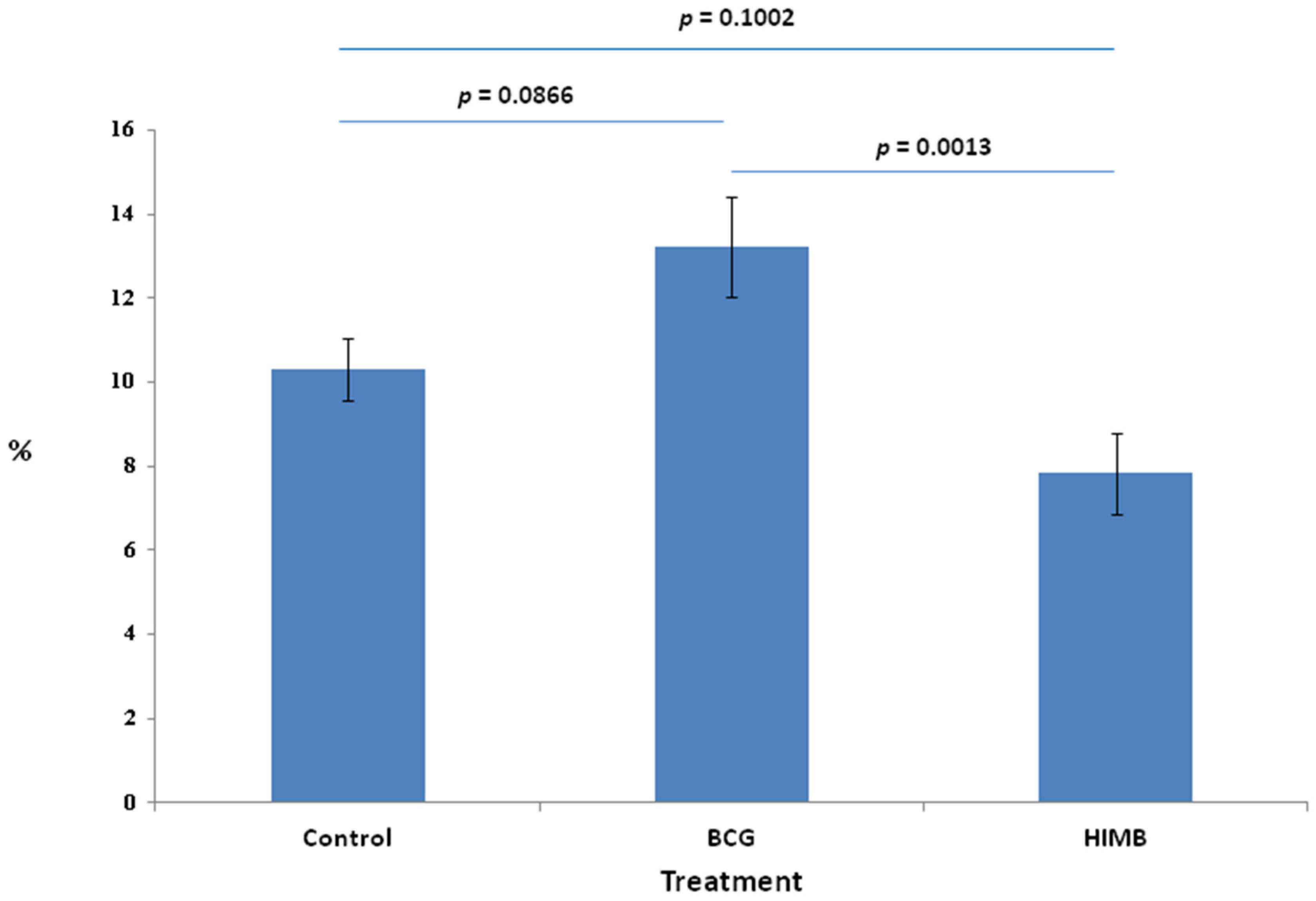
2.1. Proportion of Total Cells in Treatment Groups

Total cell proportions were significantly different between treatment groups (Figure 1). There were significant differences in the total number of cells (control, 10.30%; BCG, 13.22%; HIMB, 7.82%) between the BCG and HIMB groups (p = 0.0013), and almost significant differences between the control and BCG groups (p = 0.0866) and the HIMB (p = 0.1002) group.

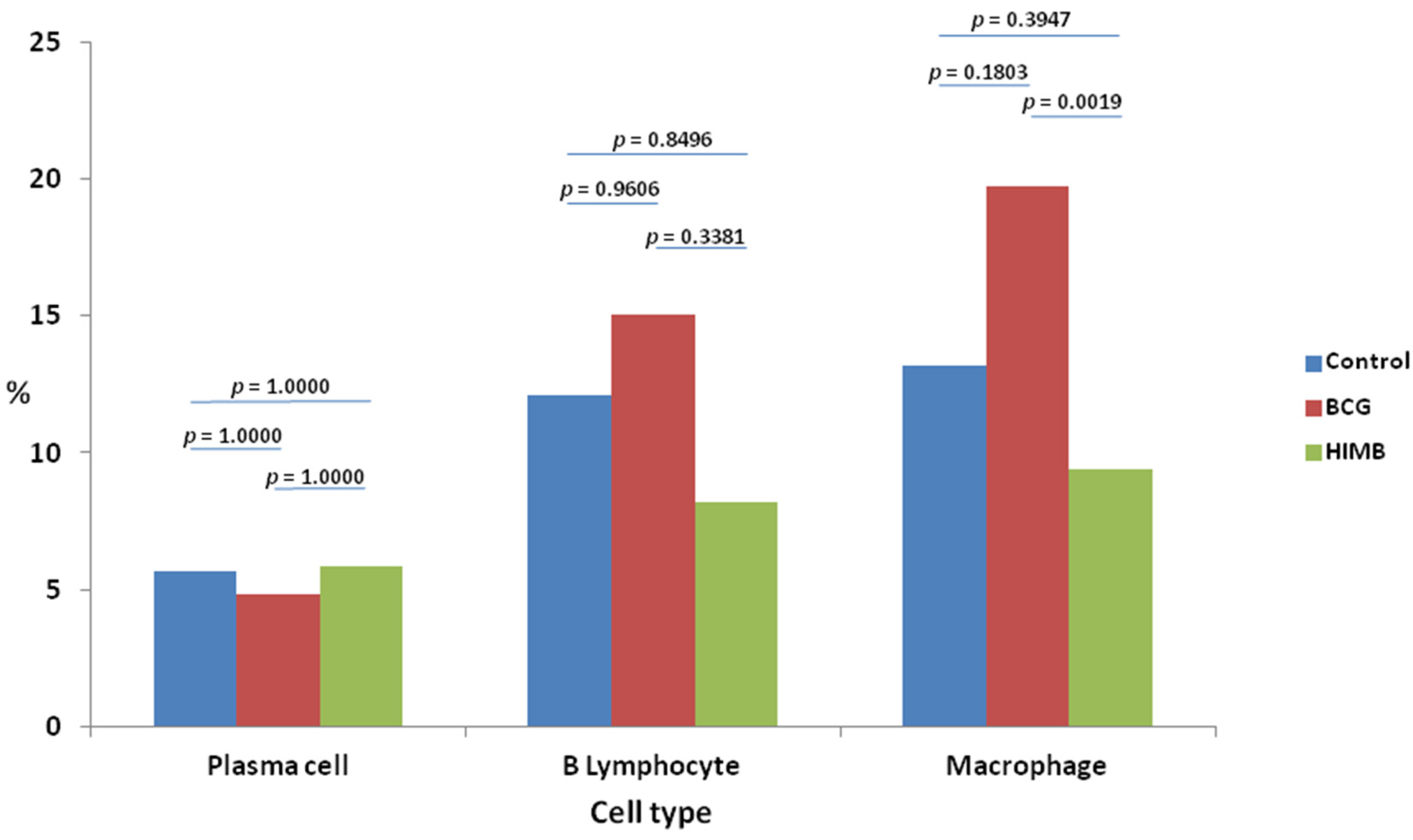
2.2. Cell Types and Differences within Treatment Groups

Macrophages were the most abundant cell type in the three treatment groups (Table 1). Significant differences in macrophages were found between BCG vaccinated and control animals (p = 0.044). In addition, there were also significant differences in macrophage proportions between BCG and HIMB vaccinated animals (p = 0.0017). The next most abundant cell type was B lymphocytes, followed by plasma cells and T lymphocytes (Figure 2), which were absent in all animals. No significant differences were found between other cell types in any of the groups.

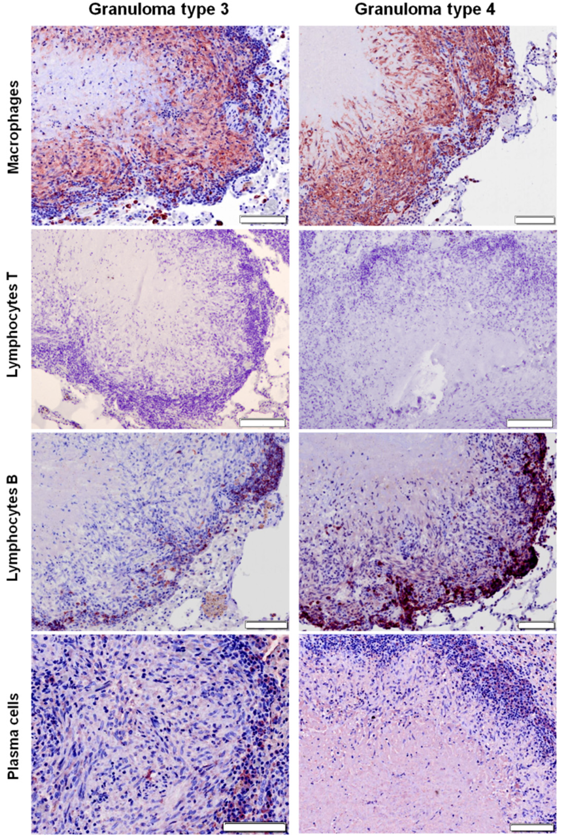
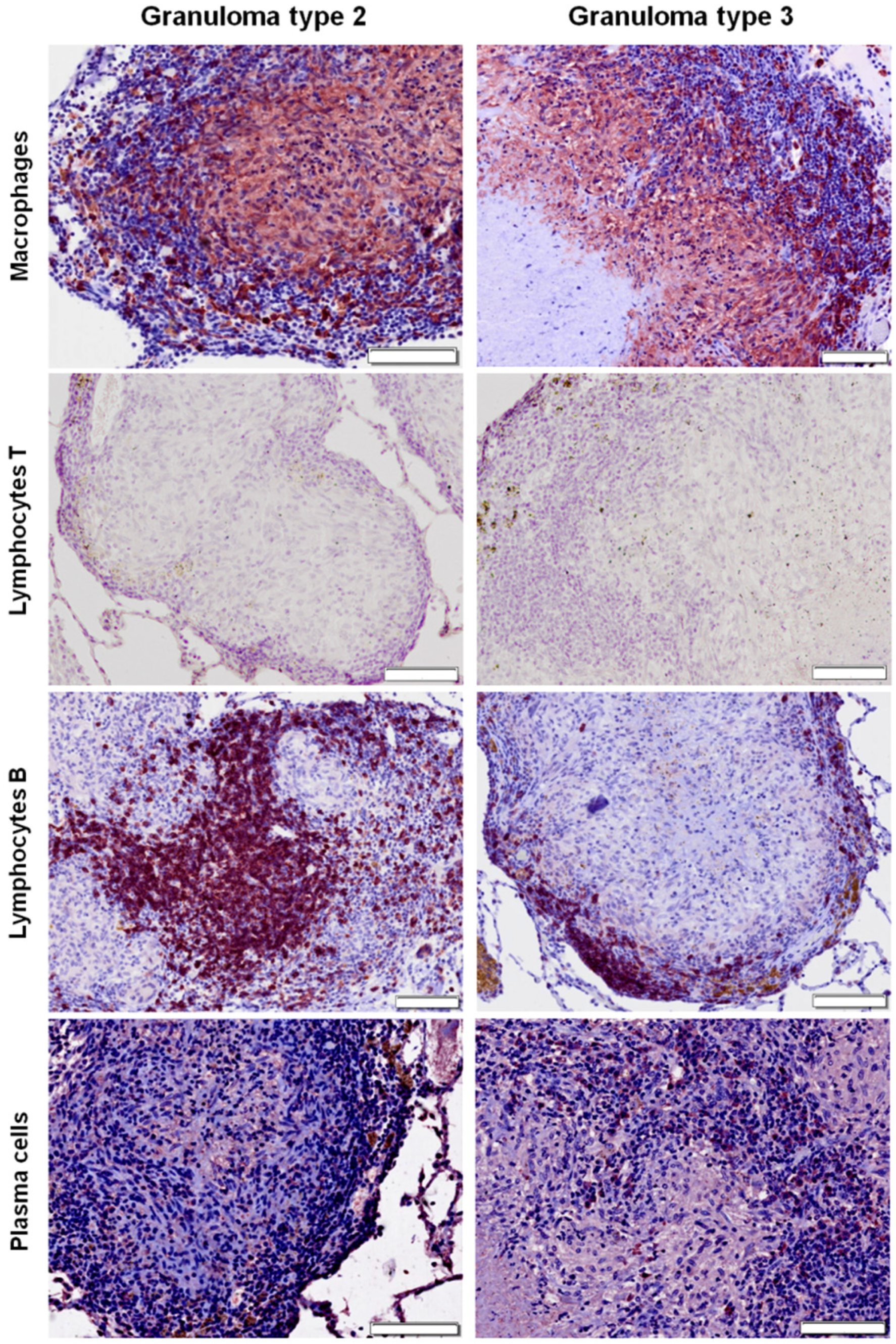
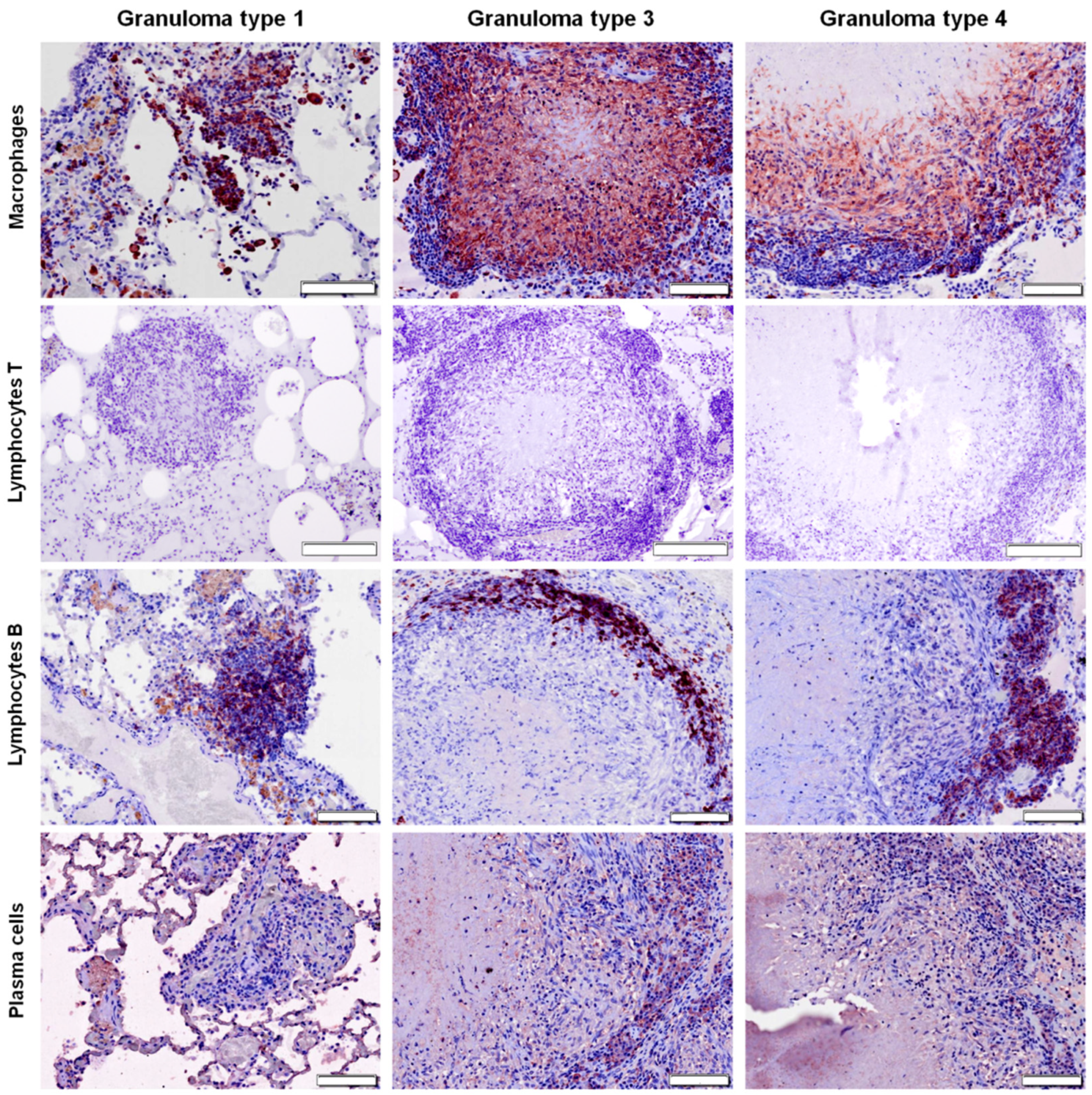
2.3. Distribution of Cells according to Granuloma Type (1 to 4)

In type 1 and type 2 granulomas (see Materials and Methods sections for details), macrophages were diffusely distributed throughout the lesion (Figure 3 and Figure 4), while they were present in the outer layers and surrounding the necrotic foci in more advanced stages (granuloma types 3 and 4) (Figure 3, Figure 4 and Figure 5). B lymphocytes were scattered within the lesion in early stages (granuloma types 1 and 2) (Figure 4 and Figure 5), and forming clusters and surrounding the necrotic area in advanced stages (granuloma types 3 and 4) (Figure 3, Figure 4 and Figure 5). IHC analysis showed very few scattered positively stained plasma cells within the granuloma types 1 and 2 (Figure 4 and Figure 5), but their number increased as the granuloma developed (types 3 and 4). They were found forming clusters or sparsely distributed between B cells and macrophages surrounding the necrotic area (Figure 3, Figure 4 and Figure 5).

2.4. Statistical Analysis in Relation with Local Response in Lung

The results of the isolation of M. bovis in mediastine showed significant differences between the three treatment groups (Figure 6). The bacterial load was not different between vaccinated groups (p = 0.9173), but was significantly reduced (98.36%; p = 0.0304) in the HIMB, and nearly so in the BCG (96.63%; p = 0.1104) groups compared to the control group.
Regarding MRI results, HIMB (42.9%) vaccinated animals tended to have smaller TB lesion areas than BCG (60%) and control (83.3%) groups. This corresponded with a reduction in the lesion volume geometric mean in the vaccinated groups compared to the control group; 78.1% for BCG (p = 0.454) and 97.7% for HIMB (p = 0.0548).

3. Discussion

In this study we have examined, for the first time, the local immune response in control (non-vaccinated), BCG and HIMB-vaccinated badgers experimentally infected with M. bovis, through cell characterization and quantification within TB lung granulomas. Regarding the number of total cells, different studies have shown that vaccination of animals is associated with a reduction in the local number and severity of lesions, as well as in the amount of inflammatory cells [33,34]. However, other studies showed that protection was related to an increase in the number of cells, i.e., T lymphocytes and macrophages [35,36,37]. In our study, both vaccinated groups with variable numbers of total cells were protected based on visible lesions.
The cell distribution pattern was similar in all treatment groups. Granulomas showed a high number of macrophages, followed by B lymphocytes and plasma cells. The composition and distribution of cells observed in this study were similar to those already described in badgers [32], except for T lymphocytes, which were not detected in any animal from our study. The absence of T lymphocytes would indicate the predominance of the humoral immune response at the time of slaughter, which could be interpreted as a rapid progression of the infection towards a more specific immune response. Since this occurred in all groups, it can be postulated that the M. bovis strain used in the challenge may have also influenced the granuloma cell composition.
T lymphocytes and their secreted cytokines activate macrophages during the cell-mediated immune response [38,39]. In the present study, no T lymphocytes were detected, therefore one possible hypothesis for this result is that the macrophages in granulomas would be those carrying out the non-specific innate response, which would be represented by phagocytic cells rather than by specific adaptative cells [29,37]. However, the lack of histological markers of adaptative immunity, including T cells, is surprising, 12 weeks post-challenge, when peripheral T cell activity was observed [26]. The higher proportion of macrophages in the BCG group may reflect (i) the greater immunogenic properties of the BCG (live vaccine) than HIMB vaccine (inactivated vaccine), or (ii) the stronger stimulation of the innate immune response of the host. It also may, on the contrary, indicate (iii) a failure of the macrophages to efficiently destroy the mycobacteria thriving within the lesion and thus inducing a further influx of inflammatory cells. The latter hypothesis might be supported by the observation that BCG induces TH1/TH17 responses against TB and other unrelated pathogens [40,41,42]. In addition, live vaccines are more immunogenic than inactivated ones [43]. It should be noted the differences between our results and those found in previous studies, in which the number of macrophages decreased in BCG vaccinated animals compared to control ones, which would be consistent with a cellular immune response against M. bovis in early stages mediated by T lymphocytes and macrophages [34]. This difference could be due in part to the type of tissue where cellular characterization was performed in each study: lymphoid tissue vs lung tissue. According to this view, the HIMB vaccine could have induced a more effective response in the lungs, which would not have stuck the macrophagic cells in the granuloma, and led to infection clearance. This could be supported by the finding that, in this study, the control badgers presented a high proportion of macrophages within granulomas in advanced stages (types 3 and 4), as seen in previous studies in badgers [32], sheep and pacu fish [28,29], but not in fallow deer, wild boar or cattle at advanced stages of the disease [6,7,30]. A detailed comparison of the role of macrophages across host species may highlight the importance of this cell type in TB inflammatory responses and the overall severity of the disease.
T lymphocytes, however, were absent in all treatment groups. T lymphocytes have constantly been described as a very abundant cell type in the TB granulomas in badgers [32,44], and also in other species, such as cattle [31,34] or fallow deer (7). In general, in badger studies, the interferon-gamma (IFN-γ) production and peripheral blood mononuclear cells (PBMC) proliferation, measured by lymphocyte transformation assays (LTA), decrease between 12 and 21 weeks post-infection [45,46]. As described in our previous study, IFN-γ producing cells responded to combined PPD-B (bovine purified protein derivate) and PPD-A (avian purified protein derivate) antigenic stimulation by whole blood IFN-γ release assay (IGRA) enzyme-linked immunosorbent assay (ELISA) and IGRA enzyme-linked immunospot assay (ELISPOT), 2 weeks post-infection, peaking at 7 weeks post-infection and then decreasing. Necropsies were performed at week 12 post-infection, which might explain the absence of T lymphocytes. In this case, our results are similar to those found in a previous study [34], since a decrease of T lymphocytes was observed after BCG vaccination, consistent with a cellular immune response against M. bovis in the early stages mediated by T lymphocytes and macrophages. If we compare our control animals with the cell types found in other animals in advanced granulomas (types 3 or 4), our results are similar to cattle and sheep, where the number of T lymphocytes decreased as the lesion progressed [29,34]. Nevertheless, we have observed great differences with fallow deer, wild boar, and with previous studies of badgers, where the amount of T lymphocytes was very high in advanced stages [6,7,32].
The number of B lymphocytes observed was higher in the BCG group and lower in the HIMB-vaccinated animals, compared to the control group. B cells are involved in the host’s early interactions with intracellular bacterial pathogens, and also participate in the induction of innate defense responses [47]. We found more B lymphocytes in BCG vaccinated badgers than in control animals, similar to what was observed in previous studies in cattle [34]. The increased expression of B lymphocytes within the granulomas, coupled with the high IFN-γ production throughout the course of the experiment, implies that a Th0 response developed in the BCG animals following infection [34]. Higher numbers of B lymphocytes in advanced stages in control animals have also been observed in cattle, sheep and fallow deer [7,29,34]; however, they were fewer in previous studies of badger and wild boar, where the number of B lymphocytes decreases as the granuloma progresses [6,32].
Interestingly, the number of plasma cells was higher in the HIMB group than in the BCG and control groups. Overall, HIMB vaccinated animals presented fewer macrophages and B lymphocytes than BCG vaccinated animals, but a greater number of plasma cells, which suggests that the HIMB vaccine induces the establishment of an adaptative humoral-type response. This fact is in agreement with a previous study in wild boar, which concluded that oral vaccination with HIMB induced both an activation of the innate immune response and an adaptative antibody response [48]. As the infection progresses and the mycobacteria persist in the lesions, the antibody responses also persist or increase [49]; thus the number of plasma cells is expected to be higher in more advanced stages than in early ones. The humoral immune response in badgers is related to the severity of lesions being more evident in advanced stages of the disease [50]. Usually, in experimentally infected badgers, the serological responses develop gradually from 4 to 6 weeks post-infection [26,51], and persist at the time of slaughter 12 weeks post-infection. This fact agrees with previous studies on IHC characterization in TB granulomas in badgers, which suggest that humoral immunity is not a major factor in the early establishment of lesions [32]. Also, the amount of plasma cells found in control badgers is similar to those observed in other species, such as fallow deer and sheep, increasing their numbers as the granuloma progresses [7,29].
It is important to highlight the significant reduction of the bacterial load in the mediastine of the vaccinated animals, with respect to the control group, mainly in the HIMB group. This could be related to the cell ratio findings discussed above, and in any case supports the idea that the HIMB vaccine could have induced a trained immune response [52] that could have better cleared the initial infection. More striking, though, is the fact that for global isolation including all tissues, the bacterial load was higher for the HIMB group than for the BCG group [26]. This shows that M. bovis induces a complex immune response highly dependent on the individual host response in each organic region. The HIMB vaccine appears to induce a greater response at the lung level compared to the lymphatic system, which could be crucial for the reduction of TB lesions, as demonstrated both by magnetic resonance image and bacterial load in this particular tissue [26], and thus for a reduction of the excretion of mycobacteria via secretions and aerosol. On the other hand, this fact also justifies in vivo experimentation, and highlights the importance of these experimental infections for understanding this complex system, where different criteria may be needed for the local or general evaluation. These differences, between lung tissue and lymphoid tissue bacterial load, as related to vaccine protection, are probably crucial and require further research.
In conclusion, this study has characterized the immunopathology of the TB granulomas in control, BCG and HIMB vaccinated European badgers, experimentally infected with M. bovis in the lung, highlighting the higher proportion of macrophages than B lymphocytes and plasma cells, and the absence of T lymphocytes, which would suggest the predominance of a non-specific innate response mediated by phagocytic cells over an adaptative humoral immune response. In this sense, the HIMB vaccine induced a greater presence of plasma cells than the BCG vaccine did, suggesting the establishment of an adaptative humoral response; although more studies would be necessary to confirm this hypothesis. However, the decrease in the bacterial load and volume of lesions at the lung level with the HIMB vaccine must be highlighted, since it disagrees with results regarding lymphoid tissue bacterial load and lesions [26], which would be an advantage in favor of its use in field conditions for reducing TB transmission and environmental contamination.

4. Materials and Methods

4.1. Ethical Statement

All methods were employed in accordance with the relevant guidelines and regulations. All experimental protocols were approved by the ethical committees from Servicio Regional de Investigación y Desarrollo Agroalimentario del Principado de Asturias (SERIDA) and from the Government of the Principality of Asturias. The license reference numbers were 010/07-01-2011, PROAE 20/2015 and PROAE 47/2018.

4.2. Animals and Tissue Samples

Samples collected in a previous experimental study were used [26]. Briefly, 24 badgers (13 males and 11 females) were trapped and randomly allocated to three treatment groups: unvaccinated controls (n = 12), oral vaccinated with live Danish BCG [108 colony-forming units (CFU), n = 5], and oral vaccinated with HIMB (107 CFU, n = 7). After a period of 13 weeks post-vaccination, all badgers were experimentally challenged by the endobronchial route in the right middle lung lobe with 1 mL of a M. bovis field strain (SB0339) [103 CFU/mL in phosphate buffer saline (PBS)] isolated from a tuberculous wild boar. After a period of 12 weeks post-challenge, badgers where euthanized and a complete collection of tissue (lymph nodes and organs) samples [26] was performed at post mortem examination (PM). In that study, 4 animals (1 control, 1 BCG vaccinated and 2 HIMB vaccinated) were positive by the P22 ELISA on the day of challenge, which would indicate a previous sensitization to M. bovis or other mycobacteria not detected using cellular immune techniques [26]. Those animals have not been included in the present study.

4.3. Histopathological, Bacteriological and Magnetic Resonance Imaging (MRI) Data

As already reported in a previous publication, samples were processed by standard protocols for histopathology [hematoxylin and eosin (HE) and Ziehl–Neelsen (ZN) staining, specifically for acid-fast bacilli (AFB)], bacteriology and MRI [26]. Only local results from lungs and mediastine have been used for the specific current statistical analyses.
The histological score was calculated for each tissue and the granulomas were classified based on cell composition, severity and number of mycobacteria from types 1 to 4. Briefly, type 1 granuloma consisted of clusters of lymphocytes, epithelioid cells and plasma cells. Granulomas type 2 and 3 presented the same cellular types as granuloma 1, but they also showed coagulative and caseous central necrosis, respectively. Finally, granuloma type 4, the most severe, showed caseous and mineralized central necrosis [26]. The histological score was based on the most severe lesion observed on the section (see [26] for details). The formalin fixed lungs were scanned by MRI in order to determine the lesion volume in lung (mm3) in each animal [26]. In Table 2, results obtained in the previous study regarding histological scores, bacterial load and MRI (mm3) of each badger are shown.

4.4. Immunohistochemistry (IHC)

The numbers of macrophages, T lymphocytes, B lymphocytes and plasma cells within the TB granulomas located in the right middle lung lobe were calculated. We selected the right middle lung lobe because that tissue was consistently affected (as it was the challenge tissue target) and had a similar size in all studied badgers. The specific details of the IHC protocols are summarized in Table 3. Formalin fixed right middle lung lobe samples were cut into 3-µm sections, dewaxed, rehydrated and rinsed with tap water at room temperature. Afterwards, epitope demasking was performed microwaving in citric acid buffer [1.48 g citric acid (Merck KGaA, Darmstadt, Germany) in 500 mL distilled water] (Table 3). The sections were then washed with tap water or tris buffered saline 1× [TBS 1×, 5 mM Tris (Merck KGaA, Darmstadt, Germany)/HCl (Panreac Química, SLU, Barcelona, Spain) pH 7.6, 136 mM NaCl (Merck KGaA, Darstadt, Germany)] in the case of Lambda light chain primary antibody. Then, slides were placed in different solutions to block endogenous peroxidase activity (Table 3) and washed with tap water or TBS 1× in the case of Lambda primary antibody. Afterwards, the samples were treated to prevent primary antibody cross-reactivity with tissue constituents over a 20-min incubation period at room temperature, with 10% normal horse serum for CD3 protocol (Vector Laboratories, Burlingame, CA, USA) or with 10% normal goat serum for CD20, IBA-1 and Lambda light chain protocols (Vector Laboratories), and 3% bovine serum albumin (BSA, Sigma-Aldrich, Saint Louis, MO, USA). Then, sections were incubated overnight at 4 °C with commercial monoclonal and polyclonal antibodies (Table 3), and then washed three times with TBS 1×. Afterwards, samples were incubated with the appropriate biotinylated secondary antibody (anti-mouse biotinylated in horse or anti-rabbit biotinylated in goat; Vector Laboratories) before being washed three times with TBS 1×. The sections were incubated for 30 min (and 40 min in the case of Lambda primary antibody) at room temperature with Avidin Biotin Complex (ABC kit Peroxidase Standard, Vector Laboratories). Finally, the sections were washed three times with TBS 1× and the signal was detected using Vector® NovaRED™ peroxidase substrate kit SK-4800 (Vector Laboratories) for 2–5 min. Samples were rinsed with tap water or TBS 1× for 5 min, before being placed in Mayer’s hematoxylin (MerckKGaA, Darmstadt, Germany) counterstain for 30 seconds. Finally, the slices were washed with tap water or TBS 1× and mounted with DPX (MerckKGaA, Darmstadt, Germany). Appropriate controls were included in each IHC run. Those included positive controls (lymph nodes from badger where the target antigen was present in the section and the specific antibody was used) (Supplementary Figure S1) and negative controls (slides with omission of the primary antibody).

4.5. Image Analysis

Immunolabelled sections were scanned at 40× using an Olympus BX51 microscope (Olympus, Tokyo, Japan) with an Olympus XC10 camera (Olympus, Tokyo, Japan). Scanned images were analyzed with digital image analysis software (Nikon NIS-Elements Br, Nikon, Japan). For each animal, five randomly selected areas were sourced as regions of interest (RI) and were analyzed using 200× magnification. The area with an immunohistochemical-positive reaction within RI was calculated by the Nikon NIS-Elements software after setting the thresholds. The results were expressed as the proportion of the positively immunolabelled area within the total area of the selected site.

4.6. Statistical Analyses

Data was analyzed using the SAS statistical package (SAS Inc., Cary, NC, USA). To normalize the data for statistical analysis, an arc sin square root transformation was applied to all the proportions. Results were submitted to analysis of variance with the geometrical linear mean (GLM) procedure to determine the statistical significance of differences in the cell type area proportions among treatment groups, and also to determine if there were differences among cell type proportions within each treatment group. Therefore, the resulting model had as independent variables (main effects) the vaccine treatment, the cell type and its interaction, and as dependent variable the stained area’s proportion. A correlation analysis was also run between cell type proportions in order to identify associations between them. The multiple comparison adjusted Tukey–Kramer test was applied for effect level comparison. Statistical significance was accepted at p > 0.05.

Supplementary Materials

The following are available online at https://www.mdpi.com/2076-0817/9/6/456/s1, Figure S1: Immunohistochemical technique in positive controls; lymph nodes from badger.

Author Contributions

A.B. and M.P. conceived and designed the experiments in the research study. C.B.V., M.P., M.B., R.A.J., F.J.S., S.L., D.D., I.Z.M., M.G.d.G.G., R.C. and A.B. performed and validated the results and analyzed the data. C.B.V., A.B. and R.A.J. wrote the initial version of the manuscript. Further reviews of the manuscript were conducted by all the authors. All authors have read and agreed to the published version of the manuscript.

Funding

This work has been funded by Ministerio de Ciencia, Innovación y Universidades (MCIU) and the Agencia Estatal de Investigación (AEI) reference project RTI2018-096010-B-C21 (FEDER co-funded) and, by PCTI 2018–2020 (GRUPIN: IDI2018-000237) and FEDER. Ms. Cristina Blanco Vázquez was granted with a predoctoral fellowship funded by INIA-CCAA (FPI-INIA) (2018 call). Ms. Ileana Z. Martínez was supported by a Fundación Carolina PhD scholarship (2017 call). We have received funds by RTI2018-096010-B-C21 (FEDER co-funded) to cover publication costs.

Acknowledgments

This work was supported by the Spanish Ministry of Agriculture, Fisheries and Food. The authors would like to thank the Vice-Ministry of the Environment of the Principado of Asturias for the permits to capture the badgers. Special thanks to the gamekeepers of the Hunting and Fishing Service assigned to the Reserve of Sueve and Parres for their invaluable help in locating the trapping areas and monitoring the cage traps. Authors also thank Red de Investigación en Sanidad Animal (RISA, Spain).

Conflicts of Interest

The authors declare no conflict of interest.

References

  1. Vordermeier, H.M.; Jones, G.J.; Buddle, B.M.; Hewinson, R.G.; Villarreal-Ramos, B. Bovine tuberculosis in Cattle: Vaccines, DIVA Tests and Host Biomarker Discovery. Annu. Rev. Anim. Biosci. 2016, 4, 87–109. [Google Scholar] [CrossRef] [PubMed]
  2. De Val Pérez, B.; López-Soria, S.; Nofrarías, M.; Martín, M.; Vordermeier, H.M.; Villareal-Ramos, B.; Romera, N.; Escobar, M.; Solanes, D.; Cardona, P.J.; et al. Experimental model of tuberculosis in the domestic goat after endobronchial infection with Mycobacterium caprae. Clin. Vaccine Immunol. 2011, 18, 1872–1881. [Google Scholar] [CrossRef] [PubMed] [Green Version]
  3. Muñoz-Mendoza, M.; Romero, B.; del Cerro, A.; Gortázar, C.; García-Marín, J.F.; Menéndez, S.; Moruelo, J.; de Juan, L.; Sáez, J.L.; Delahay, R.J.; et al. Sheep as a potential source of bovine TB: Epidemiology, pathology and evaluation of diagnostic techniques. Transbound. Emerg. Dis. 2015, 63, 635–646. [Google Scholar] [CrossRef]
  4. Jackson, R.; Cooke, M.M.; Coleman, J.D.; Morris, R.S. Naturally occurring tuberculosis caused by Mycobacterium bovis in brushtail possums (Trichosurus vulpecula): I. An epidemiological analysis of lesion distribution. N. Z. Vet. J. 1995, 43, 306–314. [Google Scholar] [CrossRef] [PubMed]
  5. Renwick, A.R.; White, P.C.; Bengis, R.G. Bovine tuberculosis in southern African wildlife: A multi-species host-pathogen system. Epidemiol. Infect. 2007, 135, 529–540. [Google Scholar] [CrossRef] [PubMed]
  6. García-Jiménez, W.L.; Salguero, F.J.; Fernández-Llario, P.; Martínez, R.; Risco, D.; Gough, J.; Ortíz-Peláez, A.; Hermoso-de-Mendoza, J.; Gómez, L. Immunopathology of granulomas produced by Mycobacterium bovis in naturally infected wild boar. Vet. Immunol. Immunopathol. 2013, 156, 54–63. [Google Scholar] [CrossRef]
  7. García-Jiménez, W.L.; Fernández-Llario, P.; Gómez, L.; Benítez-Medina, J.M.; García-Sánchez, A.; Martínez, R.; Risco, D.; Gough, J.; Ortíz-Peláez, A.; Smith, N.H.; et al. Histological and immunohistochemical characterisation of Mycobacterium bovis induced granulomas in naturally infected fallow deer (Dama dama). Vet. Immunol. Immunopathol. 2012, 149, 66–75. [Google Scholar] [CrossRef]
  8. Corner, L.A.; Murphy, D.; Gormley, E. Mycobacterium bovis infection in the Eurasian badger (Meles meles): The disease, pathogenesis, epidemiology and control. J. Comp. Pathol. 2011, 144, 1–24. [Google Scholar] [CrossRef]
  9. MAPA. 2018. Available online: http://www.mapa.gob.es/gl/ganaderia/temas/sanidad-animal-higiene-ganadera/patubes2017_3_tcm37-378321.pdf (accessed on 26 March 2020).
  10. Corner, L.A.; Clegg, T.A.; More, S.J.; Williams, D.H.; O’Boyle, I.; Costello, E.; Sleeman, D.P.; Griffin, J.M. The effect of varying levels of population control on the prevalence of tuberculosis in badgers in Ireland. Res. Vet. Sci. 2008, 85, 238–249. [Google Scholar] [CrossRef]
  11. Jenkins, H.E.; Woodroffe, R.; Donnelly, C.A. The duration of the effects of repeated widespread badger culling on cattle tuberculosis following the cessation of culling. PLoS ONE 2010, 5, e9090. [Google Scholar] [CrossRef] [Green Version]
  12. Gortázar, C.; Delahay, R.J.; McDonald, R.A.; Boadella, M.; Wilson, G.J.; Gavier-Widen, D.; Acevedo, P. The status of tuberculosis in european wild mammals. Mammal Rev. 2012, 42, 193–206. [Google Scholar] [CrossRef]
  13. Gormley, E.; Collins, J.D. The development of wildlife control strategies for eradication of tuberculosis in cattle in Ireland. Tuber. Lung Dis. 2000, 80, 229–236. [Google Scholar] [CrossRef]
  14. Gormley, E.; Costello, E. Tuberculosis and badgers: New approaches to diagnosis and control. J. Appl. Microbiol. 2003, 94, 80S–86S. [Google Scholar] [CrossRef]
  15. Chambers, M.A.; Carter, S.P.; Wilson, G.J.; Jones, G.; Brown, E.; Hewinson, R.G.; Vordermeier, M. Vaccination against tuberculosis in badgers and cattle: An overview of the challenges, developments and current research priorities in Great Britain. Vet. Rec. 2014, 175, 90–96. [Google Scholar] [CrossRef] [Green Version]
  16. Buddle, B.M.; Vordermeier, H.M.; Chambers, M.A.; de Klerk-Lorist, L.M. Efficacy and safety of BCG vaccine for control of tuberculosis in domestic livestock and wildlife. Front. Vet. Sci. 2018, 5, 259. [Google Scholar] [CrossRef] [Green Version]
  17. Nol, P.; Wehtje, M.E.; Bowen, R.A.; Robbe-Austerman, S.; Thacker, T.C.; Lantz, K.; Rhyan, J.C.; Baeten, L.A.; Juste, R.A.; Sevilla, I.A.; et al. Effects of Inactivated Mycobacterium bovis Vaccination on Molokai-Origin Wild Pigs Experimentally Infected with Virulent M. bovis. Pathogens 2020, 9, 199. [Google Scholar] [CrossRef] [Green Version]
  18. Thomas, J.; Risalde, M.A.; Serrano, M.; Sevilla, I.; Geijo, M.; Ortíz, J.A.; Fuentes, M.; Ruíz-Fons, J.F.; de la Fuente, J.; Domínguez, L.; et al. The response of red deer to oral administration oh heta-inactivated Mycobacterium bovis and challenge with a fiel strain. Vet. Microbiol. 2017, 208, 195–202. [Google Scholar] [CrossRef]
  19. Díez-Delgado, I.; Sevilla, I.A.; Romero, B.; Tanner, E.; Barasona, J.A.; White, A.R.; Lurz, P.W.W.; Boots, M.; de la Fuente, J.; Domínguez, L.; et al. Impact of piglet oral vaccination against tuberculosis in endemic free-ranging wild boar populations. Prev. Vet. Med. 2018, 155, 11–20. [Google Scholar] [CrossRef]
  20. Garrido, J.M.; Sevilla, I.A.; Beltrán-Beck, B.; Minguijón, E.; Ballesteros, C.; Galindo, R.C.; Boadella, M.; Lyashchenko, K.P.; Romero, B.; Geijo, M.V.; et al. Protection against tuberculosis in eurasian wild boar vaccinated with heat-inactivated Mycobacterium bovis. PLoS ONE 2011, 6, e24905. [Google Scholar] [CrossRef] [Green Version]
  21. Beltrán-Beck, B.; Romero, B.; Sevilla, I.A.; Barasona, J.A.; Garrido, J.M.; González-Barrio, D.; Díez-Delgado, I.; Miguijón, E.; Casal, C.; Vicente, J.; et al. Assessment of an oral Mycobacterium bovis BCG vaccine and an inactivated M. bovis preparation for wild boar in terms of adverse reactions, vaccine strain survival, and uptake by nontarget species. Clin. Vaccine Immunol. 2014, 21, 12–20. [Google Scholar] [CrossRef]
  22. Roy, A.; Risalde, M.A.; Bezos, J.; Casal, C.; Romero, B.; Sevilla, I.; Díez-Guerrier, A.; Rodríguez-Bertos, A.; Domínguez, M.; Garrido, J.; et al. Response of goats to intramuscular vaccination with heat-killed Mycobacterium bovis and natural challenge. Comp. Immunol. Microbiol. Infect. Dis. 2018, 60, 28–34. [Google Scholar] [CrossRef]
  23. Arrieta-Villegas, C.; Perálvarez, T.; Vidal, E.; Puighibet, Z.; Moll, X.; Canturri, A.; Sevilla, I.A.; Espada, Y.; Juste, R.A.; Domingo, M.; et al. Efficacy of parenteral vaccination against tuberculosis with heat-inactivated Mycobacterium bovis in experimentally challenged goats. PLoS ONE 2018, 13, e0196948. [Google Scholar] [CrossRef] [PubMed]
  24. Balseiro, A.; Altuzarra, R.; Vidal, E.; Moll, X.; Espada, Y.; Sevilla, I.A.; Domingo, M.; Garrido, J.M.; Juste, R.A.; Prieto, M.; et al. Assessment of BCG and inactivated Mycobacterium bovis vaccines in an experimental tuberculosis infection model in sheep. PLoS ONE 2017, 12, e0180546. [Google Scholar] [CrossRef] [PubMed] [Green Version]
  25. López, V.; Risalde, M.A.; Contreras, M.; Mateos-Hernández, L.; Vicente, J.; Gortázar, C.; de la Fuente, J. Heat-inactivated Mycobacterium bovis protects zebrafish against mycobacteriosis. J. Fish Dis. 2018, 41, 1515–1528. [Google Scholar] [CrossRef]
  26. Balseiro, A.; Prieto, J.M.; Álvarez, V.; Lesellier, S.; Davé, D.; Salguero, F.J.; Sevilla, I.A.; Infantes-Lorenzo, J.A.; Garrido, J.M.; Adriaensen, H.; et al. Protective effect of oral BCG and inactivated Mycobacterium bovis vaccines in European badgers (Meles meles) experimentally infected with M. bovis. Front. Vet. Sci. 2020, 7, 41. [Google Scholar] [CrossRef] [Green Version]
  27. Jones, G.J.; Steinbach, S.; Sevilla, I.A.; Garrido, J.M.; Juste, R.; Vordermeier, H.M. Oral vaccination of cattle with heat inactivated Mycobacterium bovis does not compromise bovine TB diagnostic tests. Vet. Immunol. Immunopathol. 2016, 182, 85–88. [Google Scholar] [CrossRef] [Green Version]
  28. Manrique, W.G.; Da Silva Claudiano, G.; Pardi de Castro, M.; Petrillo, T.R.; Pereira Figueiredo, M.A.; de Andrade Belo, M.A.; Quiroga Berdeal, M.I.; Rodini de Moraes, J.E.; Ruas de Moraes, F. Expression of cellular components in granulomas inflammatory response in Piaractus mesopotamicus model. PLoS ONE 2015, 10, e0121625. [Google Scholar] [CrossRef]
  29. Vallejo, R.; García Marín, J.F.; Juste, R.A.; Muñoz-Mendoza, M.; Salguero, F.J.; Balseiro, A. Immunohistochemical characterization of tuberculous lesions in sheep naturally infected with Mycobacterium bovis. BMC Vet. Res. 2018, 14, 154. [Google Scholar] [CrossRef]
  30. Wangoo, A.; Johnson, L.; Gough, J.; Ackbar, R.; Inglut, S.; Hicks, D.; Spencer, Y.; Hewinson, G.; Vordermeier, M. Advanced granulomatous lesions in Mycobacterium bovis-infected cattel are assiciated with increased expression on type I procollagen, γδ (WC1+) T cells and CD68+ cells. J. Comp. Pathol. 2005, 133, 223–234. [Google Scholar] [CrossRef]
  31. Palmer, M.V.; Waters, W.R.; Thacker, T.C. Lesion development and immunohistochemical changes in granulomas from cattle experimentally infected with Mycobacterium bovis. Vet. Pathol. 2007, 44, 863–874. [Google Scholar] [CrossRef]
  32. Canfield, P.J.; Day, M.J.; Gavier-Widen, D.; Hewinson, R.G.; Chambers, M.A. Immunohistochemical characterization of tuberculous and non-tuberculous lesions in naturally infected European badgers (Meles meles). J. Comp. Pathol. 2002, 126, 25–64. [Google Scholar] [CrossRef] [PubMed]
  33. Johnson, L.; Gough, J.; Spencer, Y.; Hewinson, G.; Vordermeier, M.; Wangoo, A. Immunohistochemical markers augment evaluation of vaccine efficacy and disease severity in bacillus Calmette-Guerin (BCG) vaccinated cattle challenged with Mycobacterium bovis. Vet. Immunol. Immunopathol. 2006, 111, 219–229. [Google Scholar] [CrossRef] [PubMed]
  34. Salguero, F.J.; Gibson, S.; García-Jiménez, W.; Gough, J.; Strickland, T.S.; Vordermeier, H.M.; Villarreal-Ramos, B. Differential cell composition and cytokine expression within lymph node granulomas from BCG-vaccinated and non-vaccinated cattle experimentally infected with Mycobacterium bovis. Transbound. Emerg. Dis. 2016, 64, 1734–1749. [Google Scholar] [CrossRef] [Green Version]
  35. Zuo, Z.; Qi, F.; Yang, J.; Wang, X.; Wu, Y.; Wen, Y.; Yuan, Q.; Zou, J.; Guo, K.; Yao, Z.B. Immunization with Bacillus Calmette-Guérin (BCG) alleviates neuroinflammation and cognitive déficits in APP/PS1 mice via the recruitment of inflammation-resolving monocytes to the brain. Neurobiol. Dis. 2017, 101, 27–39. [Google Scholar] [CrossRef]
  36. Chung, J.Y.; Lee, E.S.; Lee, W.J.; Kim, H.H.; Min, K.J.; Lee, C. Analysis of the immunologic mechanism of intravesical Bacillus Calmette-Guérin therapy for superficial bladder tumors: Distribution and function of immune cells. J. Korean Med. Sci. 1993, 8, 135–144. [Google Scholar] [CrossRef] [PubMed] [Green Version]
  37. Ehlers, S.; Schaible, U.E. The granuloma in tuberculosis: Dynamics of a host-pathogen collusion. Front. Immunol. 2013, 3, 411. [Google Scholar] [CrossRef] [PubMed] [Green Version]
  38. Flynn, J.L.; Chan, J.; Lin, P.L. Macrophages and control of granulomatous inflammation in tuberculosis. Mucosal Immunol. 2011, 4, 271–278. [Google Scholar] [CrossRef] [PubMed] [Green Version]
  39. Dannenberg, A.M., Jr.; Rook, G.A.W. Pathogenesis of pulmonary tuberculosis: An interplay of tissue-damaging and macrophages activating immune-response dual mechanisms that control bacillary multiplication. In Tuberculosis Pathogenesis, Protection, and Control; Bloom, B.R., Ed.; American Society for Microbiology Press: Washington, DC, USA, 1994; pp. 459–483. [Google Scholar]
  40. Soares, A.P.; Scriba, T.J.; Joseph, S.; Harbacheuski, R.; Murray, R.A.; Gelderbloem, S.J.; Hawkridge, A.; Hussey, G.D.; Maecker, H.; Kaplan, G.; et al. Bacille Calmette Guerin vaccination of human newborns induces T cells with complex cytokine and phenotypic profiles. J. Immunol. 2010, 180, 3569–3577. [Google Scholar] [CrossRef] [Green Version]
  41. Kleinnijenhuis, J.; Quintin, J.; Preijers, F.; Benn, C.S.; Joosten, L.A.B.; Jacobs, C.; Van Loenhout, J.; Ramnik, J.X.; Aaby, P.; Van der Meer, J.W.M.; et al. Long-lasting effects of BCG vaccination on both heterologous Th1/Th17 responses and innate trained immunity. J. Innate Immun. 2013, 6, 152–158. [Google Scholar] [CrossRef]
  42. Kagina, B.M.N.; Abel, B.; Scriba, T.J.; Hughes, E.J.; Keyser, A.; Soares, A.; Gamieldien, H.; Sidibana, M.; Hatherill, M.; Gelderbloem, S.; et al. Specific T cell frequency and cytokine expression profile do not correlate with protection against tuberculosis after Bacillus Calmette-Guérin vaccination of newborns. Am. J. Respir. Crit. Care Med. 2010, 182, 1073–1079. [Google Scholar] [CrossRef] [Green Version]
  43. Forcada Segarra, J.A. Generalidades sobre las vacunas. In Vacuna a Vacuna. Manual de Información Sobre Vacunas on-Line, 4th ed.; Pasquín, M.J.Á., Segarra, J.A.F., Eds.; Amazing books: Zaragoza, Spain, 2019. [Google Scholar]
  44. Chambers, M.A.; Aldwell, F.; Williams, G.A.; Palmer, S.; Gowtage, S.; Ashford, R.; Dalley, D.J.; Davé, D.; Weyer, U.; Salguero, F.J.; et al. The effect of oral vaccination with Mycobacterium bovis BCG on the development of tuberculosis in captive european badgers (Meles meles). Front. Cell. Infect. Microbiol. 2017, 7, 6. [Google Scholar] [CrossRef] [PubMed] [Green Version]
  45. Lesellier, S.; Corner, L.; Costello, E.; Sleeman, P.; Lyashchenko, K.; Greenwald, R.; Esfandiari, J.; Singh, M.; Hewinson, R.G.; Chambers, M.; et al. Antigen specific immunological responses of badgers (Meles meles) experimentally infected with Mycobacterium bovis. Vet. Immunol. Immunopathol. 2008, 122, 35–45. [Google Scholar] [CrossRef] [PubMed]
  46. Lesellier, S.; Corner, L.; Costello, E.; Lyashchenko, K.; Greenwald, R.; Esfandiari, J.; Singh, M.; Hewinson, R.G.; Chambers, M.; Gormley, E. Immunological responses and protective immunity in BCG vaccinated badgers following endobronchial infection with Mycobacterium bovis. Vaccine 2009, 27, 402–409. [Google Scholar] [CrossRef] [PubMed]
  47. Krocova, Z.; Plzakova, L.; Pavkova, I.; Kubelkova, K.; Marcela, A.; Ozanic, M.; Marecic, V.; Mihelcic, M.; Santic, M. The role of B cells in an early immune response to Mycobacterium bovis. Microb. Pathog. 2020, 140, 103937. [Google Scholar] [CrossRef]
  48. Beltrán-Beck, B.; de la Fuente, J.; Garrido, J.M.; Aranaz, A.; Sevilla, I.; Villar, M.; Boadella, M.; Galindo, R.C.; Pérez de la Lastra, M.; Moreno-Cid, J.A.; et al. Oral vaccination with heat inactivated Mycobacterium bovis activates the complement system to protect against tuberculosis. PLoS ONE 2014, 9, e98048. [Google Scholar] [CrossRef] [Green Version]
  49. Waters, W.R.; Whelan, A.O.; Lyashchenko, K.P.; Greenwald, R.; Palmer, M.V.; Harris, B.N.; Hewinson, R.G.; Vordermeier, H.M. Immune responses in cattle inoculated with Mycobacterium bovis, Mycobacterium tuberculosis, or Mycobacterium kansasii. Clin. Vaccine Immunol. 2010, 17, 247–252. [Google Scholar] [CrossRef] [Green Version]
  50. Chambers, M.A.; Lyashchenko, K.P.; Greenwald, R.; Esfandiari, J.; James, E.; Barker, L.; Jones, J.; Watkins, G.; Rolfe, S. Evaluation of a rapid serological test for the determination of Mycobacterium bovis infection in badgers (Meles meles) found dead. Clin. Vaccine Immunol. 2010, 17, 408–411. [Google Scholar] [CrossRef] [Green Version]
  51. Lesellier, S. Immunological responses of European badgers (Meles meles) to infection with Mycobacterium bovis. Comp. Immunol. Microbiol. Infect. Dis. 2018, 61, 9–15. [Google Scholar] [CrossRef]
  52. Taracón, R.; Domínguez-Andrés, J.; Uranga, S.; Ferreira, A.V.; Groh, L.A.; Domenech, M.; González-Camacho, F.; Riksen, N.P.; Aguilo, N.; Yuste, J.; et al. New live attenuated tuberculosis vaccine MTBVAC induces trained immunity and confers protection against experimental lethal pneumonia. PLoS Pathog. 2020, 16, e1008404. [Google Scholar]
Figure 1. Total cell proportion represented by percentage of immunohistochemical stained area in the control, Bacillus Calmette–Guérin (BCG) and heat-inactivated Mycobacterium bovis (HIMB) vaccinated badgers. Treatment mean proportion.
Pathogens 09 00456 g001
Figure 2. Cell proportion within control, Bacillus Calmette–Guérin (BCG) and heat-inactivated Mycobacterium bovis (HIMB) vaccinated animals. Mean cell type proportion within each treatment group.
Pathogens 09 00456 g002
Figure 3. Right middle lung lobe; immunohistochemical characterization (ABC standard) of cellular populations in tuberculous granulomas (types 3 and 4) in control group. Note that macrophages are the most abundant cells followed by B lymphocytes, which are forming clusters in the periphery of the granulomas. T lymphocytes are absent and plasma cells appear sparsely distributed between B cells and macrophages surrounding the necrotic areas. Bar = 100 microns.
Pathogens 09 00456 g003
Figure 4. Right middle lung lobe; immunohistochemical characterization (ABC standard) of cellular populations in tuberculous granulomas of bacillus Calmette–Guérin (BCG) vaccinated group (types 2 and 3). Note that macrophages are the most abundant cells followed by B lymphocytes, which are forming clusters in the periphery of the granuloma type 3. In granuloma type 2, cells are diffusely distributed throughout the lesion. Scattered plasma cells are observed within the granuloma types 2 and 3. T lymphocytes are absent. Brown color observed in images from T lymphocytes corresponds to pigment. Bar = 100 microns.
Pathogens 09 00456 g004
Figure 5. Right middle lung lobe; immunohistochemical characterization (ABC standard) of cellular populations in tuberculous granulomas of heat-inactivated Mycobacterium bovis (HIMB) vaccinated group. Note that macrophages are the most abundant cells, followed by B lymphocytes, which are forming clusters in the periphery of granuloma types 3 and 4. Plasma cells appear sparsely distributed between B cells and macrophages surrounding the necrotic areas. In granuloma type 1, cells are diffusely distributed throughout the lesion. T lymphocytes are absent within all types of granuloma. Bar = 100 microns.
Pathogens 09 00456 g005
Figure 6. Comparison of bacterial load in mediastine and tuberculosis lesion volume (mm3) in lungs between the three treatment groups. BCG: Bacillus Calmette–Guérin; HIMB: heat-inactivated Mycobacterium bovis vaccinated animals; CFU: colony forming units; MRI: magnetic resonance imaging.
Pathogens 09 00456 g006
Table 1. Proportion (%) of cellular types found in tuberculous granulomas.
Treatment GroupBadger IDMacrophages (%)T Lymphocytes (%)B Lymphocytes (%)Plasma Cells (%)
Control036921.86011.2429.612
Control08667.79407.5246.72
Control146425.62806.823.252
Control16613.406013.482.678
Control21916.59027.32.14
Control395912.14602.7365.366
Control52258.606012.9625.852
Control536028.176015.8566.852
Control533813.286015.0185.978
Control82487.662014.3628.146
Control97618.2608.1466.382
BCG296614.926029.7744.1125
BCG557012.098013.85.09
BCG517925.4607.9666.17
BCG953826.5208.7263.22
HIMB39023.52405.612.122
HIMB43278.10207.4267.96
HIMB72285.17208.0845.814
HIMB760321.208015.08811.542
HIMB94139.04207.484.144
BCG: Bacillus Calmette–Guérin vaccinated animals; HIMB: heat-inactivated Mycobacterium bovis vaccinated animals; ID: identification.
Table 2. Histological score, bacterial load and volume of lesion (mm3) in lungs using magnetic resonance imaging (MRI) of each badger used (n = 20) in the present study (see [26] for details).
TreatmentIDRight Middle Lung Lobe Histology ScoreLung (Mediastine) M. bovis IsolationLung MRI
(mm3)
COLGRAMSCO10
Control0369450.513.92638
Control146445314.538.58573
Control219130.000.00464
Control533841860.967.533388
Control536040.000.00836
Control0866448.033.879653
Control16614110.134.709439
Control395943400.818.1316,230
Control52254746.276.624885
Control82484263.955.581982
Control97613151.525.020
BCG296620.000.00772
BCG517930.000.001503
BCG57703428.576.064007
BCG953820.000.000
HIMB390210.000.000
HIMB432730.000.000
HIMB7228453.193.970
HIMB760340.000.003721
HIMB941330.000.004125
BCG: Bacillus Calmette–Guérin vaccinated animals; HIMB: heat-inactivated Mycobacterium bovis vaccinated animals; ID: identification; COLGRAM: colony forming units (CFU) of M. bovis per gram; SCO10: mean log10 CFU score.
Table 3. Immunohistochemical protocols used for cellular type characterization.
Primary Antibody Epitope DemaskingBlock Endogenous Peroxidase ActivitySecondary Antibody
DescriptionSpecificitySourceDilutionDescriptionSourceDilution
IBA-1MacrophagesWako Pure Chemical Industries, Osaka, Japan. Catalog number: 019-197411/1200Microwave in citrate pH 6.0 4 × 5 minMethanol (VWR, Monroeville, PA, USA) with 3% hydrogen peroxide (Sigma-Aldrich, MO, USA) 10 min room temperatureAnti-rabbit biotinilated in goatVector Laboratories, Burlingame, CA, USA1/200
CD3Pan T cellNovocastra Leica, Newcastle, UK. Catalog number: NCL-L-CD3-565. Clone: LN101/400Microwave in citrate pH 6.0 5 × 5 minMethanol (VWR, Monroeville, PA, USA) with 3% hydrogen peroxide (Sigma-Aldrich, MO, USA) 10 min room temperatureAnti-mouse biotinilated in horse Vector Laboratories, Burlingame, CA, USA1/200
CD20Pan B cellThermoFisher, Waltham, MA, USA. Catalog number: PA5-167011/500Microwave in citrate pH 6.0 4 × 5 minMethanol (VWR, Monroeville, PA, USA) with 3% hydrogen peroxide (Sigma-Aldrich, MO, USA) 10 min room temperatureAnti-rabbit biotinilated in goatVector Laboratories, Burlingame, CA, USA1/200
Lambda light chainPlasma cellsNovocastra Leica, Newcastle, UK. Catalog number: NCL-L-LAM-578. Clone: SHL531/200Microwave in citrate pH 6.0 4 × 5 minDestiled water with 0,5% hydrogen peroxide (Sigma-Aldrich, Saint Louis, MO, USA) 30 min room temperature in obscurityAnti-rabbit biotinilated in goatVector Laboratories, Burlingame, CA, USA1/200

