Article

Effect of Praziquantel on Schistosoma mekongi Proteome and Phosphoproteome

by
Peerut Chienwichai
1,
Sumate Ampawong
2,
Poom Adisakwattana
3,
Tipparat Thiangtrongjit
4,
Yanin Limpanont
5,
Phiraphol Chusongsang
5,
Yupa Chusongsang
5 and
Onrapak Reamtong
4,*
1
Faculty of Medicine and Public Health, HRH Princess Chulabhorn College of Medical Science, Chulabhorn Royal Academy, Bangkok 10210, Thailand
2
Department of Tropical Pathology, Faculty of Tropical Medicine, Mahidol University, Bangkok 10400, Thailand
3
Department of Helminthology, Faculty of Tropical Medicine, Mahidol University, Bangkok 10400, Thailand
4
Department of Molecular Tropical Medicine and Genetics, Faculty of Tropical Medicine, Mahidol University, Bangkok 10400, Thailand: [email protected]
5
Department of Social and Environmental Medicine, Faculty of Tropical Medicine, Mahidol University, Bangkok 10400, Thailand
*
Author to whom correspondence should be addressed.
Pathogens 2020, 9(6), 417; https://doi.org/10.3390/pathogens9060417
Submission received: 19 April 2020 / Revised: 23 May 2020 / Accepted: 25 May 2020 / Published: 27 May 2020
(This article belongs to the Section Human Pathogens)

Abstract

:
Schistosoma mekongi causes schistosomiasis in southeast Asia, against which praziquantel (PZQ) is the only treatment option. PZQ resistance has been reported, thus increasing the requirement to understand mechanism of PZQ. Herein, this study aimed to assess differences in proteome and phosphoproteome of S. mekongi after PZQ treatment for elucidating its action. Furthermore, key kinases related to PZQ effects were predicted to identify alternative targets for novel drug development. Proteomes of S. mekongi were profiled after PZQ treatment at half maximal inhibitory concentration and compared with untreated worms. A total of 144 proteins were differentially expressed after treatment. In parallel, immunohistochemistry indicated a reduction of phosphorylation, with 43 phosphoproteins showing reduced phosphorylation, as identified by phosphoproteomic approach. Pathway analysis of mass spectrometric data showed that calcium homeostasis, worm antigen, and oxidative stress pathways were influenced by PZQ treatment. Interestingly, two novel mechanisms related to protein folding and proteolysis through endoplasmic reticulum-associated degradation pathways were indicated as a parasiticidal mechanism of PZQ. According to kinase–substrate predictions with bioinformatic tools, Src kinase was highlighted as the major kinase related to the alteration of phosphorylation by PZQ. Interfering with these pathways or applying Src kinase inhibitors could be alternative approaches for further antischistosomal drug development.

1. Introduction

Schistosomiasis, also known as bilharzia, is a neglected tropical disease that infects over 200 million people worldwide each year, resulting in 200,000 deaths [1,2]. Schistosoma mekongi is the causative agent of intestinal schistosomiasis in the lower Mekong river region, especially in Laos and northern Cambodia [1]. Infections in other parts of the world are occasionally reported owing to human migration and travel-related illness [3,4,5]. Clinical manifestations of Mekong schistosomiasis include abdominal pain, bloody stool, diarrhea and liver enlargement, resulting in fatality in severe cases [1,6].
The only drug currently available for the treatment and prevention of S. mekongi is praziquantel (PZQ) [7,8]. PZQ has long been considered the drug of choice for schistosomiasis [7]. It has been extensively used for the treatment and control of blood fluke infections and other parasitic diseases for both medical and veterinary purposes [7,9]. PZQ shows excellent efficacy against various species of worm; however, drug resistance and low susceptibility of parasites have been recently observed [10,11]. PZQ resistance has been reported in various species of worms, including S. mansoni [12,13], S. japonicum [14], S. hematobium [15] and other cestode species [16,17]. For blood flukes, PZQ is effective only on adult and cercarial stage worms, while juvenile stage schistosomulae can retain viability after chemotherapy and progress to adulthood [18,19]. In addition, PZQ shows no protection against re-infection, making disease control difficult in high endemic areas [18]. To overcome these problems, a thorough understanding of the mechanism of action of PZQ is required and novel drug targets should be studied. Many hypotheses have been proposed to explain the helminthotoxic effect of PZQ in several Schistosoma species; for example, interference of cellular calcium homeostasis [20,21,22], damaging the tegument and exposure of worm antigen to host immunity [20,22,23] and induction of oxidative stress [22,24]. Alterations in protein phosphorylation may also be one of the effects of PZQ in blood fluke killing; however, only a few studies have focused on this [25]. Differential protein phosphorylation throughout development has been detected using anti-phospho antibodies, suggesting the importance of phosphorylation in S. mansoni [26]. Moreover, sub-lethal dose of PZQ induced over-expression of Ca2+/calmodulin-dependent protein kinase II (CamKII), key kinase for calcium homeostasis, indicating the possible link between the drug and phosphorylation [27]. Information on the effect of PZQ on protein phosphorylation in Schistosoma parasites would fill the knowledge void on the mechanism of action of this drug as well as providing some clues for future drug development targeting protein phosphorylation.
Phosphorylation is an important modification of proteins that play fundamental roles in the regulation of cellular metabolism, growth and division. Changes in phosphorylation levels can critically impact the functions proteins thereby reducing the survivability of organisms. Modern mass spectrometric (MS) technologies and phosphoprotein enrichment methods have been successfully used to elucidate the mechanism of action of a number of drugs, such as gemcitabine [28], midostaurin [29] and pyrazolo [3,4-d] pyrimidine [30]. The aim of this study was to apply proteomic and phosphoproteomic approaches to understand the mechanism of action of PZQ in S. mekongi. Moreover, the integration of phosphoproteomic and bioinformatics data can be used to predict the main kinases that control and alter of the phosphoproteome. The findings of this study could lead to a better understanding of the molecular action of PZQ in schistosome. In addition, the key kinases affected by PZQ exposure may be potential targets for antischistosomal drug development.

2. Results

2.1. Anthelmintic Assay of PZQ on S. Mekongi

To examine the effective dose of PZQ for parasiticide activity on S. mekongi, adult worms were exposed to various concentrations of PZQ for 60 min and worm viability was assessed by observing the movement of the worms under a video microscope. PZQ at 0 and 20 μg/mL had no effect with all worms moving periodically (Video S1). After 40 μg/mL-PZQ treatment, only half of the worms moved, indicating some parasiticidal activity (Figure 1, Video S2). At PZQ doses higher than 40 μg/mL, all worms were killed (no movement observed). A PZQ concentration of 40 μg/mL killed 46.7% of adult worms and was defined as IC50. Worms treated with 40 μg/mL-PZQ were further investigated for protein expression alteration.

2.2. Effects of PZQ on S. Mekongi Proteome

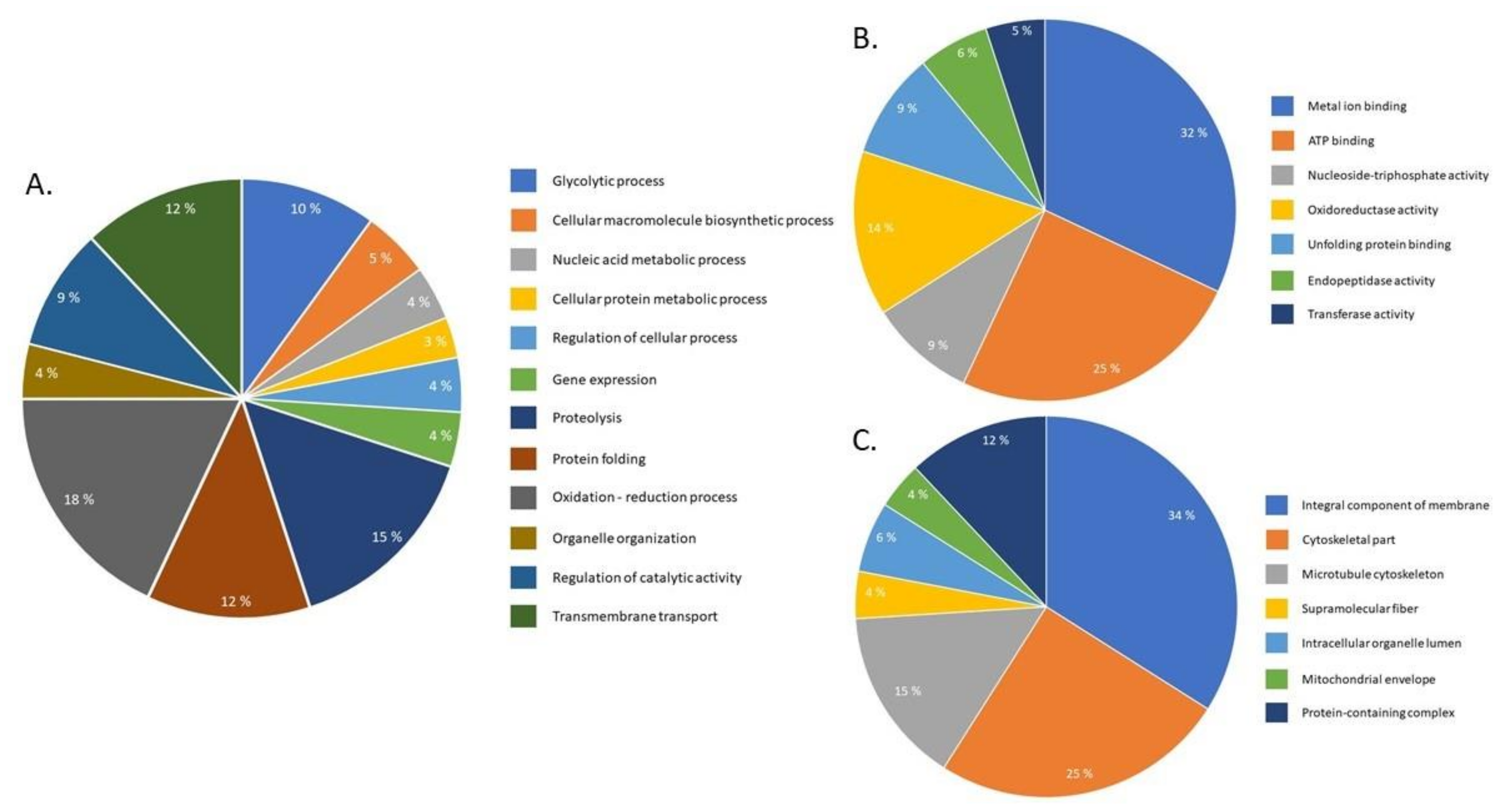
The proteomic approach identified 1076 S. mekongi proteins in which 144 proteins were differentially expressed after PZQ treatment. The abundance of 68 proteins increased (Table S1), while 76 proteins decreased (Table S2). Gene ontology analysis of differential proteins was performed using BLAST2GO bioinformatic software. According to the biologic process term, most differentially expressed proteins are involved in redox processes (18%) and proteolysis (15%) (Figure 2A). However, using a molecular function term indicated that the proteins mainly correlated to metal-ion (32%) and ATP binding (25%) (Figure 2B). In the cellular component term, most of the altered proteins related to integral membrane (34%) and cytoskeletal components (25%) (Figure 2C). The top twenty up- and downregulated S. mekongi proteins after PZQ treatment are shown in Table 1 and Table 2, respectively. Proteins related to structure, energy, protease, antioxidant, transcription and translation and antigens were highly upregulated after PZQ exposure. The structural protein paramyosin was the most upregulated protein with 5.96-fold change after treatment. However, some proteins involved in structure, energy, stress response and signaling were highly downregulated. The energy metabolism protein aldehyde dehydrogenase 1B1 precursor was the most downregulated protein after PZQ exposure (4-fold decrease). Interestingly, while none of kinases were found to be highly upregulated, there was a significant number of kinases showing decreased expression after PZQ treatment, such as pyruvate kinase PKM (A0A095B084: 2.38-fold decrease), arginine kinase (C1LFZ8: 2.27-fold decrease) and inositol hexakisphosphate and diphosphoinositol–pentakisphosphate kinase 2 (A0A095ASB2: 2-fold decrease) (Table 2).

2.3. Effects of PZQ on S. Mekongi Phosphorylation and Phosphoprotein Abundance

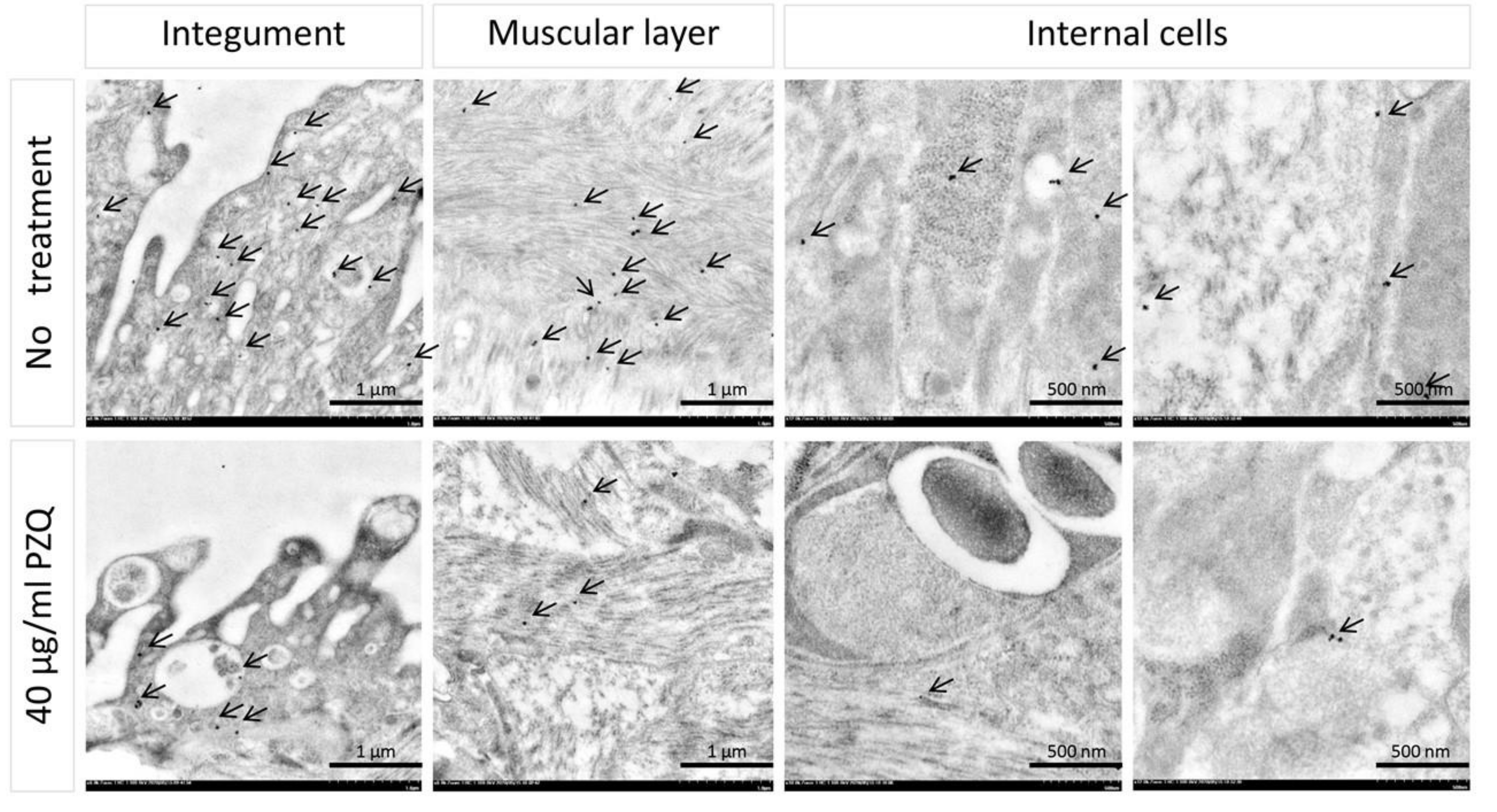
Levels of phosphorylation in PZQ-treated S. mekongi was examined by immunogold labeling and visualized by transmission electron microscope (TEM) and compared with untreated parasites. Anti-phosphoserine was used to determine phosphorylation levels and tissue localization in the parasites. In Schistosoma parasite, the main phosphorylated residues are serine (67.8%), threonine (20.1%) and tyrosine (12.1%) [31]. Phosphorylation of tegument, muscle and internal cells of the worms were markedly reduced after PZQ treatment, as indicated by the black spots in Figure 3. The reduction of phosphorylation could be one of the effects of PZQ treatment. To explore the mechanism of action of PZQ on parasite phosphorylation, phosphoprotein abundance of untreated and PZQ-treated S. mekongi were profiled using MS-based phosphoproteomics.
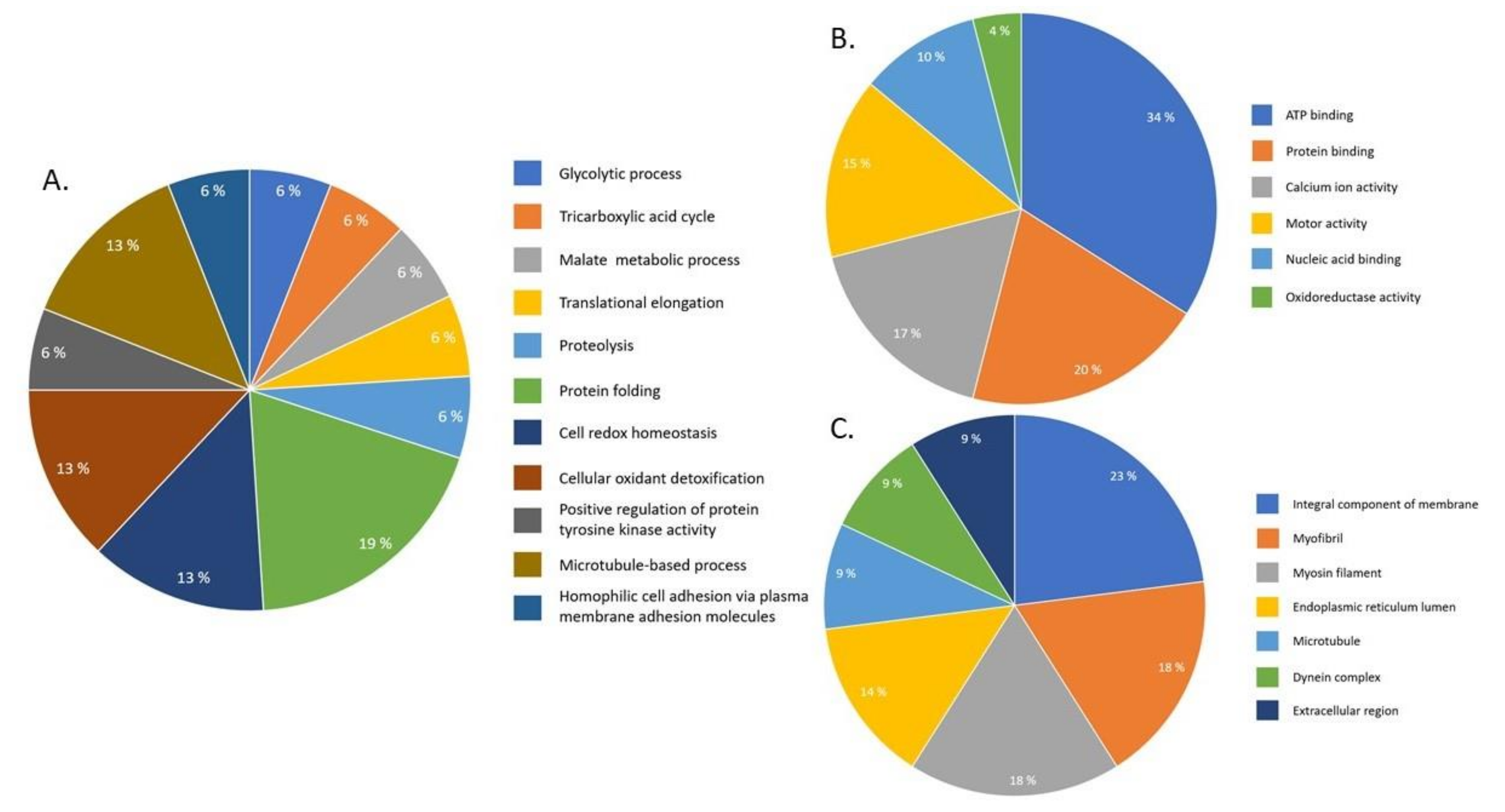
A total of 448 phosphoproteins of S. mekongi were identified using TALON PMAC Magnetic Phospho Enrichment Kit and MS. Among these, 43 phosphoproteins differed after PZQ exposure. Twenty-eight phosphoproteins were upregulated and 15 were downregulated after PZQ treatment. MS-identified phosphopeptides of up- and downregulated phosphoproteins were 124 and 44 peptides, respectively (Table 3 and Table 4). Phosphoproteins involved in structure, energy, protease, antigen, transcription and translation, immune system, stress response, transporters and antioxidants were upregulated. An unknown protein, SJCHGC01909, was the most upregulated phosphoprotein after PZQ treatment (6.13-fold increase). Some phosphoproteins involved in structure, energy, antigen, scaffold, antioxidant, transcription and translation, stress response and signaling were also downregulated. Energy metabolism protein aldolase was the most downregulated phosphoprotein (2.38-fold decrease). Gene ontology classification of differential phosphoproteins after PZQ exposure using the biologic process term showed that differential phosphoproteins mostly affected protein folding (19%), cell redox homeostasis (13%), cellular oxidant detoxification (13%) and microtubule-based process (13%) (Figure 4A). Furthermore, the molecular function term classification revealed that most altered phosphoproteins related to ATP binding (34%) and protein binding (20%) (Figure 4B). For the cellular component term, most differential phosphoproteins were integral membrane (23%), myofibril (18%) and myosin filament (18%) components (Figure 4C).
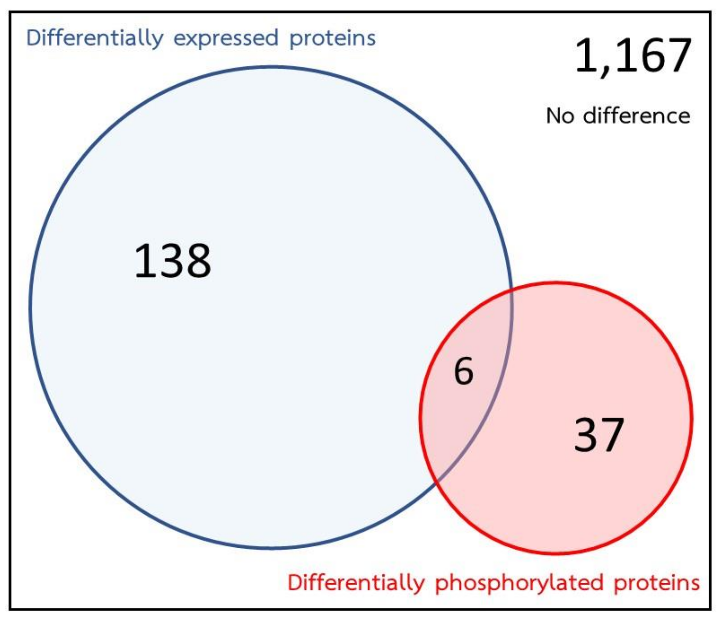
The integrated proteomic and phosphoproteomic data revealed that 1348 proteins could be identified. After S. mekongi were exposed to PZQ, a total of 144 proteins were identified by proteomics as showing differential expression and 43 phosphoproteins were altered as identified by phosphoproteomics. However, 138 proteins were found to be altered by proteomics analysis alone. These proteins may indicate the effect of PZQ at the protein expression level. Likewise, 37 proteins were found to be altered by phosphoproteomic analysis alone, which could reflect the effect of PZQ at the phosphorylation level. Only six proteins—paramyosin, cathepsin B, annexin, glutathione S-transferase, aldolase and heat shock protein—showed the alterations by both proteomic and phosphoproteomic approaches (Figure 5).

2.4. Pathway Analysis

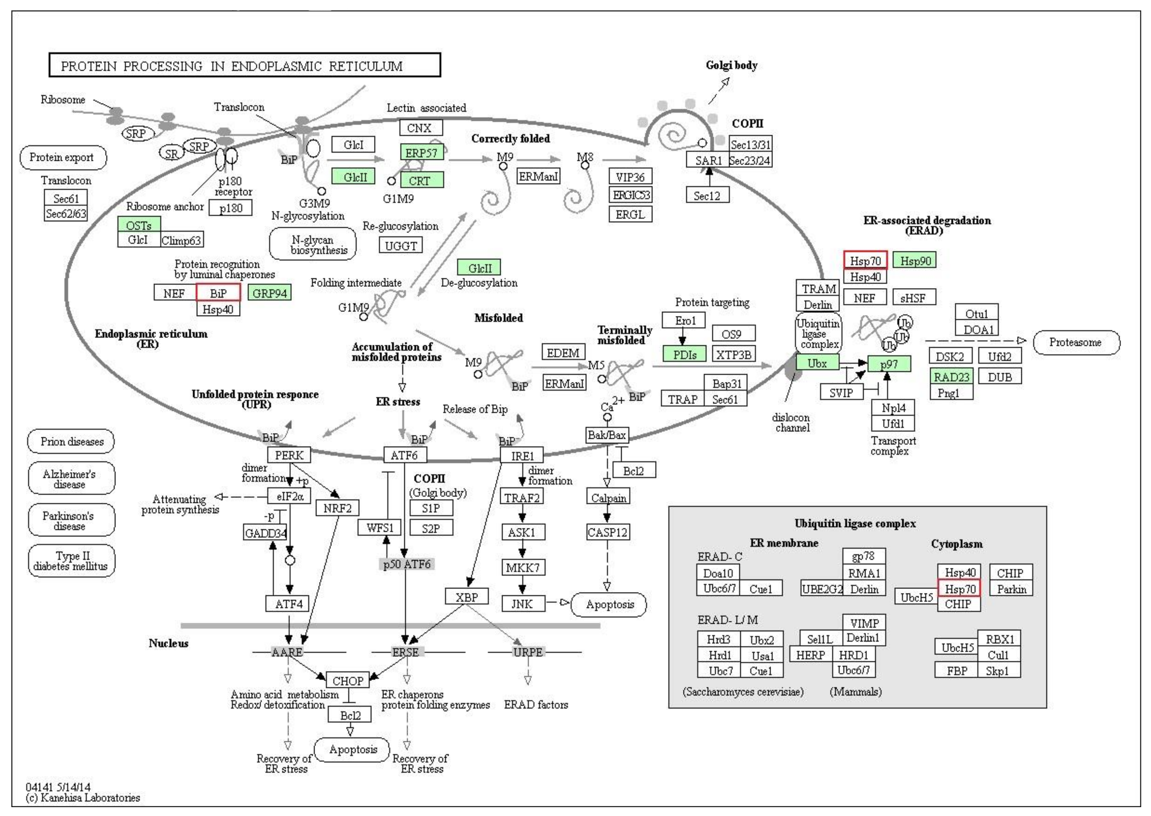
To fully understand the anthelmintic mechanisms of PZQ, the integral proteomic and phosphoproteomic data were further investigated by KAAS bioinformatic software. The result suggested that protein processing in the endoplasmic reticulum (ER) (pathway ID: 04,141) was the main enriched pathway in this study. As shown in Figure 6, differentiations of 10 proteins (green boxes) and two phosphoproteins (red borders) were detected in this pathway after treatment. Furthermore, most differential proteins and phosphoproteins affected by PZQ were mapped to protein misfolding and ER-associated degradation (ERAD). These two processes control the quality of newly synthesized proteins and degrade misfolded proteins. Findings of KAAS analysis correlated with the gene ontology classification, which demonstrated that protein folding, unfolding protein binding, proteolysis and endopeptidase activity were altered by PZQ treatment (Figure 2 and Figure 4). Therefore, alteration of protein processing in the ER, especially on the ERAD pathway, may be a possible mechanism of action of PZQ in S. mekongi. The integrated proteomic and phosphoproteomic data not only provided a novel PZQ mechanism, but also supported the putative PZQ mechanisms on schistosome including calcium homeostasis, tegument damage and induction of oxidative stress. There were 9, 10 and 28 proteins with altered regulation in calcium homeostasis, worm antigens and oxidative stress, respectively (Table 5).

2.5. Prediction of Key Kinases Effected by PZQ

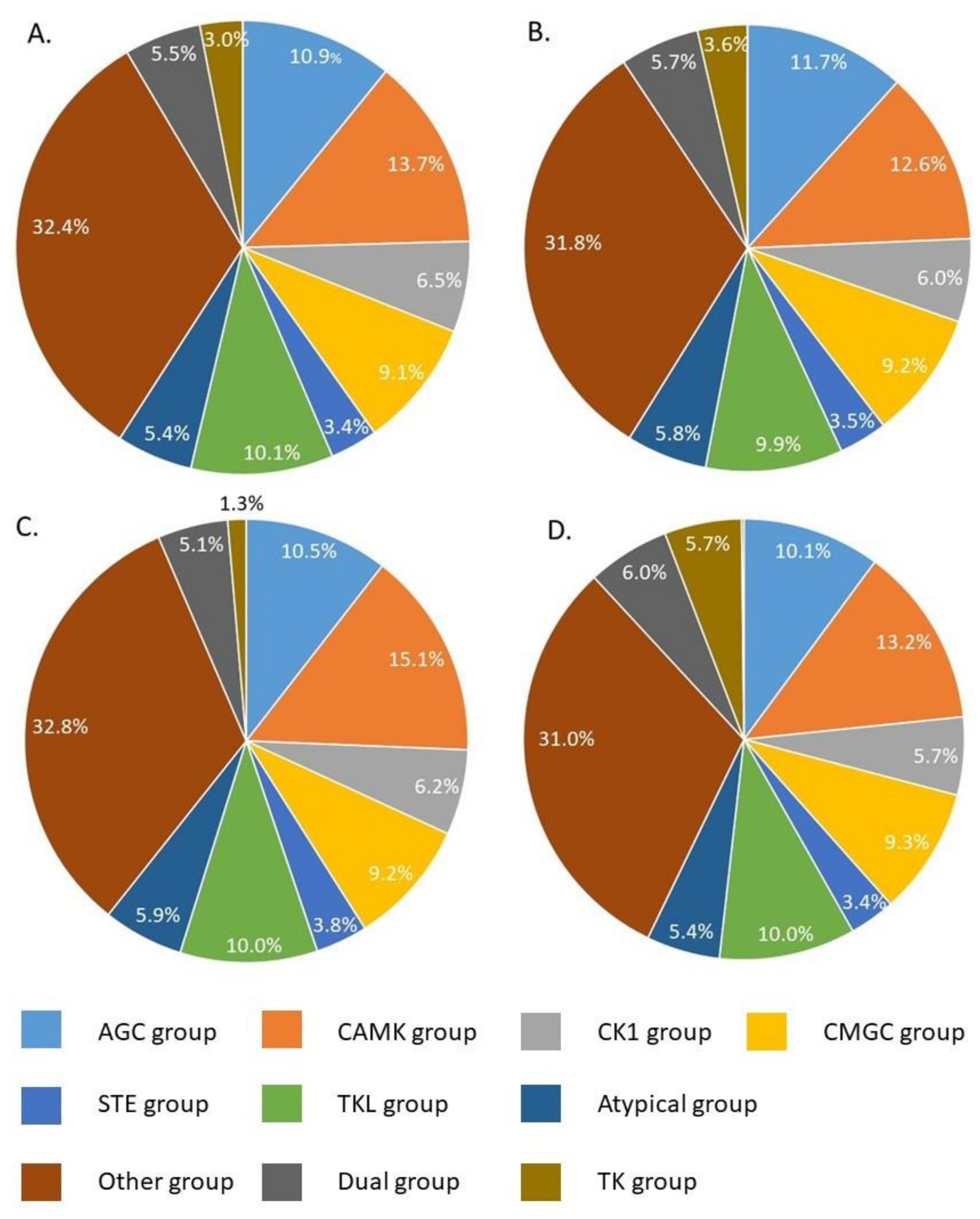
Phosphorylation of proteins in S. mekongi was strongly affected by PZQ, as shown by immunohistochemistry and phosphoproteomic analyses. Kinases are the key enzymes in the control of phosphorylation levels in cells and are known to be potential drug targets in several diseases. Using phosphopeptides and phosphorylation sites obtained from the MS results, it was possible to predict kinase–substrate interactions by the group-based prediction system. Phosphorylation data of calcium homeostasis, worm antigen, oxidative stress, protein folding, and proteolysis were subjected to kinase–substrate prediction to identify key kinases. Results showed 83 kinases (11 kinase groups) were involved in phosphorylation of proteins in these four pathways (Figure 7). Among the 83 kinases, proto-oncogene tyrosine-protein kinase Src, calcium/calmodulin-dependent protein kinase-like (CAMKL) and Wee protein kinase were the top three kinases controlling the pathways of interest (Table S3). Inhibition of these kinases would interfere with phosphorylation and signal transduction of calcium homeostasis, worm antigens, oxidative stress, protein folding and proteolysis pathways in a manner comparable to PZQ. Applying inhibitors of these kinases could therefore be an alternative means of schistosomiasis treatment.

3. Discussion

S. mekongi is a public health threat in South–East Asia with PZQ currently the only drug of choice for treatment of this blood fluke. This study aimed to understand the parasiticidal mechanisms of PZQ on S. mekongi using proteomic and phosphoproteomic approaches. To study the parasiticidal effect of PZQ, IC50 was established by administering the drug to worms and observing for their movement. PZQ at 40 μg/mL killed 46.7% of adult worms and was defined as IC50 (Figure 1). In previous studies, the IC50 of PZQ varied from 0.03 to 80 μg/mL [29,32,33,34]. Duration of exposure could have an effect on IC50. Our study exposed adult worms to PZQ for 60 min before observing the mortality rates. This correlates with data indicating that PZQ starts to affect worms a few minutes after exposure [20]. The 60 min incubation period in this study could have allowed more time for the drug to penetrate into the worms to disrupt their protein moieties.
Several studies have proposed mechanisms of action of PZQ on schistosome. However, the exact molecular mechanism of PZQ against Schistosoma spp. is still unclear. High-throughput technologies were applied to Schistosoma parasite with reduced susceptibility to PZQ for elucidating its mechanism of action. Proteomic analysis of less-susceptible S. mansoni to PZQ revealed the upregulation of 2 proteins; heat shock protein 70 and calcium ATPase 2 protein [35]. Our study found altered expression of the same protein or proteins with similar function; heat shock protein 70 (O45039) and sodium/potassium-transporting ATPase subunit alpha (A0A095AFH4, A0A094ZR23) (Table S1,2). Furthermore, gene expression profiling of less-susceptible parasite indicated a list of gene involving with calcium signaling pathway (calcium binding protein, calmodulin, calponin) and oxidative stress response (thioredoxin and superoxide dismutase) [36]. Our mass spectrometric data also found proteins in those pathways, which were 20 kDa calcium-binding protein (A0A095A3D3), putative annexin (G4VL68), voltage-dependent anion-selective channel protein (VDAC) (G4M1U8) for calcium signaling pathway and tryparedoxin peroxidase (C1LV40), glutathione-S-transferase (Q26513) for oxidative stress pathway (Table 5). The mechanisms of action of PZQ have been continuously investigated. The interference of the calcium signaling pathway is one of the putative PZQ mechanisms of schistosome [7,20,21,37,38]. Pharmacologically, PZQ induces calcium influx to worm myocytes and contraction of musculature, leading to paralysis and loss of worm movement [20,21]. Voltage-gated calcium channels have been proposed as potential targets for PZQ by interfering with the interaction of the α and β subunit of the proteins, thereby causing excessive uptake of calcium into cells [21,39]. Our MS results revealed the alteration of proteins involving in calcium signaling and interaction, such as the 20 kDa calcium-binding protein (A0A095A3D3), putative annexin (G4VL68) and voltage-dependent anion-selective channel protein (VDAC) (G4M1U8) (Table 5). VDAC controls calcium movement between the mitochondria and cytoplasm; inhibition of this protein causes apoptosis and has been used as a drug target for cancer treatment [40,41]. Therefore, alteration of calcium homeostasis is strongly suggested as one of the parasiticidal mechanisms of PZQ. Another parasiticidal mechanism of PZQ against schistosome is the increased susceptibility of the parasite to host immunity by exposing antigens on worm surface [20]. Antibody and cell-mediated immunity significantly impact the killing ability of PZQ [20,23]. PZQ was less effective against S. mansoni in T cell-deprived hosts than normal hosts [42]. Immunization of mice with worm antigens enhanced the worm-killing effect of PZQ [43]. The studies stated that the worm antigens and host immunity play important roles in the parasiticidal ability of PZQ on schistosome, although the detailed mechanism is still unknown. Our findings found that expression of several antigenic proteins increased after PZQ treatment, including 55 kDa antigen myophilin (Q86DV9) (Table 5), which may play some role in the effects of PZQ. Oxidative stress induction was also proposed as a mechanism of PZQ to parasitic worms [24,44]. The pathway for PZQ to induce oxidative stress in parasite is still unknown, although schistosome and other parasitic flatworms increased expression of antioxidants in response to PZQ [24,37,44,45]. Our study indicated that PZQ treatment increased expression of antioxidant proteins, such as tryparedoxin peroxidase (C1LV40) and glutathione-S-transferase (Q26513). In addition, phosphorylation of thioredoxin peroxidase (O97161), 26 kDa glutathione-S-transferase (G4LXF8) and peroxiredoxin-2 (A0A095A0V3) were altered (Table 5). Our findings also revealed a potential novel mechanism of PZQ: the alteration of protein folding and proteolysis through the ERAD pathway. The function of the ERAD pathway is to eliminate unfolded/misfolded proteins from the ER through a series of proteins and phosphoproteins [46]. ERAD tags unfolded/misfolded proteins with ubiquitin prior to sending them for proteasome-mediated degradation [47]. Precise activity of ERAD is crucial for quality control of structure and function of the protein repertoire [48]. Interruption of ERAD could cause the accumulation of toxic proteins, apoptosis and cell death, which has been implicated in many diseases, such as Alzheimer’s disease, Parkinson’s disease and rheumatoid arthritis [47,48]. In Trypanosoma brucei, ERAD plays important roles in the processing of antigenic variation for host immune evasion [49]. T. brucei is believed to survive in extreme stress conditions, such as blood, by using unique ERAD mechanisms to shift their surface antigens and avoid detection by the immune system [46,49]. Moreover, Plasmodium falciparum showed sensitivity to ERAD inhibitor treatment. Therefore, proteins in ERAD may be novel targets for malarial drug development [50,51]. Unlike blood-dwelling protozoa, the process of antigenic variation of schistosome is manipulated by alternative splicing of micro-exon genes [52,53]. Although the schistosome ERAD pathway has never been studied before, protein break down and degradation was proposed as drug target for schistosomiasis treatment in a previous study [54]. Here, we propose that PZQ exerts its effect by interfering with the ERAD pathway of S. mekongi.
Phosphorylation is a vital molecular process for blood fluke biology, the alteration of which affects the viability of the worms. Our immunogold labeling to phosphoserine showed an overall reduction of phosphorylation levels after PZQ treatment (Figure 3). This present study found changes of a number of phosphoproteins after PZQ treatment. However, none of target validation was performed which is the limitation of this study. In addition, the inhibition of specific kinases, protein kinase C (PKC) and extracellular signal-regulated kinases, resulted in a reduction of protein phosphorylation and decreases in attachment, muscular activity, neuromuscular coordination, pairing and reproductive function of schistosomes [26]. Recently, drugs acting on phosphorylation and protein kinases have been proposed as novel targets for schistosomiasis treatment [25,26,55,56,57]. Kinomic array of S. mansoni revealed a number of kinases those are vital for Schistosoma biology [31] in which Src kinase, Cyclin-dependent kinase (CDK), PKC were predicted from bioinformatic tool as the critical kinases for anthelminthic effects of PZQ (Table S3). Our study indicates that kinases of critical pathways in the parasite and Src kinase are important kinases for PZQ response (Figure S1, Table S3). Src kinase is a cytoplasmic tyrosine kinase that controls diverse cellular processes, including adhesion, proliferation and differentiation [25]. Only a few studies have focused on the inhibition of Src kinase in schistosomes. An inhibitor of Src kinase, Herbimycin A, suppresses the mitotic activity and egg production of female schistosome, resulting in reduced reproductive capacity of worms [58]. As a consequence, Src kinase was proposed as novel target for the future development of antischistosomal drugs.
In summary, PZQ at IC50 caused a general reduction of protein phosphorylation in S. mekongi. PZQ also altered expression and phosphorylation of many proteins in key pathways of the parasites, including calcium homeostasis, worm antigens and oxidative stress. Moreover, the differentially expressed and phosphorylated proteins indicated that PZQ interferes with protein folding and proteolysis through the ERAD pathway. Furthermore, kinases responsible for phosphorylation of previously identified pathways were predicted and Src kinase was found to be the main enzyme influenced in the four pathways of interest. Our findings suggest a novel effect of PZQ as well as indicating possible drug targets, which could be used for the treatment of Mekong schistosomiasis in the future.

4. Materials and Methods

4.1. S. Mekongi Life Cycle Maintenance, Animal Husbandry and Worm Collection

The life cycle of S. mekongi was maintained using snail Neotricula aperta as an intermediate host and mice Mus musculus as definitive hosts, as described earlier [59]. The snails were primarily collected from Mekong River in northeastern Thailand and cultured at the Applied Malacology Laboratory, Department of Social and Environmental Medicine, Faculty of Tropical Medicine, Mahidol University, Bangkok, Thailand. Eight-week-old female ICR mice were purchased from the National Laboratory Animal Center, Mahidol University. Mice were percutaneously infected with 25–30 cercariae of S. mekongi and housed in the Animal Care Unit, Faculty of Tropical Medicine, Mahidol University. Adult worms were collected at 8 weeks post-infection by hepatic perfusion with sterile 0.85% saline solution and subsequently cultured in Roswell Park Memorial Institute (RPMI) medium (Hyclone, GE Healthcare, Little Chalfont, UK) with 10% fetal calf serum (Hyclone) in a humidified 5% CO2 incubator at 37 °C until PZQ treatment.
All experiments involving animals were performed according to the ethical principles and guidelines for the use of animals at the National Research Council of Thailand (NRCT). In addition, all procedures were conducted with the permission from the Faculty of Tropical Medicine Animal Care and Use Committee (FTM-ACUC), Mahidol University, with approval number FTM-ACUC 014/2016.

4.2. PZQ Treatment and Half Maximal Inhibitory Concentration (IC50) Assessment

PZQ (Tokyo Chemical Industry, Tokyo, Japan) was mixed with Dimethyl sulfoxide (DMSO) prior to diluted with RPMI medium to concentrations of 0, 20, 40, 60, 80 and 100 μg/mL and administered for an hour to 10 pairs of S. mekongi for each concentration (three biologic replicates for each concentration). Worm movement was used as the indicator for viability. Periodic movement of the worms was observed under a video microscope (Celestron, CA, USA) at 0, 5, 10, 15, 30 and 60 min and worms that did not move after a minute of observation were identified as dead. After 1 hour of PZQ treatment, all worms were collected and kept at −80 °C for further experiments. Data on worm viability were plotted and statistical significance at 0.05 was calculated with ANOVA using SPSS for Windows version 15.0 (SPSS, Inc., Chicago, IL, USA). The concentration that gave approximately 50% viability was identified as IC50.

4.3. Protein Extraction and Phosphoprotein Enrichment

Protein extraction was performed within a week after PZQ treatment, using the frozen extraction method and phosphoprotein enrichment was performed using the TALON PMAC Magnetic Phospho Enrichment Kit (Takara, Shiga, Japan). First, 10 pairs of untreated and 40 μg/mL-PZQ-treated worms were put into a mortar and homogenized in liquid nitrogen. After the nitrogen had completely evaporated, lysis buffer (provided with the kit) was added and incubated on ice for 10 min. Protein lysate was transferred into tubes and centrifuged at 5000 × g for 5 min at 4 °C. The supernatant was transferred to new tube and the protein concentration was measured using the bicinchoninic acid (BCA) assay (Thermo Fisher Scientific, Waltham, MA, USA).
Phosphoprotein enrichment was performed according to manufacturer’s protocol. Briefly, 250 μg protein was mixed with metal ion-coated magnetic beads and mixed on a rotary shaker for 90 min at 4 °C. The supernatant was discarded, and proteins bound to beads were eluted using 75 μL of the provided elution buffer. Successful enrichment was confirmed using 12% sodium dodecyl sulfate–polyacrylamide gel electrophoresis (SDS-PAGE) and Pro-Q diamond staining (Thermo Fisher Scientific). All experiments were performed in three biologic replicates.

4.4. Protein Separation and In-Gel Digestion

For proteomic analysis, 50 μg of sample was analyzed by SDS-PAGE and resulting protein bands were visualized using Coomassie Blue G staining. For phosphoproteomic analysis, 15 μL of eluted sample was analyzed by SDS-PAGE gel and protein bands were visualized using silver staining. Thereafter protein bands from each lane were cut into small pieces for further in-gel digestion.
Coomassie dye was removed by incubating gels with 25-mM ammonium bicarbonate buffer containing 50% acetonitrile. Silver stain was removed with 30-mM potassium ferricyanide and 156-mM sodium thiosulfate in a 1:1 ratio. Gel pieces were then reduced with 4-mM dithiothreitol (Sigma-Aldrich, St. Louis, MO, USA) in 50-mM ammonium bicarbonate buffer, further alkylated with 250-mM iodoacetamide (Sigma-Aldrich) and dehydrated with 100% acetonitrile. Proteins were digested overnight with 10 ng trypsin (Sigma-Aldrich) dissolved in 200 μL of 50-mM ammonium bicarbonate buffer containing 5% acetonitrile. After digestion, peptides were extracted by adding 200 μL of acetonitrile. The supernatant containing eluted peptides was transferred to a new tube and dried using a speed vacuum, before being dissolved with 0.1% formic acid.

4.5. Protein Identification by MS

Peptide samples were analyzed using an UltiMate 3000 nano-liquid chromatography system (Thermo Fisher Scientific) coupled with a micrOTOF-Q electrospray ionization quadrupole time-of-flight MS (Bruker Daltonics, Billerica, MA, USA). Mobile phase A for sample injection was 2% acetonitrile and 0.1% formic acid in water and mobile phase B was 0.1% formic acid in acetonitrile with flow rate of 300 nl/min for 60 min. Data acquisition was performed using Hystar software (Bruker Daltonics). Spectra of the peptide covered mass ranges of m/z 400–3000 and 50–1500.

4.6. Data Analysis and Pathway Enrichment

Data from all gel pieces were merged into single Mascot generic file (.mgf) using MASCOT DEMON (Matrix Science, London, UK), which was then searched for S. mekongi proteins using an in-house transcriptome database with missed cleavages allowed [59]. Peptide tolerance was assigned to 200 ppm and the tandem MS tolerance was assigned to 0.6 Da. Methionine oxidation, cysteine carbamidomethylation, serine phosphorylation, tyrosine phosphorylation and threonine phosphorylation were identified as variable modifications. Proteins that showed differential expression or phosphorylation of more than 2-fold were selected for further pathway enrichment using BLAST2GO and KEGG Automatic Annotation Server (KAAS) applications.
BLAST2GO Version 5.2.3 (BioBam Bioinformatics, Valencia, Spain) was used to annotate sequences of differentially expressed or phosphorylated proteins according to their biologic processes, molecular function and subcellular localization with standard parameters [60]. Furthermore, protein sequences were analyzed and mapped into the KEGG Pathway using KEGG Automatic Annotation Server (KAAS: https://www.genome.jp/kegg/kaas/) with standard parameters [61]. The top 20 annotated pathways were shown, and pathways related to the effects of PZQ were presented with identified proteins highlighted.

4.7. Phosphorylation Level Investigation by Immunogold Labeling and Transmission Electron Microscope (TEM)

Phosphorylation levels of untreated and 40 μg/mL-PZQ-treated S. mekongi were assessed by phosphoserine immunogold labeling and TEM. Briefly, worms with and without treatment were fixed using 2.5% glutaraldehyde in 0.1 M sucrose phosphate buffer (SPB), pH 7.4, for an hour and subsequently washed 3 times with SPB for 10 min. Worm samples were fixed with secondary fixative 1% osmium tetroxide and dehydrated in graded ethanol. Samples were immersed in a series of LR white resin (Electron Microscopy Sciences, PA, USA) before embedding in pure LR white (Electron Microscopy Sciences) and polymerizing at 60 °C for 48 h. Sample blocks were cut into 100-nm-thick sections for further immunogold labeling.
Worm sections were initially blocked with 50-mM glycine in phosphate-buffered saline (PBS) and then with 5% bovine serum albumin (BSA) (Electron Microscopy Sciences) in PBS for 30 min each. Sections were incubated with primary antibody against phosphoserine (Merck, Darnstadt, Germany) for 1 hour, followed by washing three times with 0.1% BSA in PBS. Sections were incubated with secondary antibody conjugated with 10-nm gold particles (Electron Microscopy Sciences). After washing three times, a silver enhancement kit (Electron Microscopy Sciences) was used to improve the contrast of gold particle labeling. Finally, sections were stained with lead citrate and uranyl acetate prior to visualization in a HT7700 model TEM (Hitachi, Tokyo, Japan). The labeled gold particles were examined throughout the parasite body, especially on tegument, muscle and internal cells.

4.8. Kinase Prediction for Identified Pathways

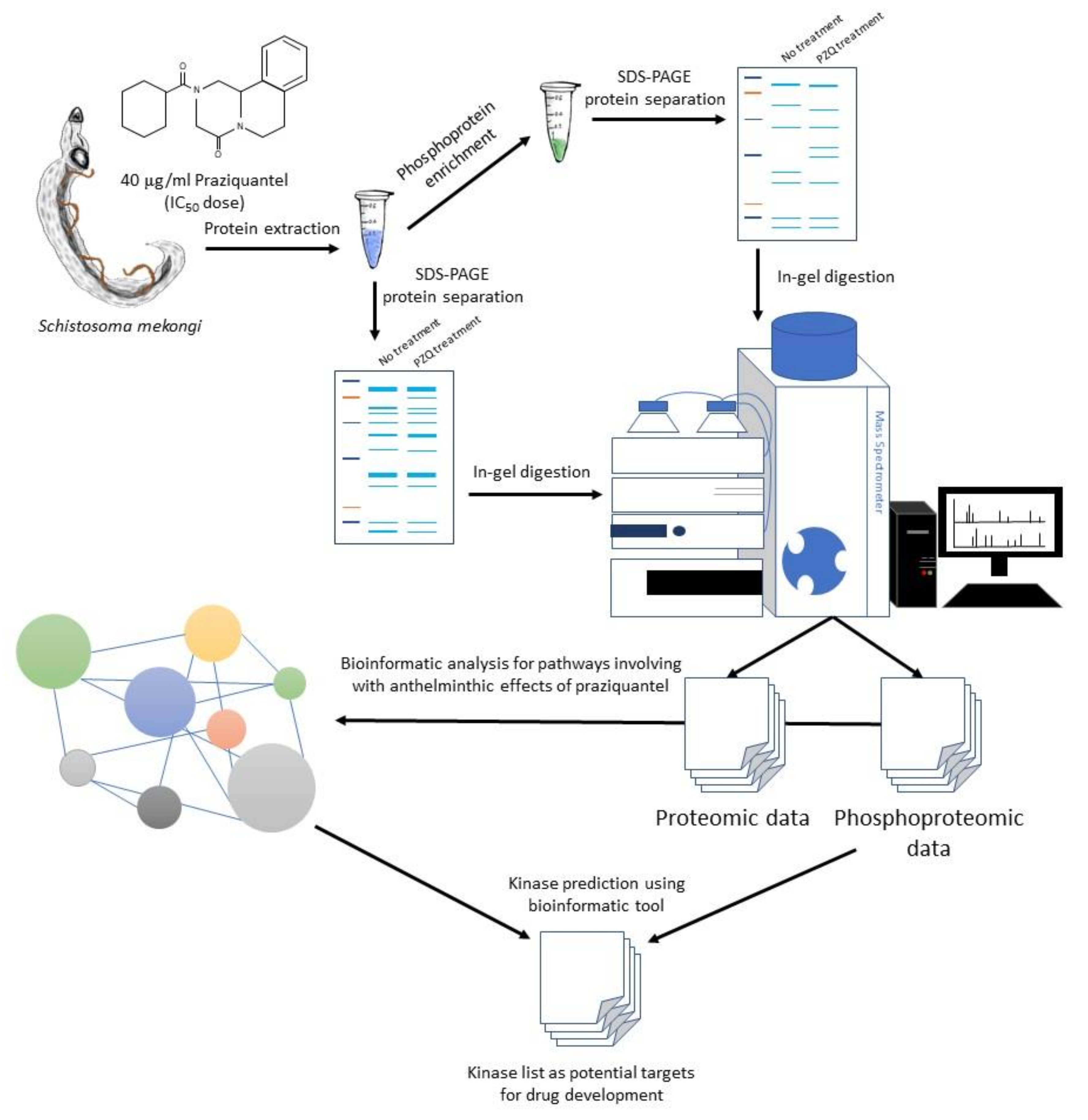
Kinases that participate in calcium homeostasis, worm antigen, oxidative stress, protein folding and proteolysis pathways were predicted using the bioinformatic tool Group-based Prediction System version 5.0 (http://gps.biocuckoo.cn/online.php). The phosphopeptide data were input and analyzed with standard settings [62]. Predicted kinases were categorized according to their group and the number and percentage of phosphorylation sites of each predicted kinase were calculated. The graphical summary of whole experiments in this study is presented in Figure 8.

Supplementary Materials

The following are available online at https://www.mdpi.com/2076-0817/9/6/417/s1, Figure S1: Src kinase–substrate prediction from phosphopeptide data. Phosphopeptide data were used to predict kinases corresponding for their phosphorylation with Group-based Prediction System. Protein substrates of Src kinase are shown with number of phosphorylation sites and pathways. Src kinase was predicted to phosphorylate 85 sites of 14 proteins involved in calcium homeostasis, worm antigen, oxidative stress and proteins folding and proteolysis; Video S1: Movement of S. mekongi without PZQ treatment; Video S2: Movement of S. mekongi after treatment with 40 μg/mL of PZQ for 60 min; Table S1: S. mekongi proteins upregulated after 40 μg/mL-PZQ treatment; Table S2: S. mekongi proteins downregulated after 40 μg/mL-PZQ treatment; Table S3: Top 25 predicted kinases responsible for phosphorylation of proteins involved in calcium binding, worm antigen, oxidative stress, protein folding and proteolysis.

Author Contributions

Conceptualization, P.A., Y.L. and O.R.; methodology, O.R.; investigation, Y.L., P.C., Y.C., P.C., T.T., S.A., and, O.R.; formal analysis, P.C.; writing—original draft preparation, P.C.; writing—review & editing, O.R. and P.C.; supervision, O.R.; funding acquisition, O.R. and P.C. All authors have read and agreed to the published version of the manuscript.

Funding

This study was supported by the Goal-oriented Research Project Grant 2016 of Mahidol University through Onrapak Reamtong and Chulabhorn Royal Academy to Peerut Chienwichai.

Acknowledgments

We thank the Department of Molecular Tropical Medicine and Genetics and Central Equipment Unit, Faculty of Tropical Medicine, Mahidol University for providing the equipment and facilities. We also express our gratitude to Department of Helminthology, Applied Malacology Laboratory, Department of Social and Environmental Medicine and Animal Care Unit, Faculty of Tropical Medicine, Mahidol University for their support for animal experiment and parasite preparation.

Conflicts of Interest

The authors declare no conflict of interest.

References

  1. World Health Organization. Schistosomiasis. Available online: https://www.who.int/en/news-room/fact-sheets/detail/%20schistosomiasis. (accessed on 18 May 2019).
  2. Adenowo, A.F.; Oyinloye, B.E.; Ogunyinka, B.I.; Kappo, A.P. Impact of human schistosomiasis in sub-Saharan Africa. Braz. J. Infect Dis. 2015, 19, 196–205. [Google Scholar] [CrossRef] [PubMed] [Green Version]
  3. Campa, P.; Develoux, M.; Belkadi, G.; Magne, D.; Lame, C.; Carayon, M.J.; Girard, P.M. Chronic Schistosoma mekongi in a traveler—A case report and review of the literature. J. Travel Med. 2014, 21, 361–363. [Google Scholar] [CrossRef] [PubMed] [Green Version]
  4. Pilsczek, F.H. Infectious diseases of Khmer immigrants in the United States: Review of published reports. J. Ayub Med. Coll. Abbottabad 2011, 23, 149–152. [Google Scholar] [PubMed]
  5. Leshem, E.; Meltzer, E.; Marva, E.; Schwartz, E. Travel-related schistosomiasis acquired in Laos. Emerg. Infect. Dis. 2009, 15, 1823–1826. [Google Scholar] [CrossRef] [PubMed]
  6. Ohmae, H.; Sinuon, M.; Kirinoki, M.; Matsumoto, J.; Chigusa, Y.; Socheat, D.; Matsuda, H. Schistosomiasis mekongi: From discovery to control. Parasitol. Int. 2004, 53, 135–142. [Google Scholar] [CrossRef] [PubMed]
  7. Silva, V.; Campos, B.; Oliveira, J.F.; Decout, J.L.; Lima, M. Medicinal chemistry of antischistosomal drugs: Praziquantel and oxamniquine. Bioorg. Med. Chem. 2017, 25, 3259–3277. [Google Scholar] [CrossRef]
  8. Lovis, L.; Mak, T.K.; Phongluxa, K.; Soukhathammavong, P.A.; Vonghachack, Y.; Keiser, J.; Vounatsou, P.; Tanner, M.; Hatz, C.; Utzinger, J.; et al. Efficacy of praziquantel against Schistosoma mekongi and Opisthorchis viverrini: A randomized, single-blinded dose-comparison trial. PLOS Negl. Trop. Dis. 2012, 6, e1726. [Google Scholar] [CrossRef] [Green Version]
  9. Albonico, M.; Levecke, B.; LoVerde, P.T.; Montresor, A.; Prichard, R.; Vercruysse, J.; Webster, J.P. Monitoring the efficacy of drugs for neglected tropical diseases controlled by preventive chemotherapy. J. Glob. Antimicrob. Resist. 2015, 3, 229–236. [Google Scholar] [CrossRef] [Green Version]
  10. Cioli, D.; Pica-Mattoccia, L.; Basso, A.; Guidi, A. Schistosomiasis control: Praziquantel forever? Mol. Biochem. Parasitol. 2014, 195, 23–29. [Google Scholar] [CrossRef]
  11. Greenberg, R.M. New approaches for understanding mechanisms of drug resistance in schistosomes. Parasitology 2013, 140, 1534–1546. [Google Scholar] [CrossRef] [Green Version]
  12. Melman, S.D.; Steinauer, M.L.; Cunningham, C.; Kubatko, L.S.; Mwangi, I.N.; Wynn, N.B.; Mutuku, M.W.; Karanja, D.M.S.; Colley, D.G.; Black, C.L.; et al. Reduced susceptibility to praziquantel among naturally occurring Kenyan isolates of Schistosoma Mansoni. PLOS Negl. Trop. Dis. 2009, 3, e504. [Google Scholar] [CrossRef] [PubMed] [Green Version]
  13. Ismail, M.; Botros, S.; Metwally, A.; William, S.; Farghally, A.; Tao, L.F.; Bennett, J.L.; Farghally, A.; Metwally, A. Resistance to praziquantel: Direct evidence from Schistosoma mansoni isolated from Egyptian villagers. Am. J. Trop. Med. Hyg. 1999, 60, 932–935. [Google Scholar] [CrossRef] [PubMed]
  14. Seto, E.Y.W.; Wong, B.K.; Lu, D.; Zhong, B. Human Schistosomiasis resistance to praziquantel in China: Should we be worried? Am. J. Trop. Med. Hyg. 2011, 85, 74–82. [Google Scholar] [CrossRef] [PubMed] [Green Version]
  15. King, C.H.; Muchiri, E.M.; Ouma, J.H. Evidence against rapid emergence of praziquantel resistance in Schistosoma haematobium, Kenya. Emerg. Infect. Dis. 2000, 6, 585–594. [Google Scholar] [CrossRef] [Green Version]
  16. Chelladurai, J.J.; Kifleyohannes, T.; Scott, J.; Brewer, M.T. Praziquantel resistance in the zoonotic cestode Dipylidium caninum. Am. J. Trop. Med. Hyg. 2018, 99, 1201–1205. [Google Scholar] [CrossRef] [Green Version]
  17. Lateef, M.; Zargar, S.A.; Khan, A.R.; Nazir, M.; Shoukat, A. Successful treatment of niclosamide- and praziquantel-resistant beef tapeworm infection with nitazoxanide. Int. J. Infect. Dis. 2008, 12, 80–82. [Google Scholar] [CrossRef] [Green Version]
  18. McManus, D.P.; Dunne, D.W.; Sacko, M.; Utzinger, J.; Vennervald, B.J.; Zhou, X. Schistosomiasis. Nat. Rev. Dis. Primers 2018, 4. [Google Scholar] [CrossRef]
  19. Siqueira, L.P.; Fontes, D.A.F.; Aguilera, C.S.B.; Timóteo, T.R.R.; Ângelosa, M.A.; Silva, L.; De Melo, C.G.; Rolim, L.A.; Da Silva, R.M.F.; Neto, P.J.R. Schistosomiasis: Drugs used and treatment strategies. Acta. Trop. 2017, 176, 179–186. [Google Scholar] [CrossRef]
  20. Chai, J.Y. Praziquantel treatment in trematode and cestode infections: An update. Infect. Chemother. 2013, 45, 32–43. [Google Scholar] [CrossRef] [Green Version]
  21. Chan, J.D.; Zarowiecki, M.; Marchant, J.S. Ca2+ channels and Praziquantel: A view from the free world. Parasitol. Int. 2013, 62, 619–628. [Google Scholar] [CrossRef] [Green Version]
  22. Andrew, P. Praziquantel: Mechanism of anti-schistosomal activity. Pharm. Ther. 1985, 29, 129–156. [Google Scholar] [CrossRef]
  23. Brindley, P.J.; Strand, M.; Norden, A.P.; Sher, A. Role of host antibody in the chemotherapeutic action of praziquantel against Schistosoma mansoni: Identification of target antigens. Mol. Biochem. Parasitol. 1989, 34, 99–108. [Google Scholar] [CrossRef]
  24. Aragon, A.D.; Imani, R.A.; Blackburn, V.R.; Cupit, P.M.; Melman, S.D.; Goronga, T.; Webb, T.; Loker, E.S.; Cunningham, C. Towards an understanding of the mechanism of action of praziquantel. Mol. Biochem. Parasitol. 2009, 164, 57–65. [Google Scholar] [CrossRef] [PubMed] [Green Version]
  25. Dissous, C.; Ahier, A.; Khayath, N. Protein tyrosine kinases as new potential targets against human schistosomiasis. BioEssays 2007, 29, 1281–1288. [Google Scholar] [CrossRef]
  26. Ressurreicao, M.; Saram, P.; Kirk, R.S.; Rollinson, D.; Emery, R.M.; Page, N.M.; Davies, A.J.; Walker, A.J. Protein kinase C and extracellular signal-regulated kinase regulate movement, attachment, pairing and egg release in Schistosoma mansoni. PLoS Negl. Trop. Dis. 2014, 8, e2924. [Google Scholar] [CrossRef] [Green Version]
  27. You, H.; McManus, D.P.; Hu, W.; Smout, M.J.; Brindley, P.J.; Gobert, G.N. Transcriptional responses of in vivo praziquantel exposure in schistosomes identifies a functional role for calcium signalling pathway member CamKII. PLoS Pathog. 2013, 9, e1003254. [Google Scholar] [CrossRef]
  28. Duan, O.; Zhao, H.; Zhang, Z.; Li, H.; Wu, H.; Shen, Q.; Wang, C.; Yin, T. Mechanistic evaluation and translational signature of gemcitabine-induced chemoresistance by quantitative phosphoproteomics analysis with iTRAQ labeling mass spectrometry. Sci. Rep. 2017, 7, 12891. [Google Scholar] [CrossRef] [Green Version]
  29. Ctortecka, C.; Palve, V.; Kuenzi, B.M.; Fang, B.; Sumi, N.J.; Izumi, V.; Novakova, S.; Kinose, F.; Rix, L.L.R.; Haura, E.B.; et al. Functional proteomics and deep network interrogation reveal a complex mechanism of action of midostaurin in lung cancer cells. Mol. Cell Proteom. 2018, 17, 2434–2447. [Google Scholar] [CrossRef] [Green Version]
  30. Bernardini, G.; Laschi, M.; Serchi, T.; Spreafico, B.; Botta, M.; Schenone, S. Proteomics and phosphoproteomics provide insights into the mechanism of action of a novel pyrazolo [3,4-d] pyrimidine Src inhibitor in human osteosarcoma. Mol. BioSyst. 2014, 10, 1305–1312. [Google Scholar] [CrossRef]
  31. Hirst, N.L.; Nebel, J.C.; Lawton, S.P.; Walker, A.J. Deep phosphoproteome analysis of Schistosoma mansoni leads development of a kinomic array that highlights sex-biased differences in adult worm protein phosphorylation. PLoS Negl. Trop. Dis. 2020, 14, e0008115. [Google Scholar] [CrossRef]
  32. Corrêa, S.; Oliveira, R.N.; Mendes, T.M.F.; Santos, K.R.; Boaventura, S.B., Jr.; Garcia, V.L.; Jeraldo, V.D.L.S.; Allegretti, S.M. In vitro and in vivo evaluation of six artemisinin derivatives against Schistosoma mansoni. Parasitol. Res. 2019, 118, 505–516. [Google Scholar] [PubMed]
  33. Kovač, J.; Vargas, M.; Keiser, J. In vitro and in vivo activity of R- and S-praziquantel enantiomers and the main human metabolite trans-4-hydroxypraziquantel against Schistosoma haematobium. Parasit. Vectors 2017, 10. [Google Scholar] [CrossRef] [Green Version]
  34. Pica-Mattoccia, L.; Cioli, D. Sex- and stage-related sensitivity of Schistosoma mansoni to in vivo and in vitro praziquantel treatment. Int. J. Parasitol. 2004, 34, 527–533. [Google Scholar] [CrossRef] [PubMed]
  35. Abou-El-Naga, I.F.; Amer, E.I.; Boulos, L.M.; El-Faham, M.H.; Abou Seada, N.M.; Younis, S.S. Biological and proteomic studies of Schistosoma mansoni with decreased sensitivity to praziquantel. Comp. Immunol. Microbiol. Infect. Dis. 2019, 66, 101341. [Google Scholar] [CrossRef]
  36. Sanchez, M.C.; Cupit, P.M.; Bu, L.; Cunningham, C. Transcriptomic analysis of reduced sensitivity to praziquantel in Schistosoma Mansoni. Mol. Biochem. Parasitol. 2019, 228, 6–15. [Google Scholar] [CrossRef]
  37. Cupit, P.M.; Cunningham, C. What is the mechanism of action of praziquantel and how might resistance strike? Future Med. Chem. 2015, 7, 701–705. [Google Scholar] [CrossRef]
  38. Xiao, S.; Sun, J.; Chen, M. Pharmacological and immunological effects of praziquantel against Schistosoma japonicum: A scoping review of experimental studies. Infect. Dis. Poverty 2018, 7, 9. [Google Scholar] [CrossRef] [Green Version]
  39. Kohn, A.B.; Roberts-Misterly, J.M.; Anderson, P.A.V.; Greenberg, R.M. Creation by mutagenesis of a mammalian Ca2+ channel b subunit that confers praziquantel sensitivity to a mammalian Ca2+ channel. Int. J. Parasitol. 2003, 33, 1303–1308. [Google Scholar] [CrossRef]
  40. Shoshan-Barmatz, V.; De, S.; Meir, A. The mitochondrial voltage-dependent anion channel 1, Ca2+ transport, apoptosis and their regulation. Front. Oncol. 2017, 7. [Google Scholar] [CrossRef] [Green Version]
  41. Ben-Hail, D.; Palty, R.; Shoshan-Barmatz, V. Measurement of mitochondrial Ca2+ transport mediated by three transport proteins: VDAC1, the Na+/Ca2+ exchanger and the Ca2+ uniporter. Cold Spring Harb. Protoc. 2014, 2, 161–166. [Google Scholar] [CrossRef]
  42. Doenhoff, M.J.; Sabah, A.A.; Fletcher, C.; Webbe, G.; Bain, J. Evidence for an immune-dependent action of praziquantel on Schistosoma mansoni in mice. Trans. R. Soc. Trop. Med. Hyg. 1987, 81, 947–951. [Google Scholar] [CrossRef]
  43. Fallon, P.G.; Doenhoff, M.J. Active immunization of mice with Schistosoma mansoni worm membrane antigens enhances efficacy of praziquantel. Parasite Immunol. 1995, 17, 261–268. [Google Scholar] [CrossRef] [PubMed]
  44. Giri, B.R.; Roy, B. Praziquantel induced oxidative stress and apoptosis-like cell death in Raillietina echinobothrida. Acta. Trop. 2016, 159, 50–57. [Google Scholar] [CrossRef] [PubMed]
  45. Zhang, L.; Liu, F.; Cui, S.; Xu, B.; Zhou, X.; Hu, W. Proteomic analysis of adult Schistosoma japonicum treated with praziquantel. Zhongguo Ji Sheng Chong Xue Yu Ji Sheng Chong Bing Za Zhi 2008, 26, 258–262. [Google Scholar] [PubMed]
  46. Manna, P.T.; Boehm, C.; Leung, K.F.; Natesan, S.K.; Field, M.C. Life and times: Synthesis, trafficking, and evolution of VSG. Trends Parasitol. 2014, 30, 251–258. [Google Scholar] [CrossRef] [PubMed] [Green Version]
  47. Guerriero, C.J.; Brodsky, J.L. The delicate balance between secreted protein folding and endoplasmic reticulum-associated degradation in human physiology. Physiol. Rev. 2012, 92, 537–576. [Google Scholar] [CrossRef] [PubMed]
  48. Printsev, I.; Curiel, D.; Carraway III, K.L. Membrane protein quantity control at the endoplasmic reticulum. J. Membr. Biol. 2017, 250, 379–392. [Google Scholar] [CrossRef]
  49. Tiengwe, C.; Muratore, K.A.; Bangs, J.D. Variant Surface Glycoprotein, Transferrin Receptor, and ERAD in Trypanos. Brucei. Cell Microbiol. 2016, 18, 1673–1688. [Google Scholar] [CrossRef] [Green Version]
  50. Chung, D.D.; Ponts, N.; Prudhomme, J.; Rodrigues, E.M.; Roch, K.G. Characterization of the ubiquitylating components of the human malaria parasite’s protein degradation pathway. PLoS ONE 2012, 7, e43477. [Google Scholar] [CrossRef]
  51. Harbut, M.B.; Patel, B.A.; Yeung, B.K.S.; McNamara, C.W.; Bright, A.T.; Ballard, J.; Supek, F.; Golde, T.E.; Winzeler, E.A.; Diagana, T.T.; et al. Targeting the ERAD pathway via inhibition of signal peptide peptidase for antiparasitic therapeutic design. Proc. Natl. Acad. Sci. USA 2012, 109, 21486–21491. [Google Scholar] [CrossRef] [Green Version]
  52. Hull, R.; Dlamini, Z. The role played by alternative splicing in antigenic variability in human endo-parasites. Parasit Vectors 2014, 7. [Google Scholar] [CrossRef] [PubMed] [Green Version]
  53. Marco, R.; Mathieson, W.; Manuel, S.J.; Dillon, G.P.; Curwen, R.S.; Ashton, P.; Ivens, A.; Berriman, M.; Verjovski-Almeida, S.; Wilson, R.A. Protein variation in blood-dwelling schistosome worms generated by differential splicing of micro-exon gene transcripts. Genome. Res. 2010, 20, 1112–1121. [Google Scholar]
  54. Liu, S.; Cai, P.; Piao, X.; Hou, N.; Zhou, X.; Wu, C.; Wang, H.; Chen, Q. Expression profile of the Schistosoma japonicum degradome reveals differential protease expression patterns and potential anti-schistosomal intervention targets. PLoS Comput. Biol. 2014, 10, e1003856. [Google Scholar] [CrossRef] [PubMed] [Green Version]
  55. Nawaratna, S.S.K.; You, H.; Jones, M.K.; McManus, D.P.; Gobert, G.N. Calcium and Ca2+/Calmodulin-dependent kinase II as targets for helminth parasite control. Biochem. Soc. Trans. 2018, 46, 1743–1751. [Google Scholar] [CrossRef]
  56. Walker, A.J.; Ressurreição, M.; Rothermel, R. Exploring the function of protein kinases in schistosomes: Perspectives from the laboratory and from comparative genomics. Front. Genet. 2014, 5. [Google Scholar] [CrossRef] [Green Version]
  57. Avelar, L.G.A.; Nahum, L.A.; Andrade, L.F.; Oliveira, G. Functional diversity of the Schistosoma mansoni tyrosine kinases. J. Signal Transduct. 2011, 2011, 603290. [Google Scholar] [CrossRef] [Green Version]
  58. Knobloch, J.; Kunz, W.; Grevelding, C.G. Herbimycin A suppresses mitotic activity and egg production of female Schistosoma mansoni. Int. J. Parasitol. 2006, 36, 1261–1272. [Google Scholar] [CrossRef]
  59. Phuphisut, O.; Ajawatanawong, P.; Limpanont, Y.; Reamtong, O.; Nuamtanong, S.; Ampawong, S.; Chaimon, S.; Dekumyoy, P.; Watthanakulpanich, D.; Swierczewski, B.; et al. Transcriptomic analysis of male and female Schistosoma mekongi adult worms. Parasit. Vectors 2018, 11. [Google Scholar] [CrossRef] [Green Version]
  60. Conesa, A.; Götz, S.; García-Gómez, J.M.; Terol, J.; Talón, M.; Robles, M. Blast2GO: A universal tool for annotation, visualization and analysis in functional genomics research. Bioinformatics 2005, 21, 3674–3676. [Google Scholar] [CrossRef] [Green Version]
  61. Moriya, Y.; Itoh, M.; Okuda, S.; Yoshizawa, A.C.; Kanehisa, M. KAAS: An automatic genome annotation and pathway reconstruction server. Nucleic Acids Res. 2007, 35, W182–W185. [Google Scholar] [CrossRef] [Green Version]
  62. Xue, Y.; Liu, Z.; Cao, J.; Ma, Q.; Gao, X.; Wang, Q.; Jin, C.; Zhou, Y.; Wen, L.; Ren, J. GPS 2.1: Enhanced prediction of kinase-specific phosphorylation sites with an algorithm of motif length selection. Protein. Eng. Des. Sel. 2011, 24, 255–260. [Google Scholar] [CrossRef] [PubMed] [Green Version]
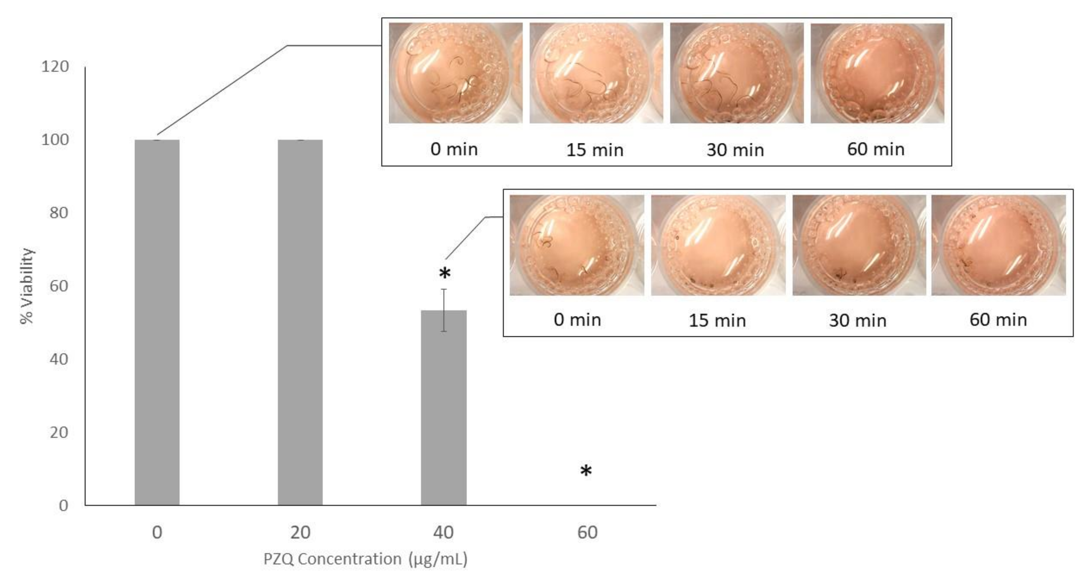
Figure 1. The 40 μg/mL concentration of praziquantel (PZQ) reduced viability of the worms by approximately 46.7%. S. mekongi were treated with increasing concentration of PZQ for an hour and parasites without movement for a minute were assumed as dead. Bar chart represents percentage of viable S. mekongi after PZQ treatment. Asterisk shows statistical significance (p value < 0.05).
Pathogens 09 00417 g001
Figure 2. Gene ontology classification of S. mekongi proteomic data after PZQ treatment. The Differential proteins after PZQ exposure were classified according to their gene ontology using Blast2Go. Three terms of gene ontologies including (A) biologic process, (B) molecular function, and (C) cellular component are presented.
Pathogens 09 00417 g002
Figure 3. Level of phosphorylation in tegument, muscle layer and internal cells decreased after PZQ treatment. Phosphorylation on untreated and PZQ-treated S. mekongi were tagged by anti-phosphoserine antibody. Immune-gold labeling and electron microscope were used to visualize parasite phosphorylation. Phosphorylation is indicated by black spots with arrows. The numbers of spots are markedly reduced in all organs after PZQ treatment.
Pathogens 09 00417 g003
Figure 4. Gene ontology classification of S. mekongi phosphoproteomic data after PZQ treatment. The differential phosphoproteins after PZQ exposure were classified according to their gene ontology using Blast2Go. Three terms of gene ontologies including (A) biologic process, (B) molecular function and (C) cellular component are presented.
Pathogens 09 00417 g004
Figure 5. Distribution of proteomic and phosphoproteomic data. Identification of proteins by mass spectrometer showed the total of 1167 proteins. Expression levels of 138 proteins changed after PZQ treatment without altering their phosphorylation. Thirty-seven phosphoproteins changed without changing their expression. Six proteins were altered both of their expression and phosphorylation.
Pathogens 09 00417 g005
Figure 6. Differentially expressed and phosphorylated proteins after PZQ treatment by the protein processing endoplasmic reticulum (ER) pathway. Differential proteins and phosphoproteins were analyzed for the potential pathways which were affected by PZQ using KEGG Automatic Annotation Server (KAAS) bioinformatic tool. Protein processing ER pathway was highlighted as the major pathway. The differential proteins and phosphorylated are indicated by green boxes and red borders, respectively.
Pathogens 09 00417 g006
Figure 7. Distribution of kinase groups involved in protein phosphorylation in vital pathways of S. mekongi. Phosphopeptides identified from differential phosphoproteins were used to predict kinases corresponding for their phosphorylation with Group-based Prediction System. Percentage of phosphorylation sites for predicted kinases in (A) calcium homeostasis, (B) worm antigen, (C) oxidative stress and (D) proteins folding and proteolysis are shown.
Pathogens 09 00417 g007
Figure 8. Graphical summary of the study. S. mekongi parasites were treated with IC50 dose of PZQ, anthelminthic drug. Protein lysate was extracted from untreated and PZQ-treated worms and subsequently enriched for phosphoproteins. Proteins and phosphoproteins were separated with 12% sodium dodecyl sulfate–polyacrylamide gel electrophoresis (SDS-PAGE) and protein bands were cut and digested by trypsin. Peptide was subjected to mass spectrometer for protein identification. Bioinformatics tools were used to analyze information from mass spectrometric data. Potential pathways involving with PZQ effects and kinases relating to those pathways were reported as possible targets for drug development.
Pathogens 09 00417 g008
Table 1. Top-20 S. mekongi proteins upregulated after 40 μg/mL-PZQ treatment.
No.Accession No. UniprotProtein NameMWpIProtein ScoreSequence CoverageAverage Fold-Change
Structural protein
1Q9Y1U7Myosin light chain18.34.53731.32
2Q26507Paramyosin, partial51.65.03522465.96
3Q26595Alpha tubulin49.94.9710417.5- 1
Energy
4A0A094ZC89L-lactate dehydrogenase A chain32.76.759719.62
5G4VP51Putative ADP,ATP carrier protein29.99.4751848.22.29
6C1LV81Aldolase39.56.5644753.43.27
7A0A095AJN4Enolase46.66.3413027.92.56
Protease
8C1L5C5Putative aminopeptidase W07G4.456.27.1436025.72.31
9A0A094ZYF3Putative aminopeptidase W07G4.457.67.5516925.32.16
10P43157Cathepsin B38.77.1452230.42.33
11A0A095B296Cathepsin B-like cysteine proteinase38.67.514338.82.05
Antioxidant
12Q26513Glutathione-S-transferase23.86.727619.4- 1
Transcription and translation
13C1LWP5Eukaryotic translation elongation factor 1 alpha 225.77.6714936.53.69
Immune system
14C1L7Y4Annexin A13 (Annexin XIII)39.55.124224.92.48
15Q86DV3Annexin36.76.1322536.43.05
16C1LKB8Prohibitin-2 (B-cell receptor-associated protein BAP37)28.89.7222638.53.29
Antigen
17 55kD antigen45.64.594020.2- 1
Unknown
18Q5DE25SJCHGC06488 protein30.25.5423225.93.54
19Q5DGY1Unknown38.77.8612321.33.22
20Q5DCK2SJCHGC06304 protein60.27.594611.62.4
1protein only identified in PZQ treatment.
Table 2. Top 20 S. mekongi proteins downregulated after 40 μg/mL-PZQ treatment.
No.Accession No.UniprotProtein NameMWpIProtein ScoreSequence CoverageAverageFold–Change
Structural protein
1G4VD36Tropomyosin, partial32.94.6261063.70 1
2Q02456Myosin heavy chain222.25.55100327.80 1
3G4M0G1Myosin heavy chain, putative182.45.687328.2−2.0
Energy
4B2LXU1Enolase46.76.7713024.7−3.12
5C1LFP4Putative aldehyde dehydrogenase 1B1 precursor53.56.0637928.9−4.0
6G4LWI3Aldehyde dehydrogenase, putative53.75.7611319.8−2.86
7B2LXU3Glyceraldehyde 3-phosphate dehydrogenase36.57.6815233.4−2.63
8C7TRL1Glyceraldehyde 3-phosphate dehydrogenase36.58.434033.7−2.63
9G4M130Sm14 fatty acid-binding protein delta E3 variant10.98.828542.901
Stress response
10G4VJ99Putative heat shock protein80.75.9132112.8−2.27
Signaling
11A0A095B084Pyruvate kinase PKM626.6313619.3−2.38
12C1LEA0TNF receptor-associated protein 180.26.243219.8−2.27
13C1LFZ8Arginine kinase80.18.465612.8−2.27
14A0A095ASB2Inositol hexakisphosphate and diphosphoinositol–pentakisphosphate kinase 21788.23348.3−2.0
Unknown
15Q5DAM7SJCHGC06305 protein61.16.4827828−3.33
16B3GUT7Unknown166.93318.20 1
17Q5DBJ9Unknown38.88.446922.2−2.0
18Q5C296SJCHGC01885 protein, partial111.85.2692437.3−2.04
19Q5DEG6SJCHGC00214 protein53.48.5630625.1−2.22
20Q5DGD4SJCHGC06677 protein90.65.086112.5−2.38
1protein only identified without treatment.
Table 3. Increased abundance of S. mekongi phosphoproteins after 40 μg/mL-PZQ treatment.
No.Accession No. UniprotProtein NamePhosphorylation SiteAverage Fold–Change
Peptide SequenceNumber
Structural protein
1A5A6F8ParamyosinQASEDRATR1 (ST)2
GVSPSTTRLESR1 (ST)
MMNHDTESHVKISR1 (ST)
TIYRGVSPSTTR5 (ST), 1 (Y)
GMRATSMM2 (ST)
ADMAERTVTVR1 (ST)
AKSSLESQVDDLK1 (ST)
LKAVSLEK1 (ST)
GVSPSTTR3 (ST)
QLTELNNAK1 (ST)
LEGLDSQLNR1 (ST)
LDEAGGSTTQTQELLKR3 (ST)
ISRTIYR2 (ST)
ATRLNNEVLR1 (ST)
ELSVTSNRGMR2 (ST)
KHAETELEEAQSR1 (ST)
2Q05870ParamyosinGVSPSTTRLESR1 (ST)2.47
MMNHDTESHVKISR1 (ST)
TIYRGVSPSTTR5 (ST), 1 (Y)
AERHAADLSYQVDALSER1 (ST)
GMRATSMM1 (ST)
ADMAERTVTVR1 (ST)
AKSSLESQVDDLK1 (ST)
VSELTIQVNTLSNDK1 (ST)
LKAVSLEK1 (ST)
GVSPSTTR3 (ST)
QLTELNNAK1 (ST)
LEGLDSQLNR1 (ST)
LDEAGGSTTQTQELLKR3 (ST)
3Q26507Paramyosin, partialATRLNNEVLR1 (ST)- 1
4A0A095BXK4Actin-1GTAASSSALEK4 (ST)- 1
MQKEISALAPSTMK1 (ST)
QEYDESGPSIVHRK1 (ST), 1 (Y)
EISALAPSTMK1 (ST)
DLYSNTVLSGGSTMYPGIADR1 (ST), 1 (Y)
5A0A094ZID2Actin-1EICNLAPTTMKIK2 (ST)- 1
KDLYSNIVLSGGSTMFPGISDR2 (ST)
Energy
6G4VBJ0Putative malate dehydrogenaseRIQEAGTEVVEAK1 (ST)2.25
AGAGSATLSMAYAGVR2 (ST), 1 (Y)
FGISFRSFLTSSK5 (ST)
LFGVTTLDVVR1 (ST)
SFLTSSKHSPK4 (ST)
AMICIITNPVNSTVPIAAEILK2 (ST)
7A0A095CGL878 kDa glucose-regulated protein, partialFDESTVQNDITHYPFSVVNKK3 (ST), 1 (Y)- 1
DIKTNK1 (ST)
EFAESYLGK1 (ST)
FDESTVQNDITHYPFSVVNKK3 (ST), 1 (Y)
Protease
8P43157Cathepsin BDIMMYGPVEAAFDVYEDFLNYK1 (Y)- 1
IAVYIVSLFTFLEAHVTTR3 (ST)
MATLGTGMR2 (ST)
AEISMLEGAVLDIRYGVSR1 (ST), 1 (Y)
IDDEINTLMTGALENPNEEITATMDK3 (ST)
YGVSRIAYSK2 (ST), 1 (Y)
MATLGTGMRCLK1 (ST)
Antigen
9Q0352822.6kd tegumental associated antigenMATTEYRLSLMEQFIR1 (ST)- 1
ATTEYR1 (ST), 1 (Y)
ATTEYRLSLMEQFIR1 (ST)
10P0841870,000 mol wt antigen/hsp70 homologue (619 AA), partialTTPSYVAFTDSER3 (ST), 1 (Y)- 1
MFSPEEISSMVLTKMK1 (ST)
11C1L7Y4Annexin A13 (Annexin XIII)IIGILGYR1 (Y)- 1
MLLTDTDKMNAR1 (ST)
Transcription and translation
12G4VAD2Putative elongation factor 1-alpha (EF-1-alpha)TEDNPKCIK1 (ST)- 1
ESGEMGKGSFK1 (ST)
KESAAK1 (ST)
MTDKPLR1 (ST)
MDCTEPPFSEDR1 (ST)
13A0A094ZN49S phase cyclin A-associated protein in the endoplasmic reticulumTASNTRSGSLISTNSR2 (ST)- 1
SGSLISTNSR2 (ST)
14G4M170Meiotic checkpoint regulator cut4, putativeGISSSSR2 (ST)- 1
HNSINMTTLR1 (ST)
GASFSSSTFR4 (ST)
CVDSLDSNKFLR1 (ST)
YTGNIVSSECQNNVGHIFQSSKR1 (ST)
GMLTFGILTGSK1 (ST)
SSGSGLFYECSRK3 (ST), 1 (Y)
WYIISNAPGTKSSYK1 (ST)
CPDQTFTDSLYHAYGR2 (ST)
VWASVIGRGMLTFGILTGSK1 (ST)
Immune system
15G4VL68Putative annexinSTSRLVSR2 (ST)- 1
CEVDMNTLKSMYR1 (ST), 1 (Y)
GDSDTFIK2 (ST)
EDTSGDFR2 (ST)
CEVDMNTLK1 (ST)
GLSTDEETITK2 (ST)
NVVITVPAYFNDSQR1 (ST)
Stress response
16A0A094ZG26Heat shock 70 kDa protein-like protein, partialRTLSSST4 (ST)- 1
LFSAEEISSMVLTK1 (ST)
17Q8MXA4Heat shock protein HSP60, partialEEMEASNSEYEKEK1 (ST), 1 (Y)- 1
DMAVGTGGIVFGDEADMYK1 (ST), 1 (Y)
SAMLVGVDILADADAVTMGPKGR1 (ST)
18O45038HSP70MKSSAESYLGK2 (ST)- 1
19G4LWI2Heat shock protein HSP60, putativeEEMEASNSEYEKEK1 (ST), 1 (Y)- 1
VGNDGTITVKMK1 (ST)
Transporter
20A0A094ZQ18Putative sodium-coupled neutral amino acid transporter 10SFQPTLSNMR3 (ST)- 1
NPPEPIDLTKIQSQMNVNNK1 (ST)
Antioxidant
21A0A095A0V3Peroxiredoxin-2 - 1
22 Glutathione S-transferaseGIPGNSSMASK2 (ST)- 1
AEISMLEGAVLDIRYGVSR1 (ST), 1 (Y)
YGVSRIAYSK2 (ST), 1 (Y)
23G4LXF826 Kd glutathione-S-transferase, Sm26, putative - 1
Unknown
24Q5DBM4SJCHGC01909 proteinMNHDSESHVK2 (ST)6.13
GVSPSTTRLESR1 (ST)
TIYRGVSPSTTR5 (ST), 1 (Y)
AERHAADLSYQVDALSER1 (ST)
GVSPSTTR3 (ST)
QLTELNNAK1 (ST)
LEGLDSQLNR1 (ST)
MNHDSESHVK2 (ST)
LDEAGGSTTQTQELLKR3 (ST)
ISRTIYR2 (ST)
25G4VM49Hypothetical protein Smp_179350WSSSSSNK1 (ST)- 1
FSSTSSTR3 (ST)
SINDNSSIEK2 (ST)
TMRSLDSCIVPR1 (ST)
ISGLTPVSTIFTGASNTTSTSTACGAVK4 (ST)
LNTSR1 (ST)
KQHSQSHDNLECSK1 (ST)
26B3GUU7Unknown - 1
27Q5DGY9SJCHGC06312 proteinMAGESGTKTESQSDESTK2 (ST)- 1
TESQSDESTK1 (ST)
DSETAESIK3 (ST)
28G4M072Hypothetical protein Smp_153120LSHSGVDDK1 (ST)- 1
QCLNLTSYSK1 (ST)
TPSSIKVYDLK1 (ST)
SKYGWAPNSLLLGK1 (Y)
MNDVTLSMVCNIR1 (ST)
TSASNTVHTIHDITVKR4 (ST)
1phosphoprotein only identified in PZQ treatment.
Table 4. Decreased abundance of S. mekongi phosphoproteins after 40 μg/mL-PZQ treatment.
No.Accession No. UniprotProtein NamePhosphorylation SiteAverage
Fold–Change
Peptide SequenceNumber
Structural protein
1G4VLW1Putative actinGYSFTTTAER2 (ST)0 1
DSYVGDEAQSK2 (ST)
GDEDVAALVIDNGSGMCK1 (ST)
YSVWIGGSILASLSTFQQMWISK1 (ST), 1 (Y)
2G4VD37Putative tropomyosinEKAEAEVAAMTR1 (ST)0 1
EESYEETIRDLTER3 (ST), 1 (Y)
LLEEDLEVSSSRLTETLTK2 (ST)
Energy
3C1LV81AldolaseHNMVTAGQACK1 (ST)−2.38
TLPTLLAERHIIPGIK1 (ST)
TLPTLLAER1 (ST)
Antigen
4C1L9U2Tegument antigen (I(H)A)EGKVSTLPLVIQIIAATMSK2 (ST)0 1
Scaffold
5G4VM27Putative zinc finger proteinDNQSSNLEESFKHECDSDK1 (ST)0 1
YIQRSGLNR1 (Y)
TFTTKSHLNK2 (ST)
IKYSCVQCYK1 (Y)
DNQSSNLEESFKHECDSDK1 (ST)
Antioxidant
6O97161Thioredoxin peroxidaseKSGGLGHMK1 (ST)0 1
Transcription and translation
7A0A094ZK92Putative ATP-dependent RNA helicase DDX56LSEVDGFRYR1 (ST)0 1
CRDVEYGVSR1 (Y)
ELCSQVASNIKCLCK1 (ST)
8G4VDN8Putative dead box ATP-dependent RNA helicaseLSEVDGFRYR1 (ST)0 1
CRDVEYGVSR1 (Y)
ELCSQVASNIKCLCK1 (ST)
Stress response
9A0A094ZZQ160 kDa heat shock protein, mitochondrialSPKITK1 (ST)0 1
GTLAPAR1 (ST)
GSKTDVDK2 (ST)
DGVTVAKGIELK1 (ST)
EESGANVAGMGGMGGMGGMGGMM1 (ST)
Signaling
10A0A095A3D320 kDa calcium-binding proteinFNNDSYK1 (Y)0 1
QCLTNHSNQSIR1 (ST)
ENSSTMR2 (ST)
SVRLSIIPLNPIYK1 (ST), 1 (Y)
11G4LZ30Calcium-binding protein, putativeLVSKENPSTMR3 (ST)0 1
SVRLSIIPLNPIYK1 (ST), 1(Y)
Unknown
12A0A095CG55Hypothetical protein MS3_10497, partialAQAVISQR1 (ST)0 1
SLLSALSTVYSHR3 (ST), 1 (Y)
13G4VH32Hypothetical protein Smp_179900SQISNSEASNFDNSK1 (ST)0 1
VVVHSTVSMPFTPSGLSTGNR2 (ST)
14C7TYF2Hypothetical proteinTVINEMSLIPSINK1 (ST)0 1
15G4V5K0Hypothetical protein Smp_057140QSSSFFMR1 (ST)0 1
YIIASARQR1 (ST)
SLLSALSTVYSHR3 (ST), 1 (Y)
HSSGSDSVVGAITTR4 (ST)
QSCSLQYKYIIASAR1 (Y)
1phosphoprotein only identified without treatment.
Table 5. Proteomic and phosphoproteomic analysis of proteins involved in calcium binding, worm antigen oxidative stress and protein folding and proteolysis pathway.
No.Accession
No. Uniprot
Protein NameStatus
ExpressionPhosphorylation
Proteins involving with calcium binding
1C1L7Y4Annexin A13 (Annexin XIII)Up
2C1LNX4Voltage-dependent anion-selective channel protein 2Down
3G4M1U8Voltage-dependent anion-selective channel, putativeDown
4G4VL68Putative annexinDownUp
5Q0352822.6kd tegumental associated antigenUp
6G4LZ30Calcium-binding protein, putativeDown
7A0A095A3D320 kDa calcium-binding proteinDown
8C1L9U2Tegument antigen (I(H)A)Down
9G4VH32Hypothetical protein Smp_179900Down
Proteins involving with worm antigens
10Q26595Alpha tubulinUp
11Q86DV9Similar to GenBank Accession Number Z29075 myophilin antigen in Echinococcus granulosusUp
12A0A094ZPJ2Putative citrate synthase 2, mitochondrialUp
13 55kD antigenUp
14Q06814AntigenDown
15Q5D9C5SJCHGC09453 proteinDown
16P0841870,000 mol wt antigen/hsp70 homolog (619 AA), partialUp
17Q0352822.6kd tegumental associated antigenUp
18C1L9U2Tegument antigen (I(H)A)Down
19A0A094ZZQ160 kDa heat shock protein, mitochondrialDown
Proteins involving with oxidative stress
20C1LV40Tryparedoxin peroxidaseUp
21Q26513Glutathione-S-transferaseUp
22A0A094ZC89L-lactate dehydrogenase A chainUp
23P37227Malate dehydrogenase, partialUp
24C1LD38Glyceraldehyde 3-phosphate dehydrogenaseUp
25A0A095AJN4EnolaseUp
26Q7Z1I3Lactate dehydrogenase-like proteinUp
27A0A094ZPJ2Putative citrate synthase 2, mitochondrialUp
28Q8WT66Phosphoglycerate mutaseUp
29C1LV81AldolaseUp
30C1LFZ8Arginine kinaseDown
31A0A095B084Pyruvate kinase PKMDown
32C7TRL1Glyceraldehyde 3-phosphate dehydrogenaseDown
33C1LD30Glyceraldehyde 3-phosphate dehydrogenaseDown
34B2LXU3Glyceraldehyde 3-phosphate dehydrogenaseDown
35B2LXU1EnolaseDown
36C1LFP4Putative aldehyde dehydrogenase 1B1 precursorDown
37G4LWI3Aldehyde dehydrogenase, putativeDown
38A0A095A4B6Aldehyde dehydrogenase X, mitochondrialDown
39Q5DAK2SJCHGC06828 proteinDown
40Q5BXP5SJCHGC05927 protein, partialDown
41Q5DAM7SJCHGC06305 proteinDown
42Q5DFZ8SJCHGC00411 proteinDown
43C1L6T7Hypothetical proteinDown
44G4VBJ0Putative malate dehydrogenaseUp
45A0A095A0 V3Peroxiredoxin-2Up
46G4LXF826 Kd glutathione-S-transferase, Sm26, putativeUp
47O97161Thioredoxin peroxidaseDown
Proteins involving with protein folding and proteolysis
48P43157Cathepsin BUpUp
49C1L5C5Putative aminopeptidase W07G4.4Up
50A0A094ZYF3Putative aminopeptidase W07G4.4Up
51A0A095B296Cathepsin B-like cysteine proteinaseUp
52C1LA34Cathepsin B-like cysteine proteinase precursorUp
53C1L8N7Putative Lysosomal Pro-X carboxypeptidase precursorUp
54G4LYP1Mitochondrial processing peptidase beta-subunit (M16 family)Up
55G4VPS5Putative chaperonin containing t-complex protein 1, theta subunit, tcpqUp
56A0A095AUU9T-complex protein 1 subunit zetaUp
57A0A095CBQ7T-complex protein 1 subunit thetaUp
58A9CBJ4DNA repair proteinUp
59Q26551Cyclophilin BUp
60Q5DC69SJCHGC01960 protein, partialUp
61Q5DDG6SJCHGC02536 proteinUp
62Q5DE64UnknownUp
63Q5C3A0UnknownUp
64Q5DGY1UnknownUp
65C1L6Z6Mitochondrial processing peptidaseDown
66C1LNT2Serine protease inhibitor serpinDown
67O45039HSP70, partialDown
68C7TZI9Heat shock protein 90kDa alpha, partialDown
69G4VJ99Putative heat shock proteinDown
70C1LEA0TNF receptor-associated protein 1Down
71Q06814CalreticulinDown
72Q5BYY8SJCHGC04813 protein, partialDown
73Q5C3 V3SJCHGC05011 protein, partialDown
74Q5DBB0SJCHGC02058 proteinDown
75Q5DGD4SJCHGC06677 proteinDown
76Q5D9K8UnknownDown
77Q5DBJ9UnknownDown
78G4LWI2Heat shock protein HSP60, putativeUp
79Q8MXA4Heat shock protein HSP60, partialUp
80Q5DGY9SJCHGC06312 proteinUp
81A0A094ZZQ160 kDa heat shock protein, mitochondrialDown