Article

First Detection of Carbapenem-Resistant Escherichia fergusonii Strains Harbouring Beta-Lactamase Genes from Clinical Samples

Department of Microbiology, College of Science and Technology, Covenant University, 112103 Ota, Nigeria
*
Author to whom correspondence should be addressed.
Pathogens 2019, 8(4), 164; https://doi.org/10.3390/pathogens8040164
Submission received: 1 April 2019 / Revised: 26 July 2019 / Accepted: 26 July 2019 / Published: 25 September 2019

Abstract

:
Recently discovered extraintestinal Escherichia fergusonii obtained from non-clinical samples has exhibited the potential for acquiring multiple beta-lactamase genes, just like many extraintestinal Escherichia coli strains. Albeit, they are often omitted or classified as E. coli. This study aimed to, therefore, identify carbapenem-resistant extended-spectrum beta-lactamase (ESBL) producing E. fergusonii isolates from clinical samples, determine their evolutionary relatedness using 16S rRNA sequencing analysis and screen for beta-lactamase genes. A total of 135 septic wound samples were obtained from patients on referral at a General Hospital in Lagos, Nigeria. For the phenotypic identification of isolates from culture-positive samples, morphological, and physiological tests were carried out. Identities of the isolates harbouring beta-lactamase genes were assigned to their genus strains using the 16S rRNA sequencing. The Kirby Bauer disc diffusion technique and double-disc synergy test were used to screen isolates for multidrug resistance and ESBL production. Carbapenem-resistant ESBL producing isolates were screened for beta-lactamase genes in a polymerase chain reaction. Three E. fergusonii isolates (CR11, CR35 and CR49) were obtained during this study. E. fergusonii strains were motile, non-lactose and non-sorbitol fermenting but positive for cellobiose and adonitol fermentation. The I6S rRNA assigned the phenotypically identified isolates to E. fergusonii species. All three isolates were multidrug-resistant, carbapenem-resistant and ESBL producers. Isolates CR11 and CR35 harboured cefotaximase (CTX-M) and temoniera (TEM) beta-lactamase genes while CR49 harboured sulfhydryl variable (SHV) beta-lactamase gene. We herein report the detection of multiple beta-lactamase genes in carbapenem-resistant ESBL producing E. fergusonii from clinical samples.

1. Introduction

Multidrug-resistant Extraintestinal pathogenic E. coli (ExPEC) clones have contributed significantly to the growing antibiotics resistance burden [1,2]. Several clinical multidrug-resistant ExPEC clones, including ST131, ST73 and ST40, reported in previous studies, have acquired resistance plasmids that encode multiple beta-lactamase genes, particularly those required for Extended-spectrum beta-lactamase (ESBL) production [3,4,5,6,7]. This facilitates their capacity in playing significant roles in the global dissemination of ESBL genes [1,8,9]. Extraintestinal E. coli ST131 clones (clade C1 and H30R) encoding CTX-M-15 genes have been reported in Japan, Thailand, Canada and Australia [10]. In Nigeria, several clones of multidrug-resistant ExPECs (ST131, ST295, ST617, ST501, ST448 and ST617) have also rapidly acquired transmissible plasmids that encode several beta-lactamase genes, including the CTX-M-15 genes [11].
Another extraintestinal human pathogen within the genus Escherichia has also shown tendencies for the dissemination of plasmid-mediated beta-lactamase genes. Escherichia fergusonii closely related to the ExPECs with about 60% similarity, was classified in 1985 as a new specie [7]. Escherichia fergusonii are opportunistic pathogens primarily associated with abdominal wounds, UTIs and bacteraemia in humans while causing septicaemia and diarrhoea in animals [7,12,13,14,15,16,17,18]. Not much is currently known about E. fergusonii. However, they are beginning to gain attention because several reports of multidrug-resistant E. fergusonii exists today. The first detection of ESBL production in clinical isolates of E. fergusonii was by Lagacé-Wiens et al. [19]. Although sulfhydryl variable (SHV-12) and CTX-M-1 conferring ESBL phenotypes have been reported in E. fergusonii from non-clinical samples, there is yet to be any report from human samples [7,20].
Most multidrug-resistant ExPEC isolated from bacterial infections in Nigeria are ESBL producers harbouring multiple beta-lactamase genes [21,22,23,24]. Moreover, recent studies on carbapenem-resistant Enterobacteriaceae infections in Nigeria have identified multidrug-resistant extraintestinal Escherichia sp. as a major causative organism [25,26,27]. However, identification of these organisms is often phenotypic based. We report herein, the 16S rDNA sequencing of carbapenem-resistant ESBL producing extraintestinal Escherichia spp. encoding multiple beta-lactamase genes.

2. Results

A total of three E. fergusonii isolates designated CR11, CR35 and CR49 were classified to distinguish them from other Escherichia species associated from wound infections. Isolates were motile, non-lactose fermenters, positive for cellobiose and adonitol but negative for sorbitol fermentation (Table 1). Following 16S rRNA sequencing analysis, the phenotypically identified isolates were assigned to E. fergusonii specie group. The isolates; CR11, CR35 and CR49 were renamed as E. fergusonii ADE4 (MH040118), E. fergusonii ADE11 (MH04009393) and E. fergusonii ADE18 (MH040100), respectively. In Figure 1, the phylogenetic tree constructed using partial 16S rDNA sequences of 1349 bp (E. fergusonii ADE4), 1390 bp (E. fergusonii ADE11) and 1385 bp (E. fergusonii ADE18), showed that all three isolates obtained from this study shared mostly the same close relatives. The sequence similarity percentages of Escherichia fergusonii ADE4 when compared with other 16S rRNA gene sequences in Genbank were 99.33% to Escherichia fergusonii ATCC 35469 (NR_027549.1), 99.33% to Shigella flexneri ATCC 29903, 99.26% to Shigella sonnei CECT 4887, 99.19% to Escherichia fergusonii NBRC 102419, 99.19% to Escherichia fergusonii ATCC 35469 (NR_074902) and 99.04% to Escherichia coli NBRC 102203. The 16S rRNA gene sequence similarity percentages of Escherichia fergusonii ADE11 were 99.56% to Escherichia fergusonii ATCC 35469 (NR_027549.1), 99.56% to Shigella flexneri ATCC 29903, 99.48% to Escherichia fergusonii ATCC 35469 (NR_074902.1), 99.48% to Shigella sonnei CECT 4887, 99.41% to Escherichia fergusonii NBRC 102419 and 99.33% to Escherichia coli NBRC 102203. For Escherichia fergusonii ADE18, sequence similarity percentages were 99.49% to Escherichia fergusonii ATCC 35469 (NR_027549.1), 99.49% to Shigella flexneri ATCC 29903, 99.42% to Escherichia fergusonii ATCC 35469 (NR_074902.1), 99.34% to Escherichia fergusonii NBRC 102419, 99.34% to Escherichia coli NBRC 102203 and 99.42% to Shigella sonnei CECT 4887.
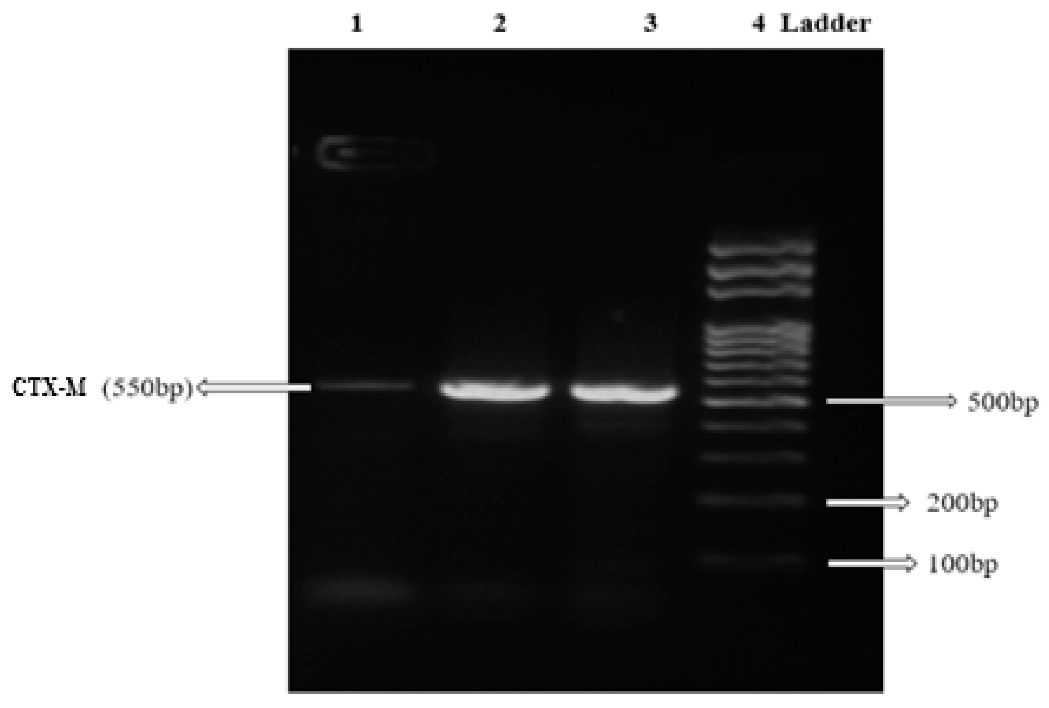
Antibiotics susceptibility patterns of CR11, CR35 and CR49 based on CLSI interpretative criteria showed multidrug resistance. All three isolates exhibited 100% resistance to all beta-lactam antibiotics, including the extended-spectrum beta-lactam (ceftriaxone, cefuroxime and ceftazidime) and carbapenem (imipenem and meropenem) antibiotics. Isolates CR11 and CR35 were also resistant to other antibiotics class such as fluoroquinolones (levofloxacin, Ofloxacin, ciprofloxacin) and nitrofurantoin. All three isolates expressed ESBL production. Polymerase chain reaction (PCR) revealed that CR11, CR35 and CR49 encoded different beta-lactamase genes (Table 2). Isolates CR11 and CR35 harboured CTX-M type beta-lactamase (Figure 2) and TEM beta-lactamase genes while CR49 harboured SHV beta-lactamase gene. No carbapenemase gene was present in the three isolates from this study.

3. Discussion

This study to the best of our knowledge, reports the first isolation of carbapenem-resistant ESBL producing E. fergusonii of clinical importance, contrary to the first published study on the isolation of E. fergusonii from drinking water sources, in Nigeria [28]. Some studies suggest that E. fergusonii had existed long before the first identification in 1985 but were often omitted or misidentified as E. coli, due to the low discriminatory power of phenotypic methods [7,28]. Previous studies have also reported several inconsistencies with the use of biochemical methods including API 20E and Vitek 2 (Vitek System; bioMérieux, Marcy l’Etoile, France and bioMerieux, Inc., Durham, NC), for identification of E. fergusonii [29,30]. Recently, the ability to ferment cellobiose and adonitol and inability to ferment lactose and sorbitol among the Escherichia sp. has been reported to be specific for E. fergusonii and can be a guide for differentiating E. fergusonii from other members of the genus Escherichia [7]. Escherichia fergusonii isolates obtained from our study were motile, utilised cellobiose and adonitol but did not ferment lactose and sorbitol. The first published biochemical profile of E. fergusonii revealed that they are motile and non-lactose fermenters, confirming our findings [31]. The study of Balqis et al. [32] and Maheux et al. [16] however reported varying patterns for lactose fermentation and motility.
Not much information on phylogenetic studies of E. fergusonii are currently available, but phylogenetic analysis from this study revealed that E fergusonii ADE4, ADE11 and ADE18 shared the same clade with the genus Escherichia and Shigella. This is similar to previous reports on Escherichia fergusonii [31,33,34]. The genus Escherichia and Shigella are considered members of the same pathogenic lineage [35,36,37,38,39]. Naveena et al. [40] reported that Shigella and E. coli are phylogenetically the same but separated based on their clinical relevance and biochemical differences. Shigella and Escherichia are closely related genera with >99% 16S rDNA sequence similarity [40]. Whole-genome phylogeny analysis carried out by Sims and Kim [41], reported clustering of several members of the genus Shigella with different Escherichia phylogroups. Maheux et al., 2014 also reported the difficulty of using gene sequence alone in distinguishing non-E. coli Escherichia spp. strains in a group comprising E. coli, Shigella spp., E. fergusonii, and E. albertii. However, the use of a polyphasic identification method that combines 16S rDNA sequencing analysis with phenotypic methods has helped to differentiate E. fergusonii from Shigella sp., in this present study. Although both genera have very similar phenotypic traits, several differences in the biochemical characteristics of E. fergusonii have helped to differentiate them from Shigella sp. Escherichia fergusonii obtained from this study were motile, with the ability to ferment cellobiose and adonitol (Table 1), whereas members of the genus Shigella are non-motile, unable to ferment cellobiose and adonitol [42]. Also, E. fergusonii from this study were non-lactose fermenters (Table 1), unlike the slow lactose-fermenting S. sonnei [36].
The three carbapenem-resistant ESBL producing E. fergusonii stains obtained in this present study were resistant to imipenem, meropenem, ceftriaxone, ceftazidime and cefuroxime antibiotics. This finding is consistent with several studies that reported multiple resistance to broad-spectrum beta-lactam antibiotics in E. fergusonii isolated from animals and humans [17,19,31,43,44,45]. Although Savini et al. [17] reported that resistance to broad-spectrum cephalosporin antibiotics by non-ESBL producing E. fergusonii from their study was due to non-compliance of the patient to the use of cephalosporin antibiotics administered, the E. fergusonii strains from this study were ESBL producers. Hence, resistance to broad-spectrum cephalosporins observed in this study was likely due to the presence of beta-lactamase resistance genes detected in the E. fergusonii strains. Escherichia fergusonii ADE4 and ADE11 strains harboured CTX-M genes (See Table 2). Our finding is similar to the first report of E. fergusonii from a farm animal in South Korea, harbouring CTX-M gene [45]. Our study could, however, be the first documented report of CTX-M gene in E. fergusonii from human samples. The SHV and TEM beta-lactamase genes were also detected in Escherichia fergusonii ADE18 and Escherichia fergusonii ADE4/ADE11, respectively. This corresponds with the study that reported SHV-12 as the first beta-lactamase gene detected in E. fergusonii from a human specimen [19]. Resistance to imipenem and meropenem antibiotics was observed as well in this study, supporting the first report on carbapenem-resistant E. fergusonii strains among 23 E. fergusonii isolates obtained from non-human primates [7]. The first report of carbapenem-resistant E. fergusonii from human samples is likely to be this study. None of the screened carbapenemase genes was present within the three E. fergusonii strains from this study. Resistance to carbapenem antibiotics could have been influenced by the beta-lactamase genes harboured by the E. fergusonii strains or was conferred by other carbapenemase gene types (e.g. OXA-48) not investigated in this study.
The limitation of this study was due to limited resources. Beta-lactamase genes detected during this study were not sequenced to determine their spectrum of activity and some carbapenemase genes were not screened. Despite these limitations, this study has shown that E. fergusonii can be a reservoir for the spread of antibiotics resistance and a threat to the effective treatment of bacterial infections they cause.

4. Materials and Methods

4.1. Culture and Identification of Clinical Isolates

4.1.1. Culture and Phenotypic Identification of Clinical Isolates

A total of 135 samples were obtained from septic wound patients on referral to Government general hospital Odan, Lagos, Nigeria. Wound swabs were obtained using sterile swab sticks, labelled appropriately and transported to the laboratory within 2-4 h of collection for further analysis. Samples were cultured on sterile MacConkey agar plates (Rapid Lab, UK), using the streak plate method and incubated for 24 h at an optimum temperature of 37 °C. Positive cultures were subcultured on MacConkey agar to obtain pure isolates [46]. All isolates were phenotypically identified following standard routine morphological and cultural characteristics, as well as biochemical tests such as Gram stain reaction, sulphur reduction, indole, motility, methyl red, voges-Proskauer, urease, oxidase, sugar fermentation and citrate utilisation test [46]. Pure isolates phenotypically identified as E. coli. were stored in 20% Luria-Bertani (LB) broth (HIMEDIA, India) at −80 °C for long-term storage while working stocks for further analysis were held at 4 °C. Escherichia sp. were further screened for the ability to utilise cellobiose, sorbitol and adonitol. Motile isolates positive for cellobiose and adonitol fermentation but negative for lactose and sorbitol utilisation were designated CR11, CR35 and CR49.

4.1.2. Genotypic Identification of Isolates

A discrete colony of CR11, CR35 and CR 49 in overnight broth cultures were used for the 16S rRNA gene sequencing method [47]. Genomic DNA of isolates CR11, CR35 and CR 49 were extracted using a commercial genomic DNA extraction kit (AidLab, China), following manufacturer’s instruction. Bacteria universal primer (27F: f5′-AGAGTTTGATCCTGGCTCAG-3′ and 1492R: r5′-GGTTACCCTTGTTACGACTT-3′) were used to amplify 16S rRNA gene of selected carbapenem-resistant ESBL producing isolates, in a simplex polymerase chain reaction (PCR), as described previously [48]. Briefly, initial denaturation of PCR mixture was carried out at 94 °C for 5 min, followed by 35 cycles at 94 °C for 30 s, 52 °C for 30 s, 68 °C for 60 s and a final extension at 68 °C for 5 min. 30 s. The PCR products were analysed by electrophoresis in Tris-borate EDTA buffer and viewed using UV fluorescence in a gel image documentation system. PCR pleural was purified using a PCR clean-up kit (Zymo Research, U.S.A.) [48] and eluted for sequencing at Macrogen Corp, Maryland, USA. Nucleotide sequences were determined using automated sequencer (Applied Biosystems SeqStudio Genetic Analyzer) and the dye-deoxy termination procedure. All 16S rRNA gene sequences obtained were edited using Bioedit (version 7.0.26) software and aligned by multiple sequence alignment technique using CLUSTAL W. The obtained bacterial DNA sequence was compared with other 16S rRNA genes in the GenBank, using the NCBI Basic Local alignment search tools BLAST-n program and a phylogenetic tree was constructed using Neighbour-Joining method in MEGA version 7.0.26 according to Saitou and Nei [49].

4.2. Antimicrobial Susceptibility Testing

Antimicrobial susceptibility patterns of CR11, CR35 and CR49 were determined by the disc diffusion method according to the Clinical and Laboratory Standards [50]. Multiple discs (Rapid Labs, UK) containing Ceftazidime (CAZ, 30 µg), Cefuroxime (CXM, 30 µg), Cefotaxime (CTX, 30 µg), Nitrofurantoin (ATM, 300 µg), Gentamicin (GEN, 10 µg), Ciprofloxacin (CIP, 5 µg), Ofloxacin (OFX, 5 µg), Augmentin (AUG, 30 µg) and Ampicillin (10 µg) antibiotics, frequently used within the community and healthcare setting in Nigeria, were tested. Single discs of Ceftriaxone (CRO, 30 µg), penicillin G (PEN G, 10 µg), imipenem (10 µg), Ertapenem (10 µg) and meropenem (10 µg) purchased from OxoidTM (United Kingdom) were also assessed. In brief, isolate suspensions diluted using 0.5 McFarland standard were streaked with a sterile cotton swab on freshly prepared Mueller Hinton agar (MHA) plates. Multiple and single antibiotics discs (Rapid Labs, UK) to be tested were carefully placed on the seeded plate with the aid of a sterile forceps. Plates were inverted and transfer into the incubator for 15–18 h at 37 °C. Escherichia coli ATCC 25922 was used as control strains following CLSI recommendation [50]. Results of antimicrobial susceptibility testing were interpreted as sensitive, intermediate or resistant, based on the zone diameter interpretive standard for Enterobacteriaceae, as published by the Clinical Laboratory Standards Institute [50].

4.3. Detection of ESBL Production

Double disc synergy test (DDST) was carried out using OxoidTM (United Kingdom) purchased augmentin (amoxicillin-clavulanate) and extended-spectrum cephalosporin antibiotics, following the recommended European Committee on Antimicrobial Susceptibility Testing breakpoint [51,52]. Ceftazidime (CAZ, 10 µg) and cefotaxime (CTX, 5 µg) disc alone and in combination with amoxicillin-clavulanate (AMX, 20/10 µg) were used to detect ESBL activity in isolates with reduced susceptibility to extended-spectrum cephalosporins. Mueller-Hinton agar (MHA) plates prepared according to manufacturer’s standard were seeded with isolate (CR11, CR35 and CR49) suspensions of 0.5 McFarland turbidity standard. Antibiotic discs were placed with sterile forceps 25mm apart, centre to centre of the plates and incubated aerobically for 18 h at 37 °C. Escherichia coli ATCC 25922 strain was used as control. ESBL results were interpreted following the zone diameter interpretive clinical standard of the European Committee on Antimicrobial Susceptibility Testing. An increased inhibition zone diameter of ≤ 20mm centre to centre, augmented in the direction of amoxicillin-clavulanate antibiotics was recorded positive for ESBL production.

4.4. DNA Extraction and Genotypic Detection of β-lactamase Genes

DNA was extracted from CR11, CR35 and CR 49. DNA extraction was carried out using TENS (Tris-HCl; 10 mM (pH 8.0), EDTA; 1 mM, NaOH; 0.1 N, SDS; 0.5% (w/v)) miniprep [53,54]. Discrete colonies of overnight cultures were inoculated into 1.5 mL of Luria Bertani (LB) broth (HIMEDIA, India) in 2 mL Eppendorf tubes and incubated overnight for 18 h at 37 °C. Eppendorf tubes containing overnight cultures were spinned in a C1008-C centrifuge (Benchmark Scientific, USA) at 10,000 rpm for 1 min, to harvest cells. Supernatants were discarded, and bacterial pellets were resuspended in 1000 μL of phosphate-buffered saline (PBS), to remove cellular debris. Tubes containing resuspended cells were centrifuged at 10,000 rpm for 1 min, and supernatants were discarded. Aliquots of 300 μL of TENS buffer were added to bacterial pellets, gently inverted 3-4 times, to lyse bacterial cells. Aliquots of 150 μL 3M NaOAc (pH 5.6) were added, and the tubes were gently inverted 3-4 times to precipitate DNA. The tubes were centrifuged at 13,000 rpm for 5 min to pellet white precipitates. Clear supernatants containing DNA were carefully removed and transferred into new sterile Eppendorf tubes, using sterile 200 μL pipettes. Aliquots of 900 μL of 95% ethanol were added to clear supernatants and tubes were inverted. The tubes were centrifuged at maximum speed for 2 min to pellet DNA. Supernatants were discarded, and 500 μL of 70% ethanol were pipetted into tubes to wash the DNA pellets. The obtained DNA pellets were centrifuged for 2 min, ethanol solutions were discarded, and DNA pellets were dried in a CentriVap DNA [53,54] concentrator 79820 (Labconco, USA). DNA was dissolved in 50 μL 10mM Tris-HCl (pH 8) and chilled on ice for further experiment. DNA was screened for eight different beta-lactamase genes, including ESBLs (SHV, TEM and CTX-M genes), Oxacillinase (OXA) and carbapenemases (IMP, VIM, KPC and NDM genes). Beta-lactamase genes were screened for using specific primers (Table 3) as previously described [55,56,57,58]. For polymerase chain reaction (PCR) amplification of beta-lactamase genes in a thermocycler C100 Touch (Bio-RAD, USA), 5 μL aliquot of DNA template was added to a 20 μL master mix. Master mix contained 0.5 μL of deoxynucleotide (dNTPs) solution (New England Biolabs, UK), 0.5 μL each of primer pairs (forward and reverse), 0.125 μL Thermus aquaticus YT-1 (Taq) DNA polymerase (New England Biolabs, UK), 2.5 μL 10x standard Taq reaction buffer (New England Biolabs, UK) and 16.375 μL nuclease-free water (New England Biolabs, UK). The PCR mixture was preheated at 94 °C for 5 min, followed by 35 cycles at 94 °C for 30 s, 30 s at annealing temperature (Table 3), 60 s at 72 °C, and 72 °C for 7 min. About 4 μL of amplified PCR mixture was resolved on agarose (1%) gel electrophoresis stained with 4 μL ethidium bromide. Expected amplicon size was visualised under a UV fluorescence in a gel image documentation system 220 (Bio-Rad, UK).

4.5. Statistical Analysis

Results from this study were analysed using the SPSS version 20.0 statistical package. Results were presented in tables, figures and dendrogram.

5. Conclusions

The results of 16S rRNA gene sequencing analysis and phylogeny were consistent with the phenotypic identification of carbapenem-resistant ESBL-producing strains obtained from this study. However, for molecular identification and determination of evolutionary relationships, the 16S rRNA gene analysis method alone was not sufficient to adequately distinguish E. fergusonii stains from other closely related species. The use of partial 16S rDNA sequences (1349–1390 bp) of the three isolates for the construction of the phylogenetic tree might have contributed to the difficulty associated with differentiating Shigella from E. fergusonii, genotypically in this study. Further molecular analysis such as DNA-DNA hybridisation or multilocus sequence typing (MLST) is recommended to complement 16S rRNA gene analysis, for revealing molecular differences at the species level. Molecular characterisation of resistance genes detected during this study will as well throws more light on their location and clinical relevance.

Author Contributions

Conceptualization, T.A.; methodology, T.A., O.N., N.D. and E.O.; investigation, T.A. O.N., N.D. and E.O.; Manuscript writing-original draft preparation, T.A.; manuscript review and editing, T.A., O.N., E.O. and O.A.

Funding

No funding was received for this study.

Acknowledgments

The authors would like to appreciate every laboratory staff in the Department of Biological Sciences (Microbiology unit), Covenant University. Authors are grateful to Covenant University for defraying the cost of this publication.

Conflicts of Interest

The authors declare no conflict of interest.

References

  1. Manges, A.R.; Johnson, J.R. Reservoirs of Extraintestinal Pathogenic Escherichia coli. Microbiol. Spectr. 2015, 3, 1–2. [Google Scholar] [CrossRef] [PubMed]
  2. Mathers, A.J.; Peirano, G.; Pitout, J.D.D. The role of epidemic resistance plasmids and international high-risk clones in the spread of multidrug-resistant Enterobacteriaceae. Clin. Microbiol. Rev. 2015, 28, 565–591. [Google Scholar] [CrossRef] [PubMed]
  3. Riley, L.W. Pandemic lineages of extraintestinal pathogenic Escherichia coli. Clin. Microbiol. Infect. 2014, 20, 380–390. [Google Scholar] [CrossRef] [PubMed]
  4. Singer, R.S. Urinary tract infections attributed to diverse ExPEC strains in food animals: Evidence and data gaps. Front. Microbiol. 2015, 6, 28. [Google Scholar] [CrossRef] [PubMed]
  5. Gomi, R.; Matsuda, T.; Matsumura, Y.; Yamamoto, M.; Tanaka, M.; Ichiyama, S.; Yoneda, M. Whole-Genome Analysis of Antimicrobial-Resistant and Extraintestinal Pathogenic Escherichia coli in River Water. Appl. Environ. Microbiol. 2017, 83, e02703-16. [Google Scholar] [CrossRef] [PubMed]
  6. Poolman, J.T.; Wacker, M. Extraintestinal Pathogenic Escherichia coli, a Common Human Pathogen: Challenges for Vaccine Development and Progress in the Field. J. Infect. Dis. 2016, 213, 6–13. [Google Scholar] [CrossRef] [PubMed]
  7. Glover, B.; Wentzel, J.; Jenkins, A.; Van Vuuren, M. The first report of Escherichia fergusonii isolated from non-human primates, in Africa. One Heal. 2017, 3, 70–75. [Google Scholar] [CrossRef] [PubMed]
  8. Kim, Y.A.; Lee, K.; Chung, J.E. Risk factors and molecular features of sequence type (ST) 131 extended-Spectrum-β-lactamase-producing Escherichia coli in community-onset female genital tract infections. BMC Infect. Dis. 2018, 18, 250. [Google Scholar] [CrossRef] [PubMed]
  9. Tsai, M.; Lee, I.; Chu, S.; Lien, R.; Huang, H.; Chiang, M.; Fu, R.; Hsu, J.; Huang, Y. Clinical and Molecular Characteristics of Neonatal Extended-Spectrum β-Lactamase-Producing Gram-Negative Bacteremia: A 12-Year Case-Control-Control Study of a Referral Center in Taiwan. PLoS ONE 2016, 11, e0159744. [Google Scholar] [CrossRef]
  10. Matsumura, Y.; Pitout, J.D.D.; Gomi, R.; Matsuda, T.; Noguchi, T.; Yamamoto, M.; Peirano, G.; DeVinney, R.; Bradford, P.A.; Motyl, M.R.; et al. Global Escherichia coli Sequence Type 131 Clade with bla CTX-M-27 Gene. Emerg. Infect. Dis. 2016, 22, 1900–1907. [Google Scholar] [CrossRef]
  11. Aibinu, I.; Odugbemi, T.; Koenig, W.; Ghebremedhin, B. Sequence Type ST131 and ST10 Complex (ST617) predominant among CTX-M-15-producing Escherichia coli isolates from Nigeria* *This study has been partially presented during the 51st Interscience Conference on Antimicrobial Agents and Chemotherapy (ICAAC) in Chicago, IL, September 2011. Clin. Microbiol. Infect. 2012, 18, E49–E51. [Google Scholar] [PubMed]
  12. Funke, G.; Hany, A.; Altwegg, M. Isolation of Escherichia fergusonii from four different sites in a patient with pancreatic carcinoma and cholangiosepsis. J. Clin. Microbiol. 1993, 31, 2201–2203. [Google Scholar] [PubMed]
  13. Gokhale, V.V.; Therese, K.L.; Bagyalakshmi, R.; Biswas, J. Detection of Escherichia fergusonii by PCR-based DNA sequencing in a case of delayed-onset chronic endophthalmitis after cataract surgery. J. Cataract Refract. Surg. 2014, 40, 327–330. [Google Scholar] [CrossRef] [PubMed]
  14. Lai, C.C.; Cheng, A.; Huang, Y.T.; Chung, K.P.; Lee, M.R.; Liao, C.H.; Hsueh, P.R. Escherichia fergusonii bacteremia in a diabetic patient with pancreatic cancer. J. Clin. Microbiol. 2011, 49, 4001–4002. [Google Scholar] [CrossRef] [PubMed]
  15. Mahapatra, A.; Mahapatra, S.; Mahapatra, A. Escherichia fergusonii: An emerging pathogen in South Orissa. Indian J. Med. Microbiol. 2005, 23, 204. [Google Scholar] [CrossRef] [PubMed]
  16. Maheux, A.F.; Boudreau, D.K.; Bergeron, M.G.; Rodriguez, M.J. Characterization of Escherichia fergusonii and Escherichia albertii isolated from water. J. Appl. Microbiol. 2014, 117, 597–609. [Google Scholar] [CrossRef] [PubMed]
  17. Savini, V.; Catavitello, C.; Talia, M.; Manna, A.; Pompetti, F.; Favaro, M.; Fontana, C.; Febbo, F.; Balbinot, A.; Di Berardino, F.; et al. Multidrug-resistant Escherichia fergusonii: A case of acute cystitis. J. Clin. Microbiol. 2008, 46, 1551–1552. [Google Scholar] [CrossRef]
  18. Weiss, A.T.A.; Lübke-Becker, A.; Krenz, M.; van der Grinten, E. Enteritis and Septicemia in a Horse Associated With Infection by Escherichia fergusonii. J. Equine Vet. Sci. 2011, 31, 361–364. [Google Scholar] [CrossRef]
  19. Lagacé-Wiens, P.R.S.; Baudry, P.J.; Pang, P.; Hammond, G. First description of an extended-spectrum-beta-lactamase-producing multidrug-resistant Escherichia fergusonii strain in a patient with cystitis. J. Clin. Microbiol. 2010, 48, 2301–2302. [Google Scholar] [CrossRef]
  20. Kola, A.; Kohler, C.; Pfeifer, Y.; Schwab, F.; Kühn, K.; Schulz, K.; Balau, V.; Breitbach, K.; Bast, A.; Witte, W.; et al. High prevalence of extended-spectrum-β-lactamase-producing Enterobacteriaceae in organic and conventional retail chicken meat, Germany. J. Antimicrob. Chemother. 2012, 67, 2631–2634. [Google Scholar] [CrossRef]
  21. Giwa, F.J.; Ige, O.T.; Haruna, D.M.; Yaqub, Y.; Lamido, T.Z.; Usman, S.Y. Extended - Spectrum Beta - lactamase Production and Antimicrobial Susceptibility Pattern of Uropathogens in a Tertiary Hospital in Northwestern Nigeria. Ann. Trop. Pathol. 2018, 9, 11–16. [Google Scholar] [CrossRef]
  22. Onyedibe, K.I.; Shobowale, E.O.; Okolo, M.O.; Iroezindu, M.O.; Afolaranmi, T.O.; Nwaokorie, F.O.; Opajobi, S.O.; Isa, S.E.; Egah, D.Z. Low Prevalence of Carbapenem Resistance in Clinical Isolates of Extended Spectrum Beta Lactamase (ESBL) Producing Escherichia coli in North Central, Nigeria. Adv. Infect. Dis. 2018, 8, 109–120. [Google Scholar] [CrossRef]
  23. Oli, A.N.; Eze, D.E.; Gugu, T.H.; Ezeobi, I.; Maduagwu, U.N.; Ihekwereme, C.P. Multi-antibiotic resistant extended-spectrum beta-lactamase producing bacteria pose a challenge to the effective treatment of wound and skin infections. Pan Afr. Med. J. 2017, 27. [Google Scholar] [CrossRef] [PubMed]
  24. Alaka, O.O.; Orimolade, E.A.; Ojo, O.O.; Onipede, A.O. The Phenotypic Detection of Carbapenem Resistant Organisms in Orthopaedic Wound nfections in Ile-Ife, Nigeria. Acta Sci. Microbiol. 2019, 2, 35–42. [Google Scholar]
  25. Yusuf, I.; Rabiu, A.T.; Haruna, M.; Abdullahi, S.A. Carbapenem-Resistant Enterobacteriaceae (CRE) in Intensive Care Units and Surgical Wards of hospitals with no history of carbapenem usage in Kano, North West Nigeria. Niger. J. Microbiol. 2015, 27, 2612–2618. [Google Scholar]
  26. Oduyebo, O.; Falayi, O.; Oshun, P.; Ettu, A. Phenotypic determination of carbapenemase producing enterobacteriaceae isolates from clinical specimens at a tertiary hospital in Lagos, Nigeria. Niger. Postgrad. Med. J. 2015, 22, 223. [Google Scholar] [PubMed]
  27. Igwe, J.C.; Olayinka, B.O.; Ehnimidu, J.O.; Onaolapo, J.A. Virulent Characteristics of Multidrug Resistant E. coli from Zaria, Nigeria. Clin. Microbiol. Open Access 2016, 5, 1–9. [Google Scholar]
  28. Olowe, B.; Oluyege, J.; Famurewa, O.; Ogunniran, A.; Adelegan, O. Molecular Identification of Escherichia coli and New Emerging Enteropathogen, Escherichia fergusonii, From Drinking Water Sources in Ado-Ekiti, Ekiti State, Nigeria. J. Microbiol. Res. 2017, 7, 45–54. [Google Scholar]
  29. Crawford-Miksza, L.K.; Himathongkham, S.; Dodd, M.L.; Badoiu, A.S.; Badoiu, O.M.; Guthertz, L.S. Misidentification of a variant biotype of Escherichia coli O157:H7 as Escherichia fergusonii by Vitek 2 Compact. J. Clin. Microbiol. 2009, 47, 872–873. [Google Scholar] [CrossRef]
  30. Rayamajhi, N.; Jung, B.Y.; Cha, S.B.; Shin, M.K.; Kim, A.; Kang, M.S.; Lee, K.M.; Yoo, H.S. Antibiotic resistance patterns and detection of blaDHA-1 in Salmonella species isolates from chicken farms in South Korea. Appl. Environ. Microbiol. 2010, 76, 4760–4764. [Google Scholar] [CrossRef]
  31. Farmer, J.J.; Fanning, G.R.; Davis, B.R.; O’Hara, C.M.; Riddle, C.; Hickman-Brenner, F.W.; Asbury, M.A.; Lowery, V.A., 3rd; Brenner, D.J. Escherichia fergusonii and Enterobacter taylorae, two new species of Enterobacteriaceae isolated from clinical specimens. J. Clin. Microbiol. 1985, 21, 77–81. [Google Scholar] [PubMed]
  32. Balqis, U.; Hambal, M.; Admi, M.; Meutia, N.; Abdullah, N.; Ferasyi, T.R.; Lubis, T.M.; Abrar, M.; Aceh, D.; Breeding, I.; et al. Escherichia fergusonii identified in preputial swabs from healthy Aceh cattle by phylogenetic 16S rRNA analysis. Malays. J. Microbiol. 2018, 14, 229–235. [Google Scholar]
  33. Nhung, P.H.; Ohkusu, K.; Mishima, N.; Noda, M.; Shah, M.M.; Sun, X.; Hayashi, M.; Ezaki, T. Phylogeny and species identification of the family Enterobacteriaceae based on dnaJ sequences. Diagn. Microbiol. Infect. Dis. 2007, 58, 153–161. [Google Scholar] [CrossRef] [PubMed]
  34. Amano, M.; Ohkusu, K.; Kusaba, K.; Ikeda, H.; Tanaka, T.; Matsuura, S.; Kawamura, Y.; Kobatake, S.; Nagasawa, Z.; Aoki, Y.; et al. Quantitative Microarray-Based DNA-DNA Hybridization Assay for Measuring Genetic Distances among Bacterial Species and Its Application to the Identification of Family Enterobacteriaceae. Microbiol. Immunol. 2005, 49, 255–263. [Google Scholar] [CrossRef] [PubMed]
  35. Pettengill, E.A.; Pettengill, J.B.; Binet, R. Phylogenetic Analyses of Shigella and Enteroinvasive Escherichia coli for the Identification of Molecular Epidemiological Markers: Whole-Genome Comparative Analysis Does Not Support Distinct Genera Designation. Front. Microbiol. 2016, 6, 1573. [Google Scholar] [CrossRef]
  36. Ud-Din, A.; Wahid, S. Relationship among Shigella spp. and enteroinvasive Escherichia coli (EIEC) and their differentiation. Braz. J. Microbiol. 2014, 45, 1131–1138. [Google Scholar] [CrossRef]
  37. Doare, K.L.; Bielicki, J.; Heath, P.T.; Sharland, M. Systematic review of antibiotic resistance rates among gram-negative bacteria in children with sepsis in resource-limited Countries. J. Pediatric Infect. Dis. Soc. 2015, 4, 11–20. [Google Scholar] [CrossRef]
  38. Gaastra, W.; Kusters, J.G.; van Duijkeren, E.; Lipman, L.J.A. Escherichia fergusonii. Vet. Microbiol. 2014, 172, 7–12. [Google Scholar] [CrossRef]
  39. Sahl, J.W.; Morris, C.R.; Emberger, J.; Fraser, C.M.; Ochieng, J.B.; Juma, J.; Fields, B.; Breiman, R.F.; Gilmour, M.; Nataro, J.P.; et al. Defining the phylogenomics of Shigella species: A pathway to diagnostics. J. Clin. Microbiol. 2015, 53, 951–960. [Google Scholar] [CrossRef]
  40. Devanga Ragupathi, N.K.; Muthuirulandi Sethuvel, D.P.; Inbanathan, F.Y.; Veeraraghavan, B. Accurate differentiation of Escherichia coli and Shigella serogroups: Challenges and strategies. New Microbes New Infect. 2018, 21, 58–62. [Google Scholar] [CrossRef]
  41. Sims, G.E.; Kim, S.-H. Whole-genome phylogeny of Escherichia coli/Shigella group by feature frequency profiles (FFPs). Proc. Natl. Acad. Sci. USA 2011, 108, 8329–8334. [Google Scholar] [CrossRef] [PubMed]
  42. Maria Dannessa Delost Introduction to Diagnostic Microbiology for the Laboratory Sciences - Maria Dannessa Delost - Google Books; Jones & Bartlett Publishers: Burlington, MA, USA, 2015; ISBN 9781284032321.
  43. Morris, D.; O’Hare, C.; Glennon, M.; Maher, M.; Corbett-Feeney, G.; Cormican, M. Extended-spectrum β-lactamases in Ireland, including a novel enzyme, TEM-102. Antimicrob. Agents Chemother. 2003, 47, 2572–2578. [Google Scholar] [PubMed]
  44. Chaudhury, A.; Nath, G.; Tikoo, A.; Sanyal, S.C. Enteropathogenicity and antimicrobial susceptibility of new Escherichia spp. J. Diarrhoeal Dis. Res. 1999, 17, 85–87. [Google Scholar] [PubMed]
  45. Rayamajhi, N.; Cha, S.B.; Shin, S.W.; Jung, B.Y.; Lim, S.; Yoo, H.S. Plasmid Typing and Resistance Profiling of Escherichia fergusonii and Other Enterobacteriaceae Isolates from South Korean Farm Animals. Appl. Environ. Microbiol. 2011, 77, 3163–3166. [Google Scholar] [CrossRef] [PubMed]
  46. Willey, M.; Sherwood, L.; Woolverton, J. Prescott, Harley, and Klein’s Microbiology, 7th ed.; Colin Wheatley/Janice Roerig-Blong: New York, NY, USA, 2008; ISBN 978-0-07-299291-5. [Google Scholar]
  47. Attallah, A.G.; EL-Shaer, H.F.A.; Abd-El-Aal, S.K.H. Research Journal of Pharmaceutical, Biological and Chemical Sciences 16S rRNA Characterization of a Bacillus Isolates From Egyptian Soil and its Plasmid Profile. Res. J. Pharm. Biol. Chem. Sci. 2014, 5, 1590–1604. [Google Scholar]
  48. El Allaoui, A.; Rhazi, F.F.; Essahale, A.; Bouchrif, B.; Karraouan, B.; Ameur, N.; Aboulkacem, A. Characterization of antimicrobial susceptibility, virulence genes and identification by 16S ribosomal RNA gene sequencing of Salmonella serovars isolated from turkey meat in Meknes, Morocco. Int. J. Microbiol. Immunol. Res. 2013, 1, 68–79. [Google Scholar]
  49. Saitou, N.; Nei, M. The neighbor-joining method: A new method for reconstructing phylogenetic trees. Mol. Biol. Evol. 1987, 4, 406–425. [Google Scholar]
  50. Jean, B.P.; Franklin, R.C.; George, M.E.; Stephen, G.J.; James, S.L.; Brandi, L. M100S Performance Standards for Antimicrobial; Clinical and Laboratory Standards Institute: Wayne, PA, USA, 2016; ISBN 1562389238. [Google Scholar]
  51. EUCAST (European Committee on Antimicrobial Susceptibility Testing). Routine and Extended Internal Quality Control for MIC Determination and Disk Diffusion as Recommended by EUCAST. Available online: http://www.korld.edu.pl/pdf/eucast/EUCAST_QC_tables_5.0_routine_and_extended.pdf (accessed on 12 September 2017).
  52. EUCAST (European Committee on Antimicrobial Susceptibility Testing). EUCAST Guidelines for Detection of Resistance Mechanisms and Specific Resistances of Clinical and/or Epidemiological Importance. Available online: http://www.eucast.org/resistance_mechanisms/ (accessed on 12 september 2017).
  53. Helmy, M.M.; Wasfi, R. Phenotypic and molecular characterization of plasmid mediated AmpC β-lactamases among Escherichia coli, Klebsiella spp., and Proteus mirabilis isolated from urinary tract infections in Egyptian hospitals. BioMed Res. Int. 2014, 2014. [Google Scholar]
  54. Greco, M.; Sáez, C.A.; Brown, M.T.; Bitonti, M.B. A simple and effective method for high quality co-extraction of genomic DNA and total RNA from low biomass Ectocarpus siliculosus, the model brown alga. PLoS ONE 2014, 9, e96470. [Google Scholar] [CrossRef]
  55. Daoud, Z.; Salem Sokhn, E.; Masri, K.; Cheaito, K.; Haidar-Ahmad, N.; Matar, G.M.; Doron, S. Corrigendum: Escherichia coli Isolated from Urinary Tract Infections of Lebanese Patients between 2005 and 2012: Epidemiology and Profiles of Resistance. Front. Med. 2015, 2, 1–11. [Google Scholar] [CrossRef]
  56. Rafiee, R.; Eftekhar, F.; Tabatabaei, S.A.; Tehrani, D.M. Prevalence of extended-spectrum and metallo β-lactamase production in AmpC β-lactamase producing Pseudomonas aeruginosa isolates from burns. Jundishapur J. Microbiol. 2014, 7, 23–28. [Google Scholar] [CrossRef] [PubMed]
  57. Wong, M.H.Y.; Li, Y.; Chan, E.W.C.; Chen, S. Functional categorization of carbapenemase- mediated resistance by a combined genotyping and two-tiered Modified Hodge Test approach. Front. Microbiol. 2015, 6, 1–6. [Google Scholar] [CrossRef] [PubMed]
  58. Nwinyi, O.C.; Ajayi, O.O.; Amund, O.O. Degradation of polynuclear aromatic hydrocarbons by two strains of Pseudomonas. Braz. J. Microbiol. 2016, 47, 551–562. [Google Scholar] [CrossRef] [PubMed] [Green Version]
  59. Hou, X.H.; Song, X.Y.; Ma, X.B.; Zhang, S.Y.; Zhang, J.Q. Molecular characterization of multidrug-resistant klebsiella pneumoniae isolates. Braz. J. Microbiol. 2015, 46, 759–768. [Google Scholar] [CrossRef] [PubMed]
  60. Lari, A.R.; Azimi, L.; Rahbar, M.; Alaghehbandan, R.; Sattarzadeh-Tabrizi, M. First report of Klebsiella pneumonia carbapenemase-producing Pseudomonas aeruginosa isolated from burn patients in Iran: Phenotypic and genotypic methods. GMS Hyg. Infect. Control 2014, 9, Doc06. [Google Scholar] [PubMed]
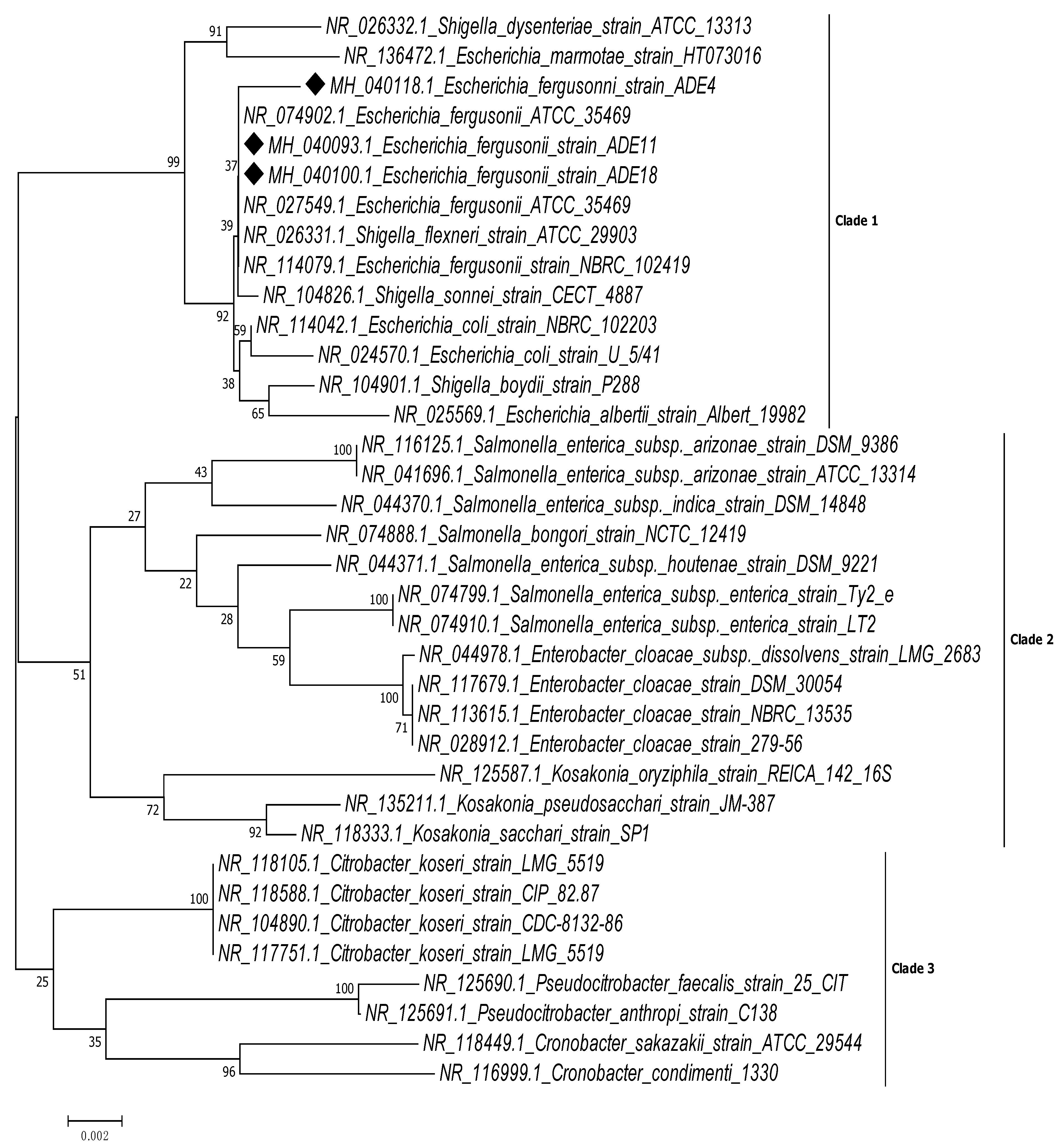
Figure 1. Phylogenetic tree showing the relationship of E. fergusonii strains ADE4, ADE11 and ADE18 from wound infection with other closely related members of Enterobacteriaceae, based on the similarities in their 16S rRNA gene sequences. The phylogenetic tree was reconstructed by comparing 36 concatenated sequences using the neighbour-joining (NJ) method while evolutionary distances were computed with the Tamura 3-parameter model, in MEGA ver. 7.0.26 software.
Pathogens 08 00164 g001
Figure 2. Agarose gel electrophoresis plate showing CTX-M extended-spectrum beta-lactamase (ESBL) genes under an ultraviolet (UV) transilluminator: Lane 1- Positive control (K. pneumoniae ATCC 7881); Lane 2- CTX-M from CR11; Lane 3- CTX-M from CR35; and Lane 4-Molecular weight marker (Norgen, 100 bp DNA Ladder, Canada).
Pathogens 08 00164 g002
Table 1. Morphological and biochemical characteristics of isolates.
Organism NameFormMarginElevationOptical PropertyAppearanceConsistencyColour on NAColour on MacCGram Stain
E. fergusoniicircularEntireFlatopaqueGlisteningMoistWhitePinkNegative
Indole productionMethyl-redVoges-ProskauerCitrateUreaseHydrogen sulphideMotilityLactoseMaltoseSucroseGlucose (A/G)MannitolCellobioseAdonitolSorbitolOxidase
+++++/++++
Keys: NA; Nutrient agar Mac; MacConkey agar A; Acid G; Gas +; positive −: negative.
Table 2. Antimicrobial susceptibility patterns and associated beta-lactamase genes of isolates CR11, CR35 and CR49.
Isolate NameOrganismAccession NumberAntibiotics Resistance PatternBeta-Lactamase Gene Encoded
CR11E. fergusoniiADE4MH040118AUG PEN G AMP CRO CRX CAZ LEV OFL CIP NIT GEN IMP MERTEM CTX-M
CR35E. fergusoniiADE11MH040093AUG PEN G AMP CRO CRX CAZ LEV OFL CIP NIT GEN IMP MERTEM CTX-M
CR49E. fergusoniiADE18MH040100AUG PEN G AMP CRO CRX CAZ IMPSHV
Key: AMP: ampicillin, PEN G; penicillin G, AUG: augmentin, CRO: cefuroxime, CRX: ceftriaxone, CAZ: ceftazidime, CTX: cefotaxime, LEV: levofloxacin, OFL: ofloxacin, GEN: gentamicin, NIT: nitrofurantoin, CIP: ciprofloxacin.
Table 3. Oligonucleotide primers used for beta-lactamase gene detection.
Primer NamePrimer Sequence 5′ ⟶ 3′Annealing TemperatureExpected Amplicon SizeReference
TEMF:5′CGCCGCATACACTATTCTCAGAATGA3′
R:5′ACGCTCACCGGCTCCAGATTTAT3′
58 °C445 pb[55]
SHVF:5′CTTTATCGGCCCTCACTCAA3′
R:5′AGGTGCTCATCATGGGAAAG3′
58 °C273 bp[55]
CTX-MF:5′CGCTTTGCGATGTGCAG3′
R:5′ACCGCGATATCGTTGGT3′
55 °C550 bp[56]
IMPF:5′-GGAATAGAGTGGCTTAAYTCTC-3′
R:5′-CCAAACYACTASGTTATCT-3′
53 °C188 bp[59]
NDMF:5′GGTTTGGCGATCTGGTTTTC
R:5′CGGAATGGCTCATCACGATC
62.8 °C621 bp[57]
OXAF:5′ACACAATACATATCAACTTCGC3′
R:5′AGTGTGTTTAGAATGGTGATC3′
62.8 °C813 bp[55]
KPCF:5′GTATCGCCGTCTAGTTCTGC3′
R:5′GGTCGTGTTTCCCTTTAGCCA3′
59.9 °C636 bp[60]
VIMF:5′-GATGGYGTTTGGTCGCATATCKCAAC3′
R:5′-CGAATGCGCAGCACCRGGATAGAA-3′
54.4 °C390 bp[57]