Genetic Diversity of Leptospira Strains Circulating in Humans and in Free-Ranging Rats Indicates That Rats Are Not Sources of Human Leptospiroses in Hungary 2022–2025
 ,
,
Abstract
1. Introduction
2. Materials and Methods
2.1. Collection and Preparation of Rat Samples
2.2. Collection and Preparation of Human Samples
2.3. PCR Assays
2.4. Dark-Field Microscopy
2.5. DNA Sequencing
2.6. Sequence Analysis
3. Results
3.1. PCR of the Rat Samples
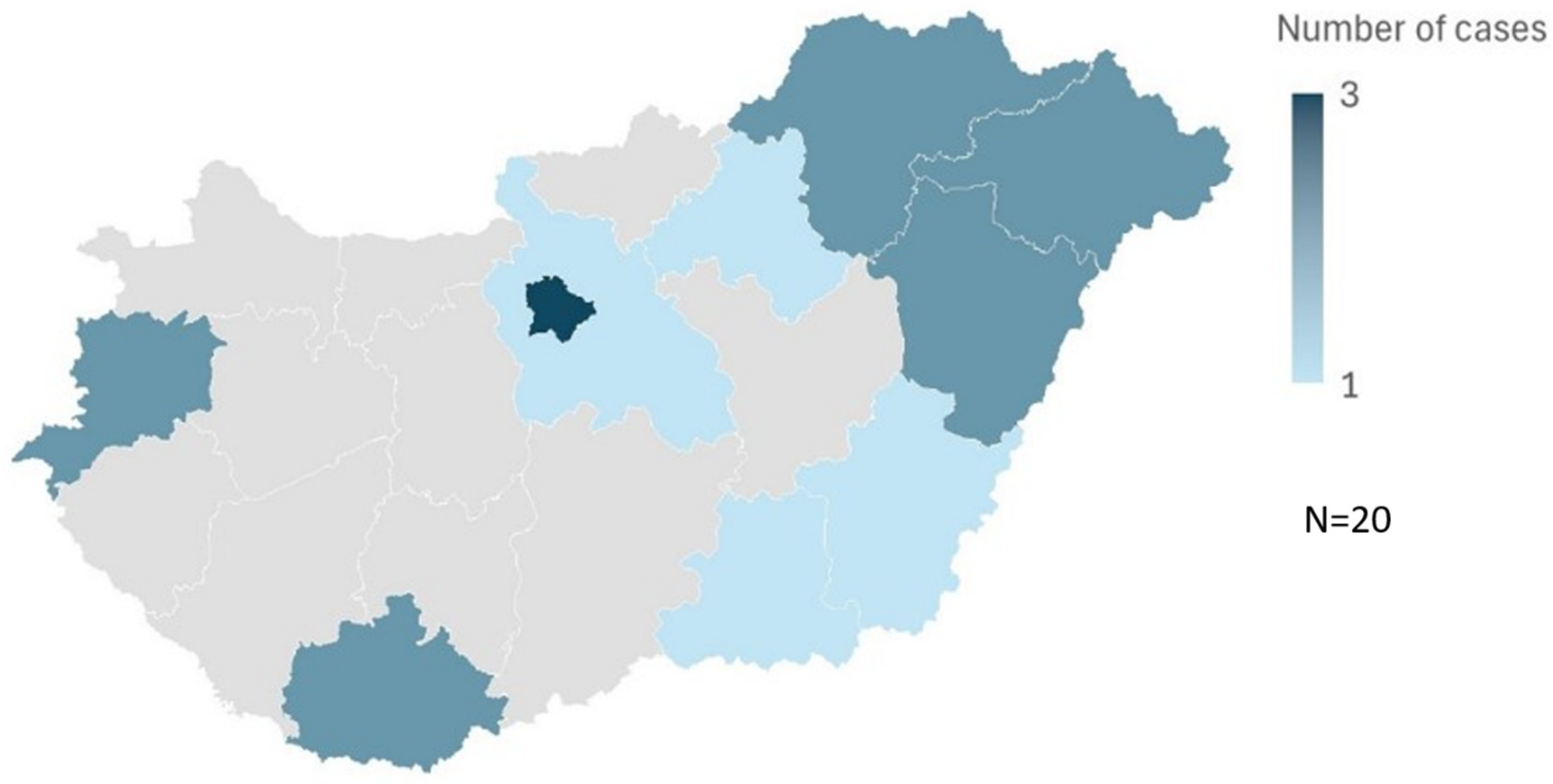
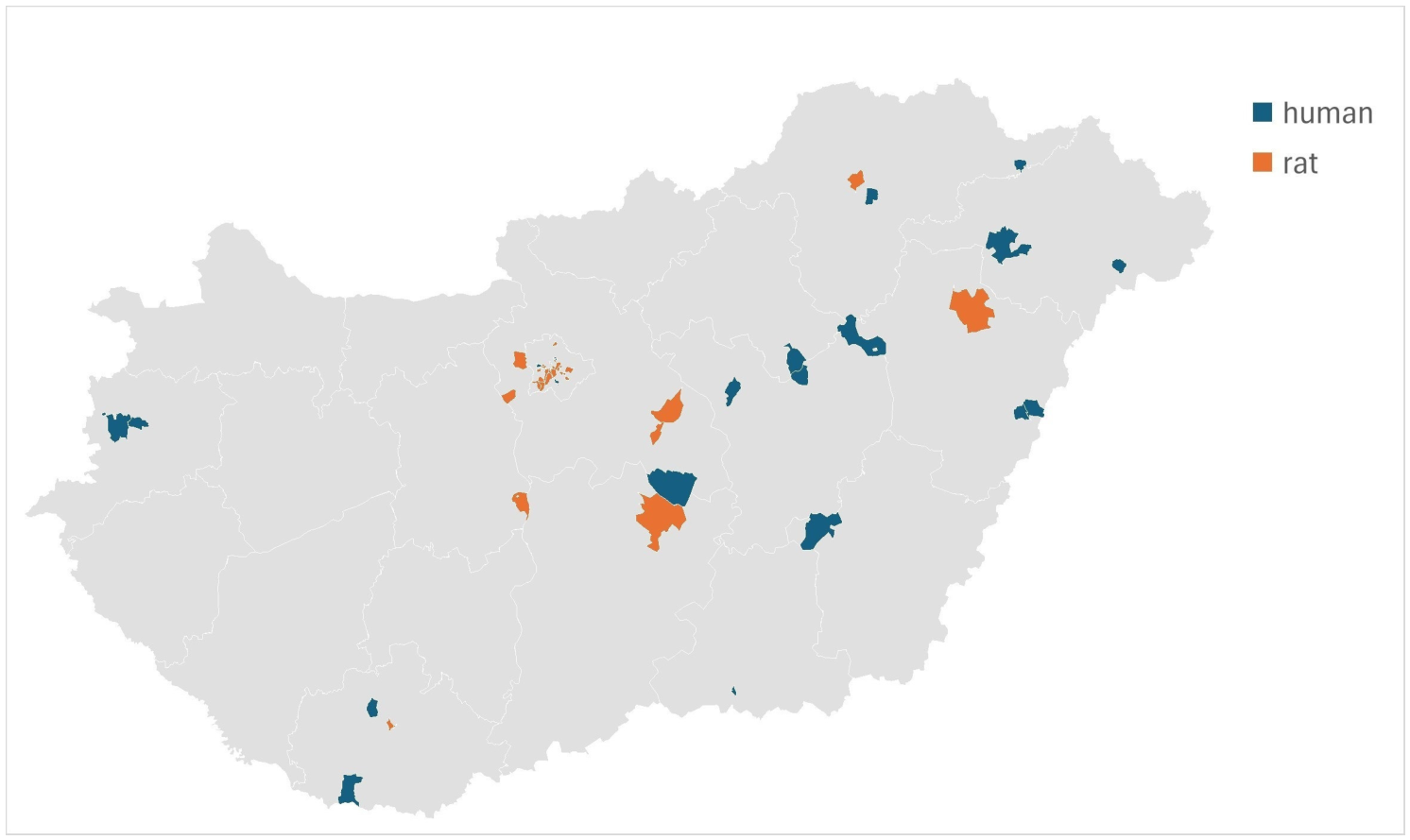
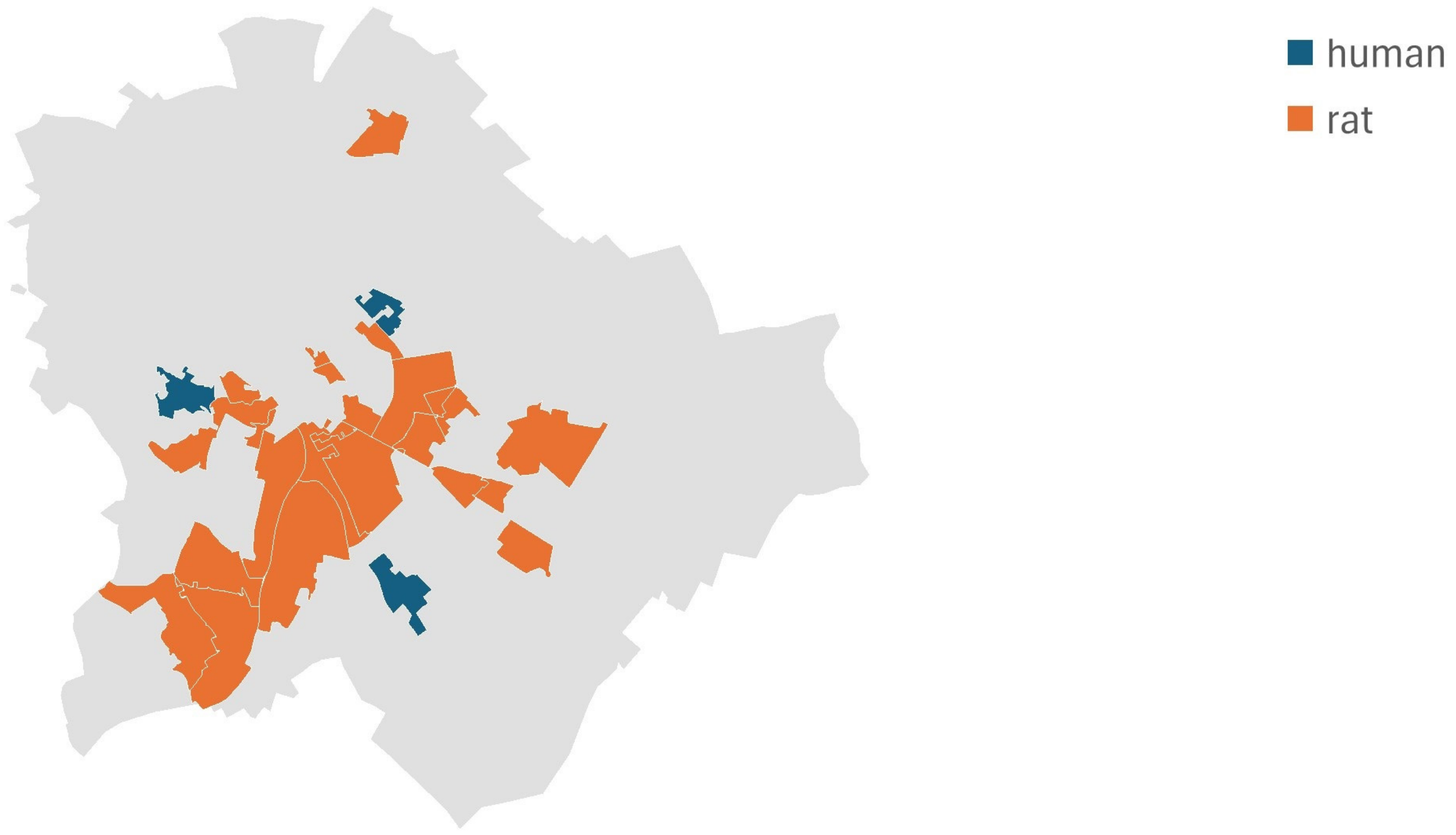
3.2. PCR, Sequencing and Epidemiology of Human Samples
3.3. Dark-Field Microscopy Studies
4. Discussion
5. Conclusions
Supplementary Materials
Author Contributions
Funding
Institutional Review Board Statement
Informed Consent Statement
Data Availability Statement
Acknowledgments
Conflicts of Interest
References
- Bharti, A.R.; Nally, J.E.; Ricaldi, J.N.; Matthias, M.A.; Diaz, M.M.; Lovett, M.A.; Levett, P.N.; Gilman, R.H.; Willig, M.R.; Gotuzzo, E.; et al. Leptospirosis: A zoonotic disease of global importance. Lancet Infect. Dis. 2003, 3, 757–771. [Google Scholar] [CrossRef]
- Karpagam, K.B.; Ganesh, B. Leptospirosis: A neglected tropical zoonotic infection of public health importance-an updated review. Eur. J. Clin. Microbiol. Infect. Dis. 2020, 39, 835–846. [Google Scholar] [CrossRef]
- Yersin, C.; Bovet, P.; Mérien, F.; Clément, J.; Laille, M.; Van Ranst, M.; Perolat, P. Pulmonary hemorrhage as a predominant cause of death in leptpspirosis in Seychelles. Trans. R. Soc. Trop. Med. Hyg. 2000, 94, 71–76. [Google Scholar] [CrossRef]
- McBride, A.J.; Athanazio, D.A.; Reis, M.G.; Ko, A.I. Leptospirosis. Curr. Opin. Infect. Dis. 2005, 18, 376–386. [Google Scholar] [CrossRef]
- Zhu, Z.; Feng, J.; Dong, Y.; Jiang, B.; Wang, X.; Li, F. Cerebral infarct induced by severe leptospirosis—A case report and literature review. BMC Neurol. 2022, 22, 506. [Google Scholar] [CrossRef] [PubMed]
- Mazzotta, E.; Bellinati, L.; Bertasio, C.; Boniotti, M.B.; Lucchese, L.; Ceglie, L.; Martignago, F.; Leopardi, S.; Natale, A. Synanthropic and wild animals as sentinels of zoonotic agents: A study of Leptospira genotypes circulating in Northeastern Italy. Int. J. Environ. Res. Public Health 2005, 20, 3783. [Google Scholar] [CrossRef] [PubMed]
- Arent, Z.; Frizzell, C.; Gilmore, C.; Allen, A.; Ellis, W.A. Leptospira interrogans serovars Bratislava and Muenchen animal infections: Implications for epidemiology and control. Vet. Microbiol. 2016, 190, 19–26. [Google Scholar] [CrossRef] [PubMed]
- Moinet, M.; Oosterhof, H.; Nisa, S.; Haack, N.; Wilkinson, D.A.; Aberdein, D.; Russell, J.C.; Vallée, E.; Collins-Emerson, J.; Heuer, C.; et al. A cross-sectional investigation of Leptospira at the wildlife-livestock interface in New Zealand. PLoS Negl. Trop. Dis. 2023, 17, e0011624. [Google Scholar] [CrossRef] [PubMed]
- Mazzotta, E.; De Zan, G.; Cocchi, M.; Boniotti, M.B.; Bertasio, C.; Furlanello, T.; Lucchese, L.; Ceglie, L.; Bellinati, L.; Natale, A. Feline susceptibility to Leptospirosis and presence immunosuppressive co-morbidities: First European report of L. interrogans serogroup australis sequence type 24 in a cat and survey of Leptospira exposure in outdoor cats. Trop. Med. Infect. Dis. 2023, 8, 54. [Google Scholar] [CrossRef] [PubMed]
- Bilung, L.M.; Tahar, A.S.; Pui, C.F.; Bakeri, M.K.S.; Su’út, L.; Ngui, R.; Kira, R.; Apun, K. Leptospira and Leptospirosis: A review of species classifications, genomes, morphological structures, antimicrobial resistances, transmissions, and clinical manifestations. Curr. Microbiol. 2026, 83, 122. [Google Scholar] [CrossRef] [PubMed]
- Daroz, B.B.; Fernandes, L.G.; Cavenague, M.F.; Kochi, L.T.; Passalia, F.J.; Takahashi, M.B.; Nascimento Filho, E.G.; Teixeira, A.F.; Nascimento, A.L. A review on host-Leptospira interactions: What we know and future expectations. Front. Cell. Infect. Microbiol. 2021, 11, 777709. [Google Scholar] [CrossRef] [PubMed]
- Bierque, E.; Thibeaux, R.; Girault, D.; Soupé-Gilbert, M.E.; Goarant, C. A systematic review of Leptospira in water and soil environments. PLoS ONE 2020, 15, e0227055. [Google Scholar] [CrossRef] [PubMed]
- Barwick, R.S.; Mohammed, H.O.; McDonough, P.L.; White, M.E. Epidemiologic features of equine Leptospira interrogans of human significance. Prev. Vet. Med. 1998, 36, 153–165. [Google Scholar] [CrossRef] [PubMed]
- Zhang, C.; Li, Z.; Xu, Y.; Zhang, Y.; Li, S.; Zhang, J.; Cui, S.; Du, Z.; Xin, X.; Chang, Y.F.; et al. Genetic diversity of Leptospira interrogans circulating isolates and vaccine strains in China from 1954–2014. Hum. Vacc. Immunother. 2019, 15, 381–387. [Google Scholar] [CrossRef] [PubMed]
- Li, S.J.; Wang, D.; Zhang, C.; Wei, X.; Tian, K.; Li, X.; Nie, Y.; Liu, Y.; Yao, G.; Zhou, J.; et al. Source tracking of human leptospirosis: Serotyping and genotyping of Leptospira isolated from rodents in the epidemic area of Guizhou province, China. BMC Microbiol. 2013, 13, 75. [Google Scholar] [CrossRef] [PubMed]
- Li, J.M.; Li, L.M.; Shi, J.F.; Li, T.; Wang, Q.; Ma, Q.X.; Zheng, W.; Feng, H.F.; Liu, F.; Du, R. Prevalence of Leptospira in murine in China: A systematic review and meta-analysis. Front. Vet. Sci. 2022, 9, 944282. [Google Scholar] [CrossRef] [PubMed]
- Esteves-Bergmann, S.B.; Gaeta, N.C.; Batista, J.M.N.; Dias, R.A.; Heinemann, M.B. Leptospira sp. infection in bats: A systematic review and meta-analysis. Transbound. Emerg. Dis. 2022, 69, e2456–e2473. [Google Scholar] [CrossRef] [PubMed]
- Adler, B.; de la Pena Moctezuma, A. Leptospira and leptospirosis. Vet. Microbiol. 2010, 140, 287–296. [Google Scholar] [CrossRef] [PubMed]
- Heuser, E.; Fischer, S.; Ryll, R.; Mayer-Scholl, A.; Hoffmann, D.; Spahr, C.; Imholt, C.; Alfa, D.M.; Fröhlich, A.; Lüschow, D.; et al. Survey for zoonotic pathogens in Norway rat populations from Europe. Pest Manag. Sci. 2017, 73, 341–348. [Google Scholar] [CrossRef] [PubMed]
- Boey, K.; Shiokawa, K.; Rajeev, S. Leptospira infection in rats: A literature review of global prevalence and distribution. PLoS Negl. Trop. Dis. 2019, 13, e0007499. [Google Scholar] [CrossRef] [PubMed]
- Colombo, V.C.; Gamietea, I.; Loffler, S.G.; Brihuega, B.F.; Beldomenico, P.M. New host species for Leptospira borgpetersenii and Leptospira interrogans serovar Copenhageni. Vet. Microbiol. 2018, 215, 90–92. [Google Scholar] [CrossRef] [PubMed]
- Cordonin, C.; Turpin, M.; Bringart, M.; Bascands, J.-L.; Flores, O.; Dellagi, K.; Mavingui, P.; Roche, M.; Tortosa, P. Pathogenic Leptospira and their animal reservoirs: Testing host specificity through experimental infection. Sci. Rep. 2020, 10, 7239. [Google Scholar] [CrossRef] [PubMed]
- Santos, A.A.N.; Ribeiro, P.D.S.; da França, G.V.; Souza, F.N.; Ramos, E.A.G.; Figueira, C.P.; Reis, M.G.; Costa, F.; Ristow, P. Leptospira interrogans biofilm formation in Rattus norvegicus (Norway rats) natural reservoirs. PLoS Negl. Trop. Dis. 2021, 15, e0009736. [Google Scholar] [CrossRef] [PubMed]
- Koizumi, N.; Muto, M.; Yamamoto, M.; Baba, Y.; Kudo, M.; Tamae, Y.; Shimomura, K.; Takatori, I.; Iwakiri, A.; Ishikawa, K.; et al. Investigation of reservoir animals of the Leptospira in the nothern part of Miyazaki prefecture. Jpn. J. Infect. Dis. 2008, 61, 465–468. [Google Scholar] [CrossRef] [PubMed]
- Muslich, L.T.; Villanueva, S.Y.; Amran, M.Y.; Segawa, T.; Saito, M.; Yoshida, S. Characterization of Leptospira infection in suckling and weaning pups. Comp. Immunol. Microbiol. Infect. Dis. 2015, 38, 47–55. [Google Scholar] [CrossRef] [PubMed]
- Krijger, I.M.; Ahmed, A.A.A.; Goris, M.G.A.; Cornelissen, J.B.W.J.; Groot Koerkamp, P.W.G.; Meerburg, B.G. Wild rodents and insectivores as carriers of pathogenic Leptospira and Toxoplasma gondii in The Netherlands. Vet. Med. Sci. 2020, 6, 623–630. [Google Scholar] [CrossRef] [PubMed]
- Richard, E.; Geslin, J.; Wurtzer, S.; Moulin, L. Monitoring of Leptospira species diversity in freshwater bathing area and in rats in Paris, France. Sci. Total Environ. 2022, 833, 155121. [Google Scholar] [CrossRef] [PubMed]
- Villanueva, S.Y.; Saito, M.; Baterna, R.A.; Estrada, C.A.; Rivera, A.K.; Dato, M.C.; Zamora, P.R.; Segawa, T.; Cavinta, L.L.; Fukui, T.; et al. Leptospira-rat-human relationship in Luzon, Philippines. Microbes Infect. 2014, 16, 902–910. [Google Scholar] [CrossRef] [PubMed]
- Stone, N.E.; Hamond, C.; Clegg, J.L.; McDonough, R.F.; Bourgeois, R.M.; Ballard, R.; Thornton, N.B.; Nuttall, M.; Hertzel, H.; Anderson, T.; et al. Host population dynamics influence Leptospira spp. transmission patterns among Rattus norvegicus in Boston, Massachusetts, US. PLoS Negl. Trop. Dis. 2025, 19, e0012966. [Google Scholar] [CrossRef] [PubMed]
- McVea, D.; Himsworth, C.G.; Patrick, D.M.; Lindsay, L.R.; Kosoy, M.; Kerr, T. Exposure to rats and rat-associated Leptospira and Bartonella species among people who use drugs in an impoverished, inner-city neighborhood of Vancouver, Canada. Vector Borne Zoonotic Dis. 2018, 18, 82–88. [Google Scholar] [CrossRef] [PubMed]
- Hu, W.; Lin, X.; Yan, J. Leptospira and leptospirosis in China. Curr. Opin. Infect. Dis. 2014, 27, 432–436. [Google Scholar] [CrossRef] [PubMed]
- Philip, N.; Bahtiar Affendy, B.; Ramli, S.N.A.; Arif, M.; Raja, P.; Nagandran, E.; Renganathan, P.; Taib, N.M.; Masri, S.N.; Yuhana, M.Y.; et al. Leptospira interrogans and Leptospira kirschneri are the dominant Leptospira species causing human leptospirosis in Central Malaysia. PLoS Negl. Trop. Dis. 2020, 14, e0008197. [Google Scholar] [CrossRef] [PubMed]
- Samrot, A.V.; Sean, T.C.; Bhavya, K.S.; Sahithya, C.S.; Chan-Drasekaran, S.; Palanisamy, R.; Robinson, E.R.; Subbiah, S.K.; Mok, P.L. Leptospiral infection, pathogenesis and its diagnosis—A review. Pathogens 2021, 10, 145. [Google Scholar] [CrossRef] [PubMed]
- Vincent, A.T.; Schiettekatte, O.; Goarant, C.; Neela, V.K.; Bernet, E.; Thibeaux, R.; Ismail, N.; Mohd Khalid, M.K.N.; Amran, F.; Masuzawa, T.; et al. Revisiting the taxonomy and evolution of pathogenicity of the genus Leptospira through the prism of genomics. PLoS Negl. Trop. Dis. 2019, 13, e0007270. [Google Scholar] [CrossRef] [PubMed]
- Mérien, F.; Truccolo, J.; Baranton, G.; Perolat, P. Identification of a 36-kDa fibronectin-binding protein expressed by a virulent variant of Leptospira interrogans serovar icterohaemorrhagiae. FEMS Microbiol. Lett. 2000, 185, 17–22. [Google Scholar] [CrossRef] [PubMed]
- Mérien, F.; Portnoi, D.; Bourhy, P.; Charavay, F.; Berlioz-Arthaud, A.; Baranton, G. A rapid and quantitative method for the detection of Leptospira species in human leptospirosis. FEMS Microbiol. Lett. 2005, 249, 139–147. [Google Scholar] [CrossRef] [PubMed]
- Perez, J.; Goarant, C. Rapid Leptospira identification by direct sequencing of the diagnostic PCR products in New Caledonia. BMC Microbiol. 2000, 10, 325. [Google Scholar] [CrossRef] [PubMed]
- García-Lopez, M.; Lorioux, C.; Soares, A.; Trombert-Paolantoni, S.; Harran, E.; Ayral, F.; Picardeau, M.; Djelouadji, Z.; Bourhy, P. Genetic diversity of Leptospira strains circulating in humans and dogs in France in 2019–2021. Front. Cell. Infect. Microbiol. 2023, 13, 1236866. [Google Scholar] [CrossRef] [PubMed]
- Podgoršek, D.; Ružić-Sabljić, E.; Logar, M.; Pavlović, A.; Remec, T.; Baklan, Z.; Pal, E.; Cerar, T. Evaluation of real-time PCR targeting the lipL32 gene for diagnosis of Leptospira infection. BMC Microbiol. 2020, 20, 59. [Google Scholar] [CrossRef] [PubMed]
- Esteves, L.M.; Bulhões, S.M.; Branco, C.C.; Carreira, T.; Vieira, M.L.; Gomes-Solecki, M.; Mota-Vieira, L. Diagnosis of human Leptospirosis in a clinical setting: Real-time PCR high resolution melting analysis for detection of Leptospira at the onset of disease. Nat. Sci. Rep. 2018, 8, 9213. [Google Scholar] [CrossRef] [PubMed]
- Sohm, C.; Willixhofer, D.; Fasching, E.; Waldner, K.; Deitzer, N.; Steiner, J.; Jöbstl, J.; Schleicher, C.; Schwarz, M.; Fuchs, R.; et al. First isolation and genotyping of pathogenic Leptospira spp. from Austria. Nat. Sci. Rep. 2024, 14, 4467. [Google Scholar] [CrossRef] [PubMed]
- Roquelo, C.; Kodjo, A.; Marié, J.L.; Davoust, B. Serological and molecular survey of Leptospira spp. infections in wild boars and red foxes from Southeastern France. Vet. World 2021, 14, 825–828. [Google Scholar] [CrossRef] [PubMed]
- Wilkinson, D.-A.; Edwards, M.; Shum, C.; Moinet, M.; Anderson, N.E.; Benschop, J.; Nisa, S. Molecular typing of Leptospira spp. in farmed and wild mammals reveals new host-serovar associations in New Zealand. N. Z. Vet. J. 2024, 72, 1–9. [Google Scholar] [CrossRef] [PubMed]
- Centers for Disease Control and Prevention (CDC). Outbreak of acute febrile illness among athletes participating in triathlons—Wisconsin and Illinois. Morb. Mortal. Wkly. Rep. 1998, 280, 1473–1474. [Google Scholar] [CrossRef]
- Xu, G.; Qiu, H.; Liu, W.; Jiang, X.; Chang, Y.-F.; Wang, J.; Li, Z.; Zhu, Y.; Zhang, C.; Xiao, F. Serological and molecular characteristics of pathogenic Leptospira in rodent populations in Fujian Province, China, 2018–2020. BMC Microbiol. 2022, 22, 151. [Google Scholar] [CrossRef] [PubMed]
- Van Crevel, R.; Speelman, P.; Gravekamp, C.; Terpstra, W.J. Leptospirosis in travelers. Clin. Infect. Dis. 1994, 19, 132–134. [Google Scholar] [CrossRef] [PubMed]



| Pairwise Distance (Mega) | |||
|---|---|---|---|
| L._interrogans_lfb1: 1 | L._interrogans_lfb1: 2 | L._interrogans_lfb1: 3 | |
| L._interrogans_lfb1: 1 | |||
| L._interrogans_lfb1: 2 | 0.01092 | ||
| L._interrogans_lfb1: 3 | 0.01091 | 0.00724 | |
| L._kirschneri_lfb1: 6 | 0.05671 | 0.06890 | 0.06880 |
| Number of Our Samples | ||||||||||||||||||||||||||||||||||||||||
|---|---|---|---|---|---|---|---|---|---|---|---|---|---|---|---|---|---|---|---|---|---|---|---|---|---|---|---|---|---|---|---|---|---|---|---|---|---|---|---|---|
| L. interrogans lfb1: 1 | 103 (rats) | A | T | T | C | T | T | C | C | T | C | G | C | C | C | T | T | C | T | A | G | T | A | T | T | T | G | G | T | T | T | T | A | T | T | T | C | A | A | C |
| L. interrogans lfb1: 2 | 12 (human) | . | . | . | . | . | . | . | . | . | . | . | . | . | . | . | . | . | . | . | . | . | . | . | . | . | . | . | . | . | . | . | . | . | . | . | . | . | . | . |
| L. interrogans lfb1: 3 | 1 (human, imported) | . | . | . | . | . | . | . | . | . | . | . | . | . | . | . | . | . | . | . | . | . | . | . | . | . | . | . | . | . | . | . | . | . | . | . | . | . | . | . |
| L. kirschneri lfb1: 6 | 1 (human) | . | . | . | . | . | . | . | . | . | . | . | . | . | . | . | . | . | . | . | . | . | . | . | . | C | . | . | . | . | . | . | . | . | . | . | . | G | . | . |
| 78 | ||||||||||||||||||||||||||||||||||||||||
| L. interrogans lfb1: 1 | 103 (rats) | C | G | C | T | T | A | C | G | C | A | C | A | G | A | T | C | G | G | T | C | A | A | A | T | C | A | A | T | C | C | T | T | C | C | T | C | C | A | T |
| L. interrogans lfb1: 2 | 12 (human) | . | T | . | . | . | . | . | . | . | . | . | . | . | . | . | . | . | . | . | . | . | . | . | . | . | . | . | . | . | . | . | . | . | . | . | . | . | . | . |
| L. interrogans lfb1: 3 | 1 (human, imported) | . | T | . | . | . | . | . | . | . | . | . | . | . | . | . | . | . | . | . | . | . | . | . | . | . | . | . | . | . | . | . | . | . | . | . | . | . | . | . |
| L. kirschneri lfb1: 6 | 1 (human) | . | . | . | . | . | . | . | . | . | . | . | . | . | . | . | . | . | . | . | . | . | . | . | . | . | . | . | . | . | . | . | . | . | T | . | . | . | . | . |
| L. interrogans lfb1: 1 | 103 (rats) | T | A | G | C | G | G | T | A | A | A | T | A | C | A | A | G | G | T | T | T | C | T | G | G | A | A | C | C | A | A | C | C | C | A | A | A | C | G | G |
| L. interrogans lfb1: 2 | 12 (human) | . | . | . | . | . | . | . | . | . | . | . | . | . | . | . | . | . | . | . | . | . | . | . | . | . | . | . | . | . | . | . | . | . | . | . | . | . | . | . |
| L. interrogans lfb1: 3 | 1 (human, imported) | . | . | . | . | . | . | . | . | . | G | . | . | . | . | . | . | . | . | . | . | . | . | . | . | . | . | . | . | . | . | . | . | . | . | . | . | . | . | . |
| L. kirschneri lfb1: 6 | 1 (human) | C | . | . | . | . | . | . | . | . | . | . | . | . | . | . | . | . | . | . | . | . | . | . | . | . | . | . | . | . | . | . | . | . | G | . | . | . | . | . |
| 156 | ||||||||||||||||||||||||||||||||||||||||
| L. interrogans lfb1: 1 | 103 (rats) | T | T | C | C | T | C | T | T | A | C | A | A | C | G | G | T | A | G | C | G | T | T | A | C | G | A | T | C | T | C | T | C | A | A | T | C | T | A | A |
| L. interrogans lfb1: 2 | 12 (human) | . | . | . | . | . | . | . | . | . | . | . | . | . | . | . | . | . | . | . | . | . | . | . | . | . | . | . | . | . | . | . | . | . | . | . | . | . | . | . |
| L. interrogans lfb1: 3 | 1 (human, imported) | . | . | . | . | . | . | . | . | . | . | . | . | . | . | . | . | . | . | . | . | . | . | . | . | . | . | . | . | . | . | . | . | . | . | . | . | . | . | . |
| L. kirschneri lfb1: 6 | 1 (human) | . | . | . | . | . | . | . | . | . | . | G | G | . | . | . | . | . | . | . | . | . | . | . | . | . | . | . | T | . | . | . | G | . | . | . | . | . | . | . |
| L. interrogans lfb1: 1 | 103 (rats) | C | G | G | A | G | A | A | T | A | C | C | T | T | T | T | T | A | C | C | T | G | G | A | C | G | G | T | C | G | C | T | G | G | T | C | A | A | A | C |
| L. interrogans lfb1: 2 | 12 (human) | . | . | . | . | . | . | . | . | . | . | . | . | . | . | . | . | . | . | . | . | . | . | . | . | . | . | . | T | . | . | . | . | . | . | . | . | . | . | . |
| L. interrogans lfb1: 3 | 1 (human, imported) | . | . | . | . | . | . | . | . | . | . | . | . | . | . | . | . | . | . | . | . | . | . | . | . | . | . | . | . | . | . | . | . | . | . | . | . | . | . | . |
| L. kirschneri lfb1: 6 | 1 (human) | . | . | . | . | . | . | . | . | . | . | . | . | . | . | . | . | . | . | . | . | . | . | . | . | . | . | . | . | . | . | C | . | . | . | . | . | . | . | . |
| 234 | ||||||||||||||||||||||||||||||||||||||||
| L. interrogans lfb1: 1 | 103 (rats) | T | T | T | C | A | C | A | G | G | A | A | C | C | G | G | A | A | C | C | C | T | T | G | A | A | G | G | T | A | C | T | A | C | T | T | T | G | A | C |
| L. interrogans lfb1: 2 | 12 (human) | . | . | . | . | . | . | . | . | . | . | . | . | . | . | . | . | . | . | . | . | . | . | . | . | . | . | . | . | . | . | . | . | . | . | . | . | . | . | . |
| L. interrogans lfb1: 3 | 1 (human, imported) | . | . | . | . | . | . | . | . | . | . | . | . | . | . | . | . | . | . | . | . | . | . | . | . | . | . | . | . | . | . | . | . | . | . | . | . | . | . | . |
| L. kirschneri lfb1: 6 | 1 (human) | . | . | . | . | . | . | . | . | . | . | . | . | . | . | . | . | . | . | . | . | . | . | . | . | . | . | . | . | . | . | . | . | . | . | . | . | . | . | . |
| L. interrogans lfb1: 1 | 103 (rats) | A | G | T | A | G | A | T | T | G | G | G | G | A | G | A | A | A | C | A | G | A | A | C | C | G | G | T | A | A | T | C | T | A | T | G | A | A | G | T |
| L. interrogans lfb1: 2 | 12 (human) | . | . | . | . | . | . | . | . | . | . | . | . | . | . | . | . | . | . | . | . | . | . | . | . | A | . | . | . | . | . | . | . | . | . | . | . | . | . | . |
| L. interrogans lfb1: 3 | 1 (human, imported) | . | . | . | . | . | . | . | . | . | . | . | . | . | . | . | . | . | . | . | . | . | . | . | . | A | . | . | . | . | . | . | . | . | . | . | . | . | . | . |
| L. kirschneri lfb1: 6 | 1 (human) | . | . | . | . | . | . | . | . | . | . | . | . | . | . | . | G | G | T | T | . | . | . | . | . | . | . | . | G | . | . | . | . | . | . | . | . | . | . | . |
| 278 | ||||||||||||||||||||||||||||||||||||||||
| L. interrogans lfb1: 1 | 103 (rats) | A | A | A | A | A | ||||||||||||||||||||||||||||||||||
| L. interrogans lfb1: 2 | 12 (human) | . | . | . | . | . | ||||||||||||||||||||||||||||||||||
| L. interrogans lfb1: 3 | 1 (human, imported) | . | . | . | . | . | ||||||||||||||||||||||||||||||||||
| L. kirschneri lfb1: 6 | 1 (human) | . | . | . | . | . |
| Sample Types | Rat Samples | Human Samples |
|---|---|---|
| Urine (dark-field microscopy negative) | 23 | 165 |
| Tissue | 190 | 5 |
| Blood | - | 181 |
| Tracheal secretion | - | 2 |
| Samples analyzed by commercial PCR kit | 190 | 353 |
| PCR-positive samples | 103 | 29 |
| Samples successfully sequenced | 103 | 20 samples (14 patients) |
| lfb1 genotype distribution | lfb1 type 1: 103 | lfb1 type 2: 12 patients |
| lfb1 type 3: 1 patient | ||
| lfb1 type 6: 1 patient |
Disclaimer/Publisher’s Note: The statements, opinions and data contained in all publications are solely those of the individual author(s) and contributor(s) and not of MDPI and/or the editor(s). MDPI and/or the editor(s) disclaim responsibility for any injury to people or property resulting from any ideas, methods, instructions or products referred to in the content. |
© 2026 by the authors. Licensee MDPI, Basel, Switzerland. This article is an open access article distributed under the terms and conditions of the Creative Commons Attribution (CC BY) license.
Share and Cite
Locsmándi, G.; Kádár-Hürkecz, E.; Kienle, Z.; Tárnoki-Boross, K.; Sima, K.; Erdősi, T.; Egyed, L. Genetic Diversity of Leptospira Strains Circulating in Humans and in Free-Ranging Rats Indicates That Rats Are Not Sources of Human Leptospiroses in Hungary 2022–2025. Pathogens 2026, 15, 729. https://doi.org/10.3390/pathogens15070729
Locsmándi G, Kádár-Hürkecz E, Kienle Z, Tárnoki-Boross K, Sima K, Erdősi T, Egyed L. Genetic Diversity of Leptospira Strains Circulating in Humans and in Free-Ranging Rats Indicates That Rats Are Not Sources of Human Leptospiroses in Hungary 2022–2025. Pathogens. 2026; 15(7):729. https://doi.org/10.3390/pathogens15070729
Chicago/Turabian StyleLocsmándi, Gabriella, Enikő Kádár-Hürkecz, Zsuzsa Kienle, Katalin Tárnoki-Boross, Krisztina Sima, Tímea Erdősi, and László Egyed. 2026. "Genetic Diversity of Leptospira Strains Circulating in Humans and in Free-Ranging Rats Indicates That Rats Are Not Sources of Human Leptospiroses in Hungary 2022–2025" Pathogens 15, no. 7: 729. https://doi.org/10.3390/pathogens15070729
APA StyleLocsmándi, G., Kádár-Hürkecz, E., Kienle, Z., Tárnoki-Boross, K., Sima, K., Erdősi, T., & Egyed, L. (2026). Genetic Diversity of Leptospira Strains Circulating in Humans and in Free-Ranging Rats Indicates That Rats Are Not Sources of Human Leptospiroses in Hungary 2022–2025. Pathogens, 15(7), 729. https://doi.org/10.3390/pathogens15070729