Article

Epidemiological Insights into Endoparasites of Brown Bears (Ursus arctos) in Greece

by
Antonios Synapalos
1,
Anastasia Diakou
2,* and
Stefanos Sgardelis
1,*
1
Department of Ecology, School of Biology, Aristotle University of Thessaloniki, 54124 Thessaloniki, Greece
2
School of Veterinary Medicine, Faculty of Health Sciences, Aristotle University of Thessaloniki, 54124 Thessaloniki, Greece
*
Authors to whom correspondence should be addressed.
Pathogens 2026, 15(7), 671; https://doi.org/10.3390/pathogens15070671
Submission received: 23 May 2026 / Revised: 19 June 2026 / Accepted: 24 June 2026 / Published: 25 June 2026
(This article belongs to the Section Parasitic Pathogens)

Abstract

Brown bear populations in Greece face multiple threats, and parasitic infections may pose an additional risk to these vulnerable animals. This study represents the first comprehensive assessment of endoparasite occurrence, prevalence, and seasonality in brown bears in Greece, in relation to geographical location and the animal’s different physiological phases. A total of 918 faecal samples were collected over a three-year period from regions with brown bear presence in Greece. For each sample, the date of collection and the coordinates of the site were recorded. Samples were examined using sedimentation, flotation, and McMaster techniques, while the Baermann method was additionally applied to a subset of 195 samples. Spatial and temporal patterns in parasite occurrence and diversity were analysed using generalised additive models (GAMs). Ten parasitic taxa were identified, with Baylisascaris transfuga being the most prevalent (39.8%), followed by Crenosoma spp. (26%), Uncinaria spp. (18.09%), and Dicrocoelium dendriticum (14.38%). Less prevalent taxa included Eucoleus aerophilus, Sarcocystis spp., Toxascaris leonina, Eimeria spp., Linguatula serrata, and Taeniidae. Μixed infections, involving two or more parasites, were detected in 22% of the samples. The prevalence of B. transfuga was higher in late autumn, with high-risk infection areas identified in both late summer and autumn. In contrast, Uncinaria spp. and D. dendriticum showed no seasonal variation, while D. dendriticum exhibited spatial clustering patterns similar to B. transfuga but without clear seasonal trends. These findings highlight the widespread occurrence and complexity of parasitic infections in Greek brown bears. Continued long-term monitoring is essential to improve understanding of transmission dynamics and the ecological processes shaping parasite distribution in this animal species.

1. Introduction

The brown bear (Ursus arctos) population in Greece shows promising signs of recovery over the last 15 years [1,2]. The species is distributed in two distinct subpopulations located in northwestern (Pindos-Peristeri mountain range) and northeastern Greece (Rhodope mountain complex) [3] with approximately 500 individuals [2,4]. However, bears in Greece still face significant challenges, including deforestation and poaching, which have been characterised as “high-intensity” threats. Other challenges, such as infrastructure (roads, dams, wind farms), poisoned baits, and climate change, are categorised as medium-intensity threats [5].
Although human activities and environmental deterioration pose the major threats to wildlife, under certain conditions, infectious diseases may also adversely affect small or isolated populations [6]. Among the agents of infectious diseases, parasites are generally understudied but can impair the health status and overall fitness of bears. Furthermore, climate change, land-use change, and food scarcity in natural habitats force animals to move closer to human settlements. Consequently, bears, domestic animals, and humans share sympatric zones that serve as a terrain of possible bridging infections [7,8].
People and wildlife in Europe coexist in an anthropogenically modified landscape that favours the circulation of pathogens among wild animals, domestic animals, and people [1,8]. Therefore, there is merit in investigating brown bear parasites and assessing their effect and importance on this animal species, domestic animals, and public health [9,10].
Studies on brown bear endoparasites in Europe are scarce, and information about their impact is limited. Most of the recent studies come from Slovakia [11,12], Croatia [13], Romania [14], Estonia [15], Spain [16,17,18], and the East Carpathians Biosphere Reserve (Slovakia/Poland/Ukraine) [19]. In Greece, two case reports have been published, reporting two parasites, i.e., Dirofilaria immitis and Thelazia callipaeda, which are common in carnivores and other mammals [20,21]. The aim of the present study was to investigate, for the first time, the endoparasite fauna of brown bears in Greece and to examine the seasonality of parasitism, in relation to geographical location and the animal’s different physiological phases throughout the year.

2. Materials and Methods

2.1. Study Area and Sampling Sites

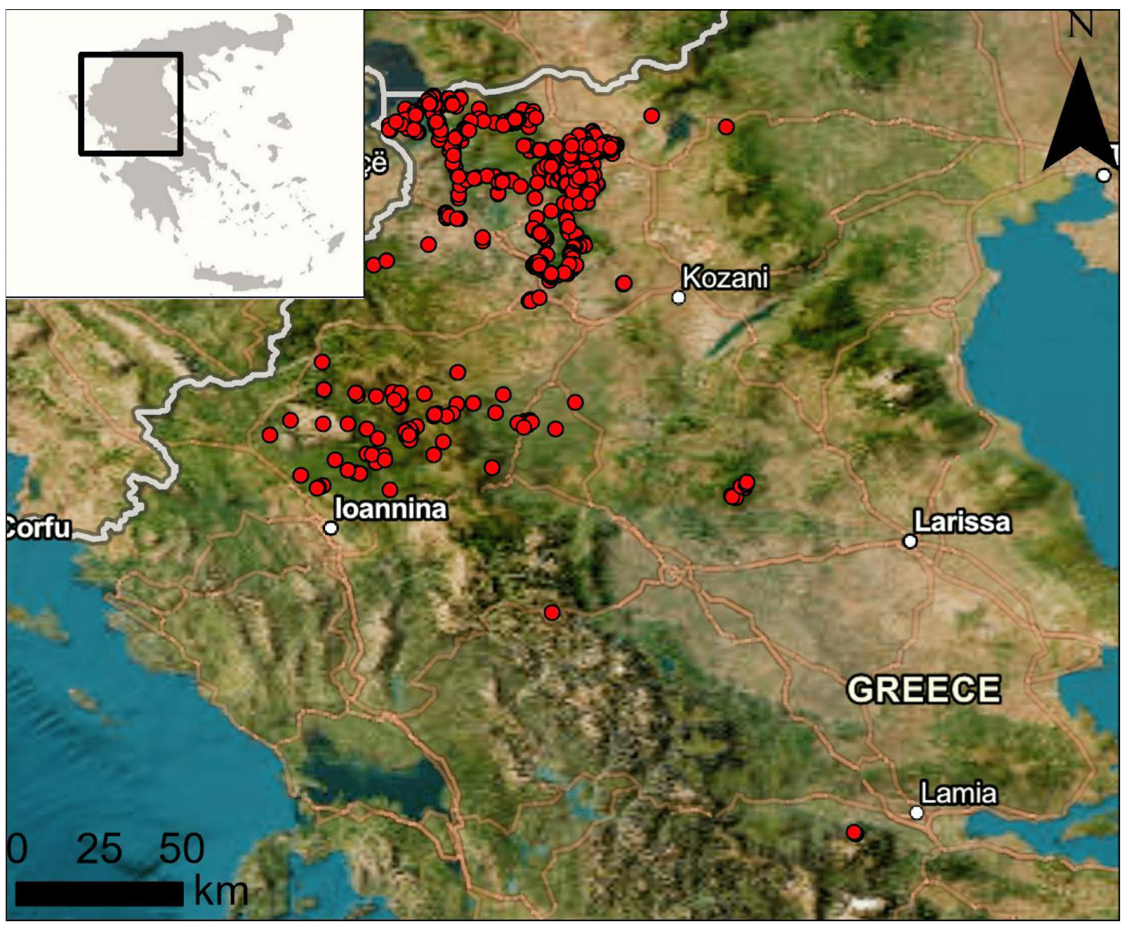
Sampling took place mostly in regions with national parks, i.e., Western Macedonia (Prespes National Park) and Northern Pindos (Northern Pindos National Park), as well as a few additional locations with documented brown bear presence in Greece (Figure 1). All regions have a continental Mediterranean climate, with cold winters and mild summers, and altitudes ranging from 400 m to 2524 m. Throughout the study area, there are mountain ranges, lakes, rivers, semi-mountainous areas, hills, and lowlands. Forests are mainly broadleaf, coniferous, or a combination of the above [22]. Four major cities (Kozani, Florina, Grevena, and Kastoria) and numerous towns and villages create a mosaic of urban, rural, and natural landscapes. Animal husbandry and various forms of agriculture (e.g., crops, orchards, vineyards) constitute the main rural activities in the area. Sampling sites were chosen based on telemetry data [22] (Figure 1).

2.2. Sampling

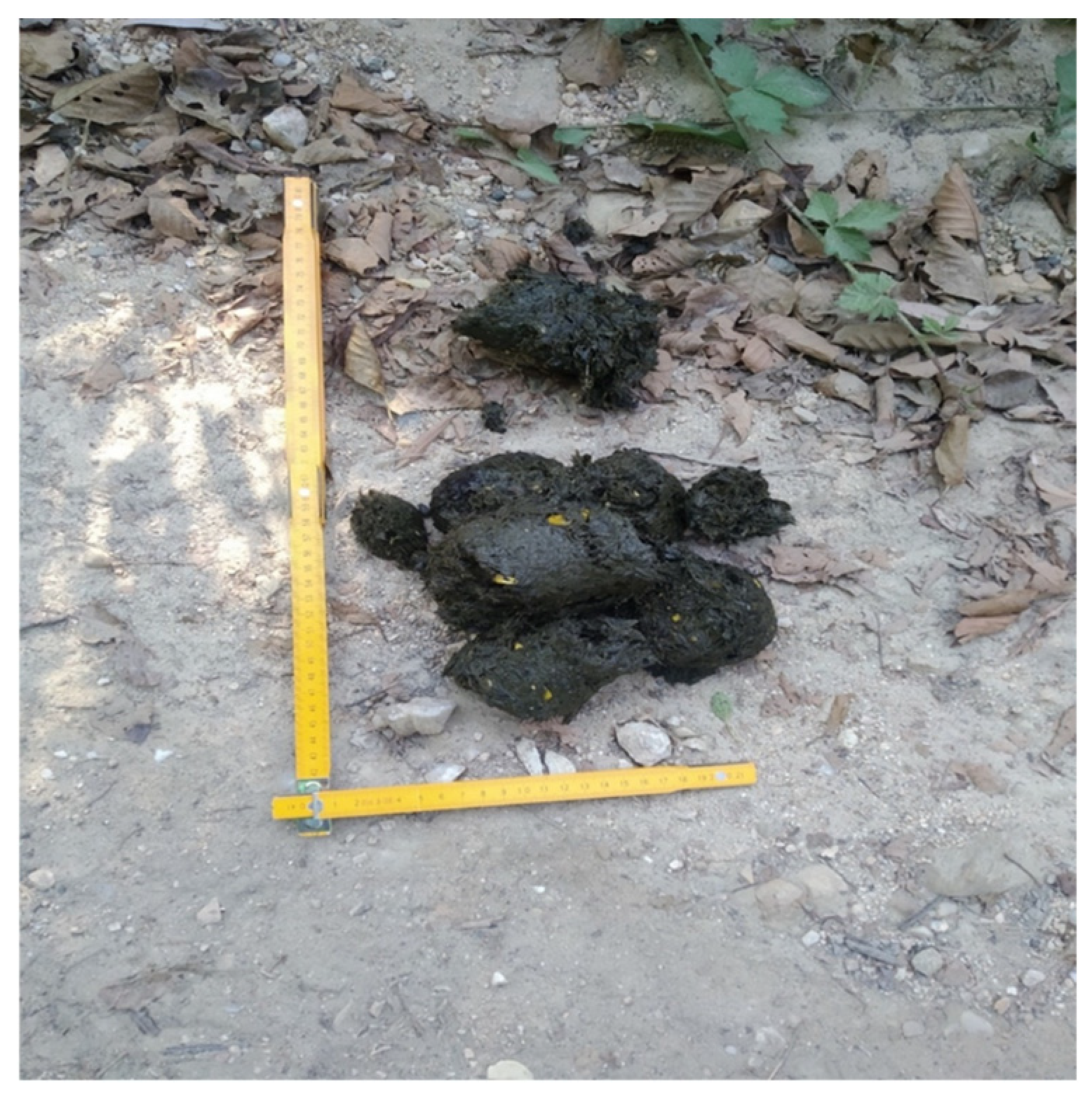
Brown bear faecal samples were collected during 5-day sampling trips conducted on a monthly to bimonthly basis during 2020–2022 and 2024, mainly along forest roads, around feeding sites, and in areas of bear activity (e.g., abandoned orchards, corn fields). Overall, 918 brown bear faecal samples, identified by their morphological characteristics (size, shape, visible food components), were collected [23]. Each sample was photographed (Figure 2) and then placed in a single-use, airtight plastic bag. To limit sampling bias caused by sampling the same individual multiple times, each site was visited only once during each sampling trip, and the sites were several kilometres apart.
Each plastic bag was tagged with a unique code. For each sample, location, GPS coordinates (Garmin Instinct Solar watch, New Taipei City, Taiwan), and collection date were recorded. The samples were stored at 4 °C until examination.

2.3. Parasitological Examinations

All faecal samples were transferred to the Laboratory of Parasitology and Parasitic Diseases, School of Veterinary Medicine, Aristotle University of Thessaloniki, and examined within 3−7 days of collection. All samples were examined by two standard parasitological methods: (i) ZnSO4 flotation with centrifugation [24] and Telemann (formaldehyde, ether) sedimentation [25] for the detection of parasitic elements. Samples deemed sufficiently fresh (i.e., retaining their shape and moisture and showing no visible fungal growth) were also examined using the Baermann method to detect first-stage larvae (L1) [24]. The preparations were examined under the microscope (100×, 400×), and the parasitic elements were identified based on their morphological and morphometric characteristics to the lowest taxon possible, i.e., species, genus, or family, depending on the presence of distinguishing morphological traits [26,27]. Positive samples were further analysed by the modified McMaster method [28] to quantify the parasite eggs per gram of faeces (EPG).

2.4. Statistical Analysis

Spatial and temporal patterns in parasite occurrence and diversity were analysed using generalised additive models (GAMs), selected for their ability to flexibly model non-linear relationships and spatial structure. Specifically, we used a tensor-product smooth of sample coordinates to model spatial clustering of infection, thus accounting for possible spatial autocorrelation of the data. Sample coordinates were available for a subset of 859 samples out of the original 918, and this subset was used for modelling.
Presence–absence responses were modelled using a binomial error distribution with a logit link. All models included spatial location, represented by longitude and latitude, and temporal parameters, i.e., season and year of study. To examine the seasonality of parasitological findings, seasons based on bear activities, as defined by de Gabriel Hernando et al., 2015 [2], were used. Seasons were defined as emergence (EM), 1 March–21 April; mating (MA), 22 April–7 August; early hyperphagia (EH), 8 August–7 October; and late hyperphagia (LH), 8 October–15 December. To allow spatial patterns to differ among seasons, spatial smooths were fitted using season-specific smooth terms.
The model selection was guided by the Unbiased Risk Estimator (UBRE) score of the binomial GAM. All models were fitted using Restricted Maximum Likelihood (REML). Spatial terms were interpreted as evidence of geographic clustering when clear spatial structure was present. The model fit was assessed using the proportion of deviance explained and the adjusted R2 values and Area Under the Curve (AUC).
Predicted spatial patterns were visualised using contour plots based on model predictions on a regular spatial grid. For presence–absence models, points indicating observed parasite presence were overlaid to illustrate the correspondence between predicted high-risk infection areas (“hotspots”) and observed infections. Only seasons exhibiting clear spatial structure are presented in the results herein.
All analyses were conducted in R (version 4.5.0) [29], primarily using the mgcv package [30] for model fitting, oddsratio [31] for the calculation of odds ratios for GAMs, DHARMa [32] for residual diagnostics based on 1000 simulations, pROC [33] for AUC estimation, and lattice/grid graphics for visualisation [34].

3. Results

3.1. Intestine Parasite Taxa and Their Prevalence

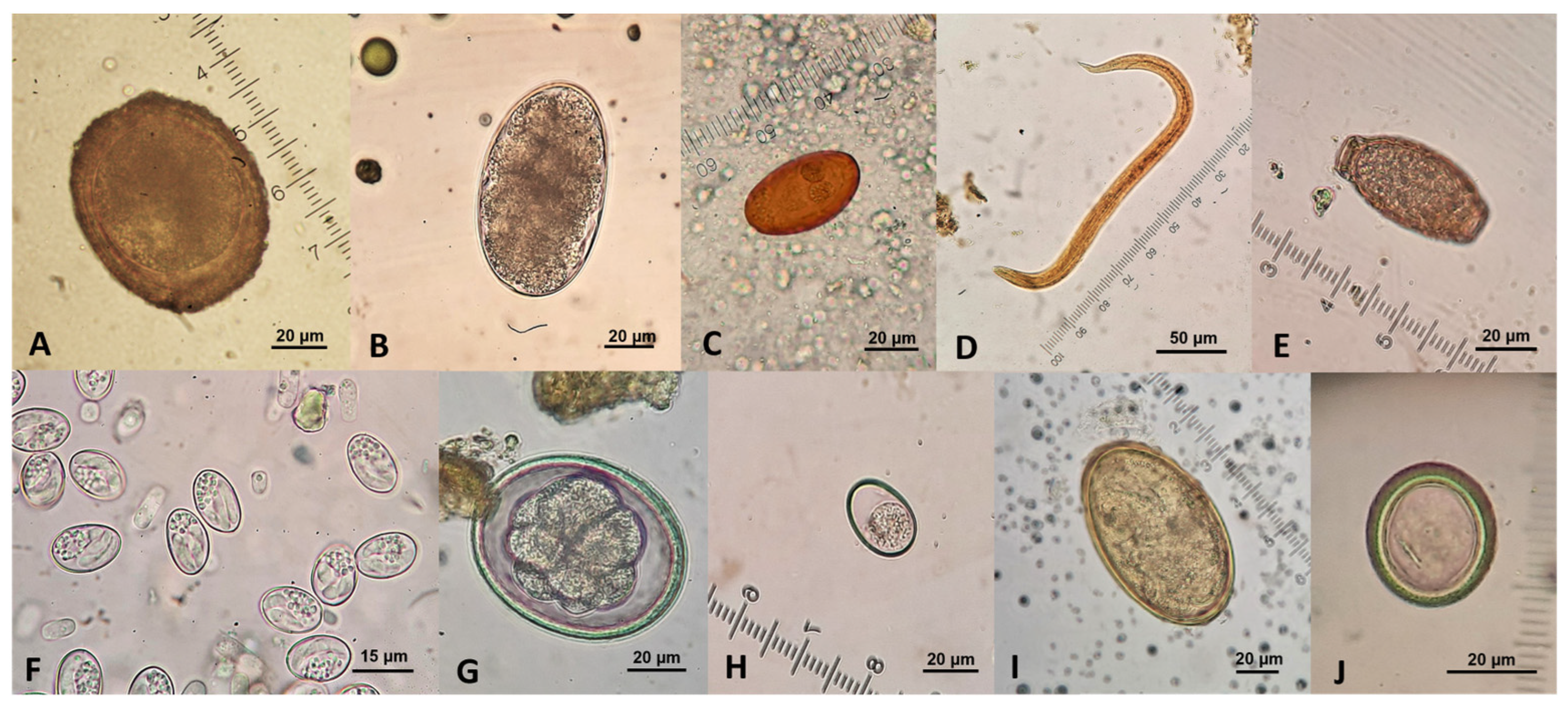
Of the 918 faecal samples,134 were collected in EM, 192 in MA, 294 in EH, and 298 in LH. Overall, 514 (56%) were positive for parasitic elements, i.e., eggs, oocysts, sporocysts, or larvae. Ten different parasitic taxa were found, of which three, i.e., Baylisascaris transfuga, Uncinaria spp., and Dicrocoelium dendriticum, were the most common, with prevalences of 39.98%, 18.09%, and 14.38%, respectively. The remaining seven taxa showed a prevalence of 2% or less. The prevalence of Crenosoma spp. (26.05%) was determined based on Baermann method results from 195 samples (Table 1, Figure 3).
The McMaster method yielded a positive result (≥50 EPG) only for B. transfuga eggs (detailed results in Section 3.2).

3.2. Spatio-Temporal Variation in Prevalence

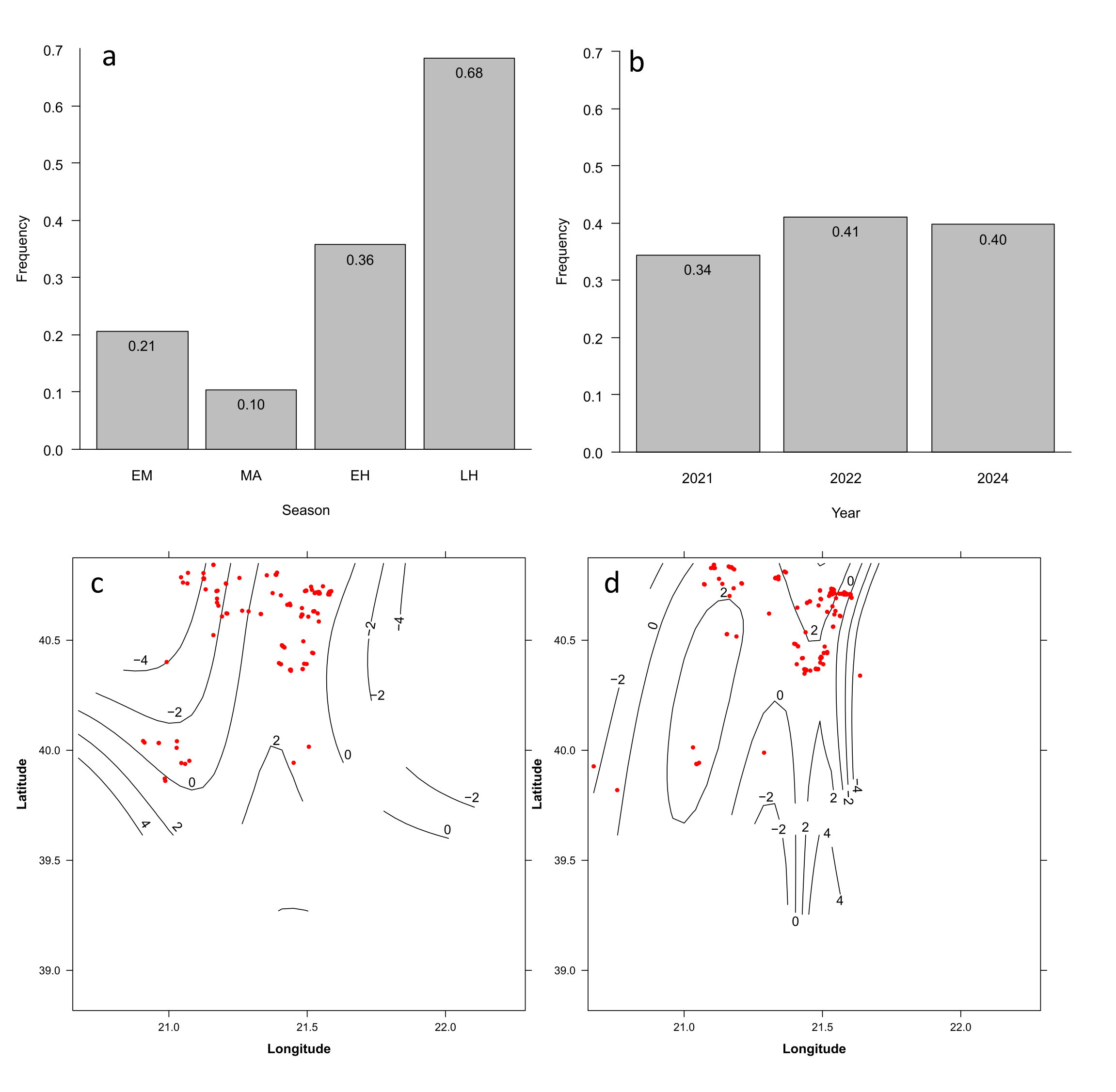
Prevalence of B. transfuga varied strongly with both season and year (Figure 3A,B, Table 2). Its overall prevalence was lower during MA compared to EM and significantly higher during LH. No significant difference was detected for EH relative to EM (p = 0.58). Prevalence was significantly higher in 2022 and 2024 compared to 2021 (Figure 4b, Table 2).
Spatial clustering of positive samples was season-dependent (Figure 3C,D and Table 2), namely, a limited spatial structure was observed during EM and MA, whereas clear and persistent geographic hotspots emerged during EH (edf = 11.0, p = 0.023) and LH (edf = 5.6, p = 0.022) (Figure 3C,D). The model explains 25.3% of the total deviance with an adjusted R2 of 0.267 and AUC = 0.83.
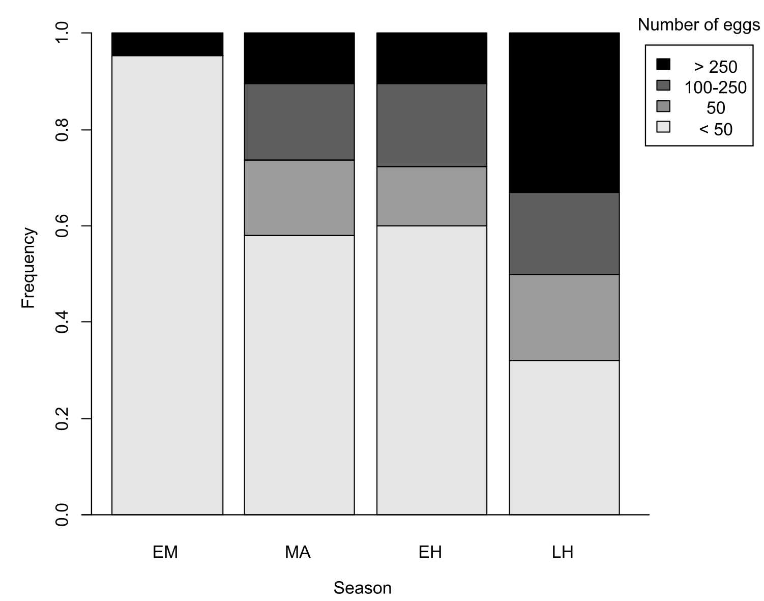
Infection intensity (EPG) was estimated in samples positive for B. transfuga. The EPG values ranged from 50 to 2000 with a median of 150. In EH the infection intensity was low. Most positive samples had fewer than 50 EPG (below the McMaster method’s detection threshold), while a few samples had more than 250 EPG. MA and EH showed similar distributions of infection intensity with an almost even representation of the three classes with more than 50 EGP and a slightly higher representation of samples with less than 50 EPG. Finally, the LH distribution shows a shift towards samples with >250 EPG (Figure 5).
The prevalence of Uncinaria spp. infection in different seasons varied between 15.6 % in MA and 21.5% in LH, but this variation was not statistically significant (Table 3). There was a tendency for the prevalence to decrease over the years. Furthermore, no significant spatial clustering of Uncinaria spp. positive samples were observed. The model explained 11.9% of the total deviance with an adjusted R2 of 0.07 and AUC = 0.7.
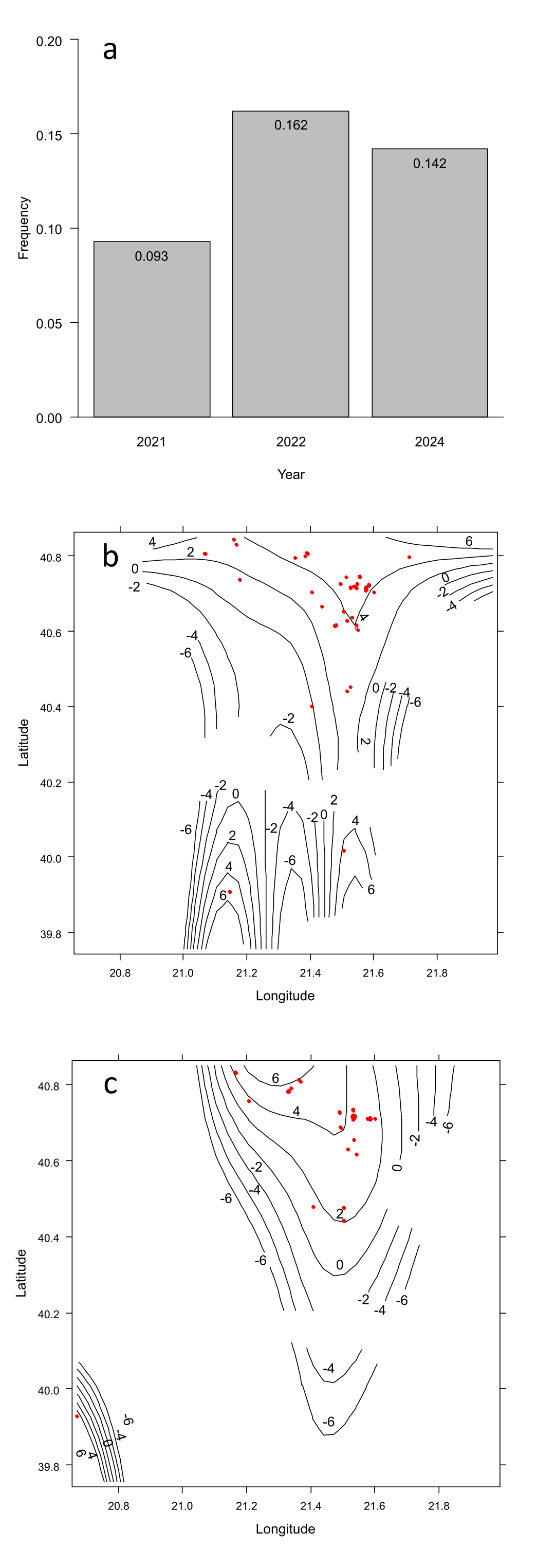
Similarly to Uncinaria spp., D. dendriticum showed no statistically significant seasonal variation, showing a minimum during MA (9.4%) and a maximum during LH (16.8 %). However, there was a significant variation on a year-by-year basis, with higher prevalence during 2022 and 2024 compared to 2021 (Figure 5 and Table 4). As in the case of B. transfuga, there was a significant spatial clustering of D. dendriticum-positive samples during both EH and LH (Figure 6). The model explained 21.1% of the total deviance with an adjusted R2 of 0.14, AUC = 0.79.
Finally, no statistical analysis was performed for Ε. aerophilus, Sarcocystis spp., T. leonina, Eimeria spp., L. serrata, and Taeniidae due to their low prevalence, and in the case of Crenosoma spp. due to the selective sampling (only fresh samples) that could bias any statistical result.

3.3. Mixed Infections

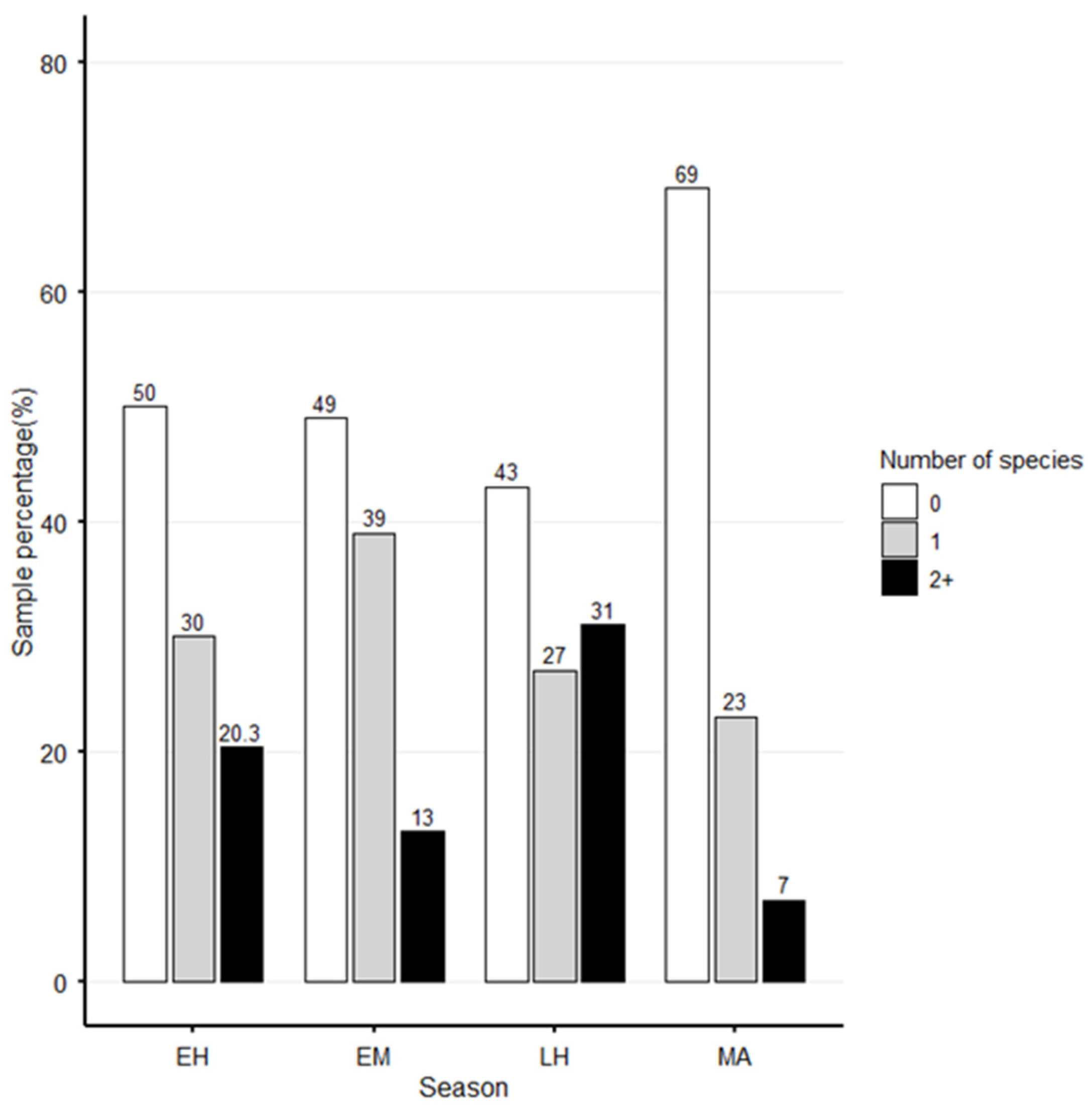
Mixed infections with two or more parasite taxa were recorded in 194 (22%) samples (Figure 7). The most common co-infection documented was B. transfuga with Uncinaria spp. (41%), followed by B. transfuga with D. dendriticum (26%), and B. transfuga with Crenosoma spp. (16%). Mixed infections with more than two different parasite taxa were less prevalent.

4. Discussion

This study presents the first systematic investigation of endoparasites in brown bears and their relationship to the species’ ecology in Greece. A total of ten parasitic taxa were identified from faecal samples, including protozoa, trematodes, cestodes, nematodes, and pentastomids.
Morphological identification of brown bear scat is a highly reliable method due to the absence of other bear species on the continent [23,35]. Although substantial effort was undertaken to reduce the probability of repeatedly sampling the same individual during a single sampling trip, faecal samples were collected opportunistically from the environment without genetic identification of individuals. As a result, repeated sampling of some bears cannot be excluded. Such pseudoreplication may have introduced bias into the baseline prevalence estimates. Moreover, because these observations were subsequently used in the predictive modelling framework, the same bias may have propagated into the model predictions.
Morphological identification to the species level was possible for five parasites, i.e., B. transfuga, D. dendriticum, E. aerophilus, T. leonina, and L. serrata, due to the characteristics of their eggs that distinguish them from similar species. These characteristics include the shape and size of the eggs, the morphology of the eggshell (e.g., thickness, surface texture, presence/absence of operculum), and the nature and morphology of the egg content (e.g., zygote, larva, or miracidium) [25,36,37]. On the other hand, the genera Uncinaria, Crenosoma, Sarcocystis, and Eimeria include different species that share common morphological characteristics in their diagnostic stages (eggs, larvae, sporocysts, and oocysts, respectively); thus, identification was restricted to the genus level [27,38]. Finally, most of the species included in the family Taenidae have morphologically similar eggs; thus, these eggs were identified to the family level [25].
Ascarid nematodes of the genera Baylisascaris and Toxascaris were recorded in the samples examined. Baylisascaris was the most prevalent parasite found. Although molecular identification of the species was not performed, the most likely species involved is B. transfuga, a parasite of ursids documented in all eight bear species [39]. Previous studies in Europe have reported varying prevalences, with higher rates in Slovakia (52.9%) [40], Estonia (51%) [15], and Spain (44.8%) [18], whereas in Croatia, Romania, and Italy the prevalences were lower (<20%) [13,14,41]. The variation in prevalence observed across these studies may be attributed to differences in sampling season, with a consistent finding of higher B. transfuga prevalence during autumn [15,17,18,19]. Though rare, clinical manifestations of baylisascariosis can include anorexia, poor coat condition, and intestinal obstruction [42]. While B. transfuga has not been confirmed to infect non-ursid species in Europe [10], all members of the genus are considered potentially zoonotic and may cause visceral, ocular, or neural larva migrans in humans [40]. The parasite’s life cycle is not fully described, but it is most likely similar to that of other ascarids, with infection occurring through the ingestion of embryonated eggs or paratenic hosts [14]. Toxascaris leonina is a common roundworm reported in domestic and wild canids and felids all around the world [43]. Eggs morphologically compatible with T. leonina have been reported in bears before [44,45]. Bears are not a confirmed host of this parasite, and the scenario of pseudoparasitism, i.e., eggs appearing in bears’ faeces as a result of consuming an infected carnivore, cannot be ruled out [42].
Eggs of the family Ancylostomatidae (hookworms) have been recorded in bears from Europe [12,13,35] and South America [46]. Within this family, two species of the genus Uncinaria have been identified in North America in American black bears (Ursus americanus) and grizzly bears (Ursus horribilis), i.e., Uncinaria yukonensis and Uncinaria rauschi [47,48]. The species Uncinaria stenocephala has been identified in brown bears in Russia [49]. Finally, another species, Uncinaria ursi, has been found in polar bears [50]. Recent genetic studies in North America suggest a close relationship between these Uncinaria species and hypothesise similar infection patterns [51]. Namely, the third-stage larvae from the environment enter a suitable host by skin penetration or oral ingestion and complete their life cycle in the host’s small intestine [52]. In Greece, Uncinaria spp. has been detected in both wild and domestic carnivores that share the same habitat as brown bears [20,53]. In the present study, the parasite shows the second-highest prevalence among bear gastrointestinal parasites (19%), which agrees with findings from Croatia (10%) [13]. A higher prevalence of Ancylostomatidae has been recorded in Slovakia (33%) and Italy (51%) [12,41]. Genetic research is needed to identify the species of Uncinaria in European brown bears and to determine its impact on the health of domestic and wild carnivores.
The trematode D. dendriticum was found here at a prevalence of 14%, while prevalences in recent studies in Europe vary widely, from 0.4% in Romania [14] to very high percentages in the Cantabrian Mountains, Spain (32–71%) [16,17,18]. Adult D. dendriticum resides in the bile ducts and gall bladder of its definitive hosts, causing lesions in the liver and, in severe cases, hepatitis and cirrhosis. The parasite’s eggs are shed in the faeces of the definitive host, and its life cycle involves two intermediate hosts: terrestrial gastropods as the first and ants as the second intermediate host [53]. Bears consume ants as part of their natural diet [54], which may lead to infection by this parasite. However, it cannot be ruled out that the detection of D. dendriticum eggs in some cases may be due to pseudoparasitism resulting from the consumption of infected definitive hosts’ (e.g., ruminants or lagomorphs) liver tissue.
Two nematodes affecting the respiratory system were found: Eucoleus aerophilus and Crenosoma spp. Eucoleus aerophilus (syn. Capillaria aerophila) has been reported in American black bears in the Southeastern United States [55] in a relatively high prevalence (10%). The presence of Eucoleus has been documented in brown bears in Slovakia [12,56], Italy, and Russia [41,57]. In Greece, the parasite has been detected in European wildcats and domestic cats [58,59]. This nematode has a direct life cycle and resides in the lungs (bronchi and trachea) of the hosts. The female releases eggs, which are transported by the mucociliary escalator to the pharynx, where they are swallowed and excreted in faeces. New hosts subsequently become infected by ingesting larvated eggs or, potentially, infected earthworms [60,61,62]. Clinical symptoms include dry and productive cough, sneezing, distress, dyspnea, and trachypnea, and range from mild to severe [62,63]. However, there is no evidence of clinical capilariosis in wild bears.
The diagnostic stage of Crenosoma spp. in the host’s faeces is the L1, which is less resistant to dryness than eggs and oocysts. Therefore, to obtain results closer to the actual prevalence of this parasite, only samples deemed sufficiently fresh were submitted to the specific larval retrieval method (Baermann). Crenosoma spp. have been previously reported in both American black bears [49,55,64] and brown bears in Romania [14], with lower prevalence than that recorded in the present study (5.45%). However, the gold-standard method for larval detection, i.e., the Baermann method [24], was not used in those studies, suggesting that the true prevalence of the parasite in those areas may be higher. Other animal species, such as wild and domestic canids, are also susceptible to Crenosoma spp. infections [65,66] and can act as reservoirs. The parasite uses gastropods as intermediate hosts, while several small vertebrates (e.g., amphibians, rodents, and reptiles) can act as paratenic hosts [67]. Adult parasites reside in the bronchi and can cause cough, bronchitis, and, in severe cases, pneumonia [68,69]. There is one clinical case in bears involving an orphaned black bear cub with mild symptoms [70]. It is unclear whether adult bears in the wild develop symptoms due to crenosomosis.
Protozoan parasites had a low prevalence in the present study, with only two genera, i.e., Sarcocystis spp. (0.5%) and Eimeria spp. (0.4%), recorded. Sarcocystis spp. have an indirect life cycle: definitive hosts acquire the infection by consuming intermediate hosts bearing tissue “sarcocysts”. In the definitive host, the parasite multiplies in the enterocytes and is shed into the environment via faeces, from which the intermediate host becomes infected [71]. Bears can act both as intermediate and as definitive hosts [72,73] due to their varied dietary habits: as herbivores and omnivores, they can act as intermediate hosts for some Sarcocystis species, whereas, as carnivores, they act as definitive hosts for other species when they consume an animal bearing sarcocysts. Both intermediate and definitive hosts can develop a variety of clinical symptoms depending on the parasite’s species, the infective dose, and the host’s immune status [74]. Recent studies on Sarcocystis-associated diseases, i.e., hepatic sarcocystosis and encephalitis, include two fatalities of a grizzly cub and a juvenile American black bear [75,76], showcasing the danger that the parasite can pose for young bears. Higher parasite prevalence was reported in older studies, namely, American black bears (11%) [55] and European brown bears in Slovakia (15.38%) [12]. Regarding Greek wildlife, Sarcocystis spp. have been detected in the faeces of golden jackals, red foxes, and European wildcats (definitive hosts) and in muscle tissue of wild boars (intermediate hosts) [59,73,74].
Two species of Eimeria have been identified in American black bears in previous studies, i.e., Eimeria albertensis and Eimeria borealis [77]. Eimeria spp. have also been found in grizzly bears [78], brown bears in Europe [12,13], and spectacled bears (Tremarctos ornatus) [79], with no species identification. Eimeria spp. infection in brown bears seems to have a low prevalence in Croatia (1.1%) and Slovakia (7.69%), in accordance with the present study (0.4%) [12,13]. Eimeria spp. are usually host-specific and infect a great variety of animals [80]. In Greece, the parasite has been detected in hares at a very high prevalence (64.28%) [81], as well as in poultry [82] and other domestic animals [83]. Eimeriosis mainly affects young animals, causing mainly enteritis and diarrhoea [81,84]. The presence of Eimeria spp. oocysts in the bears’ faecal samples may reflect true parasitism; however, cases of pseudoparasitism resulting from the consumption of infected prey are also likely. Species identification by molecular tools is needed to determine if bears are enzootic hosts of the taxon.
At least two cestode species of the Taeniidae family have been identified in ursids in the Northern Hemisphere, i.e., Taenia krabbei [48] and, in more recent genetic studies in Alaska and Finland, the Holarctic species Taenia arctos [85,86,87]. Both species have cervids as intermediate hosts. In Greece, there are only two endemic cervid species, i.e., Capreolus capreolus and Cervus elaphus, but there are no data on the presence of T. arctos in the country. The low prevalence recorded for Taeniidae (0.2%) in the present study is consistent with findings from other countries, where taenids were found by necropsy, as in the case of Finland and Canada, with a prevalence of 1.9% and 7.7%, respectively [85,87], or by combined faecal and post mortem examination as in Transylvania, with a prevalence of 3.2% [14]. In general, faecal examination is not very sensitive for detecting cestode infection, as infected animals often shed only a few free eggs and mostly release eggs within mature proglottids [59]. Furthermore, the diet of bears in Greece, which is mostly non-carnivorous [88], may explain the low prevalence of Taeniidae infection in the examined animals [85].
Eggs of the pentastomid Linguatulla serrata were recorded in two samples. Linguatulla serrata is a cosmopolitan zoonotic parasite affecting both herbivores and carnivores. Recent studies in Europe [89,90] show the presence of the parasite in both grey wolves (Canis lupus) and domestic ruminants. Studies from Greece also show the presence of the parasite in dogs, small ruminants, hares, and a variety of hospitalised wild mammals [81,90,91,92,93]. The parasite uses herbivores as an intermediate host and is transmitted when final hosts (carnivores) consume infected meat or internal organs [94]. Brown bears in Greece frequently consume ruminants [88] and might get infected. This is the first record of the parasite in brown bears, to the best of current knowledge.
Seasonal dynamics of brown bear parasites have been mentioned in several studies, both in North America [51,78,95] and in Europe [15,17,18,19], all reporting a seasonal high in parasite prevalence in autumn and a low in spring. The present results show a similar seasonal trend, with a peak in parasite prevalence in autumn (LH) and a low in early summer (MA). The seasonal trend is driven by the most prevalent species, B. transfuga, whose occurrence varied strongly across seasons. Spatial hotspots were detected primarily during EH and LH, whereas no statistically significant hotspots were observed during EM and MA, suggesting that increased host movement during these phases reduces localised environmental contamination and thus infection pressure. However, the lower sample size in EM (n = 134) may limit the model’s ability to detect subtle spatial structures during this period. Although the prevalence was higher during EH than EM, the model results indicated that this difference was largely associated with spatial clustering, highlighting the importance of location-specific processes during EH and LH.
Dicrocoelium dendriticum infections showed relatively low overall prevalence but clear spatial structuring. The probability of infection increased significantly in later years, although this effect was modest compared with spatial patterns. While no significant spatial structure was detected during EM or MA seasons, suggesting a more homogeneous distribution of infections during these periods, a strong and significant spatial clustering emerged in both EH and LH, indicating that transmission becomes more spatially aggregated during these seasons. This pattern likely reflects seasonal changes in host foraging behaviour or environmental exposure, leading to localised infection hotspots.
Dicrocoelium dendriticum shows significant and consistent spatial clustering across both EH and LH, implying a more persistent hotspot trait throughout the hyperphagia phase. In contrast, hotspots of B. transfuga appear more strongly concentrated in LH, aligning with its pronounced seasonal increase in prevalence and suggesting a sharper, more temporally focused aggregation of infection risk. Although the spatial hotspot analyses were based on modelled prevalence rather than raw case counts, sampling intensity was uneven across the study area. Areas with lower sampling intensity may therefore be associated with greater uncertainty in predicted prevalence. Consequently, the identified hotspots should be interpreted as regions of elevated infection risk within the limits of the available sampling coverage.
Uncinaria spp. showed no seasonal trend or spatial structure in any season, suggesting that other factors may affect its prevalence.
Finally, all three parasites (B. transfuga, D. dendriticum, and Uncinaria spp.) exhibited significant differences in prevalence among study years. Such variation may be associated with several factors, including sampling variation, climatic variation, fluctuations in intermediate-host availability, host population density, and habitat change; however, these factors were not evaluated in the present study because the necessary data were unavailable.
There is no definitive explanation why this seasonal trend is observed in brown bears. Several studies hypothesise that bears manage to void their intestines of helminth parasites before hibernation and get reinfected in spring [19,42,48,95]. Still, there is no clear evidence of the mechanism involved in this event. According to the present results, faecal samples positive for parasites were collected in early spring, suggesting that parasitism persisted through the hibernation period. However, the present results may be biassed due to several factors. Bears in milder climates, such as Greece, may not fully hibernate [22], and as a result, parasites persist year-round, potentially leading to biassed seasonal prevalence in the area. Furthermore, the low sample count during late spring–early summer (ΜA) may have led to an underestimation of parasite prevalence. Quantitative analysis of bear faeces to assess the abundance and excretion intensity of B. transfuga eggs has only been conducted in two studies from Slovakia [19,42]. Both studies show a peak in autumn, consistent with the seasonal pattern observed in the present study, where intensity of parasitism showed a progressive seasonal shift, with predominantly low egg counts (<50 EPG) in the first season, followed by an increase in intermediate EPG classes, i.e., 50–100 and 100–250 EPG in MA and EH seasons, respectively. The highest infection intensities were recorded in LH, where a substantial number of samples scored >250 EPG, while cases of low intensity infections declined markedly. Mixed infections with two or more different taxa were present in all seasons. The seasonal trend we observed above also seems to hold true for polyparasitism. Samples positive for more than one parasite species were most frequently observed during early and late hyperphagia, with parasite diversity being lowest during emergence and the mating period. Polyparasitism, although common in wild animals, remains under-researched and may have cumulative effects on the host’s immune system [96].
Additional limitations beyond those discussed above should be considered when interpreting the results of this study. First, pseudoparasitism cannot be excluded for certain parasite taxa, particularly given the omnivorous diet of the brown bear. Second, morphological examination alone was insufficient to identify several parasitic elements at the species level; therefore, molecular analyses would be required to confirm their taxonomic identity. Furthermore, the available data were insufficient to fully explain the temporal patterns detected by the GAMs. Spatial predictions generated by GAMs are based on smoothed surfaces and do not account for fine-scale environmental variables, such as vegetation structure, soil moisture, and microclimatic conditions, which may influence parasite distribution.
Despite these limitations, this study has several notable strengths, including the largest dataset of brown bear faecal samples examined for endoparasites in Europe to date. To the best of the authors’ knowledge, this is also the first study to employ the Baermann technique, the gold-standard coprological method for lungworm detection, thereby providing robust estimates of lungworm prevalence in the brown bear population studied. The findings provide important insights into the composition of the species’ endoparasite fauna and the seasonal variation in several parasite taxa. Future studies should incorporate genetic identification of individual bears to reduce the risk of pseudoreplication and should be complemented by parasitological examinations of carcasses to obtain a more comprehensive assessment of parasite diversity.

5. Conclusions

Ten endoparasite taxa were identified by faecal examination of brown bears in Greece. The results of the present study are consistent with findings from studies on brown bears in both Europe and North America. The sample size of the present study allows for the drawing of reasonably robust conclusions regarding the species composition and prevalence of endoparasites in the brown bear population of the study area, as well as the health risks these parasites may pose to this species, an aspect that warrants consideration given its vulnerable conservation status. However, to date, there is no unequivocal evidence of the actual impact parasitoses have on the brown bear population in Greece. Furthermore, some of the identified parasites may also infect other sympatric wild and domestic animals; therefore, brown bears may be considered among the hosts that contribute to the maintenance and transmission of these parasites within the study area. A seasonal trend in B. transfuga infection was confirmed herein, a finding that could be of relevance for brown bears’ general fitness status throughout their activity season. Further research is required to fully elucidate the life cycle of B. transfuga, its zoonotic potential, and its impact on the species’ general health. Additional genetic analyses are needed to further identify the parasites and eliminate potential bias arising from repeated sampling of the same individuals. Finally, long-term, systematic monitoring of bear parasites is essential to detect emerging changes, improve understanding of transmission dynamics, assess potential impacts on population health, and clarify the ecological processes that shape parasite distribution in this species.

Author Contributions

Conceptualization, A.D. and S.S.; methodology, A.D. and S.S.; software A.S. and S.S.; validation, A.D. and S.S.; formal analysis, A.S. and S.S.; investigation, A.D. and A.S.; resources, A.D., S.S., and A.S.; data curation, A.D., S.S., and A.S.; writing—original draft preparation, A.S.; writing—review and editing, A.D. and S.S.; visualisation, S.S., A.S.; supervision, A.D. and S.S.; project administration, A.D., S.S. All authors have read and agreed to the published version of the manuscript.

Funding

This research received no external funding.

Institutional Review Board Statement

Not applicable.

Informed Consent Statement

Not applicable.

Data Availability Statement

The raw data supporting the conclusions of this article will be made available by the authors on request.

Acknowledgments

The authors would like to thank the personnel of the three National Parks (Pindos, Prespes, and Rhodope) and the two NGOs (Arcturos and Callisto) for their help in collecting part of the samples.

Conflicts of Interest

The authors declare no conflicts of interest.

Abbreviations

The following abbreviations are used in this manuscript:
GAMsGeneralised Additive Models
GPS Garmin Instinct Solar
EMEmergence
MAMating Season
EHEarly Hyperphagia Season
LHLate Hyperphagia Season
UBREUnbiassed Risk Estimator
REMLRestricted Maximum Likelihood
AUCArea Under the Curve

References

  1. Chapron, G.; Kaczensky, P.; Linnell, J.D.C.; Von Arx, M.; Huber, D.; Andrén, H.; López-Bao, J.V.; Adamec, M.; Álvares, F.; Anders, O.; et al. Recovery of Large Carnivores in Europe’s Modern Human-Dominated Landscapes. Science 2014, 346, 1517–1519. [Google Scholar] [CrossRef] [Scilit] [PubMed]
  2. Karamanlidis, A.A.; de Gabriel Hernando, M.; Krambokoukis, L.; Gimenez, O. Evidence of a Large Carnivore Population Recovery: Counting Bears in Greece. J. Nat. Conserv. 2015, 27, 10–17. [Google Scholar] [CrossRef] [Scilit]
  3. Mertzanis, G.A. Brown Bear in Greece: Distribution, Present Status: Ecology of a Northern Pindus Subpopulation. Bears Their Biol. Manag. 1994, 9, 187–197. [Google Scholar] [CrossRef] [Scilit]
  4. Pylidis, C.; Anijalg, P.; Saarma, U.; Dawson, D.A.; Karaiskou, N.; Butlin, R.; Mertzanis, Y.; Giannakopoulos, A.; Iliopoulos, Y.; Krupa, A.; et al. Multisource Noninvasive Genetics of Brown Bears (Ursus arctos) in Greece Reveals a Highly Structured Population and a New Matrilineal Contact Zone in Southern Europe. Ecol. Evol. 2021, 11, 6427–6443. [Google Scholar] [CrossRef] [Scilit] [PubMed]
  5. Edozoume.gr. Available online: https://edozoume.gr/wp-content/uploads/2025/02/LIFE-IP-4-NATURA-Del.A.1.6.10-Action-Plan-Ursus-Arctos-OGG.pdf (accessed on 23 May 2026).
  6. Thompson, R.C.A.; Lymbery, A.J.; Smith, A. Parasites, Emerging Disease and Wildlife Conservation. Int. J. Parasitol. 2010, 40, 1163–1170. [Google Scholar] [CrossRef] [Scilit] [PubMed]
  7. Murray, D.L.; Kapke, C.A.; Evermann, J.F.; Fuller, T.K. Infectious Disease and the Conservation of Free-Ranging Large Carnivores. Anim. Conserv. 1999, 2, 241–254. [Google Scholar] [CrossRef] [PubMed]
  8. Daszak, P.; Cunningham, A.A.; Hyatt, A.D. Emerging Infectious Diseases of Wildlife—Threats to Biodiversity and Human Health. Science 2000, 287, 443–449. [Google Scholar] [CrossRef] [Scilit] [PubMed]
  9. Choquette, L.P.E. Significance of Parasites in Wildlife. Can. J. Comp. Med. Vet. Sci. 1956, 20, 418–426. [Google Scholar] [PubMed]
  10. Veronesi, F.; Deak, G.; Diakou, A. Wild Mesocarnivores as Reservoirs of Endoparasites Causing Important Zoonoses and Emerging Bridging Infections across Europe. Pathogens 2023, 12, 178. [Google Scholar] [CrossRef] [Scilit] [PubMed]
  11. Ravaszova, P.; Halanova, M.; Goldova, M.; Valencakova, A.; Malcekova, B.; Hurníková, Z.; Halan, M. Occurrence of Cryptosporidium spp. in Red Foxes Brown Bear Slovak Republic. Parasitol. Res. 2012, 110, 469–471. [Google Scholar] [CrossRef] [Scilit] [PubMed]
  12. Orosová, T.; Goldová, M.; Ciberej, J.; Štrkolcová, G. Parasitofauna of Brown Bear (Ursus arctos) in the Protected Landscape Area CHKO—Poľana. Folia Vet. 2016, 60, 20–24. [Google Scholar] [CrossRef] [Scilit]
  13. Aghazadeh, M.; Elson-Riggins, J.; Reljić, S.; De Ambrogi, M.; Huber, Đ.; Majnarić, D.; Hermosilla, C. Gastrointestinal Parasites and the First Report of Giardia spp. in a Wild Population of European Brown Bears (Ursus arctos) Croatia. Vet. Arh. 2015, 85, 201–210. [Google Scholar]
  14. Borka-Vitális, L.; Domokos, C.; Földvári, G.; Majoros, G. Endoparasites of Brown Bears in Eastern Transylvania, Romania. Ursus 2017, 28, 20–30. [Google Scholar] [CrossRef] [Scilit]
  15. Laanep, M.; Tull, A.; Tammeleht, E.; Valdmann, H.; Hindrikson, M.; Saarma, U. Seasonal Dynamics of Brown Bear Parasites: Opposite Trends for Nematodes and Protozoa. J. Helminthol. 2025, 99, e106. [Google Scholar] [CrossRef] [Scilit] [PubMed]
  16. Costa, H.; Hartasánchez, R.; Santos, A.R.; Camarão, A.; Cruz, L.; Nascimento, M.; Gomes, L.; Madeira de Carvalho, L.M. Preliminary Findings on the Gastrointestinal Parasites of the Brown Bear (Ursus arctos) in the Cantabrian Mountains, Spain. Vet. Parasitol. Reg. Stud. Rep. 2022, 28, 100681. [Google Scholar] [CrossRef] [Scilit] [PubMed]
  17. Remesar, S.; Busto, C.; Díaz, P.; Rivas, Ó.; López-Bao, J.V.; Ballesteros, F.; García-Dios, D. Presence of Gastrointestinal and Bronchopulmonary Parasites in Cantabrian Brown Bears. Eur. J. Wildl. Res. 2024, 70, 23. [Google Scholar] [CrossRef] [Scilit]
  18. Valderrábano Cano, E.; Penteriani, V.; Vega, I.; del Mar Delgado, M.; González-Bernardo, E.; Bombieri, G.; Zarzo-Arias, A.; Sánchez-Andrade Fernández, R.; Paz-Silva, A. Influence of Seasonality and Biological Activity on Infection by Helminths in Cantabrian Bear. Int. J. Parasitol. Parasites Wildl. 2024, 23, 100916. [Google Scholar] [CrossRef] [Scilit] [PubMed]
  19. Molnár, L.; Königová, A.; Major, P.; Vasilková, Z.; Tomková, M.; Várady, M. Seasonal Pattern of Prevalence and Excretion of Eggs of Baylisascaris transfuga in the Brown Bear (Ursus arctos). Animals 2020, 10, 2428. [Google Scholar] [CrossRef] [Scilit] [PubMed]
  20. Papadopoulos, H.; Himonas, C.; Papazahariadou, M.; Antoniadou-Sotiriadou, K. Helminths of foxes and other wild carnivores from rural areas in Greece. J. Helminthol. 1997, 71, 227–231. [Google Scholar] [CrossRef] [Scilit]
  21. Papadopoulos, E.; Komnenou, A.; Karamanlidis, A.A.; Bezerra-Santos, M.A.; Otranto, D. Zoonotic Thelazia callipaeda Eyeworm in Brown Bears (Ursus arctos): A New Host Record in Europe. Transbound. Emerg. Dis. 2022, 69, 235–239. [Google Scholar] [CrossRef] [Scilit] [PubMed]
  22. Mertzanis, G.; Mazaris, A.D.; Sgardelis, S.; Aravidis, E.; Giannakopoulos, A.; Godes, C.; Riegler, S.; Riegler, A.; Tragos, A. Telemetry as a Tool to Study Spatial Behaviour and Patterns of Brown Bears as Affected by the Newly Constructed Egnatia Highway—N. Pindos, Greece. In Modern Telemetry; IntechOpen: London, UK, 2011. [Google Scholar]
  23. Bang, P.; Dahlstrøm, P.; Walters, M. Animal Tracks and Signs; Oxford University Press: Oxford, UK, 2001. [Google Scholar]
  24. Faust, E.C.; D’Antoni, J.S.; Odom, V.; Miller, M.J.; Peres, C.; Sawitz, W.; Thomen, L.F.; Tobie, J.; Walker, J.H. A Critical Study of Clinical Laboratory Technics for the Diagnosis of Protozoan Cysts and Helminth Eggs in Feces: I. Preliminary Communication. Am. J. Trop. Med. Hyg. 1938, s1-18, 169–183. [Google Scholar] [CrossRef] [Scilit]
  25. Thienpont, D.; Rochette, F.; Vanparijs, O.F.J. Diagnosing Helminthiasis by Coprological Examination; Janssen Research Foundation: Beerse, Belgium, 1986. [Google Scholar]
  26. Taylor, M.A.; Coop, R.L.; Wall, R.L. Veterinary Parasitology, 3rd ed.; Blackwell Publishing: Oxford, UK, 2007. [Google Scholar]
  27. Levine, N.D. Veterinary Protozoology; Iowa State University Press: Ames, IA, USA, 1985. [Google Scholar]
  28. Coles, G.C.; Bauer, C.; Borgsteede, F.H.M.; Geerts, S.; Klei, T.R.; Taylor, M.A.; Waller, P.J. World Association for the Advancement of Veterinary Parasitology (W.A.A.V.P.) Methods for the Detection of Anthelmintic Resistance in Nematodes of Veterinary Importance. Vet. Parasitol. 1992, 44, 35–44. [Google Scholar] [CrossRef] [Scilit] [PubMed]
  29. R Core Team. R: A Language and Environment for Statistical Computing; R Foundation for Statistical Computing: Vienna, Austria, 2025. [Google Scholar]
  30. Wood, S.N. Generalized Additive Models: An Introduction with R, 2nd ed.; CRC Press: Boca Raton, FL, USA, 2017. [Google Scholar]
  31. Schratz, P. pat-s/oddsratio: Odds Ratio Calculation for GAM(M)s & GLM(M)s, version 2.0.1; R Foundation for Statistical Computing: Vienna, Austria, 2017. [CrossRef]
  32. Hartig, F. DHARMa: Residual Diagnostics for Hierarchical (Multi-Level/Mixed) Regression Models, version 0.5.0; R Foundation for Statistical Computing: Vienna, Austria, 2026.
  33. Robin, X.; Turck, N.; Hainard, A.; Tiberti, N.; Lisacek, F.; Sanchez, J.C.; Müller, M. pROC: An open-source package for R and S+ to analyze and compare ROC curves. BMC Bioinform. 2011, 12, 77. [Google Scholar] [CrossRef] [Scilit] [PubMed]
  34. Sarkar, D. Lattice: Multivariate Data Visualization with R; Springer: New York, NY, USA, 2008. [Google Scholar]
  35. Rojas, A.; Germitsch, N.; Oren, S.; Sazmand, A.; Deak, G. Wildlife parasitology: Sample collection and processing, diagnostic constraints, and methodological challenges in terrestrial carnivores. Parasites Vectors 2024, 17, 127. [Google Scholar] [CrossRef] [Scilit] [PubMed]
  36. Moudgil, A.D.; Singla, L.D.; Singh, M.P. First report on molecular identification and fenbendazole resistance against Baylisascaris transfuga infection in Melursus ursinus (sloth bear). Helminthologia 2014, 51, 262–268. [Google Scholar] [CrossRef] [Scilit]
  37. Nagamori, Y.; Ramachandran, A.; Kuzma, C.; Nafe, L.; Johnson, E.M. A zoonotic parasite, Linguatula serrata, infection in a dog imported from Ethiopia to the United States. Vet. Parasitol. Reg. Stud. Rep. 2019, 16, 100273. [Google Scholar] [CrossRef] [Scilit] [PubMed]
  38. Zajac, A.M.; Conboy, G.A.; Little, S.E.; Reichard, M.V. Veterinary Clinical Parasitology, 9th ed.; Wiley-Blackwell: Hoboken, NJ, USA, 2021. [Google Scholar]
  39. Schaul, J.C. Baylisascaris transfuga in Captive and Free-Ranging Populations of Bears (Family: Ursidae). Master’s Thesis, Ohio State University, Columbus, OH, USA, 2006. [Google Scholar]
  40. Štrkolcová, G.; Goldová, M.; Šnábel, V.; Špakulová, M.; Orosová, T.; Halán, M.; Mojžišová, J. A Frequent Roundworm Baylisascaris transfuga in Overpopulated Brown Bears (Ursus arctos) in Slovakia: A Problem Worthy of Attention. Acta Parasitol. 2018, 63, 167–174. [Google Scholar] [CrossRef] [Scilit] [PubMed]
  41. Paoletti, B.; Iorio, R.; Traversa, D.; Di Francesco, C.E.; Gentile, L.; Angelucci, S.; Amicucci, C.; Bartolini, R.; Marangi, M.; Di Cesare, A. Helminth Infections in Faecal Samples of Apennine Wolf (Canis lupus italicus) and Marsican Brown Bear (Ursus arctos marsicanus) in Two Protected National Parks of Central Italy. Ann. Parasitol. 2017, 63, 205–212. [Google Scholar] [CrossRef] [PubMed]
  42. Finnegan, S. Seasonal Dynamics in the Prevalence of Baylisascaris transfuga Ova in the Faeces of the Brown Bear (Ursus arctos) in Slovakia. Bachelor’s Thesis, University of Veterinary Medicine in Košice, Košice, Slovakia, 2009. [Google Scholar]
  43. Xie, Y.; Li, Y.; Gu, X.; Liu, Y.; Zhou, X.; Wang, L.; He, R.; Peng, X.; Yang, G. Molecular Characterization of Ascaridoid Parasites from Captive Wild Carnivores in China Using Ribosomal and Mitochondrial Sequences. Parasites Vectors 2020, 13, 382. [Google Scholar] [CrossRef] [Scilit] [PubMed]
  44. Kurnosova, O.P.; Khrustalev, A.V.; Illarionova, N.A.; Odoevskaya, I.M. A Survey of Helminths of Polar Bears in the Russian Arctic. Czech Polar Rep. 2017, 7, 164–168. [Google Scholar] [CrossRef] [Scilit]
  45. Todorov, V.; Yankov, Y.; Tsingarska, E.; Tonev, A.S. First Record of Toxascaris leonina and Eucoleus aerophilus in Population of Brown Bears (Ursus arctos) in Bulgaria. In Proceedings of the International Scientific Conference “Tradition and Modernity in Veterinary Medicine”, Stara Zagora, Bulgaria, 27–29 April 2023. [Google Scholar]
  46. Figueroa, J. New Records of Parasites in Free-Ranging Andean Bears from Peru. Ursus 2015, 26, 21–27. [Google Scholar] [CrossRef] [Scilit]
  47. Olsen, O.W. Uncinaria rauschi (Strongyloidea: Nematoda), a New Species of Hookworms from Alaskan Bears. Can. J. Zool. 1968, 46, 1113–1117. [Google Scholar] [CrossRef] [Scilit]
  48. Choquette, L.P.E.; Gibson, G.G.; Pearson, A.M. Helminths of the Grizzly Bear, Ursus arctos L., in Northern Canada. Can. J. Zool. 1969, 47, 167–170. [Google Scholar] [CrossRef] [Scilit] [PubMed]
  49. Rogers, L.L.; Rogers, S.M. Parasites of Bears: A Review. Bears Their Biol. Manag. 1976, 3, 411–430. [Google Scholar] [CrossRef] [Scilit]
  50. Kilinç, Ö.O.; Göz, Y.; Yılmaz, A.B.; Aslan, L. First Report on Heavy Uncinaria (Dochmoides) sp. (Nematoda: Ancylostomatidae) Infection in Brown Bear (Ursus arctos) Cub, in Van Province, Eastern Anatolian Region of Turkey. Kafkas Univ. Vet. Fak. Derg. 2015, 21, 295–297. [Google Scholar] [CrossRef] [Scilit]
  51. Catalano, S.; Lejeune, M.; Van Paridon, B.; Pagan, C.A.; Wasmuth, J.D.; Tizzani, P.; Duignan, P.J.; Nadler, S.A. Morphological Variability and Molecular Identification of Uncinaria spp. (Nematoda: Ancylostomatidae) from Grizzly and Black Bears: New Species or Phenotypic Plasticity? J. Parasitol. 2015, 101, 182–192. [Google Scholar] [CrossRef] [Scilit] [PubMed]
  52. Anderson, R.C. Nematode Parasites of Vertebrates: Their Development and Transmission; CABI Publishing: Wallingford, UK, 2000. [Google Scholar] [CrossRef] [Scilit]
  53. Manga-González, M.Y.; González-Lanza, C.; Cabanas, E.; Campo, R. Contributions to and Review of Dicrocoeliosis, with Special Reference to the Intermediate Hosts of Dicrocoelium dendriticum. Parasitology 2001, 123, 91–114. [Google Scholar] [CrossRef] [Scilit] [PubMed]
  54. Große, C.; Kaczensky, P.; Knauer, F. Ants: A Food Source Sought by Slovenian Brown Bears (Ursus arctos)? Can. J. Zool. 2003, 81, 1996–2005. [Google Scholar] [CrossRef] [Scilit]
  55. Crum, J.M.; Nettles, V.F.; Davidson, W.R. Studies on endoparasites of the black bear (Ursus americanus) in the Southeastern United States. J. Wildl. Dis. 1978, 14, 178–186. [Google Scholar] [CrossRef] [Scilit] [PubMed]
  56. Goldová, M.; Ciberej, J.; Rigg, R. Brown Bear (Ursus arctos) and Parasitic Zoonoses. Folia Venatoria 2003, 33, 123–127. (In Slovak) [Google Scholar]
  57. Bugmyrin, S.V.; Tirronen, K.F.; Panchenko, D.V.; Kopatz, A.; Hagen, S.B.; Eiken, H.G.; Kuznetsova, A.S. Helminths of Brown Bears (Ursus arctos) in the Kola Peninsula. Parasitol. Res. 2017, 116, 1755–1760. [Google Scholar] [CrossRef] [Scilit] [PubMed]
  58. Diakou, A.; Migli, D.; Dimzas, D.; Morelli, S.; Di Cesare, A.; Youlatos, D.; Lymberakis, P.; Traversa, D. Endoparasites of European Wildcats (Felis silvestris) in Greece. Pathogens 2021, 10, 594. [Google Scholar] [CrossRef] [Scilit] [PubMed]
  59. Symeonidou, I.; Gelasakis, A.I.; Arsenopoulos, K.; Angelou, A.; Beugnet, F.; Papadopoulos, E. Feline Gastrointestinal Parasitism in Greece: Emergent Zoonotic Species and Associated Risk Factors. Parasites Vectors 2018, 11, 227. [Google Scholar] [CrossRef] [Scilit] [PubMed]
  60. Traversa, D.; Di Cesare, A. Feline Lungworms: What a Dilemma. Trends Parasitol. 2013, 29, 423–430. [Google Scholar] [CrossRef] [Scilit] [PubMed]
  61. Traversa, D.; Di Cesare, A.; Lia, R.P.; Castagna, G.; Meloni, S.; Heine, J.; Strube, K.; Milillo, P.; Otranto, D.; Meckes, O.; et al. New Insights into Morphological and Biological Features of Capillaria aerophila (Trichocephalida, Trichuridae). Parasitol. Res. 2011, 109, 97–104. [Google Scholar] [CrossRef] [Scilit] [PubMed]
  62. Morelli, S.; Diakou, A.; Colombo, M.; Di Cesare, A.; Barlaam, A.; Dimzas, D.; Traversa, D. Cat Respiratory Nematodes: Current Knowledge, Novel Data and Warranted Studies on Clinical Features, Treatment and Control. Pathogens 2021, 10, 454. [Google Scholar] [CrossRef] [Scilit] [PubMed]
  63. Traversa, D.; Di Cesare, A. Diagnosis and Management of Lungworm Infections in Cats: Cornerstones, Dilemmas and New Avenues. J. Feline Med. Surg. 2016, 18, 7–20. [Google Scholar] [CrossRef] [Scilit] [PubMed]
  64. Addison, E.M.; Fraser, G.A. Life Cycle of Crenosoma petrowi (Nematoda: Metastrongyloidea) from Black Bears (Ursus americanus). Can. J. Zool. 1994, 72, 300–302. [Google Scholar] [CrossRef] [Scilit]
  65. Latrofa, M.S.; Lia, R.P.; Giannelli, A.; Colella, V.; Santoro, M.; D’Alessio, N.; Campbell, B.E.; Parisi, A.; Dantas-Torres, F.; Mutafchiev, Y.; et al. Crenosoma vulpis in Wild and Domestic Carnivores from Italy: A Morphological and Molecular Study. Parasitol. Res. 2015, 114, 3611–3617. [Google Scholar] [CrossRef] [Scilit] [PubMed]
  66. Tolnai, Z.; Széll, Z.; Sréter, T. Environmental Determinants of the Spatial Distribution of Angiostrongylus vasorum, Crenosoma vulpis and Eucoleus aerophilus in Hungary. Vet. Parasitol. 2015, 207, 355–358. [Google Scholar] [CrossRef] [Scilit] [PubMed]
  67. Karkamo, V.; Castren, L.; Näreaho, A. Fox lungworm, Crenosoma vulpis, in the dog-literature review and a case report. Suom. Eläinlääkäril. 2012, 118, 67–72. [Google Scholar]
  68. Stockdale, P.H.; Hulland, T.J. The Pathogenesis Route of Migration, and Development of Crenosoma vulpis in the Dog. Pathol. Vet. 1970, 7, 28–42. [Google Scholar] [CrossRef] [Scilit] [PubMed]
  69. Segeritz, L.; Westhoff, K.M.; Schaper, R.; Hermosilla, C.; Taubert, A. Angiostrongylus vasorum, Aelurostrongylus abstrusus, Crenosoma vulpis and Troglostrongylus brevior Infections in Native Slug Populations of Bavaria and Baden-Wuerttemberg in Germany. Pathogens 2022, 11, 747. [Google Scholar] [CrossRef] [Scilit] [PubMed]
  70. Mahjoub, H.A.; Murphy, N.; Mather, P.M.; Greenwood, S.J.; Conboy, G.A. Clinical Crenosomosis in a Black Bear (Ursus americanus). Vet. Parasitol. Reg. Stud. Rep. 2020, 20, 100380. [Google Scholar] [CrossRef] [Scilit] [PubMed]
  71. Dubey, J.P. The Evolution of the Knowledge of Cat and Dog Coccidia. Parasitology 2009, 136, 1469–1475. [Google Scholar] [CrossRef] [Scilit] [PubMed]
  72. Decker Franco, C.; Schnittger, L.; Florin-Christensen, M. Sarcocystis. In Parasitic Protozoa of Farm Animals and Pets; Springer: Cham, Switzerland, 2018; pp. 103–124. [Google Scholar] [CrossRef] [Scilit]
  73. Xiang, Z.; Chen, X.; Yang, L.; He, Y.; Jiang, R.; Rosenthal, B.M.; Luan, P.; Attwood, S.W.; Zuo, Y.; Zhang, Y.P.; et al. Non-Invasive Methods for Identifying Oocysts of Sarcocystis spp. from Definitive Hosts. Parasitol. Int. 2009, 58, 293–296. [Google Scholar] [CrossRef] [Scilit] [PubMed]
  74. Tenter, A.M. Current Research on Sarcocystis Species of Domestic Animals. Int. J. Parasitol. 1995, 25, 1311–1330. [Google Scholar] [CrossRef] [Scilit] [PubMed]
  75. Britton, A.P.; Bidulka, J.; Scouras, A.; Schwantje, H.; Joseph, T. Fatal Hepatic Sarcocystosis in a Free-Ranging Grizzly Bear Cub Associated with Sarcocystis canis–like Infection. J. Vet. Diagn. Investig. 2019, 31, 303–306. [Google Scholar] [CrossRef] [Scilit] [PubMed]
  76. Greenfield, J.B.; Anderson, M.V.; Dorey, E.A.; Redman, E.; Gilleard, J.S.; Nemeth, N.M.; Rothenburger, J.L. Molecular Characterization of Sarcocystis spp. as a Cause of Protozoal Encephalitis in a Free-Ranging Black Bear. J. Vet. Diagn. Investig. 2022, 34, 146–152. [Google Scholar] [CrossRef] [Scilit] [PubMed]
  77. Hair, J.D.; Mahrt, J.L. Eimeria albertensis n.sp. and E. borealis n.sp. (Sporozoa: Eimeriidae) in Black Bears Ursus americanus from Alberta. J. Protozool. 1970, 17, 663–664. [Google Scholar] [CrossRef] [Scilit] [PubMed]
  78. Gau, R.J.; Kutz, S.; Elkin, B.T. Parasites in Grizzly Bears from the Central Canadian Arctic. J. Wildl. Dis. 1999, 35, 618–621. [Google Scholar] [CrossRef] [Scilit] [PubMed]
  79. Zárate Rodriguez, P.T.; Collazos-Escobar, L.F.; Benavides-Montaño, J.A. Endoparasites Infecting Domestic Animals and Spectacled Bears (Tremarctos ornatus) in the Rural High Mountains of Colombia. Vet. Sci. 2022, 9, 537. [Google Scholar] [CrossRef] [Scilit] [PubMed]
  80. Bangoura, B.; Bhuiya, M.A.I.; Kilpatrick, M. Eimeria Infections in Domestic and Wild Ruminants with Reference to Control Options in Domestic Ruminants. Parasitol. Res. 2022, 121, 2207–2232. [Google Scholar] [CrossRef] [Scilit] [PubMed]
  81. Diakou, A.; Sokos, C.; Papadopoulos, E. Endoparasites Found in European Brown Hares (Lepus europaeus) Hunted in Macedonia, Greece. Helminthologia 2014, 51, 345–351. [Google Scholar] [CrossRef] [Scilit]
  82. Andreopoulou, M.; Chaligiannis, I.; Sotiraki, S.; Daugschies, A.; Bangoura, B. Prevalence and Molecular Detection of Eimeria Species in Different Types of Poultry in Greece and Associated Risk Factors. Parasitol. Res. 2022, 121, 2051–2063. [Google Scholar] [CrossRef] [Scilit] [PubMed]
  83. Himonas, C.A.; Antoniadou-Sotiriadou, K.S.; Sotiraki, S.T.; Papazachariadou, M.G. Intestinal Protozoa of Animals in Macedonia. J. Hell. Vet. Med. Soc. 2018, 49, 300–306. [Google Scholar] [CrossRef] [Scilit]
  84. Daugschies, A.; Najdrowski, M. Eimeriosis in Cattle: Current Understanding. J. Vet. Med. Ser. B Infect. Dis. Vet. Public Health 2005, 52, 417–427. [Google Scholar] [CrossRef] [Scilit] [PubMed]
  85. Catalano, S. Morphological and Molecular Insights into the Biodiversity of Gastrointestinal Parasites from Canadian Grizzly (Ursus arctos horribilis) and Black Bears (Ursus americanus). Master’s Thesis, University of Calgary, Calgary, AB, Canada, 2014; 90p. [Google Scholar]
  86. Haukisalmi, V.; Lavikainen, A.; Laaksonen, S.; Meri, S. Taenia arctos n. sp. (Cestoda: Cyclophyllidea: Taeniidae) from Its Definitive (Brown Bear Ursus arctos Linnaeus) and Intermediate (Moose/Elk Alces spp.) Hosts. Syst. Parasitol. 2011, 80, 217–230. [Google Scholar] [CrossRef] [Scilit] [PubMed]
  87. Lavikainen, A.; Laaksonen, S.; Beckmen, K.; Oksanen, A.; Isomursu, M.; Meri, S. Molecular Identification of Taenia spp. in Wolves (Canis lupus), Brown Bears (Ursus arctos) and Cervids from North Europe and Alaska. Parasitol. Int. 2011, 60, 289–295. [Google Scholar] [CrossRef] [Scilit] [PubMed]
  88. Paralikidis, N.P.; Papageorgiou, N.K.; Kontsiotis, V.J.; Tsiompanoudis, A.C. The Dietary Habits of the Brown Bear (Ursus arctos) in Western Greece. Mamm. Biol. 2010, 75, 29–35. [Google Scholar] [CrossRef] [Scilit]
  89. Raele, D.A.; Petrella, A.; Troiano, P.; Cafiero, M.A. Linguatula serrata (Fröhlich, 1789) in Gray Wolf (Canis lupus) from Italy: A Neglected Zoonotic Parasite. Pathogens 2022, 11, 1523. [Google Scholar] [CrossRef] [Scilit] [PubMed]
  90. Kouam, M.K.; Diakou, A.; Kantzoura, V.; Feidas, H.; Theodoropoulou, H.; Theodoropoulos, G. An Analysis of Seroprevalence and Risk Factors for Parasitic Infections of Economic Importance in Small Ruminants in Greece. Vet. J. 2014, 202, 146–152. [Google Scholar] [CrossRef] [Scilit] [PubMed]
  91. Haralabidis, S.T.; Papazachariadou, M.G.; Koutinas, A.F.; Rallis, T.S. A Survey on the Prevalence of Gastrointestinal Parasites of Dogs in the Area of Thessaloniki, Greece. J. Helminthol. 1988, 62, 45–49. [Google Scholar] [CrossRef] [Scilit] [PubMed]
  92. Dimzas, D.; Morelli, S.; Colombo, M.; Di Cesare, A.; Martinis, D.; Dourdas, G.; Diakou, A. Unusual Parasitoses in Urban Dogs: Urinary Capillariosis and Linguatulosis, Cases Report. Vet. Parasitol. Reg. Stud. Rep. 2024, 47, 100949. [Google Scholar] [CrossRef] [Scilit] [PubMed]
  93. Liatis, T.K.; Monastiridis, A.A.; Birlis, P.; Prousali, S.; Diakou, A. Endoparasites of Wild Mammals Sheltered in Wildlife Hospitals and Rehabilitation Centres in Greece. Front. Vet. Sci. 2017, 4, 220. [Google Scholar] [CrossRef] [Scilit] [PubMed]
  94. Hajipour, N.; Tavassoli, M. Prevalence and Associated Risk Factors of Linguatula serrata Infection in Definitive and Intermediate Hosts in Iran and Other Countries: A Systematic Review. Vet. Parasitol. Reg. Stud. Rep. 2019, 16, 100288. [Google Scholar] [CrossRef] [Scilit] [PubMed]
  95. Frechette, J.L. Seasonal Changes in the Prevalence of Ova of Diphyllobothrium ursi and Baylisascaris transfuga in the Feces of the Black Bear (Ursus americanus). J. Wildl. Dis. 1978, 14, 342–344. [Google Scholar] [CrossRef] [Scilit] [PubMed]
  96. Bordes, F.; Morand, S.; Kelt, D.A.; Van Vuren, D.H. Home Range and Parasite Diversity in Mammals. Am. Nat. 2009, 173, 467–474. [Google Scholar] [CrossRef] [Scilit] [PubMed]
Figure 1. Study area and brown bear faecal sampling sites (red dots). The upper cluster of sites corresponds to Western Macedonia (Prespes National Park) and the lower to Northern Pindos (Northern Pindos National Park).
Pathogens 15 00671 g001
Figure 2. Brown bear faecal sample before collection.
Pathogens 15 00671 g002
Figure 3. Diagnostic stages of parasites found in brown bear faecal examinations in Greece. (A) Baylisascaris transfuga egg; (B) Uncinaria spp. egg; (C) Dicrocoelium dendriticum egg; (D) Lugol-stained Crenosoma spp. L1; (E) Eucoleus aerophilus egg; (F) Sarcocystis spp. sporocysts; (G) Toxascaris leonina egg; (H) Eimeria spp. oocyst; (I) Linguatulla serrata egg; (J) Taeniidae egg.
Pathogens 15 00671 g003
Figure 4. Baylisascaris transfuga prevalence in brown bears in Greece per season (a) and per year of study (b). Spatial structure during EH (c) and LH (d). EM: emergence season, MA: mating season, EH: early hyperphagia season, LH: late hyperphagia season. Panels (c,d): red points denote observed positive samples. Contour lines represent centred partial effects. Zero-level contours represent the average model prediction. Areas enclosed by higher contour levels (positive effect) correspond to regions of greater predicted infection.
Pathogens 15 00671 g004
Figure 5. Frequency of Baylisascaris transfuga eggs per gram (EPG) classes per season. The first EPG class (<50) includes samples with egg counts per gram below the McMaster method’s detection threshold. EM: emergence season, MA: mating season, EH: early hyperphagia season, LH: late hyperphagia season.
Pathogens 15 00671 g005
Figure 6. Dicrocoelium dendriticum prevalence per year of study (a) and spatial structure during EH (b) and LH (c) seasons. EM: emergence season, MA: mating season, EH: early hyperphagia season, LH: late hyperphagia season. Panels (b,c): red points denote observed positive samples. Contour lines represent centred partial effects. Zero-level contours represent the average model prediction. Areas enclosed by higher contour levels (positive effect) correspond to regions of greater predicted infection.
Pathogens 15 00671 g006
Figure 7. Percentage of faecal samples with 0–2+ parasite species per season. EM: emergence season, MA: mating season, EH: early hyperphagia season, LH: late hyperphagia season.
Pathogens 15 00671 g007
Table 1. Parasite taxa and their prevalence in copromicroscopical examination of brown bears in Greece.
ParasiteNo Positive/
Examined Samples
Prevalence (%)95% Confidence
Interval
Baylisascaris tranfsuga367/91839.9836.91–43.12
Uncinaria spp.166/91818.0915.74–20.71
Dicrocoelium dendriticum132/91814.3812.24–16.85
Crenosoma spp.51/19526.1519.9–32.2
Eucoleus aerophilus19/9182.071.33–3.20
Sarcocystis spp.5/9180.540.23–1.26
Toxascaris leonina4/9180.440.17–1.13
Eimeria spp.4/9180.440.17–1.13
Linguatulla serrata2/9180.220.06–0.80
Taeniidae1/9180.110.02–0.62
Table 2. Summary statistics of the GAM binomial model used to predict the prevalence of Baylisascaris transfuga according to the season, the year of study, and the sample location defined by the coordinates of the sample. EM: emergence season, MA: mating season, EH: early hyperphagia season, LH: late hyperphagia season.
Parametric
Coefficients
EstimateSEOdds Ratioz-ValuePr(>|z|)Sig.
Intercept−2.550.52 −4.957.48 × 10−7***
SeasonMA−1.641.030.27−1.600.109112
EH0.260.950.960.270.784762
LH2.760.484.195.691.26 × 10−8***
Year20220.640.251.902.590.009526**
20241.020.272.783.720.000202***
Approximate Significance of Smooth TermsedfRef.dfX2p-ValueSig.
te (Long, Lat): Season = EM5.135.878.710.1661
te (Long, Lat): Season = MA10.6712.3610.890.5687
te (Long, Lat): Season = EH11.0912.5124.180.0247*
te (Long, Lat): Season = LH3.003.019.190.0268*
R2(adj) = 0.267 Deviance explained = 25.3%, n = 859
SE: standard error; edf: estimated degrees of freedom (smooth terms), Ref.df: reference degrees of freedom for tests, Pr: p-value. * p < 0.05, ** p < 0.01, *** p < 0.001.
Table 3. Summary statistics of the gam binomial model used to predict the prevalence of Uncinaria spp. infections by year of study and sample location, defined by sample coordinates. EM: emergence season, MA: mating season, EH: early hyperphagia season, LH: late hyperphagia season.
Parametric Coefficients:EstimateSEOdds Ratioz-ValuePr(>|z|)Sig.
Intercept −1.360.27 −4.995.95 × 10−7***
Year2022−0.250.270.78−0.940.346366
2024−1.020.310.36−3.320.000891***
Approximate Significance of Smooth Terms:edfRef.dfX2p-ValueSig.
te (Long, Lat): Season = EM8.619.9714.290.149
te (Long, Lat): Season = MA8.408.8213.630.206
te (Long, Lat): Season = EH7.888.5212.450.132
te (Long, Lat): Season = LH11.5213.7212.160.564
R2(adj) = 0.07 Deviance explained = 11.9%, n = 859
SE: standard error; edf: estimated degrees of freedom (smooth terms), Ref.df: reference degrees of freedom for tests, Pr: p-value. *** p < 0.001.
Table 4. Summary statistics of the gam binomial model used to predict the prevalence of Dicrocoelium dendriticum. infections by year of study and sample location, defined by sample coordinates. EM: emergence season, MA: mating season, EH: early hyperphagia season, LH: late hyperphagia season.
Parametric Coefficients:EstimateSEOdds Ratioz-ValuePr(>|z|)Sig.
Intercept −5.641.30 −4.331.49 × 10−5***
Year20221.060.392.892.690.0072**
20240.920.422.512.180.029*
Approximate Significance of Smooth Terms:edfRef.dfX2p-ValueSig.
te (Long, Lat): Season = EM6.567.289.270.3017
te (Long, Lat): Season = MA6.147.059.820.2029
te (Long, Lat): Season = EH12.6813.9634.210.0018**
te (Long, Lat): Season = LH9.2510.3529.450.0014**
R2(adj) = 0.14 Deviance explained = 21.1%, n = 859
SE: standard error; edf: estimated degrees of freedom (smooth terms), Ref.df: reference degrees of freedom for tests, Pr: p-value. * p < 0.05, ** p < 0.01, *** p < 0.001.
Disclaimer/Publisher’s Note: The statements, opinions and data contained in all publications are solely those of the individual author(s) and contributor(s) and not of MDPI and/or the editor(s). MDPI and/or the editor(s) disclaim responsibility for any injury to people or property resulting from any ideas, methods, instructions or products referred to in the content.