1. Introduction
Toxoplasmosis is a systemic infection caused by
Toxoplasma gondii (Nicolle & Manceaux, 1908), a protozoan parasite classified within the phylum Apicomplexa. This microorganism has an exceptionally broad host range, infecting various species, including birds and both aquatic and terrestrial mammals, humans among them. In individuals with a competent immune system,
Toxoplasma gondii infection is typically asymptomatic and self-resolving, as it is effectively controlled by both cellular and humoral immune responses [
1].
Congenital toxoplasmosis (CT) occurs when T. gondii is vertically transmitted to the fetus from a mother who acquired the primary infection during pregnancy or shortly before.
According to the literature, the risk of fetal infection increases with gestational age: at 6, 18, and 30 weeks of pregnancy, the risk of embryonic/fetal infection is reported as 2.2%, 23.0%, and 56.0% [
2], respectively. However, it has been proven that prompt treatment of the newly born and multidisciplinary management can help reduce these percentages [
3]. On the other hand, the severity of disease in the fetus is inversely proportional to the time of maternal infection. Although the likelihood of transmission is higher in late gestation, infections acquired earlier in pregnancy are generally associated with more severe clinical outcomes in the fetus, including neurological and ocular damage. Therefore, the timing of maternal infection is a critical factor both for transmission probability and for the severity of fetal disease.
The consequences of fetal infection vary and may include spontaneous abortion, stillbirth, permanent neurological or visual sequelae, as well as completely asymptomatic infections. The clinical presentation of congenital toxoplasmosis depends on several factors, including the virulence of the Toxoplasma gondii strain involved, and the parasitic load transmitted to the fetus.
Regarding prenatal diagnosis by Polymerase Chain Reaction (PCR) on amniotic fluid, this is a sensitive and specific technique; however, it is not always performed, making highly sensitive tests in the newborn necessary [
3].
Early diagnosis of CT is crucial to enable the timely initiation of appropriate therapy, which helps prevent long-term damage that could affect the child’s development. Therefore, identifying the infection in newborns in the first months of life is essential for effective medical intervention [
4].
One of the investigated markers of CT is IgM in the serum of newborns, as IgM does not cross the placenta and therefore indicates a fetal immune response.
In the diagnostic algorithm of CT, the IgM ISAGA (BioMérieux, Marcy-l’Étoile, France) test has played a crucial role, due to its high sensitivity and specificity. However, due to changes in European in vitro diagnostic regulations, the production and marketing of IgM ISAGA have been stopped in 2024. This removal posed a significant challenge in diagnosing CT, as a positive IgM ISAGA result is considered a sensitive and specific marker of the disease [
5,
6,
7].
The aim of this retrospective observational study is to assess an alternative diagnostic algorithm, designed to determine the sensitivity and specificity of the comparative Western blot (CWB–LBDIO Diagnostic, Lyon, France) in the diagnosis of congenital toxoplasmosis.
2. Materials and Methods
This retrospective observational study was conducted in an Italian referral hospital: Fondazione IRCCS Policlinico San Matteo, Pavia.
At Fondazione IRCCS Policlinico San Matteo, a dedicated multidisciplinary team manages infections during pregnancy, from laboratory diagnosis to the clinical care of at-risk pregnancies and clinical/serological follow-up of newborns.
The study included 44 newborns who were referred to our center and were born to mothers with documented T. gondii infection during pregnancy between 2019 and 2022; for some of these mothers, the pregnancies had been managed at other centers. In accordance with Italian guidelines, pregnant women diagnosed with toxoplasmosis received spiramycin (3 million units, three times daily) as first-line therapy until delivery. When seroconversion occurred after the 21st week of gestation, treatment was switched to a combination of pyrimethamine–sulfadiazine with folinic acid. The pyrimethamine–sulfadiazine regimen was also initiated in cases of positive T. gondii PCR on amniotic fluid; in these situations, spiramycin was temporarily discontinued and subsequently reintroduced as monotherapy approximately three weeks prior to delivery.
Only newborns with available one-year follow-up data were included to confirm or exclude congenital infection.
At birth, each infant underwent a comprehensive clinical and neurological evaluation, together with targeted serological testing. Ocular involvement was assessed by direct and indirect dilated fundoscopy to identify chorioretinitis or other toxoplasmosis-related lesions, while transfontanellar ultrasound was performed to rule out ventricular dilatation or intracranial calcifications.
Serological testing was performed monthly during the first three months of life and subsequently at two-month intervals until one year of age.
Newborns were classified into two groups: (i) with congenital toxoplasmosis (CT) and (ii) without congenital toxoplasmosis (Non-congenital Toxoplasmosis—NCT).
The absence of congenital infection was confirmed by the negativization of specific IgG antibodies within the first year of life in the absence of therapy. Conversely, an increase in IgG levels or the emergence of IgM and/or IgA antibodies indicates congenital infection.
According to the international and Italian guidelines [
2,
8,
9], diagnosis of congenital toxoplasmosis was established if at least one of the following conditions was present:
- -
Positive IgM or IgA for Toxoplasma gondii;
- -
IgG/IgM synthesized by newborn, identified through CWB;
- -
IgG rebound and/or persistence at 12 months of age;
- -
Positive Interferon Gamma Release Assay (IGRA) as confirmatory test.
In case of confirmed CT, treatment of infected newborns with pyrimethamine-sulfadiazine was promptly started.
The following exams were performed on each sample:
- -
VIDAS® Toxo IgG II (BioMérieux, Marcy-l’Étoile, France);
- -
CLIA LIAISON XL® Toxo IgG (DiaSorin, Saluggia, Italia);
- -
CLIA LIAISON XL® Toxo IgM (DiaSorin, Saluggia, Italia);
- -
IgM ISAGA (BioMérieux, Marcy-l’Étoile, France);
- -
IgA Enzyme-Linked Immunosorbent Assay (ELISA–NovaLisa, Dietzenbach, Germany)
- -
Comparative Western blot (CWB–LBDIO Diagnostic, Lyon, France) for mother/newborn at birth or newborn/newborn at the second control (LBDIO Diagnostic, Lyon, France);
- -
Interferon-Gamma Release Assay (IGRA in-house test) [
10].
All congenital infections were confirmed by a positive IGRA test and by the presence of IgG antibodies at the one-year follow-up.
All tests were performed following the manufacturer’s Instructions for Use (IFUs) [
11,
12], and the WB was carried out in a semi-automated manner.
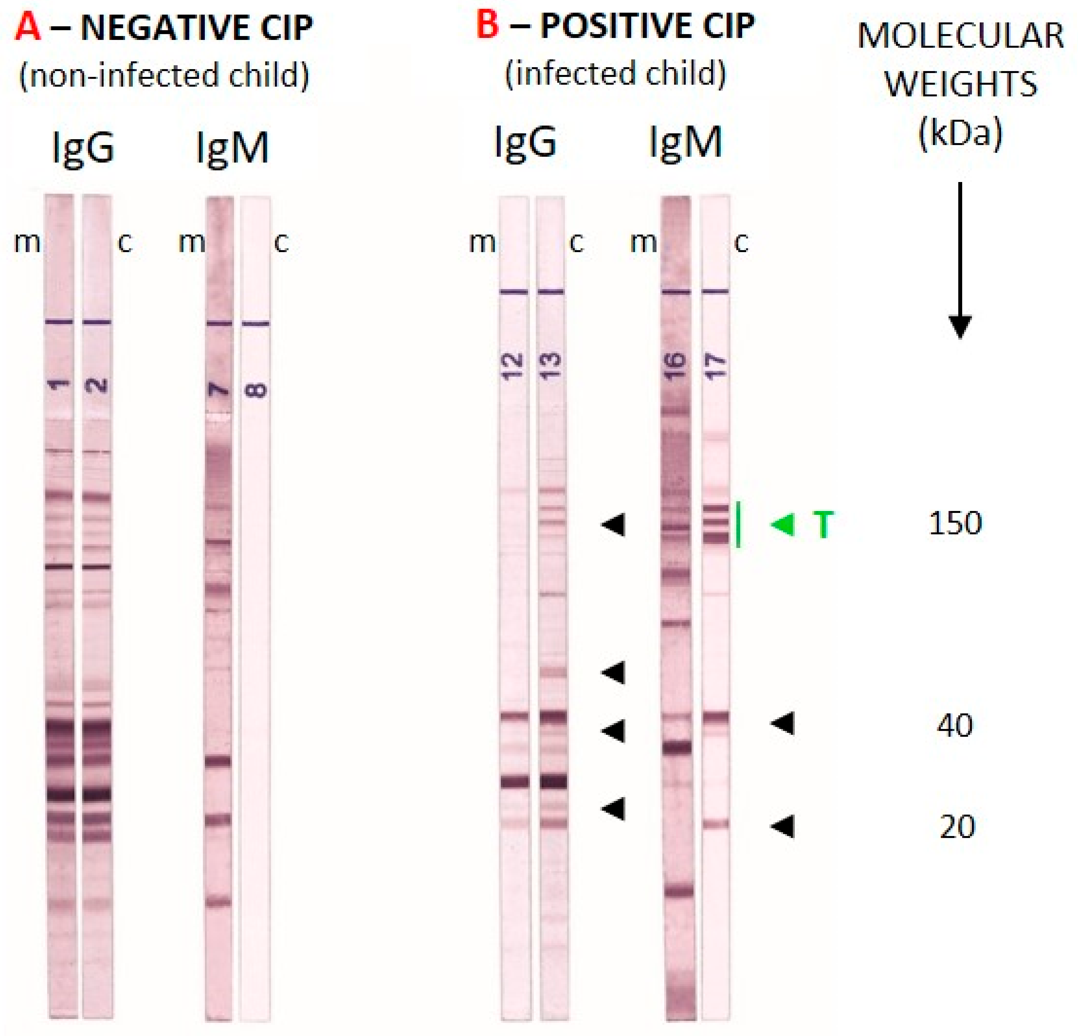
Any well-defined band with a molecular weight between 20 kDa and <120 kDa was analyzed visually by comparing maternal and neonatal samples collected at birth. For the IgG strip, any additional band present in the neonatal serum but absent in the maternal serum was considered positive. Because IgM does not cross the placenta and therefore reflects a fetal immune response, any IgM band detected in the neonatal serum was considered positive. The same criteria were applied at the one-month follow-up, with comparisons made between the two neonatal sera (at birth and at one month). An example of comparative WB analysis for a NCT and a CT newborn is shown in
Figure 1.
The study obtained ethical approval from the Ethics Committee of Fondazione IRCCS Policlinico San Matteo (protocol number 0012545125).
3. Results
A total of 44 serum samples from 44 newborns were tested at birth: 25 cases of NCT (56.8%) and 19 of CT (43.2%). Data concerning mothers with an acute T. gondii infection during pregnancy were available for 43 patients (97.7%), as one case in the CT group involved an abandoned infant for whom no maternal sample was available.
Within the NCT group, each newborn was tested twice: at birth and again one month later. Among the CT cases, all the 19 newborns were tested at birth, but only 11 newborns (57.9%) were tested at both time points.
All newborns from the NCT group exhibited IgG CLIA and IgG ELFA positivity at birth (100.0%), with 2/25 (8.0%) showing IgG negativization at the time of the second sampling. No cases of IgM positivity were recorded by IgM CLIA or ISAGA, nor was any comparative CWB IgG positivity observed at birth (0%). However, 2/25 (8.0%) infants presented equivocal CWB IgM findings at one-month follow-up. In both cases, a negative IGRA test excluded congenital infection. Thus, the overall specificity of the comparative Western blot was 100% at birth and 92% at the one-month follow-up.
Within the CT group, all newborns showed IgG positivity by both CLIA and ELFA at birth (100.0%). Eight out of 19 newborns (42.1%) tested positive for IgM by CLIA, 2/19 (10.5%) showed an equivocal IgM CLIA result, while 11 out of 19 (57.9%) were positive by IgM ISAGA. Only 2 out of 18 infants (11.1%) had completely negative IgG and IgM WB results, whereas the remaining newborns displayed various combinations of IgG and/or IgM reactivity, ranging from positive to equivocal. Accordingly, the overall sensitivity of the CWB was 88.9% (
Table 1 and
Table 2). Comparison using McNemar’s test showed that Western blot IgG/IgM had significantly higher sensitivity in detecting positive cases at birth compared to both IgM CLIA (
p = 0.008) and IgM ISAGA (
p = 0.014).
At one month follow-up, 1 newborn out of the 11 CT newborns examined (9.1%) tested positive for IgM by CLIA, 1/11 (9.1%) showed an equivocal IgM CLIA result, while 5 out of 11 (45.5%) were positive by IgM ISAGA.
Two out of 11 infants (18.2%) had negative CWB results for both IgG and IgM (these two infants were already negative at birth), whereas the remaining newborns displayed various combinations of IgG and/or IgM reactivity, ranging from positive to equivocal. (
Table 3 and
Table 4)
Accordingly, the overall sensitivity of the CWB at one month was 81.8%. Comparison using McNemar’s test showed that Western blot IgG/IgM had significantly higher sensitivity in detecting positive cases at birth compared to both IgM CLIA (p = 0.014) and IgM ISAGA (p = 0.046).
4. Discussion
The IgM ISAGA test has long represented a diagnostic cornerstone of congenital toxoplasmosis (CT) due to its sensitivity and specificity. Its withdrawal from the market has caused a significant diagnostic gap, prompting a search for alternative diagnostic algorithms. IgM ISAGA has high sensitivity, ease of use, and low cost, and as such has been adopted for routine diagnostics across many laboratories [
5,
6,
7].
The comparative Western blot (CWB) assay also demonstrates excellent diagnostic performance, and our study highlights its very high sensitivity at birth, which makes it a reliable tool for the early identification of congenital infection. The observed reduction in sensitivity at one month of life should not be considered an intrinsic limitation of the method, but rather the expected consequence of the prompt initiation of pyrimethamine–sulfadiazine therapy in congenitally infected newborns, which impairs antibody production.
As a referral center for the diagnosis of toxoplasmosis, Fondazione IRCCS Policlinico San Matteo in Pavia routinely uses comparative WB testing, which has proven to be a valid alternative to the discontinued IgM ISAGA. Comparative evaluation in both uninfected neonates and CT cases confirmed the excellent specificity of WB at birth (100.0%), with only a minor decrease at one month (92.0%) due to equivocal IgM results. The high sensitivity and specificity of this test allow for the prompt initiation of treatment in newborns with congenital infection, preventing delays that could lead to poor clinical outcomes.
It is also important to highlight the crucial role of in house IGRA test in excluding congenital infection in two cases with equivocal Western blot and in confirming it in four cases with false-negative IgM ISAGA and Western blot. The implementation of a diagnostic algorithm including Western blot and IGRA test in reference laboratories could further reduce the clinical impact of congenital toxoplasmosis by shortening the time to diagnosis and enabling earlier initiation of therapy in infected newborns.
This study has some limitations that should be acknowledged. The sample size was relatively small, reflecting the limited number of confirmed cases of congenital toxoplasmosis with complete follow-up. In addition, some heterogeneity in treatment and follow-up protocols may have occurred, as patients were managed in different clinical settings, reflecting real-life practice.
In conclusion, comparative Western blot represents a valuable diagnostic tool that appears fundamental in the algorithm for congenital toxoplasmosis, particularly in the context of IgM ISAGA commercial discontinuation. Its main limitations remain the relatively high costs, the need for trained personnel for both performance and interpretation, and the requirement for paired maternal–infant samples.
Author Contributions
Conceptualization, A.B. (Alice Bonetti), A.V., D.L., M.F., A.A., A.R., C.V., A.B. (Alessia Bressan), C.P., F.B. and V.M.; formal analysis, A.B. (Alice Bonetti), A.V., A.D.S. and G.G.; investigation, A.B. (Alice Bonetti), A.V. and L.L.G.; data curation, A.B. (Alice Bonetti), A.V.; writing—original draft preparation, A.B.; writing—review and editing, A.V., V.M., M.F., D.L., A.A., A.R., A.B. (Alessia Bressan), C.V. and C.P.; supervision, V.M. and F.B. All authors have read and agreed to the published version of the manuscript.
Funding
This work was supported by Ministero della Salute, Ricerca Corrente (grant no. 08074882).
Institutional Review Board Statement
The study was conducted in accordance with the Declaration of Helsinki and approved by the Ethics Committee of Fondazione IRCCS Policlinico San Matteo (protocol code 0012545125) approved on 6 May 2025.
Informed Consent Statement
Informed consent was obtained from all subjects involved in the study.
Data Availability Statement
The data that support the findings of this study are available on request from the corresponding author. The data are not publicly available due to privacy or ethical restrictions.
Acknowledgments
The authors would like to thank Enrico Brunetti for his valuable assistance with the English revision of the manuscript publication.
Conflicts of Interest
The authors declare no conflicts of interest.
Abbreviations
The following abbreviations are used in this manuscript:
| CT | Congenital toxoplasmosis |
| ISAGA | Immunosorbent Agglutination Assay |
| CLIA | Chemiluminescent Immunoassay |
| PCR | Polymerase Chain Reaction |
| CWB | Comparative Western Blot |
| NCT | Non-congenital Toxoplasmosis |
| IGRA | Interferon Gamma Release Assay |
| ICU | Instructions for Use |
References
- Robert-Gangneux, F.; Dardé, M.L. Epidemiology of and diagnostic strategies for toxoplasmosis. Clin. Microbiol. Rev. 2012, 25, 583. [Google Scholar] [CrossRef] [Scilit]
- Peyron, F.; L’Ollivier, C.; Mandelbrot, L.; Wallon, M.; Piarroux, R.; Kieffer, F.; Hadjadj, E.; Paris, L.; Garcia-Meric, P.G. Maternal and congenital toxoplasmosis: Diagnosis and treatment recommendations of a French multidisciplinary working group. Pathogens 2019, 8, 24. [Google Scholar] [CrossRef] [Scilit] [PubMed]
- Bonetti, A.; Comelli, A.; Chiesa, A.; Spinoni, V.; Vola, A.; Prefumo, F.; Valcamonico, A.; Bonfanti, C.; Caligaris, S.; Tomasoni, L.R.; et al. Risk of congenital toxoplasmosis in newborns from mothers with documented infection: Experience from two referral centres. Pathogens 2025, 14, 157. [Google Scholar] [CrossRef] [Scilit] [PubMed]
- Bollani, L.; Auriti, C.; Achille, C.; Garofoli, F.; De Rose, D.U.; Meroni, V.; Salvatori, G.; Tzialla, C. Congenital toxoplasmosis: The state of the art. Front. Pediatr. 2022, 10, 894573. [Google Scholar] [CrossRef] [Scilit] [PubMed]
- Arkhis, S.; Rouges, C.; Dahane, N.; Guegan, H.; Yéra, H.; Robert-Gangneux, F. Could PLATELIA Toxo IgM be the new gold standard for the serological diagnosis of congenital toxoplasmosis? A French multicenter study. J. Clin. Microbiol. 2024, 62, e0122223. [Google Scholar] [CrossRef] [Scilit] [PubMed]
- Montoya, J.G. In defense of children’s brains: Reshuffling the laboratory toolbox for the diagnosis of congenital toxoplasmosis. J. Clin. Microbiol. 2024, 62, e01697-23. [Google Scholar] [CrossRef] [Scilit] [PubMed]
- Deleplancque, A.S.; Fricker-Hidalgo, H.; Pomares, C.; L’Ollivier, C.; Lemoine, J.P.; Cimon, B.; Paris, L.; Houzé, S.; Villena, I.; Pelloux, H.; et al. Comparative performance of IgM ISAGA and ELISA assays for the diagnosis of maternal and congenital Toxoplasma infections: Which technique could replace IgM ISAGA? Parasite 2024, 31, 7. [Google Scholar] [CrossRef] [Scilit] [PubMed]
- Associazione Microbiologi Clinici Italiani (AMCLI). Italian Guidelines for Toxoplasmosis. Available online: https://amcli.it/percorsi-diagnostici-218/ (accessed on 8 February 2026).
- Wallon, M.; Peyron, F.; Cornu, C.; Vinault, S.; Abrahamowicz, M.; Bonithon-Kopp, C.; Binquet, C. Congenital Toxoplasma infection: Monthly prenatal screening decreases transmission rate and improves clinical outcome at age 3 years. Clin. Infect. Dis. 2013, 56, 1223–1231. [Google Scholar] [CrossRef] [Scilit] [PubMed]
- Mahmoudi, S.; Mamishi, S.; Suo, X.; Keshavarz, H. Early detection of Toxoplasma gondii infection using an interferon-gamma release assay: A review. Exp. Parasitol. 2017, 172, 39–43. [Google Scholar] [CrossRef] [Scilit] [PubMed]
- Rilling, V.; Dietz, K.; Krczal, D.; Knotek, F.; Enders, G. Evaluation of a commercial IgG/IgM Western blot assay for early postnatal diagnosis of congenital toxoplasmosis. Eur. J. Clin. Microbiol. Infect. Dis. 2003, 22, 174–180. [Google Scholar] [CrossRef] [Scilit] [PubMed]
- LDBIO Diagnostics. TOXOPLASMA Western Blot IgG IgM (CIP-WB): Instructions for Use. Lyon, France: LDBIO Diagnostics. Available online: https://ldbiodiagnostics.com/en/our-products/immunoblots/toxoplasma-western-blot-igg-igm/ (accessed on 8 February 2026).
| Disclaimer/Publisher’s Note: The statements, opinions and data contained in all publications are solely those of the individual author(s) and contributor(s) and not of MDPI and/or the editor(s). MDPI and/or the editor(s) disclaim responsibility for any injury to people or property resulting from any ideas, methods, instructions or products referred to in the content. |




 ,
,