1. Introduction
Respiratory syncytial virus (RSV) is a leading cause of acute lower respiratory tract infection in infants, young children, elderly adults, and adults with comorbidities worldwide, producing a clinical spectrum that ranges from mild upper respiratory disease to life-threatening bronchiolitis and pneumonia, with the highest risk of severe outcomes and death in infants under six months of age [
1,
2,
3]. Before widespread availability of RSV prophylaxis and vaccines, the global pediatric burden was substantial: in 2019 an estimated 33.0 million RSV-associated acute lower respiratory tract infections (LRTIs), 3.6 million RSV-associated LRTI hospital admissions, and more than 100,000 deaths in children under five were attributed to RSV [
3]. Post-COVID changes in viral circulation have not eliminated this burden: 2022 saw persistently elevated RSV mortality in young children at 9.76 deaths per 100,000 children under 5, and the Global Burden of Disease Study continues to register RSV among leading causes of pediatric morbidity and mortality, with the burden disproportionately concentrated in low- and middle-income countries (LMICs) [
3,
4]. These data underscore an ongoing, unmet medical need for effective RSV therapeutics to supplement the current vaccines and prophylactic antibodies, particularly options that can treat established infection and reduce severe outcomes [
5,
6,
7,
8,
9,
10,
11,
12].
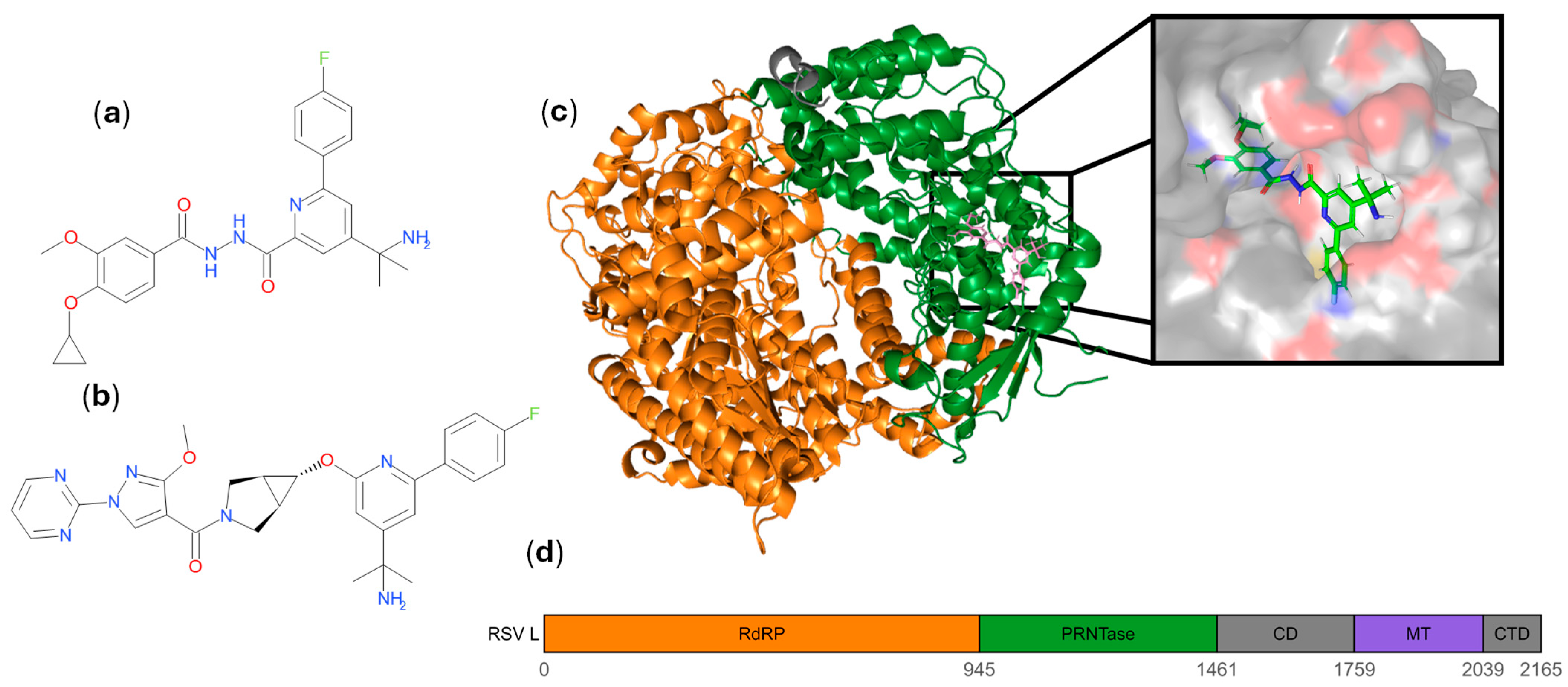
The RSV viral polymerase complex is a compelling antiviral target because it is essential for both viral transcription and genome replication and contains multiple enzymatic functions that can be inhibited pharmacologically. Central to this complex is the large polymerase (L) protein. The multifunctional L protein is composed of several discrete domains: an RNA-dependent RNA polymerase (RdRP) domain that mediates RNA chain synthesis; a polyribonucleotidyltransferase (PRNTase) domain which facilitates 5′ cap addition to viral mRNAs; a connector domain (CD); a methyltransferase (MT) domain involved in cap methylation; and a C terminal domain (CTD) [
13,
14,
15,
16,
17,
18,
19]. High-resolution structural studies have defined these domain arrangements and suggested conformational changes that underlie transitions between initiation, elongation, and mRNA capping during the viral transcription–replication cycle [
16,
17,
18].
Multiple antiviral strategies targeting the L protein have been explored [
8,
20,
21,
22]. Nucleoside analogues inhibit the RdRP domain by acting as chain terminators or mutagens [
23,
24]. In contrast, non-nucleoside inhibitors (NNIs) act allosterically by binding to regulatory pockets within the L protein, typically the CD and PRNTase domain, to block conformational transitions required for distinct stages of polymerase function [
25,
26,
27,
28,
29,
30,
31]. These different mechanisms offer complementary opportunities to suppress viral replication. However, a critical consideration in antiviral development is the potential for preexisting resistance due to natural polymorphisms in circulating viral populations. Sequence variation at or near drug-binding sites can compromise antiviral efficacy, as demonstrated by the clinical failure of suptavumab, a monoclonal antibody targeting the RSV F protein, due to widespread circulation of RSV B strains harboring mutations that abrogated antibody binding [
8,
29].
Given the importance of assessing preexisting resistance, we focused on a class of NNIs exemplified by MRK-1 and MRK-2, which target a conserved pocket within the PRNTase domain of the RSV L protein [
28,
29]. Prior PKPD evaluation of MRK-1 in cotton rat and African green monkey models of RSV infection showed lack of drug-specific resistance mutation at I1381 and L1421 [
30]. Our study evaluated sequence variability of the PRNTase domain and the MRK-1/2 binding pocket across globally circulating RSV strains. We performed a comprehensive residue-level analysis of 28,140 RSV L protein sequences obtained from public databases, mapped observed variants onto structural models of the polymerase, and conducted in vitro dose escalation experiments to identify resistance-conferring mutations. Our findings demonstrate that the PRNTase domain, and specifically the MRK-1/2 binding pocket, is highly conserved, with minimal natural variation and no evidence of preexisting resistance mutations in circulating strains. These results support the continued development of PRNTase-targeting NNIs as promising therapeutic candidates for RSV infection.
2. Materials and Methods
2.1. Sequence Collection
We retrieved 13,499 RSV L protein sequences from the NCBI Virus browser by filtering the dataset for RSV proteins with sequence lengths of 2150 to 2170 [
32]. The subtypes for the NCBI sequences were identified using the metadata, including organism and isolate names. We also retrieved 45,550 RSV A and 33,729 RSV B genome sequences from GISAID EpiRSV [
33]. To isolate and identify whole L protein sequences, the genomes were compared with L protein sequences from reference sequences (RSV A: NC_001803; RSV B: NC_001781) with the NCBI BLAST+ (2.2.28) command line application, and hits that covered the whole sequence were extracted to be analyzed [
34].
2.2. Sequence Year and Location Isolation
The location and collection-year data were pulled from the metadata associated with the sequences within the NCBI and GISAID databases. For sequences without explicit location and collection-year metadata entries, the information was pulled from other metadata or associated publications where available. All location data that was at the city level was mapped back to the country of origin.
2.3. Residue Variation
For each of the four groups of proteins, the sequences were aligned using the ClustalO command line tool [
35]. The residue frequency at each position was calculated using the EMBOSS Prophecy command line application [
36]. The frequency tables were used to identify consensus residues (residues that show up in ≥50% of sequences with non-ambiguous amino acids at that position), major variants (<50% and ≥1%), and minor variants (<1% and ≥0.1%).
2.4. Mapping the Residue Variation with Respect to the Binding Site
The cryo-EM structure of the truncated RSV L protein bound to the inhibitor MRK-1 has been solved (PDB ID: 8FPI resolution 2.52 A with domain boundaries noted in reference) [
31]. The 5 A binding site was defined using PyMOL (3.1.6.1) with the command “elect nearligm byres chain A within 5 of 9001”. The 31 residues in the binding site are P1002, H1120, G1218, V1219, T1220, S1221, I1241, S1266, L1337, H1338, R1345, P1346, F1349, T1365, I1368, N1369, L1372, T1373, Y1376, G1377, D1378, E1379, D1380, I1381, D1382, I1383, V1384, F1385, Q1386, C1388, and M1422. To obtain the docking pose for the MRK-2 structure, we used a closely related cryo-EM bound structure in which the bound small molecule had a few atom differences with respect to MRK-2, albeit with the same binding site as MRK-1. This provided strong confidence in the predicted pose in this pocket. Subsequently, these structures were used to map the residues with major and minor variations while highlighting their positions with respect to the binding site. MOE and PyMol were used for docking and visualization analysis [
37,
38].
2.5. Potency Evaluation
For all compound potency assays, we used an RSV GFP reporter virus that was described previously [
29]. MRK-1 was assayed against RSV A2 GFP and MRK-2 against RSV B Wash GFP. The compounds were dissolved in DMSO and titrated in an 11-point 3-fold serial dilution in a 96-well tissue culture plate. The dilutions were mixed with 2 × 10
4 Hep-2 cells in DMEM with 10% heat inactivated fetal bovine serum (FBS) infected at a 0.1 MOI. After 2 days of incubation at 37 °C, the fluorescence was measured using an Acumen Cellista plate reader. EC
50 and EC
90 concentrations were calculated by fitting a dose-response curve to the data using GraphPad Prism (10.2.2).
2.6. Resistance Selection
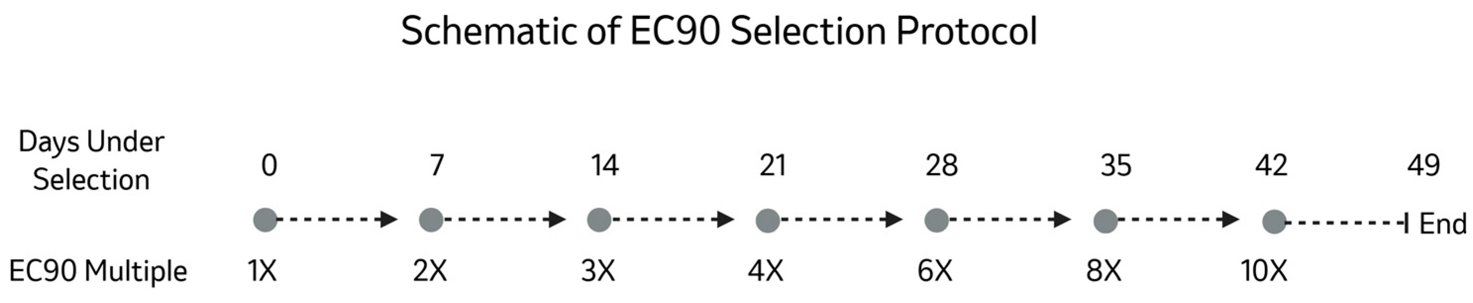
(Detailed in
Scheme 1). 2 × 10
4 HEp-2 cells in 10% FBS-DMEM were added to each well of a 96-well cell culture plate. DMSO solutions of MRK-1 and MRK-2 were added to each well of the first 11 columns at their respective EC
90 concentrations. The plates were infected with RSV A2 GFP and RSV B Wash GFP, respectively, at an MOI of 0.1. After 3 to 4 days of incubation at 37 °C, the fluorescence was read in an Acumen Cellista plate reader. The plate was then passaged by preparing plates with 2 × 10
4 HEp-2 cells/well and MRK-1 or MRK-2 and transferring 10 μL of supernatant from each well on the old plates into the corresponding well on the new plates. The concentration was increased on every other passage going from 1× EC
90 to 2×, 3×, 4×, 6×, 8× and finally 10× the EC
90.
2.7. Sequencing
The viral RNA for each resistant well was extracted using the QIAGEN QIAamp Viral RNA Mini Kit. The RNA was converted to cDNA using the SuperScript IV UniPrime 1-Step RT-PCR kit. The PCR and sequencing primers used were TGACACAATTCAAAATTTTCTACAACA (F) and ATTCCTCCAAGATTAAAATGATAACTTTAG (R) for RSV A2; TCCACAAAAGCATAACCATAAGCA (F) and TGTCTCGTTGTGTTGTAAATGCA (R) for RSV B Wash.
2.8. Variant Potency Shift
RSV variants were generated by mutating the pSmart RSV A2 GFP via Gibson assembly. The introduction of the mutation was verified by sequencing. The recombinant viruses were recovered through transfection into BSR-T7 via Mirus TransIt-LT1 of a mixture of the mutated pSmart BAC along with pcDNA3.1, M2-1, N, P, and L in a 2:2:2:2:1 ratio.
DMSO solutions of MRK-1 and MRK-2 were titrated in an 11-point 3-fold serial dilution in a 96-well tissue culture plate. The dilutions were mixed with 2 × 10⁴ Hep-2 cells in DMEM with 10% heat inactivated fetal bovine serum (FBS) infected with 10 μL of supernatant from the resistant wells. After 2 days of incubation at 37 °C, the fluorescence was read in an Acumen Cellista plate reader. EC50 and EC90 concentrations were calculated by fitting a dose-response curve to the data using GraphPad Prism.
3. Results
3.1. Sequence Gathering
We pulled 13,499 L protein sequences from NCBI and 79,279 genomes (45,550 RSV A and 33,729 RSV B) from GISAID (EPI_SET ID: EPI_SET_251031go). NCBI did have L protein sequences available, but they were not all assigned to a subtype. After checking the other metadata, there were 6284 RSV A and 5582 RSV B sequences; the rest had insufficient data to conclude their subtype and were therefore discarded.
GISAID has a subtype assigned to all RSV genomes but does not have the L protein sequences readily available. After running a BLAST to extract the L protein sequence from their genome sequences, 13,279 RSV A and 14,945 RSV B sequences were identified as complete. Due to the lack of metadata to cross-reference between the two databases, it was not possible to determine the overlap between GISAID and NCBI submissions. For this reason, we have kept the sequences from each database in distinct groups, creating four (4) datasets (
Table S1).
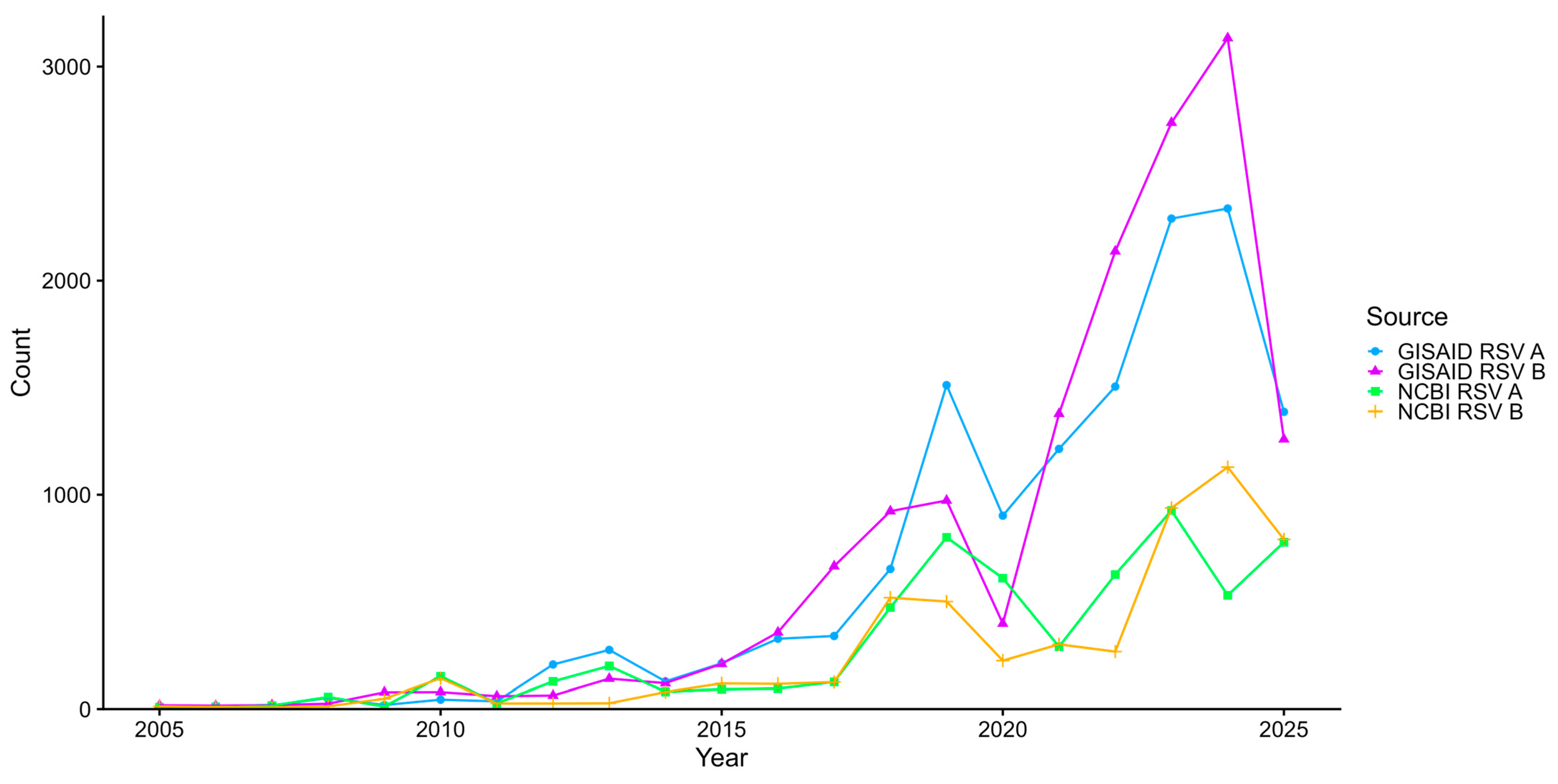
These datasets are contemporary, with 56.4% of the sequences collected between 2022 and 2025 (
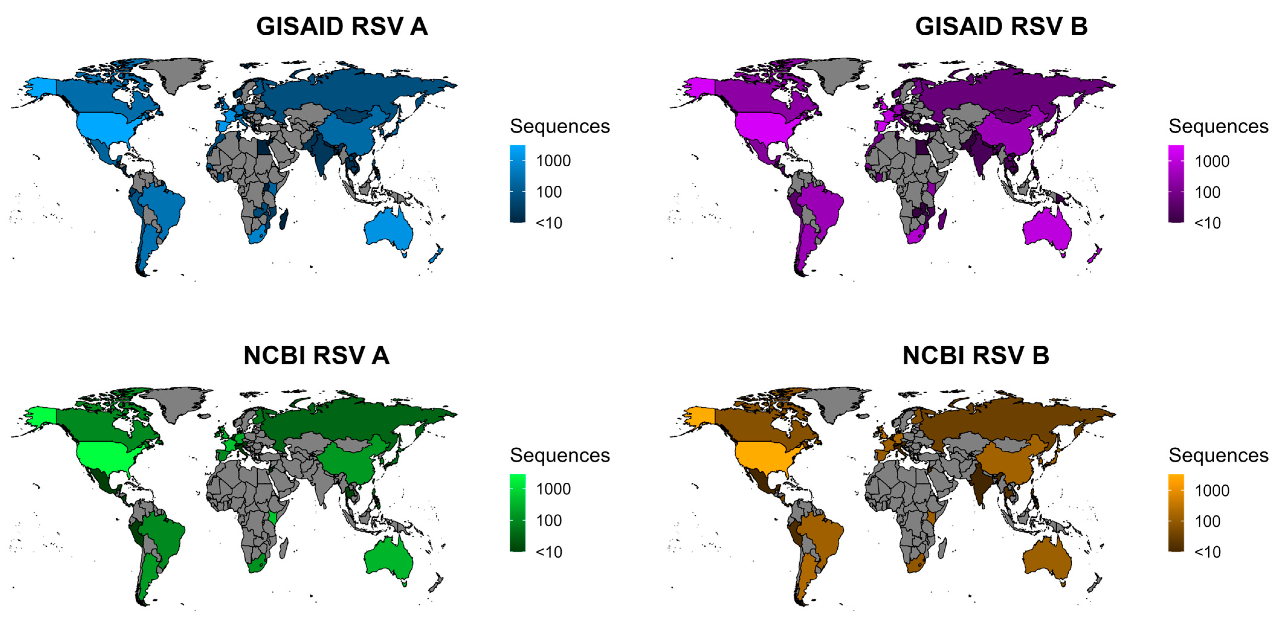
Figure 1). The data are unfortunately not globally diverse, with LMICs being underrepresented (
Figure 2). The Americas, Western Europe, and East Asia have good sequencing coverage. Australia, New Zealand, South Africa, and Kenya individually have high sequencing coverage, but the rest of Africa and Oceania have little to no sequencing.
3.2. Low Variance in PRNTase
To understand which regions of the RSV L protein are conserved across the global population, the datasets were aligned, and variability was calculated at the positional level (
Tables S2–S5). For each position, the amino acids were categorized into 3 categories: consensus, accounting for 50+% of the residues aligned to that position; major variant, making up at least 1% but less than 50% of the aligned residues; and minor variant, making up between 0.1% and 1% of the residues. All amino acids that made up less than 0.1% of the residues aligned to that position were considered insignificant. If the consensus amino acid at a position makes up less than 90% of the residues aligned to that position, that position is labeled as highly variable. Most positions have low variability, with only 11 (0.51%) positions in RSV A and 10 (0.46%) positions in RSV B being labeled as highly variable (
Table 1). For both RSV A and RSV B, there are 4 highly variable positions in the RdRP, 4 in the CD and 1 in the CTD. There are 2 highly variable positions in the MT in RSV A and 1 in RSV B. The PRNTase is the least variable domain of the L protein, with no highly variable positions in either subtype.
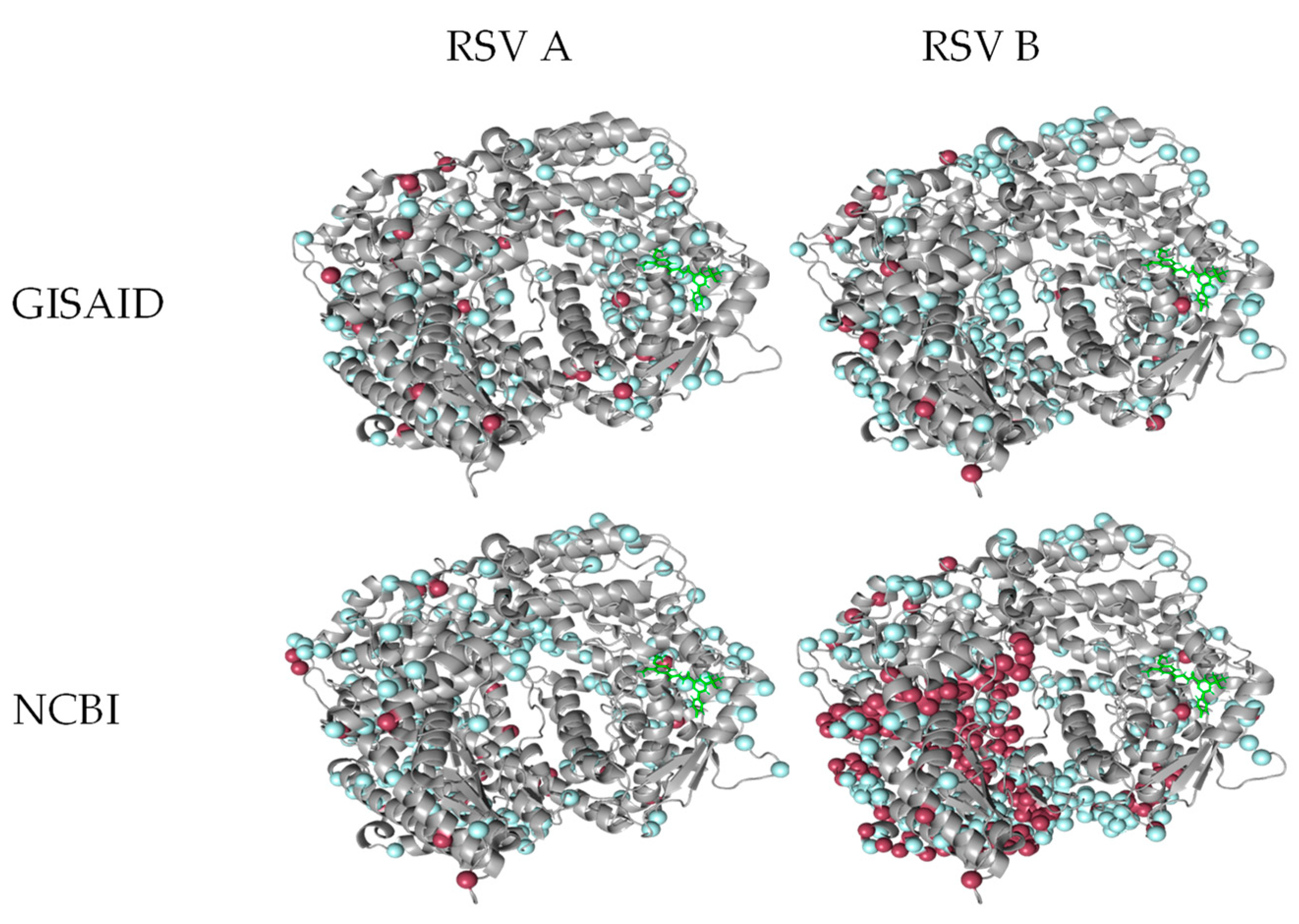
The PRNTase domain is also the least covered by significant variants (
Table 2). Minor variants cover 0.14–0.22% of the whole RSV L protein compared to 0.09–0.14% coverage of the PRNTase domain. Excluding the NCBI RSV B data, major variants cover 0.02–0.04% of the RSV L protein. For the NCBI RSV B dataset, that number jumps to 0.14%, but the variant coverage for the PRNTase domain remains at 0.01% for all groups. The PRNTase domain shows low variation as compared to the rest of the L protein.
3.3. Low Variance in MRK-1/MRK-2 Binding Pocket
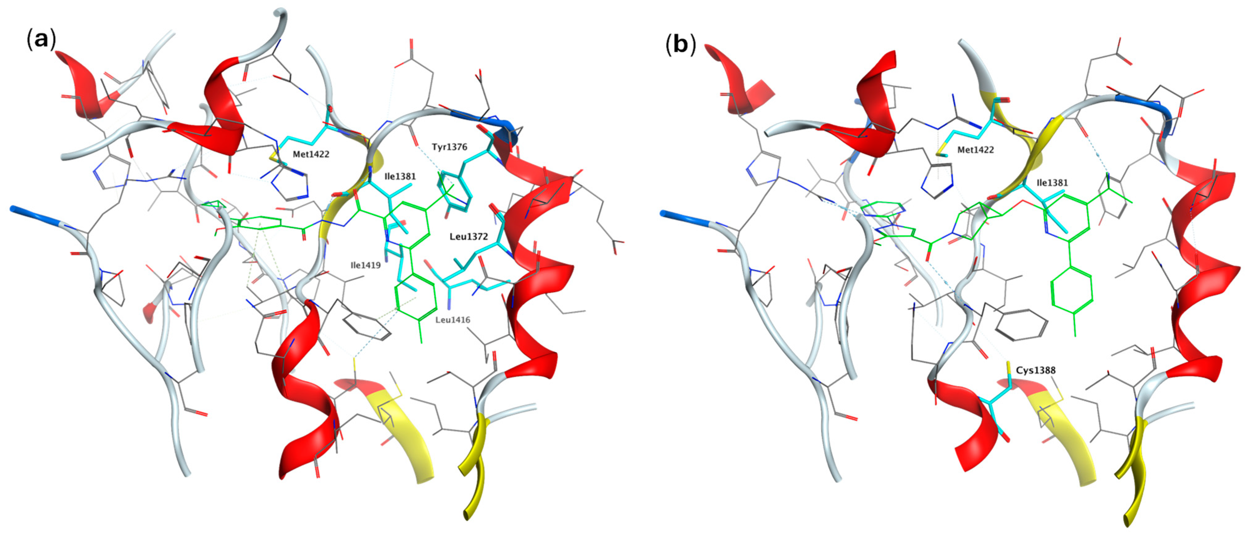
MRK-1 binds in the PRNTase domain (
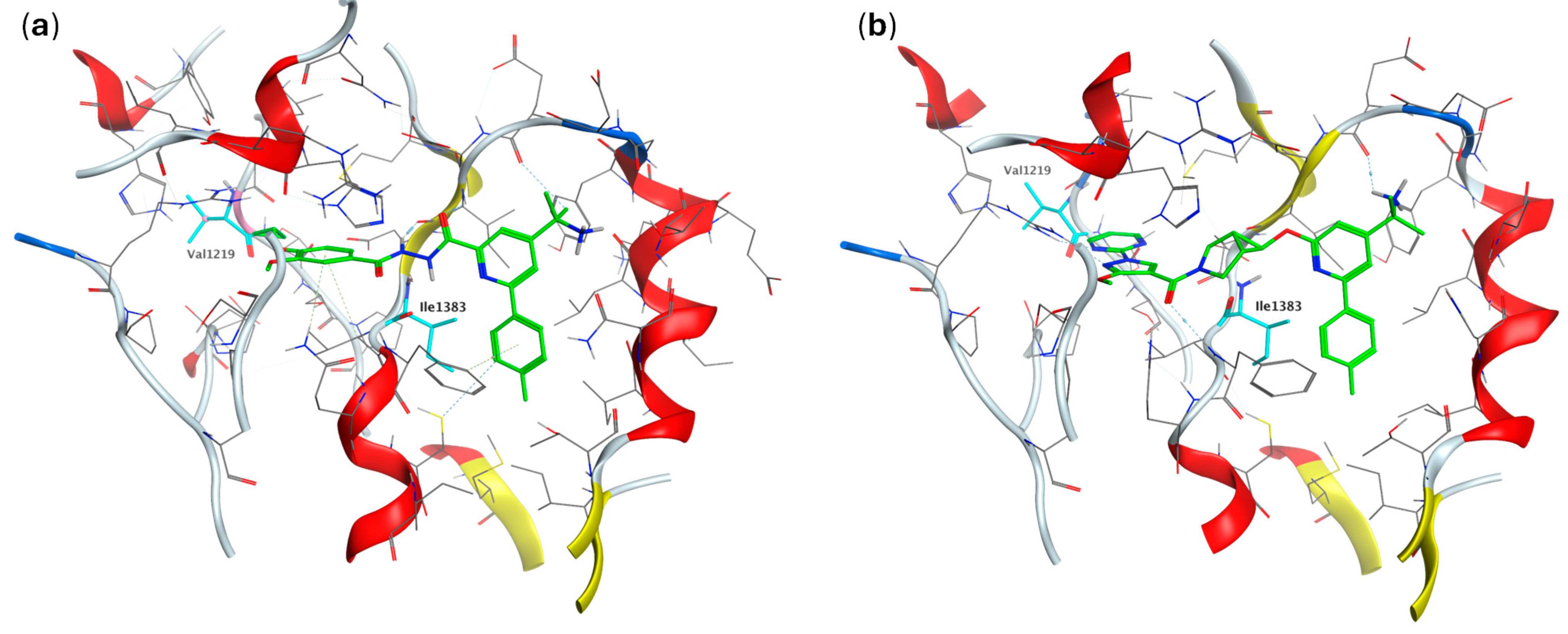
Figure 3) of the RSV L protein. MRK-2 is structurally close to MRK-1 and binds to the same binding site as MRK-1. The docked model shows MRK-2 fits well into the pocket and interacts with similar residues. Though there are minor variations in the residue positions, the overall shape and structure of the pocket is well maintained (
Figure 4).
None of the residues directly interacting with the small molecule in the binding site show significant variation in the surveillance data (
Figure 5). Inspecting the binding pocket residues also reveals that the consensus amino acid is identical between RSV A and RSV B with no major variants (
Table S6). There are, however, only two (2) minor variants within the binding pocket in the surveillance data (V1219I [0.2%] and I1383V [0.1%]) for RSV A (
Figure 4). While the I1383 side chain has a hydrophobic interaction with the ligand in the binding site, both observed variants are considered conserved and are expected to have minimal effect on the binding affinity of the small molecule inhibitor. Therefore, it can be inferred that the MRK-1/2 binding pocket is highly conserved, and the lack of variation in the binding pocket implies that the collected strains should not show resistance to either NNI, assuming all resistance-conferring mutations occur in the binding pocket.
3.4. Dose Escalation Selects for Resistance Mutations Within the Binding Pocket
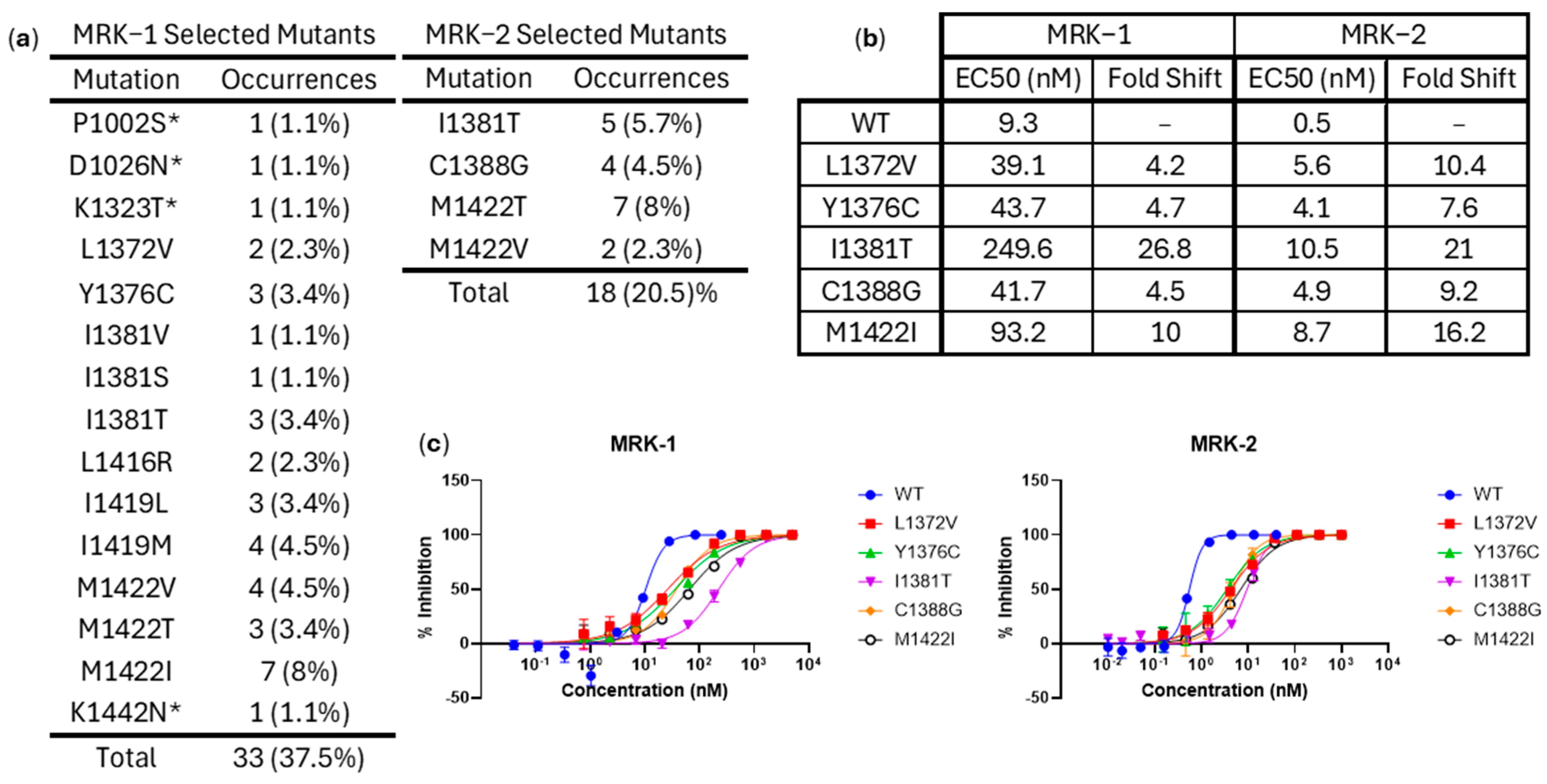
To find mutations likely to confer resistance, we performed a dose escalation experiment. Of the eighty-eight (88) replicates, MRK-2 selected for resistance in eighteen (18) and MRK-1 for thirty-three (33). Six (6) of the MRK-1 resistant samples showed double mutations. Sequencing these resistant samples showed there were mutations at three (3) distinct positions across the samples selected against MRK-2 and at ten (10) for those against MRK-1 (4 only in samples with multiple mutations). There were two (2) positions where resistant mutants were selected against both NNIs (I1381 and M1422). MRK-2 also selected for a mutant in C1388, and MRK-1 selected for potentially resistance-conferring mutations at L1372, Y1376, L1416, and I1419 (
Figure 6). Of these seven (7) positions, five (5) are in the binding pocket, directly interacting with the small molecule inhibitor (the first shell), and the other two (2) are interacting with the residues making up the binding site (the second shell). Neither of the second shell positions show significant variation in the surveillance data (
Tables S2–S5).
Five of the generated mutations were selected for evaluation to verify their resistance to the NNIs. I1381T and M1422I were particularly selected because they arose for both NNIs in the dose escalations. C1388G only showed up in the escalation against MRK-2, while L1372V and Y1376C only showed up in the escalation against MRK-1. These mutations were expressed in recombinant viruses. All 5 of these mutants showed resistance to the NNIs, with the EC
50 of each being ≥3 fold greater than that of the wild-type RSV A2. The fold shift against MRK-2 was generally larger than against MRK-1, but MRK-2 was more potent against every tested virus (
Figure 7).
Most of the positions identified through the dose escalations are within the identified binding pocket. None of the identified positions show significant variants.
4. Discussion
The currently available RSV surveillance data show that the PRNTase domain of RSV L protein, and the MRK-1/2 binding pocket in particular, is highly conserved. There are thousands of L protein sequences from both RSV A and B, with the majority being contemporary. GISAID has more than double the available RSV L protein sequences compared to NCBI, and much of that discrepancy is explained by GISAID having more sequence submissions post-2020 than NCBI. There are more RSV nucleotide sequences available than RSV L protein sequences, as submitted sequences are often partial genomes and the L protein is not typically a priority target for sequencing in genotyping and surveillance studies of RSV. With the available full-length L protein sequences, we have demonstrated that the residues within 5 Å of the binding pocket exhibit minimal variation across the current global RSV population.
To confirm that low variation in the MRK-1/2 binding pocket means that there is no preexisting resistance to these NNIs in the circulating RSV strains, we performed dose escalation experiments against each NNI to select for resistance-conferring mutations. Some mutations were selected during both dose escalations, and the rest were selected during only individual assays. A handful of these mutations were tested and shown to confer resistance to both NNIs. Most of these mutations occurred within the MRK-1/2 binding pocket. The mutations that occurred outside but close to the pocket are at positions that show no variation in the circulating RSV. None of the selected mutants were seen as variants in the surveillance data. Given this data, it is highly unlikely that there is any preexisting resistance to these PRNTase-targeting NNIs within the global RSV population.
There are limitations to this analysis. Though the sequences are contemporary, they cannot be said to be globally diverse. The sequences mostly originate from high-income countries, with data from LMICs being comparatively sparse. This is important, as the burden of RSV is higher in LMICs. This perennial problem extends past this study. LMICs are understudied and have poor surveillance. Without investment in public health surveillance in these countries, it will be challenging to gain a comprehensive understanding of the complete variation landscape of any virus. However, since there are data from Kenya, Egypt, South Africa, India, and Vietnam, countries that are and border LMICs, some of the variation prevalent in LMICs is represented in the dataset.
Another limitation, and potential avenue for future research, is that the list of resistance-conferring mutations generated from the dose escalations is not exhaustive. Only one dose escalation was performed against each NNI, while each assay has eighty-eight (88) replicates, which is not enough to find every resistance-conferring mutation. This is demonstrated by the presence of unselected mutations in the dose escalation against MRK-1, which exhibited resistance, and similarly for MRK-2. Although more resistance-conferring mutations are likely to be identified with further dose escalation assays, they are likely to be located within the binding pocket or adjacent to it. Neither limitation impacts the conclusion of this investigation.
This form of surveillance analysis enables the further development of NNIs targeting PRNTase without concern that existing strains may be resistant. Such foresight is enabled by the increases in public health surveillance and data sharing through publicly accessible databases that have occurred after the COVID-19 pandemic.
5. Conclusions
A substantial amount of data indicates high conservation within the RSV L protein PRNTase domain, particularly within the MRK-1/2 binding pocket. No mutations that could confer resistance to either MRK-1 or MRK-2 have been identified in the surveillance data. As such, this structurally defined binding pocket is a promising target that is unlikely to have any RSV strains with pre-existing resistance.
Supplementary Materials
The following supporting information can be downloaded at:
https://www.mdpi.com/article/10.3390/pathogens15010085/s1, Table S1: All accession numbers for sequences used; Table S2: Variation in RSV A L protein sequences collected from GISAID EpiRSV at each position; Table S3: Variation in RSV A L protein sequences collected from NCBI Virus at each position; Table S4: Variation in RSV B L protein sequences collected from GISAID EpiRSV at each position; Table S5: Variation in RSV B L protein sequences collected from NCBI Virus at each position; Table S6: Variation in MRK-1/2 Binding Pocket.
Author Contributions
Conceptualization, R.P. and J.A.H.; methodology, R.P. and E.M.; software, R.P.; validation, R.P. and D.D.N.; formal analysis, R.P.; investigation, R.P., E.M., D.D.N., M.Y. and B.A.; data curation, R.P.; writing—original draft preparation, R.P.; writing—review and editing, M.Y., N.M. and J.A.H.; visualization, R.P.; supervision, N.M. and J.A.H.; project administration, R.P. All authors have read and agreed to the published version of the manuscript.
Funding
This research was funded by Merck Sharp & Dohme LLC, a subsidiary of Merck & Co., Inc., Rahway, NJ, USA.
Institutional Review Board Statement
Not applicable.
Informed Consent Statement
Not applicable.
Data Availability Statement
The surveillance sequences are available at GISAID under the EPI Set ID EPI_SET_251031go.
Acknowledgments
We gratefully acknowledge all data contributors, i.e., the Authors and their Originating laboratories responsible for obtaining the specimens, and their Submitting laboratories for generating the genetic sequence and metadata and sharing via the GISAID Initiative and NCBI GenBank, on which this research is based. We thank Christopher Warren for his assistance in visualization design.
Conflicts of Interest
All authors are current employees of Merck Sharp & Dohme LLC, a subsidiary of Merck & Co., Inc., Rahway, NJ, USA, and potentially own stock and/or hold stock options in Merck & Co., Inc., Rahway, NJ, USA.
Abbreviations
The following abbreviations are used in this manuscript:
| RSV | Respiratory syncytial virus |
| LRTI | Lower respiratory tract infections |
| LMIC | Low- and middle-income country |
| L | Large polymerase protein |
| RdRP | RNA dependent RNA polymerase |
| PRNTase | Polyribonucleotidyltransferase |
| CD | Connector domain |
| MT | Methyltransferase |
| CTD | C-terminal domain |
| NNI | Non-nucleoside inhibitor |
| NCBI | National Center for Biotechnology Information |
| GISAID | Global Initiative on Sharing All Influenza Data |
| BLAST | Basic local alignment search tool |
| Cryo-EM | Cryogenic electron microscopy |
| MOE | Molecular Operating Environment |
| GFP | Green fluorescent protein |
| DMSO | Dimethyl sulfoxide |
| DMEM | Dulbecco’s Modified Eagle Medium |
| FBS | Fetal bovine serum |
References
- Fearns, R.; Liang, B. Introduction to Respiratory Syncytial Virus. Methods Mol. Biol. 2025, 2948, 1–17. [Google Scholar] [CrossRef] [Scilit]
- Burkart, K.; Liang, C.; Rafferty, Q.; Gillespie, C.W.; McLaughlin, S.; Oros, A.; Suba, J.; Bruno, D.; Fahey, M.; Grajales, A.G.; et al. Respiratory syncytial virus-attributable hospitalizations among adults in high- and middle-income countries: Application of the Global Burden of Disease framework. EclinicalMedicine 2025, 85, 103292. [Google Scholar]
- Li, Y.; Wang, X.; Blau, D.M.; Caballero, M.T.; Feikin, D.R.; Gill, C.J.; Madhi, S.A.; Omer, S.B.; Simoes, E.A.F.; Campbell, H.; et al. Global, regional, and national disease burden estimates of acute lower respiratory infections due to respiratory syncytial virus in children younger than 5 years in 2019: A systematic analysis. Lancet 2022, 399, 2047–2064. [Google Scholar] [CrossRef] [Scilit] [PubMed]
- GBD 2023 Causes of Death Collaborators. Global burden of 292 causes of death in 204 countries and territories and 660 subnational locations, 1990-2023: A systematic analysis for the Global Burden of Disease Study 2023. Lancet 2025, 406, 1811–1872. [Google Scholar] [CrossRef] [Scilit]
- Alandijany, T.A.; Qashqari, F.S. Evaluating the efficacy, safety, and immunogenicity of FDA-approved RSV vaccines: A systematic review of Arexvy, Abrysvo, and mResvia. Front. Immunol. 2025, 16, 1624007. [Google Scholar] [CrossRef] [Scilit]
- Jones, J.M.; Fleming-Dutra, K.E.; Prill, M.M.; Roper, L.E.; Brooks, O.; Sanchez, P.J.; Kotton, C.N.; Mahon, B.E.; Meyer, S.; Long, S.S.; et al. Use of Nirsevimab for the Prevention of Respiratory Syncytial Virus Disease Among Infants and Young Children: Recommendations of the Advisory Committee on Immunization Practices—United States, 2023. MMWR Morb. Mortal. Wkly. Rep. 2023, 72, 920–925. [Google Scholar] [CrossRef] [Scilit]
- Kelleher, K.; Subramaniam, N.; Drysdale, S.B. The recent landscape of RSV vaccine research. Ther. Adv. Vaccines Immunother. 2025, 13, 25151355241310601. [Google Scholar] [CrossRef] [Scilit]
- Liang, X.; Yin, Y.; Lin, Y.; Chen, S.; Qian, Q.; Yuan, J.; Yang, L.; Yang, Y. Molecular and Cellular Mechanisms of Respiratory Syncytial Viral Infection: Its Implications for Prophylactic and Therapeutic Pharmaceuticals. MedComm 2025, 6, e70403. [Google Scholar] [CrossRef] [Scilit] [PubMed]
- The IMpact-RSV Study Group. Palivizumab, a humanized respiratory syncytial virus monoclonal antibody, reduces hospitalization from respiratory syncytial virus infection in high-risk infants. Pediatrics 1998, 102, 531–537. [Google Scholar] [CrossRef] [Scilit]
- Tang, A.; Chen, Z.; Cox, K.S.; Su, H.P.; Callahan, C.; Fridman, A.; Zhang, L.; Patel, S.B.; Cejas, P.J.; Swoyer, R.; et al. A potent broadly neutralizing human RSV antibody targets conserved site IV of the fusion glycoprotein. Nat. Commun. 2019, 10, 4153. [Google Scholar] [CrossRef] [Scilit]
- Topalidou, X.; Kalergis, A.M.; Papazisis, G. Respiratory Syncytial Virus Vaccines: A Review of the Candidates and the Approved Vaccines. Pathogens 2023, 12, 1259. [Google Scholar] [CrossRef] [Scilit]
- Yang, G.; Jiang, G.; Jiang, J.; Li, Y. Advances in development of antiviral strategies against respiratory syncytial virus. Acta Pharm. Sin. B 2025, 15, 1752–1772. [Google Scholar] [CrossRef] [Scilit]
- Paesen, G.C.; Collet, A.; Sallamand, C.; Debart, F.; Vasseur, J.J.; Canard, B.; Decroly, E.; Grimes, J.M. X-ray structure and activities of an essential Mononegavirales L-protein domain. Nat. Commun. 2015, 6, 8749. [Google Scholar] [CrossRef] [Scilit] [PubMed]
- Gilman, M.S.A.; Liu, C.; Fung, A.; Behera, I.; Jordan, P.; Rigaux, P.; Ysebaert, N.; Tcherniuk, S.; Sourimant, J.; Eleouet, J.F.; et al. Structure of the Respiratory Syncytial Virus Polymerase Complex. Cell 2019, 179, 193–204.e114. [Google Scholar] [CrossRef] [Scilit]
- Ogino, T.; Green, T.J. RNA Synthesis and Capping by Non-Segmented Negative Strand RNA Viral Polymerases: Lessons From a Prototypic Virus. Front. Microbiol. 2019, 10, 1490. [Google Scholar] [CrossRef] [Scilit] [PubMed]
- Sutto-Ortiz, P.; Eleouet, J.F.; Ferron, F.; Decroly, E. Biochemistry of the Respiratory Syncytial Virus L Protein Embedding RNA Polymerase and Capping Activities. Viruses 2023, 15, 341. [Google Scholar] [CrossRef] [Scilit] [PubMed]
- Te Velthuis, A.J.W.; Grimes, J.M.; Fodor, E. Structural insights into RNA polymerases of negative-sense RNA viruses. Nat. Rev. Microbiol. 2021, 19, 303–318. [Google Scholar] [CrossRef] [Scilit]
- Cao, D.; Gao, Y.; Roesler, C.; Rice, S.; D’Cunha, P.; Zhuang, L.; Slack, J.; Domke, M.; Antonova, A.; Romanelli, S.; et al. Cryo-EM structure of the respiratory syncytial virus RNA polymerase. Nat. Commun. 2020, 11, 368. [Google Scholar] [CrossRef] [Scilit]
- Collins, P.L.; Fearns, R.; Graham, B.S. Respiratory syncytial virus: Virology, reverse genetics, and pathogenesis of disease. Curr. Top. Microbiol. Immunol. 2013, 372, 3–38. [Google Scholar] [CrossRef] [Scilit]
- Bonneux, B.; Jacoby, E.; Ceconi, M.; Stobbelaar, K.; Delputte, P.; Herschke, F. Direct-acting antivirals for RSV treatment, a review. Antivir. Res. 2024, 229, 105948. [Google Scholar] [CrossRef] [Scilit]
- Cockerill, G.S.; Good, J.A.D.; Mathews, N. State of the Art in Respiratory Syncytial Virus Drug Discovery and Development. J. Med. Chem. 2019, 62, 3206–3227. [Google Scholar] [CrossRef] [Scilit] [PubMed]
- Shi, Z.; Ge, A.; Li, X.; Wu, J.; Wang, Z.; Kang, D.; Liu, X. Recent advances of the respiratory syncytial virus inhibitors. Bioorganic Med. Chem. Lett. 2025, 129, 130365. [Google Scholar] [CrossRef] [Scilit] [PubMed]
- Deval, J.; Hong, J.; Wang, G.; Taylor, J.; Smith, L.K.; Fung, A.; Stevens, S.K.; Liu, H.; Jin, Z.; Dyatkina, N.; et al. Molecular Basis for the Selective Inhibition of Respiratory Syncytial Virus RNA Polymerase by 2’-Fluoro-4’-Chloromethyl-Cytidine Triphosphate. PLoS Pathog. 2015, 11, e1004995. [Google Scholar] [CrossRef] [Scilit]
- Yoon, J.J.; Toots, M.; Lee, S.; Lee, M.E.; Ludeke, B.; Luczo, J.M.; Ganti, K.; Cox, R.M.; Sticher, Z.M.; Edpuganti, V.; et al. Orally Efficacious Broad-Spectrum Ribonucleoside Analog Inhibitor of Influenza and Respiratory Syncytial Viruses. Antimicrob. Agents Chemother. 2018, 62, e00766-18. [Google Scholar] [CrossRef] [Scilit] [PubMed]
- Bonneux, B.; Shareef, A.; Tcherniuk, S.; Anson, B.; de Bruyn, S.; Verheyen, N.; Thys, K.; Conceicao-Neto, N.; Van Ginderen, M.; Kwanten, L.; et al. JNJ-7184, a respiratory syncytial virus inhibitor targeting the connector domain of the viral polymerase. Antivir. Res. 2024, 227, 105907. [Google Scholar] [CrossRef] [Scilit]
- Elmore, K.; DeVincenzo, J.; Rhodin, M.H.J.; Rottinghaus, S.T.; Ahmad, A. EDP-323, a First-In-Class, Once-Daily, Oral L-Protein Inhibitor for the Treatment of RSV: Results From a Phase 1 Study in Healthy Adults. Clin. Transl. Sci. 2025, 18, e70231. [Google Scholar] [CrossRef] [Scilit]
- Wolf, J.D.; Sirrine, M.R.; Cox, R.M.; Plemper, R.K. Structural basis of paramyxo- and pneumovirus polymerase inhibition by non-nucleoside small-molecule antivirals. Antimicrob. Agents Chemother. 2024, 68, e0080024. [Google Scholar] [CrossRef] [Scilit]
- Yu, X.; Abeywickrema, P.; Bonneux, B.; Behera, I.; Anson, B.; Jacoby, E.; Fung, A.; Adhikary, S.; Bhaumik, A.; Carbajo, R.J.; et al. Structural and mechanistic insights into the inhibition of respiratory syncytial virus polymerase by a non-nucleoside inhibitor. Commun. Biol. 2023, 6, 1074. [Google Scholar] [CrossRef]
- Simoes, E.A.F.; Forleo-Neto, E.; Geba, G.P.; Kamal, M.; Yang, F.; Cicirello, H.; Houghton, M.R.; Rideman, R.; Zhao, Q.; Benvin, S.L.; et al. Suptavumab for the Prevention of Medically Attended Respiratory Syncytial Virus Infection in Preterm Infants. Clin. Infect. Dis. 2021, 73, e4400–e4408. [Google Scholar] [CrossRef] [Scilit]
- Citron, M.P.; Zang, X.; Leithead, A.; Meng, S.; Rose, W.A., II; Murray, E.; Fontenot, J.; Bilello, J.P.; Beshore, D.C.; Howe, J.A. Evaluation of a non-nucleoside inhibitor of the RSV RNA-dependent RNA polymerase in translatable animals models. J. Infect. 2024, 89, 106325. [Google Scholar] [CrossRef] [Scilit]
- Kleiner, V.A.; Fischmann, T.O.; Howe, J.A.; Beshore, D.C.; Eddins, M.J.; Hou, Y.; Mayhood, T.; Klein, D.; Nahas, D.D.; Lucas, B.J.; et al. Conserved allosteric inhibitory site on the respiratory syncytial virus and human metapneumovirus RNA-dependent RNA polymerases. Commun. Biol. 2023, 6, 649. [Google Scholar] [CrossRef] [Scilit] [PubMed]
- GenBank, B.M. NCBI Virus. National Library of Medicine (US), National Center for Biotechnology Information: Bethesda, MD, USA, 2004. Available online: https://www.ncbi.nlm.nih.gov/labs/virus/vssi/#/ (accessed on 7 August 2025).
- Elbe, S.; Buckland-Merrett, G. Data, disease and diplomacy: GISAID’s innovative contribution to global health. Glob. Chall. 2017, 1, 33–46. [Google Scholar] [CrossRef] [Scilit] [PubMed]
- Camacho, C.; Coulouris, G.; Avagyan, V.; Ma, N.; Papadopoulos, J.; Bealer, K.; Madden, T.L. BLAST+: Architecture and applications. BMC Bioinform. 2009, 10, 421. [Google Scholar] [CrossRef] [Scilit] [PubMed]
- Sievers, F.; Higgins, D.G. Clustal Omega for making accurate alignments of many protein sequences. Protein Sci. 2018, 27, 135–145. [Google Scholar] [CrossRef] [Scilit]
- Rice, P.; Longden, I.; Bleasby, A. EMBOSS: The European Molecular Biology Open Software Suite. Trends Genet. 2000, 16, 276–277. [Google Scholar] [CrossRef] [Scilit]
- Molecular Operating Environment (MOE); 2024.0601; Chemical Computing Group: Montreal, QC, Canada, 2025.
- The PyMOL Molecular Graphics System; Version 3.1.6.1; Schrödinger, LLC.: New York, NY, USA.
| Disclaimer/Publisher’s Note: The statements, opinions and data contained in all publications are solely those of the individual author(s) and contributor(s) and not of MDPI and/or the editor(s). MDPI and/or the editor(s) disclaim responsibility for any injury to people or property resulting from any ideas, methods, instructions or products referred to in the content. |



 ,
,