Article

Morphometric and Molecular Insights into Hepatozoon spp. in Wild and Synanthropic Rodents from Southern and Southeastern Brazil

by
Tatiana Pádua Tavares de Freitas
1,2,
Bernardo Rodrigues Teixeira
2,*,
Eduarda de Oliveira Silva Lima Machado
1,
Isaac Leandro Lira Pinto
1,
Laís da Silva de Oliveira
1,
Karina Varella
2,
Huarrisson Azevedo Santos
3,
Fernando de Oliveira Santos
2,
Liliani Marilia Tiepolo
4,
Carlos Luiz Massard
1 and
Maristela Peckle
1
1
Department of Animal Parasitology, Veterinary Institute, Federal Rural University of Rio de Janeiro-UFRRJ, Seropédica 23890-000, RJ, Brazil
2
Laboratory of Biology and Parasitology of Reservoir Wild Mammals (LABPMR), Oswaldo Cruz Institute-FIOCRUZ, Rio de Janeiro 21040-900, RJ, Brazil
3
Department of Epidemiology and Public Health, Veterinary Institute, Federal Rural University of Rio de Janeiro-UFRRJ, Seropédica 23890-000, RJ, Brazil
4
Laboratory of Analysis and Monitoring of Atlantic Forest, Federal University of Paraná, Campus Litoral, Matinhos 83260-000, PR, Brazil
*
Author to whom correspondence should be addressed.
Pathogens 2025, 14(8), 756; https://doi.org/10.3390/pathogens14080756 (registering DOI)
Submission received: 25 April 2025 / Revised: 24 June 2025 / Accepted: 25 June 2025 / Published: 31 July 2025
(This article belongs to the Special Issue Vector Control and Parasitic Infection in Animals)

Abstract

Small rodents are known hosts of various pathogens, including Hepatozoon, but until now, in Brazil, only Hepatozoon milleri has been described in these animals. In this study, liver samples and blood smears were obtained from 289 rodents belonging to 14 Cricetidae and two Muridae species that had been captured in municipalities of the states of Paraná and Rio de Janeiro. Smears were stained with Giemsa, and gametocytes were detected via microscopy in 10.72% (n = 31/289) of samples, with these individuals representing three rodent species. Significant morphometric differences were observed in gametocyte measurements in Akodon rodents. Using conventional PCR, Hepatozoon spp. 18S rDNA fragments were amplified in 24.91% (n = 72/289) of samples, with those individuals representing seven rodent species. Phylogenetic analyses clustered 41 sequences from this study into a subclade with other sequences from small mammals in Brazil, identifying four distinct haplotypes, and, for the first time, a relationship between Hepatozoon haplotype and gametocyte length was observed. Based on phylogenetic analysis, this study reinforces the trophic relationship between rodents and reptiles as a possible link in the Hepatozoon transmission cycle in South America. Furthermore, our findings expand knowledge on Hepatozoon spp. hosts, describing Oxymycterus nasutus and Oxymycterus quaestor as new host species and identifying two novel circulating haplotypes in rodents from Paraná State, southern Brazil.

Graphical Abstract

1. Introduction

Since the early 20th century, apicomplexan parasites of the genus Hepatozoon (Miller, 1908) (Adeleorina, Hepatozoidae) have been identified in a wide range of hosts, including rodents [1]. Hepatozoon are obligate heteroxenous parasites that infect mammals, reptiles, and amphibians, which serve as their intermediate hosts. In these vertebrate hosts, the parasites undergo merogony and the formation of gametocytes. In contrast, sexual reproduction and sporogony take place within hematophagous invertebrate hosts, such as ticks, mites, sand flies, tsetse flies, mosquitoes, fleas, lice, reduviid bugs, and leeches [2,3].
Transmission occurs through the ingestion of infected hematophagous ectoparasites [2,4]. Within the intestine of the arthropod vector, the ingested gametocytes undergo sexual reproduction, resulting in the formation of a zygote, which crosses the intestinal wall and migrates to the hemocoel, where it develops into a polysporocystic oocyst containing sporozoites. Infection of the vertebrate host occurs when the host ingests the infected arthropod, releasing the sporozoites and allowing the parasitic cycle to continue within its tissues [1,2].
In vertebrate hosts, vermicules penetrate membranes to reach the circulation and organs, where merogony occurs. Merozoites disseminate through the bloodstream, remaining in a quiescent stage (cystic stage) and invading leukocytes, where they differentiate into gametocytes [2,5,6]. The ingestion of ectoparasites is associated with the grooming behavior observed in mammals and plays a crucial role in the trophic transmission of certain parasites [7]. It has also been suggested that transmission may occur through the predation of vertebrates acting as paratenic intermediate hosts [2,8,9,10], and there are reports of vertical transmission in dogs [11] and rodents [12].
Wild rodents infected with Hepatozoon spp. have been reported in various regions of the world, including South America, with records in Brazil [13,14,15,16,17,18,19] and Chile [20,21,22]. It has been suggested that they play a role as paratenic hosts of Hepatozoon spp. due to the presence of parasitic cysts in their tissues and their participation in the trophic network as prey [6,10,23]. Few studies have described health alterations in rodents infected with Hepatozoon spp. Miller (1908) observed that rodents with massive infections exhibited anemia, which could result in the animals’ deaths [1]. More recently, in a study of rodents naturally infected with Hepatozoon sp. in Pakistan, a significant increase in oxidative stress markers was detected in vital organs (heart, kidney, lung, and liver), suggesting tissue damage, along with alterations in hematological parameters [24].
In Brazil, the first report of infection by this parasite in wild rodents, the morphological description of Hepatozoon muris in Akodon fuliginosus (=Thaptomys nigrita), dates back to 1915 and originated from the state of São Paulo [25]. More recently, the first molecular detection of Hepatozoon sp. infection in the rodent species Calomys callosus was recorded in the state of Mato Grosso [14], and a new parasite species, Hepatozoon milleri, was described in Akodon montensis rodents in São Paulo [6]. Other reports of molecular detection of Hepatozoon spp. in small wild rodents have been recorded in the states of Bahia [17], Mato Grosso [17], Mato Grosso do Sul [16,17,18,19], São Paulo [6,19], and Rio de Janeiro [17]. Although Demoner et al. [6] described a new Hepatozoon species parasitizing small rodents, most studies on wild and synanthropic rodents do not identify the parasite at the species level, suggesting that there may be a high diversity of Hepatozoon species within this mammalian population.
This study aimed to expand knowledge on Hepatozoon spp. in wild and synanthropic rodents captured in municipalities of the southern and southeastern regions of Brazil, integrating gametocyte morphometric analysis, genetic-diversity analysis, and phylogenetic analysis for species characterization.

2. Materials and Methods

2.1. Rodent Capture and Handling

In order to capture the rodents used in this study, Sherman® (H.B. Sherman Traps, Inc., Tallahassee, FL, USA) and Tomahawk® (Tomahawk Live Trap Co., Tomahawk, WI, USA) traps were set up over a period of four to six consecutive nights in the municipalities of Ponta Grossa (PG: 25°11′58.82″ S, 49°57′10.33″ W), Cruz Machado (CM: 26°4′25.7″ S, 51°25′0.5″ W), and Lidianópolis (LD: 24°8′15.02″ S, 51°38′25.60″ W) in the state of Paraná, as well as in Nova Friburgo (NF: 22°13′23″ S, 42◦38′16″ W) and Iguaba Grande (IG: 22°50′21.01″ S, 42°13′44.00″ W) in the state of Rio de Janeiro, all of which are situated in the southern and southeastern regions of Brazil.
The study areas in the southern region (Paraná State) are located within the Atlantic Forest biome, with Araucaria angustifolia pine tree vegetation (mixed ombrophilous forest) in CM and A. angustifolia vegetation with natural grasslands in PG [26]. The municipality of LD is located in the seasonal semideciduous forest region, and all three study areas have mesothermal temperate subtropical climates [26]. NF is situated in the mountainous region of the Atlantic Forest in Rio de Janeiro, which is characterized by a high-altitude tropical climate [27]. In contrast, IG is located in a region called “Região dos Lagos,” where rodent capture took place in vegetation typical of the restinga ecosystem, along the coastal area of the Atlantic Forest biome.
The expeditions took place between 2019 and 2022. In NF, four expeditions were conducted during this period, whereas in the other municipalities, only one expedition was carried out (Table 1). The traps were inspected and baited daily with a mixture of banana, oats, peanuts, and bacon, and captured specimens were transferred to a field laboratory categorized as biosafety level 3 [28,29]. All procedures were conducted on humanely euthanized rodents in accordance with the experimental protocol approved by the Ethics Committee for Animal Use at the Oswaldo Cruz Institute (L-036/2018). Blood and liver samples were collected to prepare blood smears and conduct molecular analysis, respectively. Subsequently, the rodents were taxidermized, identified through morphological, cytogenetic, and molecular analyses, and deposited in the Collection for Reservoir Wild Mammals (COLMASTO) at the Oswaldo Cruz Institute—FIOCRUZ-RJ [30]. The collection of small mammals was authorized by SISBIO-ICMBIO through licenses 13373 and 63846.

2.2. Preparation of Blood Smears for Gametocytes Research and Analysis

Two blood smears were prepared per animal, air-dried, and fixed in absolute methanol for 10 min. The slides were then stained with Giemsa in a 1:10 dilution and examined under an Olympus BX-51 optical microscope (Olympus Corporation, Tokyo, Japan) at a 1000× magnification.
For the morphometric analysis, cellSens Version Standard 2.1 software (Olympus Corporation, Tokyo, Japan) was used to measure the total lengths and widths of the gametocytes and their nuclei [6]. Individual averages, the overall mean, and the standard deviation were calculated for each parameter.

2.3. Statistical Analysis

The length and width of 10 gametocytes and their respective nuclei were measured for each host. These data were organized into tables for descriptive analysis, including the calculation of arithmetic means and standard deviations, and the normality of the data was assessed using the Lilliefors test.
To evaluate potential morphometric differences in gametocytes among predefined groups (such as rodent species, localities, and haplotypes), Tukey’s test and the Kruskal–Wallis test were applied, with all statistical analyses performed using BioEstat 5.3 software (Instituto Mamirauá, Belém, Brazil) [31].

2.4. DNA Extraction

Liver samples were preserved in RNAlater® (Thermo Fisher Scientific, Waltham, MA, USA) and subsequently stored at −80 °C until DNA extraction. Genomic DNA was extracted from 25 mg liver samples from rodents using a DNeasy Blood and Tissue Kit (Qiagen, Hilden, Germany), following the manufacturer’s recommended protocol. The extracted DNA samples were then quantified using a Nanodrop® spectrophotometer (Thermo Fisher Scientific Inc., Wilmington, MA, USA) and standardized to a concentration of 60 ng/μL.

2.5. Molecular Analysis

To confirm the DNA extraction, a fragment of the mammalian CytB gene (~800 bp) was amplified from each sample using polymerase chain reaction (PCR) with the primers MVZ05 and MVZ16 [32,33].
To detect the presence of Hepatozoon spp. DNA, a modified protocol developed by Ujvari et al. [34] was used; this protocol involves amplification of a fragment of the 18 SrDNA (~600 bp) through conventional PCR. The total PCR volume was 25 μL, with that volume containing 1U Platinum® Taq DNA Polymerase (Invitrogen™, Thermo Fisher Scientific, Waltham, MA, USA), 1.5 mM MgCl2, 1× Buffer, 10 pmol of each primer (HepF300—5′ GTTTCTGACCTATCAGCTTTCGACG 3′ and Hep900—5′ CAAATCTAAGAATTTCACCTCTGAC 3′), and 0.4 mM dNTPs. The thermal cycling conditions were 94 °C for 4 min, followed by 35 cycles of 94 °C for 30 s, 55 °C for 30 s, and 72 °C for one minute, with a final extension at 72 °C for 7 min. Each PCR reaction used 3 μL of DNA. Negative controls (ultrapure water) and positive controls from a Hepatozoon canis-infected dog, with gametocytes visualized on a stained blood smear slide, were included in each reaction.
Subsequently, to obtain larger fragments of the 18S rDNA (~1600 bp), DNA samples that had tested positive in the previous reaction, representing each rodent species by locality, were subjected to a nested PCR (nPCR) reaction. For the first reaction (PCR), the primers HAM 1F (5′ GCCAGTAGTCATATGCTTGTC 3′) and HPF 2R (5′ GACTTCTCCTTCGTCTAAG 3′) [35] were used, and for the second reaction (nPCR), the primers EF-M (5′ AAAACTGCAAATGGCTCATT 3′) [36,37] and Hep1615R (5′ AAAGGGCAGGGACGTAATC 3′) [38] were employed, using a Promega® Master Mix kit (Madison, WI, USA). Each reaction consisted of 1× Master Mix (Taq DNA polymerase 50 units/mL, dATP 400 μM, dGTP 400 μM, dCTP 400 μM, dTTP 400 μM, MgCl2 3 mM), 10 pmol of each primer, 3 μL of DNA (PCR) or 1 μL of the amplified product (nPCR), and ultrapure water to complete the final volume of 25 μL. The cycling parameters were 95 °C for 5 min, followed by 40 cycles of 95 °C for 45 s, 52 °C (PCR) or 55 °C (nPCR) for 45 s, 72 °C for 1 min, and a final extension at 72 °C for 7 min.

2.6. Electrophoresis of Reactions, Purification of Amplified Products, and Sequencing

The PCR products were subjected to electrophoresis on a 1.5% agarose gel with GelRed Nucleic Acid Stain (Biotium, Hayward, CA, USA) for one hour and observed under ultraviolet light using a transilluminator. The amplified products were purified using an Illustra GFX kit (Cytiva, Marlborough, MA, USA), and the samples were prepared for sequencing using a BigDye Terminator Cycle Sequencing Ready Reaction Kit v. 3.1 (Applied Biosystems, Foster, CA, USA) on an ABI3730xl DNA Analyzer (Applied Biosystems™) by capillary electrophoresis (SANGER) at the Fiocruz Technology Platforms Network RPT01A (Rio de Janeiro, Brazil).

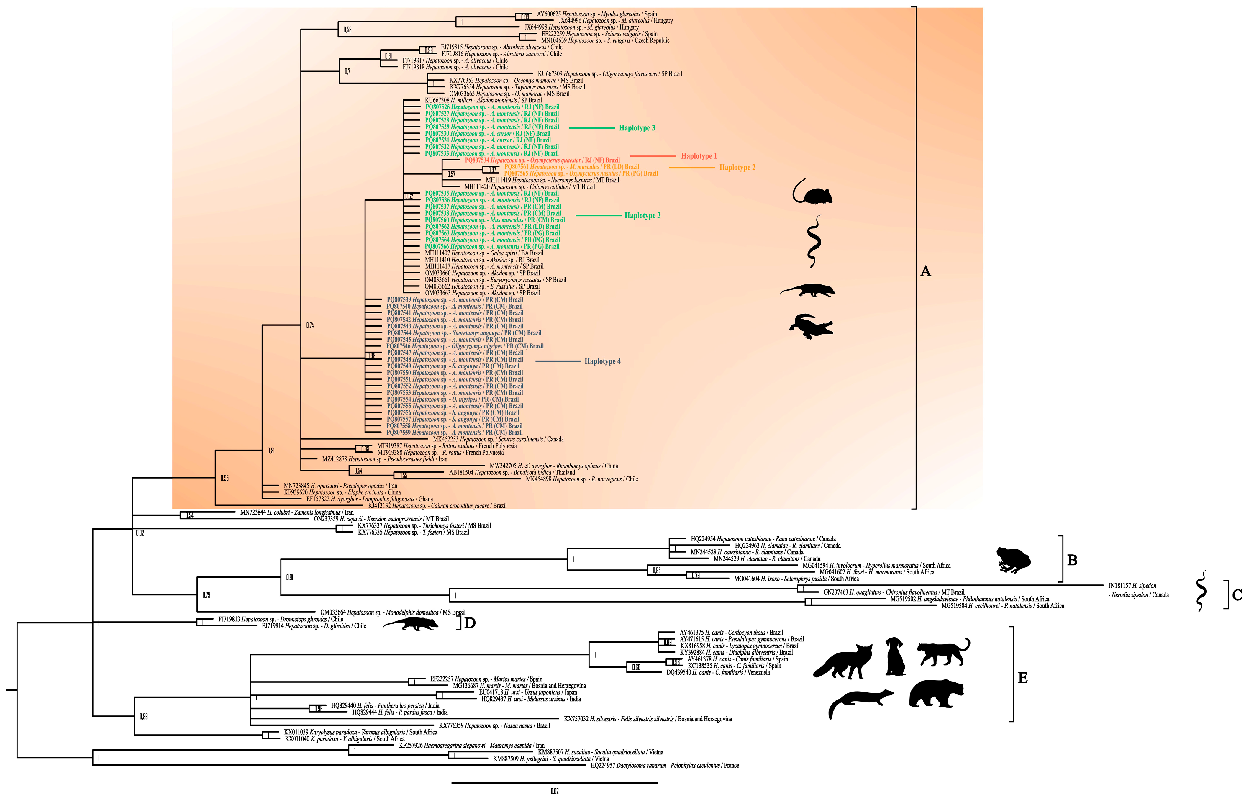
2.7. Phylogenetic Analysis

The sequences from this study were assembled and edited using Geneious Prime 2025.0.3 (Biomatters Ltd., Auckland, New Zealand) by comparison with homologous sequences through the BLASTn tool (https://blast.ncbi.nlm.nih.gov/Blast.cgi accessed on 26 September 2024) [39]. After analysis, they were deposited in GenBank. The consensus sequences generated, including those from this study (PQ807526-66) and others retrieved from GenBank, were aligned using the Clustal W method in MEGA 11 (Pennsylvania State University, State College, PA, USA) [40] and trimmed to obtain two phylogenetic trees: one created using short sequences (>500 bp) and another created using long sequences (>1600 bp). A database was created with Hepatozoon spp. sequences selected according to the following criteria: the included sequences were (a) obtained from vertebrates, (b) from the same region of the 18S rDNA gene as the sequences in this study, and (c) of a size greater than 500 bp. Additionally, sequences obtained from GenBank of other members of the suborder Adeleorina, namely Hemogregarina spp., Karyolysus paradoxa, and Dactylosoma spp. (outgroup), were added to the database (Table S1).
For the Bayesian inference (BI), in both phylogenetic analyses, a nexus file was created from the alignment in the software Mesquite 3.81 (Mesquite Project, University of California, Davis, CA, USA) [41] for MrBayes 3.2.7 (University of Oslo, Oslo, Norway) [42] using the Markov Chain Monte Carlo (MCMC) method with the GTR + I + G model for 10,000,000 generations. BI phylogenetic reconstructions were performed using the MrBayes program, which was executed on the CIPRES Science Gateway platform (San Diego Supercomputer Center, University of California San Diego, La Jolla, CA, USA) [43]. The trees were summarized with a 25% burn-in discarded, and the effective sample size (ESS) of the MCMCs was analyzed using Tracer 1.7.2 (Andrew Rambaut, University of Edinburgh, Edinburgh, UK) [44]. The consensus trees were visualized in FigTree 1.4.1 (Andrew Rambaut, University of Edinburgh, Edinburgh, UK), exported as SVG files, and edited in Inkscape 1.3 (Inkscape Project, community-developed software).
The overall evolutionary divergence, as well as the intergroup (between different clades) and intragroup (within clades) evolutionary divergence between Hepatozoon spp. sequences were estimated using the p-distance method (gamma distribution = 1) in MEGA11 software [40]. Additionally, polymorphism metrics such as the number of polymorphic sites (S), the number of haplotypes (H), haplotype diversity (Hd), nucleotide diversity (π), and haplotype frequency among the sequences of this study were obtained using DnaSP 6.12.03 software (Julio Rozas, Universitat de Barcelona, Barcelona, Spain) [45]. The analyses excluded gaps and considered invariant sites. Subsequently, to determine the haplotype frequency of Hepatozoon spp. in rodents from Brazil and Chile, a haplotype network was constructed with sequences of 519 bp using the median-joining (MJ) method in PopART v.1.7 software (Leigh & Bryant, School of Biological Sciences, University of Auckland, Auckland, New Zealand). Analysis of molecular variance (AMOVA) [46] and the fixation index (Fst) [47] were calculated for the sequences from Brazil and Chile using the Arlequin software package, version 3.5.2.2 (Laurent Excoffier and Stéphane Lischer, University of Bern, Bern, Switzerland) [48]. AMOVA was used to test genetic variability between and within these groups, and the fixation index was employed to measure levels of genetic differentiation between haplotypes in two countries (Brazil and Chile).

3. Results

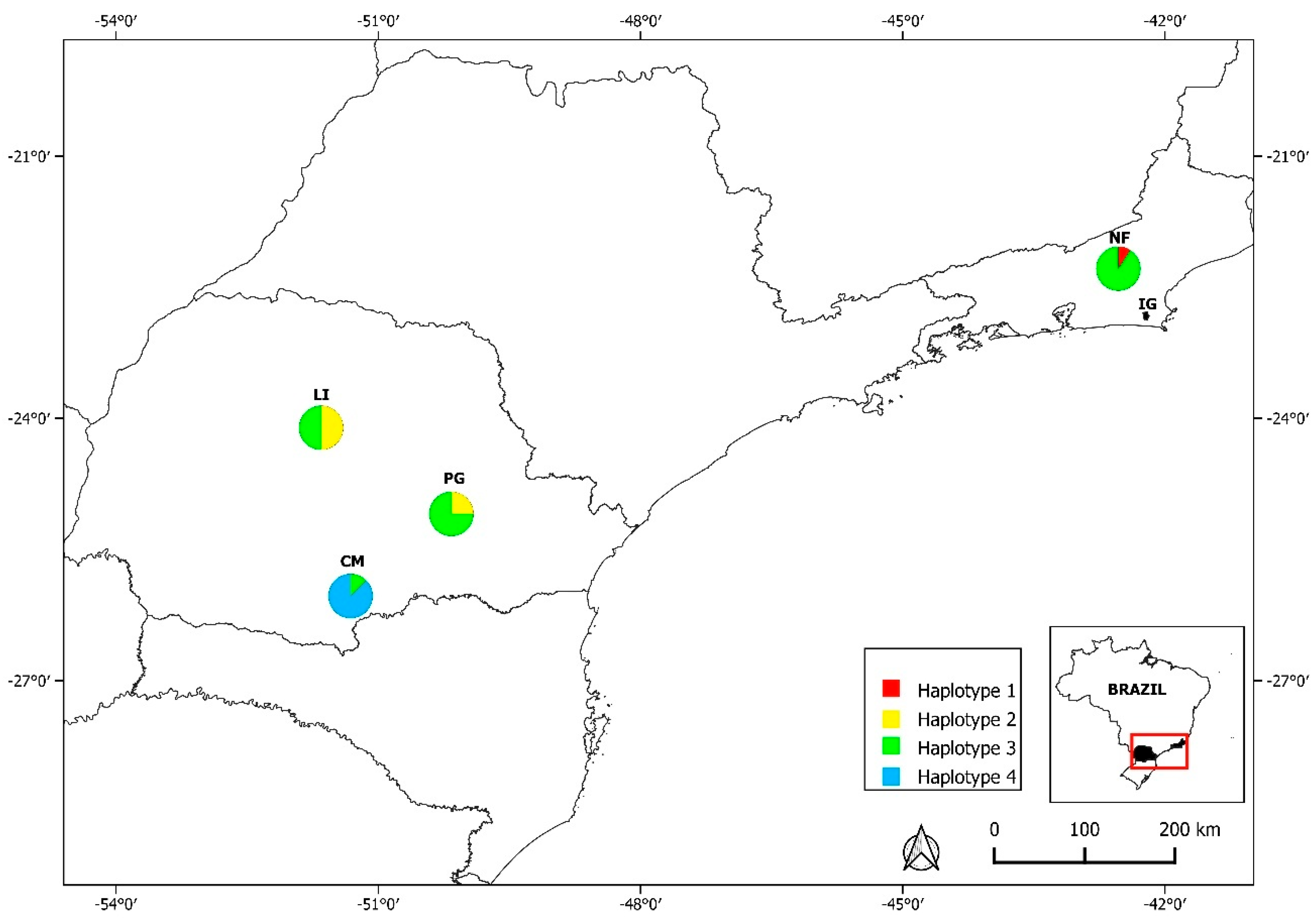
3.1. Distribution of Rodents

A total of 289 rodents were analyzed, with this sample comprising fourteen species of wild rodents from the Cricetidae family and two synanthropic species from the Muridae family. A total of 134 specimens were collected in the state of Rio de Janeiro, with 25 from Iguaba Grande (IG) and 109 from Nova Friburgo (NF), while 155 specimens were collected in the state of Paraná, including 52 from Cruz Machado (CM), 48 from Lidianópolis (LD), and 55 from Ponta Grossa (PG). Ponta Grossa exhibited the highest rodent diversity, with ten different species in the sample, whereas Iguaba Grande had the lowest, with only one. Among the wild rodents, Akodon montensis was the most abundant species (n = 106), followed by Oligoryzomys nigripes (n = 40) and Akodon cursor (n = 29), while Mus musculus was the most abundant of the synanthropic species (n = 42) (Table 1).

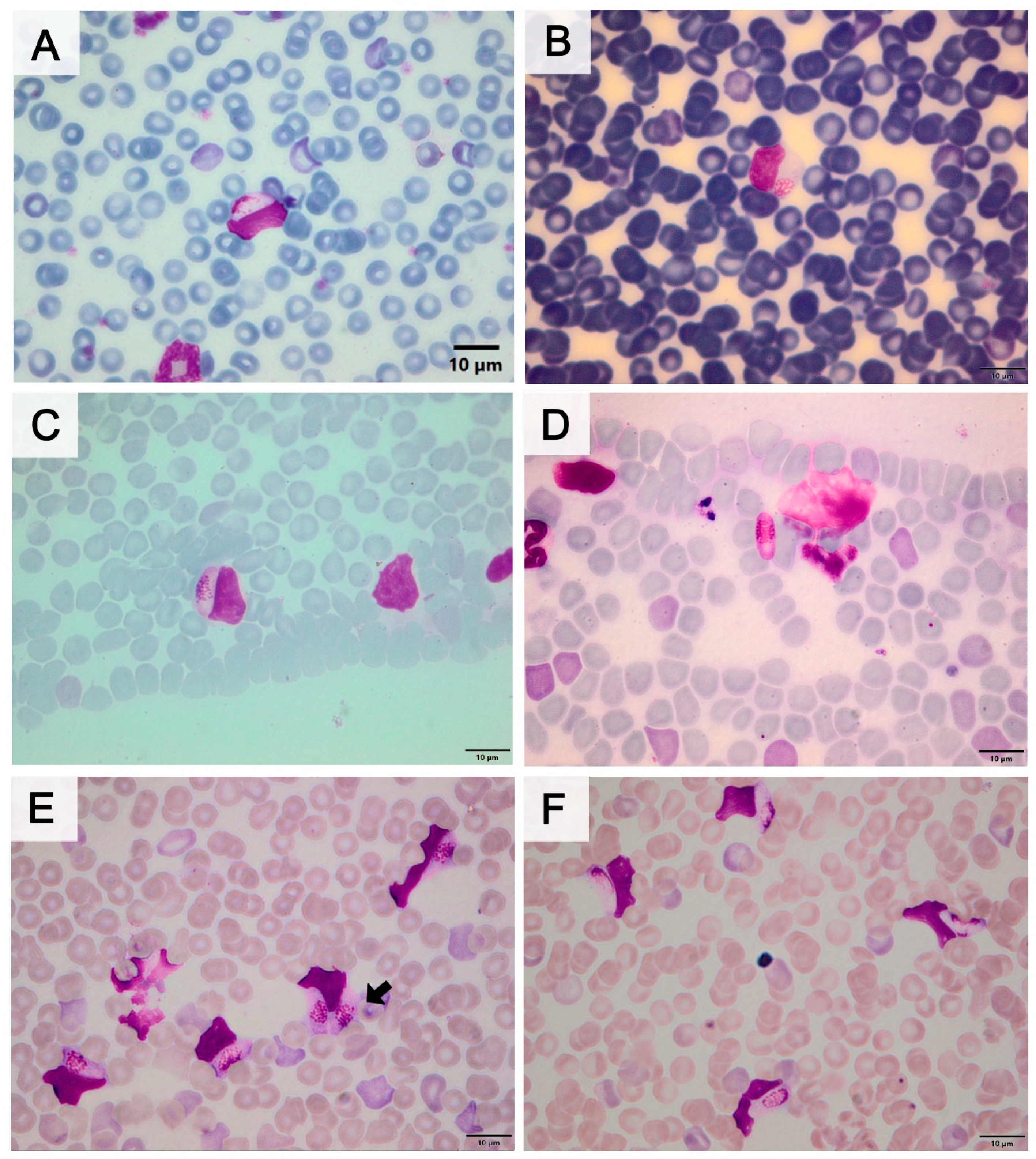
3.2. Morphometric Analysis of Gametocytes of Hepatozoon spp.

Gametocytes of Hepatozoon spp. were detected in blood smears from 10.72% of the sampled rodents (n = 31/289), with the positive rodents belonging to the species A. cursor (n = 2, NF), A. montensis (n = 28: two individuals from PG, nine from NF, and 17 from CM), and O. nigripes (n = 1, CM). The gametocytes exhibited an elongated and ovoid shape and light blue coloration and were located either inside leukocytes or free in the bloodstream. Their nuclei were slightly condensed and granular in appearance and were positioned either centrally or at one of the extremities. A case of double infection within a leukocyte was observed in the cytological analysis of an A. montensis specimen from CM (Figure 1).
The morphometric measurements of Hepatozoon gametocytes and their nuclei obtained in this study were compared with data previously reported for rodent hosts in other geographic regions (Table S2). In this study, capsule lengths of Hepatozoon spp. found in Akodon spp. and Oligoryzomys nigripes ranged from 10.13 µm to 10.95 µm, while widths varied between 4.40 µm and 4.77 µm. Nuclear lengths ranged from 5.83 µm to 6.54 µm, and nuclear widths ranged from 2.96 µm to 3.64 µm. The gametocytes detected in A. montensis specimens from the three localities (CM, NF, and PG) exhibited capsule measurements that were within a narrow range (10.23–10.95 µm in length; 4.44–4.69 µm in width), as did their nuclear dimensions. Overall, the values obtained in this study fall within the range of variation previously reported for rodent-associated Hepatozoon species in Brazil and Canada (Table S2).
The morphometric analysis of A. montensis gametocytes revealed that those observed in specimens from CM were significantly larger than those from NF (p < 0.05) and PG (p < 0.01). The nuclear lengths of gametocytes from PG were significantly greater than those from CM (p < 0.05) and NF (p < 0.01), and significant differences were also observed in the mean nuclear widths between gametocytes from CM and NF (p < 0.05), CM and PG (p < 0.01), and NF and PG (p < 0.01) (Table S3).

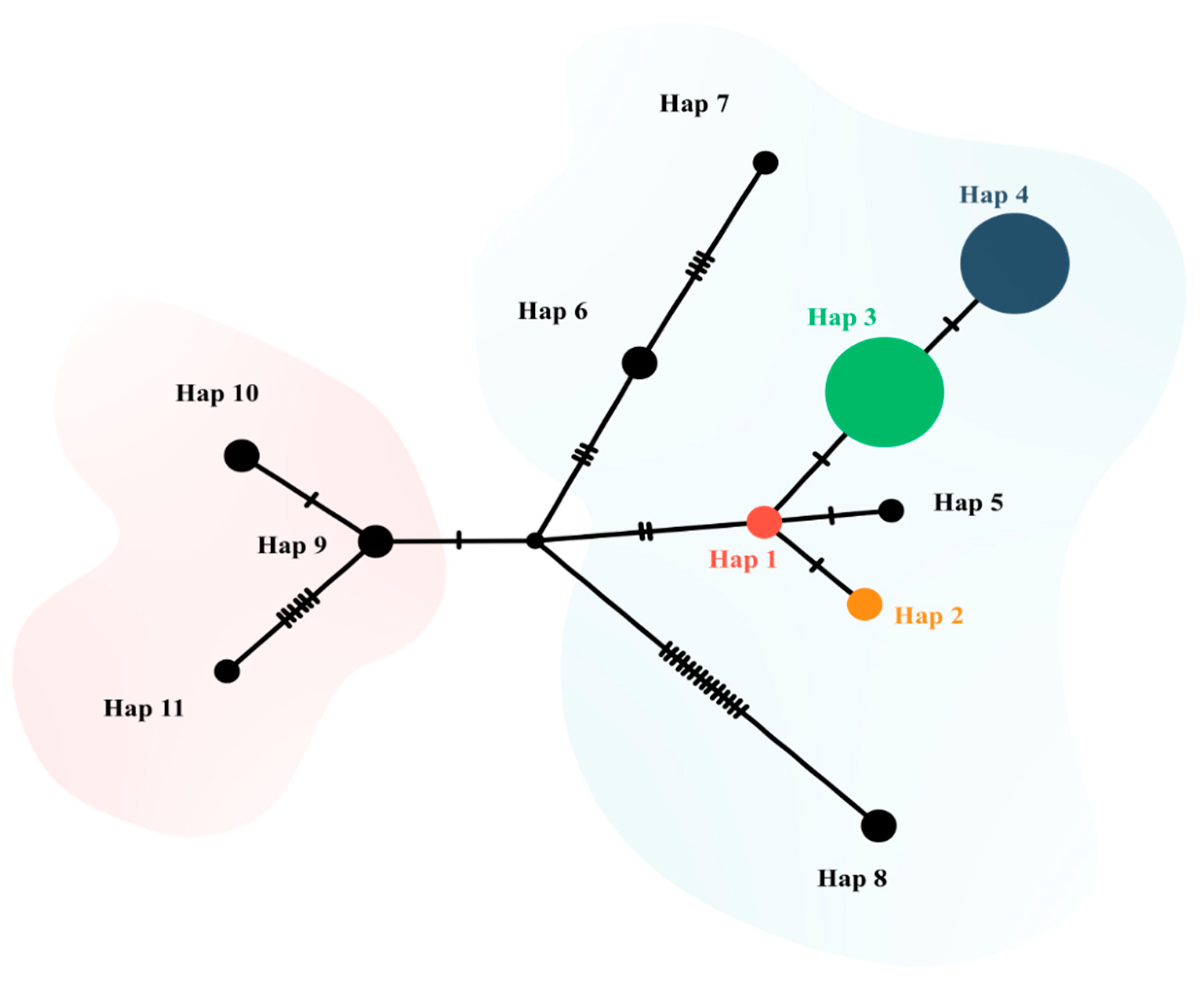
3.3. Detection and Phylogenetic Analysis of Hepatozoon spp.

In the PCR assay for the detection of Hepatozoon spp., 72 out of the 289 analyzed samples originating from seven species (Table 1) yielded an amplified fragment (~600 bp) of Hepatozoon sp. 18S rDNA (24.91%). These positive samples were from rodents collected in PG (4/55, 7.27%), NF (36/109, 33%), CM (30/52, 57.69%), and LD (2/48, 4.16%). The localities with the highest prevalence rates were CM (57.69%) and NF (33%), while samples from LD had the lowest prevalence (4.16%). Akodon montensis was the rodent species with the greatest number of positive individuals (46.23%, 49/106) (Table 1).
From the 72 Hepatozoon spp. amplifications, 41 sequences were obtained, including 31 from animals in which gametocytes had been detected in blood smears and 10 from animals with negative cytological analysis. The sequences from this study were deposited in GenBank under accession number PQ807526-66.
The BLASTn analysis of the representative haplotype sequences from this study displayed similarity to sequences previously obtained from rodents in Brazil, with identity values ranging from 99.81% to 100% (Table 2).
The BI analyses yielded ESS values greater than 200, demonstrating the robustness of the sampling. In the phylogenetic tree reconstructed with 114 partial 18S rDNA sequences (BI 1), the 108 Hepatozoon spp. sequences were grouped into five clades: Clade A—Hepatozoon spp. from rodents and reptiles from various regions worldwide, as well as from a marsupial in Brazil (KX776354) (posterior probability (pp) = 0.92); Clade B—Hepatozoon spp. from amphibians (pp = 1); Clade C—Hepatozoon spp. from snakes (pp = 1); Clade D—Hepatozoon spp. from marsupials in Chile (pp = 1); and Clade E—Hepatozoon spp. from carnivores (pp = 1) (Figure 2).
Figure 2. Phylogenetic relationships among Hepatozoon spp. based on 519 bp of 18S rDNA. The phylogenetic tree was inferred using Bayesian inference (BI 1) and the GTR + G + I model with 10,000,000 generations. Sequences detected in this study are highlighted in bold. Dactylosoma ranarum was used as an outgroup. The sequences obtained in this study clustered within Clade A, forming a subclade with other sequences previously obtained from rodents in Brazil (pp = 0.98), including the representative sequence of H. milleri (KU667308). The polymorphism analysis of the 41 Hepatozoon spp. sequences (519 bp) obtained in this study revealed the following values: S = 3, Hd = 0.577, π = 0.00151; and four haplotypes: Hap 1 (n = 1), Hap 2 (n = 2), Hap 3 (n = 17), and Hap 4 (n = 21). The colors of each haplotype are the same as those used in Figure 3, indicating their respective areas of occurrence. The frequency of the different haplotypes in the studied areas is also presented in Figure 3. The overall evolutionary divergence was 3%, while the genetic variability between Clade A and the other clades was 5% for Clades B and E, 6% for Clade C, and 3% for Clade D.
Pathogens 14 00756 g002
Figure 3. Map of part of the southern and southeastern regions of Brazil showing the frequency of Hepatozoon spp. haplotypes detected in rodents. The haplotype graphs were laid out over the areas sampled in the states of Paraná (LI, CM, and PG) and Rio de Janeiro (IG and NF). Abbreviations: CM, Cruz Machado; IG, Iguaba Grande; LI, Lidianópolis; NF, Nova Friburgo; PG, Ponta Grossa. This Map was created using QGIS version 3.42.0 [https://qgis.org/ (accessed on 14 March 2025)].
Pathogens 14 00756 g003
The second BI (2) analysis, conducted with 37 fragments of 1605 bp, revealed a tree topology similar to the previous analysis. The representative sequences of haplotypes 1, 3, and 4 from this study clustered closely with sequences obtained from rodents in Brazil (OM033660–OM033663), with strong statistical support (pp = 1) (Figure S1). It was not possible to obtain a long sequence for haplotype 2. In this analysis, the overall genetic distance was 4%, while the genetic distance between Clade A and the other clades was 6% for Clade B, 5% for Clades C and E, and 3% for Clade D.
The haplotype network was constructed using the sequences from this study (n = 41) along with 20 sequences obtained in GenBank from rodents in Brazil (n = 15) and Chile (n = 5) (Figure 4, Table S4). The genetic diversity analysis of these 61 sequences yielded S = 32, Hd = 0.718, and π = 0.00641, and identified 11 haplotypes. The haplotypes analyzed formed four haplogroups originating from an ancestral sequence (not sampled) (Figure 4).
Based on haplotype formation, the gametocyte length of haplotype 4 (n = 153, mean = 10.951, SD = 0.726) was statistically greater than that of haplotype 3 (n = 96, mean = 10.682, SD = 0.817) (p < 0.01) (Table 3). No gametocytes were detected in samples of haplotypes 1 and 2. The genetic variation between countries was 56.78% (Fst = 0.567; p < 0.01), and the genetic variation within countries was 43.22%. Fst values revealed significant genetic differences between countries (Fst = 0.567; p < 0.01).

4. Discussion

This study detected Hepatozoon spp. infection in liver samples from six species of wild rodents from the Atlantic Forest biome and one synanthropic species in southern and southeastern regions of Brazil, specifically the states of Paraná and Rio de Janeiro. This is the first report of Oxymycterus nasutus and Oxymycterus quaestor as hosts of Hepatozoon spp. in Brazil and the first description of Hepatozoon spp. infection in wild rodents in Paraná.
Currently, few studies employ cytological analysis of blood smear slides as a diagnostic tool for Hepatozoon sp. and for the characterization of its gametocytes in animals [1,6,23,25,49,50]. When comparing the mean measurements of Hepatozoon sp. gametocytes (capsules and nuclei) obtained in this study with those reported in the literature, we found morphometric similarities to Hepatozoon milleri as described by Demoner et al. [6] in Akodon montensis, and to Hepatozoon griseisciuri as described by Leveille et al. [50] in the squirrel Sciurus carolinensis, although S. carolinensis does not occur in Brazil [50] (Table S2). The morphological analysis did not reveal any unique characteristics that could differentiate the gametocytes observed in this study.
Two gametocytes were observed within the same leukocyte in a specimen of A. montensis from CM (Figure 1). This finding, referred to as “double infection,” is commonly detected in reptiles parasitized by Hepatozoon spp. [51]. The presence of multiple Hepatozoon spp. gametocytes infecting the same leukocytes has been reported in cases of massive infections in snakes [2]. Based on this, we infer that a similar phenomenon may have occurred in the A. montensis specimen from CM, which could explain our observations. No reports of Hepatozoon sp. double infection within the same leukocyte in a rodent were found in the literature.
The morphometric analyses in this study revealed significant differences among the A. montensis gametocytes captured in CM, NF, and PG in terms of the gametocyte length and the nuclear length and width (Table S3). Similar findings have not been previously reported in the literature; however, based on these results and the detection of haplotypes, we compared the mean gametocyte measurements of those haplotypes for which the greatest numbers of gametocytes were analyzed. We observed that the mean gametocyte length of haplotype 4 (n = 153 gametocytes) was significantly greater than that of haplotype 3 (n = 96 gametocytes) (p < 0.01).
The PCR results demonstrated an overall prevalence of Hepatozoon spp. infection of 24.91% (72/289) in the analyzed liver samples, with A. montensis being the rodent species with the highest prevalence of infection (46.23%, 49/106), as well as the most abundant species in this study. This rodent species has previously been reported as being infected with Hepatozoon spp. [17,19] and identified as a host of H. milleri [6] in prior studies conducted in Brazil. The second-highest prevalence was detected in Sooretamys angoyua (45.45%, 5/11) in CM. This wild rodent species was previously reported to be infected with Hepatozoon sp. in São Paulo [13].
The prevalence detected in CM (A. montensis, M. musculus, O. nigripes, and S. angouya) was similar to the rate of positive tests for Hepatozoon spp. reported in the Botucatu region, São Paulo State (55.2% in Akodon sp., Necromys lasiurus, Oligoryzomys flavescens, O. nigripes, and S. angouya) [13]. It was also higher than the rate of positive tests (21.8% in Thrichomys fosteri and Oecomys mamorae) in the Pantanal region, Mato Grosso do Sul State [16], and rate of positive tests in Calomys callosus rodents in Mato Grosso State (7.1%) [14]. However, the highest prevalence found in this study was lower than that reported in Valdivia, Chile, where 82.43% of rodents (Abrothrix longipilis, A. olivaceus, M. musculus, Oligoryzomys longicaudatus, Rattus norvegicus, and R. rattus) were infected with Hepatozoon spp. [21].
A low prevalence (7.27%, 4/55) was detected in the municipality of PG, where the highest diversity (10 species) of wild rodents was found. This may be attributed to the fact that it is an area less impacted by humans, in contrast to the findings of Weck et al. [19], who detected Hepatozoon spp. infection only in areas with a high diversity of small mammals and ticks. Their findings suggest that the parasite’s life cycle occurs through a complex interaction between vertebrates and invertebrates.
Rodents of the species A. montensis showed the highest overall prevalence of infection compared to other rodent species and were found to be infected with Hepatozoon spp. in all areas where they were present, with significant prevalence values, which suggests that A. montensis may serve as a common intermediate host for Hepatozoon spp. in the Brazilian Atlantic Forest. This association may also reflect a possible parasite–host co-evolution event, as observed by Santodomingo et al. [22] in Chile, bearing in mind that most parasite species co-evolve with only a single host species [52]. The second-highest prevalence was detected in Sooretamys angoyua in CM. This wild rodent species was previously reported to be infected with Hepatozoon sp. in São Paulo [13].
BLASTn analyses revealed a close similarity (99.81% to 100%) between the Hepatozoon spp. sequences found in this study and sequences obtained from Akodon sp. and Euryoryzomys russatus rodents from the Atlantic Forest in the state of São Paulo [19]. Unfortunately, the lack of some species-level identification of the rodents prevents direct comparisons, particularly due to the occurrence of sympatry among Akodon species in that area [53]. The BLASTn analysis with the lowest similarity was observed for the sequences representing haplotype 1, which exhibited 99.81% similarity with the best BLAST hit sequence obtained from Hepatozoon sp. infecting Akodon sp. and Euryoryzomys russatus (OM033660–OM033663) from the state of São Paulo [19].
The sequences from this study are included in Clade A of Hepatozoon spp., which comprises sequences obtained from rodents and reptiles from different parts of the world, as well as from marsupials in the Americas. This clade remains distinct from Clade E, which includes sequences obtained from carnivores, a pattern also observed in previous studies [10,17,19]. A genetic divergence of 5% was observed between the sequences of these clades.
Our analyses support the association of Hepatozoon spp. parasitism between nonflying small mammals and reptiles, confirming the existence of an enzootic cycle in which the former act as intermediate or paratenic hosts, being preyed upon by reptiles within the trophic network. This hypothesis was experimentally demonstrated by Sloboda et al. [54], who successfully infected the snake Python regius with Hepatozoon ayorgbor through the ingestion of infected rodent tissues. However, some Hepatozoon species recovered from snakes remained isolated within Clade C, with a genetic distance of 6% (BI 1) to 5% (BI 2), suggesting that not all species of this parasite participate in the rodent-reptile enzootic cycle. Another possible route for the transmission of this parasitism could be the accidental ingestion of the definitive host by the reptile, as inferred for the snake Xenodon matogrossensis, which was found to be infected with Hepatozoon cepavii (ON237359) despite not preying on rodents [55]. We suggest that further studies on this parasitism involving these vertebrate orders should be encouraged to expand knowledge on these trophic relationships, which influence the transmission dynamics of Hepatozoon spp.
The Hepatozoon spp. sequences detected in this study correspond to four haplotypes. Haplotype 1 was identified in the mountainous region of the Atlantic Forest in the state of Rio de Janeiro in a specimen of Oxymycterus quaestor (PQ807534); however, it had previously been described in Calomys callidus rodents (MH111420) from the state of Mato Grosso. The circulation of the same Hepatozoon sp. haplotype among rodents from the Central–West and coastal regions of Brazil was reported in a previous study [17].
Although these two ecoregions have highly diverse landscapes, a recent study described the potential gene flow of another rodent species, Necromys lasiurus, occurring between the Atlantic Forest in Rio de Janeiro and the arid region of South America, including the state of Mato Grosso, since the start of the Holocene. As hosts migrate, their associated parasites circulate with them [56]. Infection by Hepatozoon sp. has been reported in a specimen of N. lasiurus [17] from Mato Grosso, whose sequence (MH111419) is a representative of haplotype 5, circulating among rodents in Brazil (Figure 4).
Haplotype 3 was previously described in rodents from the Atlantic Forest biome in the state of São Paulo, including in Akodon sp., A. montensis, and Euryoryzomys russatus [6,19], as well as in Galea spixii from the state of Bahia [17]. In this study, it was detected in A. cursor and A. montensis in the mountainous region of Rio de Janeiro (NF), as well as in A. montensis and Mus musculus in the three sampled municipalities in the state of Paraná (CM, LD, and PG). This was the most widespread haplotype, being detected in all analyzed areas where the parasite was present. In the phylogenetic tree reconstructed using the shorter 18S rDNA sequences, it demonstrated a close relationship with the representative sequence of H. milleri (KU667308). However, haplotypes 2 and 4 grouped separately, potentially representing new Hepatozoon spp. haplotypes present in rodents from the Atlantic Forest of Paraná. Haplotype 2 was detected in Oxymycterus nasutus and M. musculus in the municipalities of PG and LD, respectively, while haplotype 4 was found in A. montensis, O. nigripes, and Sooretamys angouya in CM. Finally, this study detected two distinct Hepatozoon spp. haplotypes infecting rodents, with varying prevalences across the analyzed municipalities (Figure 3). The discovery of new haplotypes is extremely valuable for filling gaps in the epidemiology of this parasite and may support the hypothesis of high genetic diversity of Hepatozoon spp. among wild rodent populations in Brazil [17,57].
In this study, two distinct haplotypes were detected in rodents of the species A. montensis (haplotypes 3 and 4) and M. musculus (haplotypes 2 and 3). The detection of different haplotypes within the same rodent species has been previously reported in Brazil [17] and Chile [21], but it was not possible to detect any association between a specific haplotype and a particular rodent group (wild or synanthropic), as was observed in Chile [21,22].
The genetic distance based on 18S rDNA among the sequences within Clade A (intragroup), which included our sequences, was very small (1%) and was insufficient to infer possible distinctions among parasite species. This was observed despite the inclusion of two of the three most hypervariable regions (V2 and V4) of the Hepatozoon spp. 18S rDNA gene in the 1600 bp sequences analyzed.
The nucleotide polymorphism analysis of Hepatozoon spp. sequences from this study revealed moderate haplotype diversity (Hd = 0.577)—higher than that reported in a study using sequences obtained from rodents across various Brazilian biomes (Hd = 0.426 [17]. However, it was lower than the diversity detected in rodents from Chile (Hd = 0.933 [21]. When analyzing the sequences from Brazil and Chile included in the haplotype network (n = 61), we observed high haplotype diversity (Hd = 0.701). In the haplotype network, sequences obtained from rodents in Brazil and Chile clustered into four haplogroups, all originating from an ancestral haplotype that has not yet been described (represented by the black circle in the center). The haplogroups were not shared between the two countries, with haplotypes 1 to 8 being detected in Brazil and haplotypes 9 to 11 being detected in Chile, suggesting a geographic structure [58]. These findings were supported by significant AMOVA and Fst results, which revealed genetic differences between the two countries; however, there is a gap in information between these sampled regions.
According to Ferreri et al. [58], the frequency, distribution, and arrangement of haplotypes can reveal a history of phylogeographic events related to coalescence theory. Undoubtedly, additional Hepatozoon spp. haplotypes exist in these countries; however, some of the detected sequences were not included in our analysis because they did not meet the size criterion (>500 bp) or corresponded to different regions of the 18S rDNA fragment.
The present study detected significant differences in the average sizes of gametocytes observed in rodents, establishing for the first time a significant relationship between Hepatozoon spp. haplotype and gametocyte length (p < 0.01). This reinforces the importance of optical microscopy as a complementary tool in molecular studies. In addition, two new hosts for this parasite among Brazilian wild rodents are described, namely Oxymycterus nasutus and Oxymycterus quaestor, and two novel Hepatozoon spp. haplotypes were detected among Brazilian rodents, i.e., haplotype 2 (O. nasutus and M. musculus) and haplotype 4 (A. montensis, O. nigripes, and S. angouya). Based on the haplotype network analysis, we suggest that there is geographic structuring among Hepatozoon haplogroups associated with rodents from Brazil and Chile (Fst = 0.567; p < 0.01). The application of additional molecular markers, including mitochondrial [36,50] and nuclear genes [59], may contribute to a more precise characterization of species and a deeper understanding of the diversity and evolution of parasitism within the order Rodentia.

Supplementary Materials

The following supporting information can be downloaded at: https://www.mdpi.com/article/10.3390/pathogens14080756/s1, Table S1: List of 18S rDNA sequences used in the phylogenetic analysis of Hepatozoon spp. The first column shows the GenBank accession, followed by the identification of the parasite, host species, host order, and country of origin. Table S2: Morphometric data on the capsule and nucleus of different Hepatozoon species detected in rodents found in the literature and in this study. Table S3: Average measurements of gametocyte length and width and nucleus length and width of Hepatozoon spp. observed in rodents in this study, according to locality. Table S4: List of Hepatozoon spp. haplotypes used in the haplotype network (Figure 4), relating the respective haplotype to the GenBank accession, intermediate host, and country of origin. Figure S1: Phylogenetic relationship among Hepatozoon spp. based on a final dataset of 1605 bp sequences of 18S rDNA. The phylogenetic tree was inferred using Bayesian inference (BI 2) and the GTR + G + I model with 10,000,000 generations. Sequences detected in this study are highlighted in bold. Dactylosoma kermiti was used as an outgroup.

Author Contributions

Conceptualization, T.P.T.d.F., B.R.T., C.L.M. and M.P.; methodology, T.P.T.d.F., E.d.O.S.L.M., I.L.L.P., L.d.S.d.O., K.V., B.R.T. and M.P.; software, T.P.T.d.F., E.d.O.S.L.M., K.V., F.d.O.S. and M.P.; investigation, T.P.T.d.F., B.R.T. and M.P.; writing—original draft preparation, T.P.T.d.F.; writing—review and editing, T.P.T.d.F., B.R.T., E.d.O.S.L.M., I.L.L.P., L.d.S.d.O., K.V., H.A.S., F.d.O.S., L.M.T., C.L.M. and M.P.; visualization, T.P.T.d.F., E.d.O.S.L.M., K.V. and F.d.O.S.; supervision, B.R.T., C.L.M. and M.P.; project administration, T.P.T.d.F., B.R.T. and M.P.; funding acquisition, B.R.T., L.M.T. and M.P. All authors have read and agreed to the published version of the manuscript.

Funding

This study also received financial support from the Carlos Chagas Filho Foundation for Research Support of the State of Rio de Janeiro (FAPERJ Processes JCNE E-26/200.234/2023, ARC E-26/211.249/2019 and APQ1 E-26/211.615/2021), and the Brazilian National Council for Scientific and Technological Development (CNPq, Processes 421292/2017-2 and 421866/2023-3).

Institutional Review Board Statement

The collection of small mammals was authorized by SISBIO-ICMBIO through licenses 13373 (Permanent license for the collection of zoological material since 19 November 2007) and 63846 (25 September 2018).

Informed Consent Statement

Not applicable.

Data Availability Statement

The datasets supporting the conclusions of this article are included within the article.

Acknowledgments

We are grateful to Federal Rural University of Rio de Janeiro (UFRRJ), Oswaldo Cruz Institute/Oswaldo Cruz Foundation (IOC/FIOCRUZ) and Federal University of Paraná (UFPR) for support and financing this research, as well as ICMBio for authorizing fieldwork. We also thank Jorge Pinto da Silva, Adilson Benedito de Almeida, and the teams from LABPMR/IOC/FIOCRUZ, LHV/UFRRJ and UFPR for their assistance during fieldwork and Leila Gatti, Eugenio, and team for their valuable support in the field work at NEIG/UFF. Additionally, we acknowledge the support of the state health secretaries of Rio de Janeiro and Paraná, along with the municipal health secretaries of Nova Friburgo, Cruz Machado, Lidianópolis, and Ponta Grossa, as well as the 3rd, 6th, and 22nd regional health secretaries of the State of Paraná.

Conflicts of Interest

The authors declare no conflicts of interest.

Abbreviations

The following abbreviations are used in this manuscript:
DNADeoxyribonucleic acid
PCRPolymerase chain reaction
CytbCytochrome b
µMMicrometer
SDStandard deviation

References

  1. Miller, W.W. Hepatozoon perniciosum (n. g., n. sp.); A haemogregarine pathogenic for white rats; With a description of the sexual cycle in the inter mediate host, a mite (Lelaps echidninus). Hygienic Lab. Bull. 1908, 46, 51–123. [Google Scholar]
  2. Smith, T.G. The genus Hepatozoon (apicomplexa: Adeleina). J. Parasitol. 1996, 82, 565–585. [Google Scholar] [CrossRef] [PubMed]
  3. Baneth, G. Perpectives on canine and feline hepatozoonosis. Vet. Parasitol. 2011, 181, 3–11. [Google Scholar] [CrossRef] [PubMed]
  4. Maia, J.P.; Carranza, S.; Harris, D.J. Comments on the Systematic Revision of Adeleid Haemogregarines: Are More Data Needed? J. Parasitol. 2016, 102, 549–552. [Google Scholar] [CrossRef]
  5. Baneth, G.; Samish, M.; Shkap, V. Life Cycle of Hepatozoon canis (Apicomplexa: Adeleorina: Hepatozoidae) in the Tick Rhipicephalus sanguineus and Domestic Dog (Canis familiaris). J. Parasitol. 2007, 93, 283–299. [Google Scholar] [CrossRef]
  6. Demoner, L.C.; Silva, M.R.L.; Magro, N.M.; O’Dwyer, L.H. Hepatozoon milleri sp. nov. (Adeleorina: Hepatozoidae) in Akodon montensis (Rodentia: Cricetidae: Sigmodntinae) from southeastern Brazil. Parasitology 2018, 146, 662–669. [Google Scholar] [CrossRef]
  7. Johnson, P.T.J.; Dobson, A.; Lafferty, K.D.; Marcogliese, D.J.; Memmott, J.; Orlofske, S.A.; Poulin, R.; Thieltges, D.W. When parasites become prey: Ecological and epidemiological significance of eating parasites. Trends Ecol. Evol. 2010, 25, 362–371. [Google Scholar] [CrossRef]
  8. Johnson, E.M.; Allen, K.E.; Breshears, M.A.; Panciera, R.J.; Little, S.E.; Ewing, S.A. Experimental transmission of Hepatozoon americanum to rodents. Vet. Parasitol. 2008, 151, 164–169. [Google Scholar] [CrossRef]
  9. Jonhson, E.M.; Panciera, R.J.; Allen, K.E.; Sheets, M.E.; Beal, J.D.; Ewing, S.A.; Little, S.E. Alternate Pathway of Infection with Hepatozoon americanum and the Epidemiologic Importance of Predation. J. Vet. Intern. Med. 2009, 23, 1315–1318. [Google Scholar] [CrossRef]
  10. Maia, J.P.; Álvares, F.; Boratynski, Z.; Brito, J.C.; Leite, J.V.; Harris, J. Molecular assessment of Hepatozoon (Apicomplexa: Adeleorina) infections in wild canids and rodents from North Africa, with implications for transmission dynamics across taxonomic groups. J. Wildl. Dis. 2014, 50, 837–848. [Google Scholar] [CrossRef]
  11. Murata, T.; Inoue, M.; Tateyama, S.; Taura, Y.; Nakama, S. Vertical transmission of Hepatozoon canis in dogs. J. Vet. Med. Sci. 1993, 55, 867–868. [Google Scholar] [CrossRef] [PubMed]
  12. Tolkacz, K.; Kowalec, M.; Alsarraf, M.; Grzybek, M.; Dwuznik-Sazare, D.; Behnke, J.M.; Bajer, A. Candidatus Neoehrlichia mikurensis and Hepatozoon sp. in voles (Microtus spp.): Occurrence and evidence for vertical transmission. Sci. Rep. 2023, 13, 1733. [Google Scholar] [CrossRef] [PubMed]
  13. Demoner, L.C.; Magro, N.M.; Silva, M.R.L.; Antunes, J.A.P.; Calabuig, C.I.P.; O’Dwyer, L.H. Hepatozoon spp. infections in wild rodents in an area of endemic canine hepatozoonosis in southeastern Brazil. Tick Tick-Borne Dis. 2016, 7, 859–864. [Google Scholar] [CrossRef] [PubMed]
  14. Wolf, R.W.; Aragona, M.; Muñoz-Leal, S.; Pinto, L.B.; Melo, A.L.T.; Braga, I.A.; Costa, J.S.; Martins, T.F.; Marcili, A.; Pacheco, R.C.; et al. Novel Babesia and Hepatozoon agents infecting non-volant small mammals in the Brazilian Pantanal, with the first record of the tick Ornithodoros guaporensis in Brazil. Tick Tick-Borne Dis. 2016, 7, 449–456. [Google Scholar] [CrossRef]
  15. Gomes, L.A.; Moraes, L.A.; Aguiar, D.C.F.; Dias, H.L.T.; Ribeiro, A.S.S.; Rocha, H.P.C.; Nunes, M.R.T.; Gonçalves, E.C. Genetic diversity of Hepatozoon spp. in Hydrochoerus hydrochaeris and Pecari tajacu from eastern Amazon. Ticks Tick-Borne Dis. 2018, 9, 314–318. [Google Scholar] [CrossRef]
  16. Sousa, K.C.M.; Fernandes, M.P.; Herrera, H.M.; Benevenute, J.L.; Santos, F.M.; Barreto, W.T.G.; Macedo, G.C.; Campos, J.B.; Martins, T.F.; Pinto, P.C.E.A.; et al. Molecular detection of Hepatozoon spp. in domestic dogs and wild mammals in southern Pantanal, Brazil with implications in the transmission route. Vet. Parasitol. 2017, 237, 37–46. [Google Scholar] [CrossRef]
  17. Perles, L.; Roque, A.L.R.; D’Andrea, P.S.; Lemos, E.R.S.; Santos, A.F.; Morales, A.C.; Machado, R.Z.; André, M.R. Genetic diversity of Hepatozoon spp. in rodents from Brazil. Sci. Rep. 2019, 9, 10122. [Google Scholar] [CrossRef]
  18. Santos, F.M.; Sousa, K.C.M.; Sano, N.Y.; Nantes, W.A.G.; Liberal, S.C.; Machado, R.Z.; André, M.R.; Herrera, H.M. Relationships between vector-borne parasites and free-living mammals at the Brazilian Pantanal. Parasitol. Res. 2021, 120, 1003–1010. [Google Scholar] [CrossRef]
  19. Weck, B.C.; Serpa, M.C.A.; Ramos, V.N.; Luz, H.R.; Ramirez, D.G.; Benatti, H.R.; Piovezan, U.; Szabó, M.P.J.; Marcili, A.; Krawczak, F.S.; et al. Novel genotypes of Hepatozoon spp. in small mammals, Brazil. Parasite Vector 2022, 15, 87. [Google Scholar] [CrossRef]
  20. Merino, S.; Vásquez, R.A.; Martínez, J.; Celis-Diez, J.L.; Gutiérrez-Jimenes, L.; Ippi, S.; Sánchez-Monsalvez, I.; La Puente, J.M. Molecular characterization of an ancient Hepatozoon species parasitizing the ‘living fossil’ marsupial ‘Monito del Monte’ Dromiciops gliroides from Chile. Biol. J. Linn. Soc. 2009, 98, 568–576. [Google Scholar] [CrossRef]
  21. Alabi, A.S.; Monti, G.; Otth, C.; Sepulveda-Garcia, P.; Perles, L.; Machado, R.Z.; André, M.R.; Bittencourt, P.; Muller, A. Genetic diversity of Hepatozoon spp. in rodents from Chile. Braz. J. Vet. Parasitol. 2021, 30, e012721. [Google Scholar] [CrossRef] [PubMed]
  22. Santodomingo, A.M.; Thomas, R.S.; Quintero-Galvis, J.F.; Echeverry-Berrio, D.M.; Silva-de la Fuente, M.C.; Moreno-Salas, L.; Muñoz-Leal, S. Apicomplexans in small mammals from Chile, with the first report of the Babesia microti group in South American rodents. Parasitol. Res. 2022, 121, 1009–1020. [Google Scholar] [CrossRef] [PubMed]
  23. Jonhson, E.M.; Allen, K.E.; Panciera, R.J.; Ewing, S.A.; Little, S.E.; Reichard, M.V. Field survey of rodents for Hepatozoon infections in na endemic focus of American canine hepatozoonosis. Vet. Parasitol. 2007, 150, 27–32. [Google Scholar] [CrossRef]
  24. Ljaz, M.; Khan, A.U.; Ali, M.; Naeem, M.; Ibenmoussa, S.; Dawoud, R.M.; Khan, A.; Said, M.B.; Iqbal, F. First report of Hepatozoon and Lankesterella spp. infections in wild rodents from Pakistan, and their potential impact on blood parameters and oxidative stress markers in vital organs. Vet. Res. Commun. 2025, 49, 45. [Google Scholar]
  25. Carini, A.; Maciel, J. Sur une hémogrégarine et um trypanosome d’un Muridé (Akodon fuliginosus). Bull. Soc. Pathol. Exot. 1915, 8, 165–169. [Google Scholar]
  26. Kottek, M.; Grieser, J.; Beck, C.; Rudolf, B.; Rubel, F. World Map of the Köppen-Geiger climate classification updated. Metereol. Z. 2006, 15, 259–263. [Google Scholar] [CrossRef]
  27. Peel, M.C.; Finlayson, B.L.; Mcmahon, T.A. Update world map of the Köppen-Geiger climate classification. Hydrol. Earth Syst. Sci. 2007, 11, 1633–1644. [Google Scholar] [CrossRef]
  28. Mills, J.N.; Childs, J.E.; Ksiazek, T.G.; Velleca, W.M. Methods for Trapping and Sampling Small Mammals for Virologic Testing. Department of Health & Human Services; Centers for Disease Control and Prevention: Atlanta, GA, USA, 1995; pp. 1–61. [Google Scholar]
  29. Lemos, E.R.S.; D’Andrea, P.S. Trabalho de Campo com Animais: Procedimentos, Riscos e Biossegurança, 1st ed.; Fiocruz: Rio de Janeiro, Brazil, 2014; 180p. [Google Scholar]
  30. D’Andrea, P.S.; Teixeira, B.R.; Gonçalves-Oliveira, J.; Dias, D.; Vilela, R.V.; Lúcio, C.S.; Santos, F.O.; Costa, G.C.; Carvalhaes, J.G.; Santos, M.M.; et al. The Mammal Collection of the Laboratory of Biology and Parasitology of Reservoir Wild Mammals—Oswaldo Cruz Foundation. Braz. J. Mammal. 2021, 90, e90202119. [Google Scholar] [CrossRef]
  31. Ayres, M.; Ayres, J.M.; Aures, D.L.; Santos, A.D.A. Aplicações Estatísticas nas Áreas das Ciências Bio-Médicas; Instituto Mamirauá: Belém, Brazil, 2007; Volume 364. [Google Scholar]
  32. Smith, M.F.; Patton, J.L. Phylogenetic relationships and the radiation of sigmodontine rodents in South America: Evidence from Cytochrome b. J. Mamm. Evol. 1999, 6, 89–128. [Google Scholar] [CrossRef]
  33. Smith, M.F.; Patton, J.L. The diversification of South American murid rodents: Evidence from mitochondrial DNA sequence data for the akodontine tribe. Biol. J. Linn. Soc. 1993, 50, 149–177. [Google Scholar] [CrossRef]
  34. Ujvari, B.; Madsen, T.; Olsson, M. High prevalence Hepatozoon spp. (Apicomplexa, Hepatozoidae) infection in water pythons (Liasis fuscus) from tropical Australia. J. Parasitol. 2004, 90, 670–672. [Google Scholar] [CrossRef] [PubMed]
  35. Criado-Fornelio, A.; Ruas, J.L.; Casado, N.; Farias, N.A.R.; Soares, M.P.; Müller, G.; Brum, J.G.W.; Berne, M.E.A.; Buling-Saraña, A.; Barba-Carretero, J.C. New molecular data on mammalian Hepatozoon species (Apicomplexa: Adeleorina) from Brazil and Spain. J. Parasitol. 2006, 92, 93–99. [Google Scholar] [CrossRef] [PubMed]
  36. Hrazdilová, K.; Cervená, B.; Blanvillain, C.; Foronda, P.; Modrý, D. Quest for the type species of the genus Hepatozoon—phylogenetic position of hemogregarines of rats and consequences for taxonomy. Syst. Biodivers. 2021, 19, 622–631. [Google Scholar] [CrossRef]
  37. Kvicerová, J.; Pakandl, M.; Hypsa, V. Phylogenetic relationships among Eimeria spp. (Apicomplexa, Eimeriidae) infecting rabbits: Evolutionary significance of biological and morphological features. Parasitolology 2008, 135, 443–452. [Google Scholar] [CrossRef]
  38. Quilfeldt, P.; Romeike, T.; Masello, J.F.; Reiner, G.; Willems, H.; Bedolla-Guzmán, Y. Molecular survey of coccidian infections of the side-blotched lizard Uta stansburiana on San Benito Oeste Island, Mexico. Parasite 2018, 25, 43. [Google Scholar] [CrossRef]
  39. Benson, D.A.; Karsch-Mizrachi, I.; Lipman, D.J.; Ostell, J.; Rapp, B.A.; Wheeler, D.L. GenBank. Nucleic Acids Res. 2000, 28, 15–18. [Google Scholar] [CrossRef]
  40. Tamura, K.; Stecher, G.; Kumar, S. MEGA11: Molecular Evolutionary Genetics Analysis version 11. Mol. Biol. Evol. 2021, 38, 3022–3027. [Google Scholar] [CrossRef]
  41. Maddison, W.P.; Maddison, D.R. Mesquite: A Modular System for Evolutionary Analysis; Version 3.81; University of California: Davis, CA, USA, 2023. [Google Scholar]
  42. Ronques, F.; Teslenko, M.; Mark, V.; Darling, A.; Höhna, S.; Larget, B.; Suchard, A.; Huelsenbeck, J.P. MrBayes 3.2: Efficient Bayesian phylogenetic inference and model choice across a large model space. Syst. Biol. 2012, 61, 539–542. [Google Scholar] [CrossRef]
  43. Miller, M.A.; Pfeiffer, W.; Schwartz, T. Creating the CIPRES Science Gateway for inference of large phylogenetic trees. In Proceedings of the Gateway Computing Environments Workshop (GCE), New Orleans, LA, USA, 14 November 2010; pp. 1–8. [Google Scholar]
  44. Rambaut, A.; Drummond, A.J.; Xie, D.; Baele, G.; Suchard, M.A. Posterior summarisation in Bayesian phylogenetics using Tracer 1.7. Syst. Biol. 2018, 67, 901–904. [Google Scholar] [CrossRef]
  45. Rozas, J.; Ferrer-Mat, A.; Sánchez-DelBarrio, J.C.; Guirao-Rico, S.; Librado, P.; Ramos-Onsins, S.E.; Sánchez-Garcia, A. DnaSP v6: DNA Sequence Polymorphism Analysis of Large Datasets. Mol. Biol. Evol. 2017, 34, 3299–3302. [Google Scholar] [CrossRef]
  46. Excoffier, L.; Smouse, P.E.; Quattro, P.E. Analysis of molecular variance inferred from metric distances among DNA haplotypes: Application to human mitochondrial DNA restriction data. Genetics 1992, 131, 479–491. [Google Scholar] [CrossRef] [PubMed]
  47. Wright, S. Evolution and the Genetics of Populations. Variability Within and Among Natural Populations; University of Chicago Press: Chicaco, IL, USA, 1978; Volume 4. [Google Scholar]
  48. Excoffier, L.; Lischer, H.E.L. Arlequin suite ver 3.5: A new series of programs to perform population genetics analyses under Linux and Windows. Mol. Ecol. Resour. 2010, 10, 564–567. [Google Scholar] [CrossRef] [PubMed]
  49. Karbowiak, G.; Rychlik, L.; Nowakowski, W.; Wita, I. Natural infections of small mammals with blood parasites on the borderland of boreal and temperature forest zones. Acta Theriol. 2005, 50, 31–42. [Google Scholar] [CrossRef]
  50. Leveille, N.A.; Skhawy, N.E.; Barta, J.R. Multilocus sequencing of Hepatozoon cf. griseisciuri infections in Ontario Eastern gray squirrels (Sciurus carolinensis) uncovers two genotypically distinct sympatric parasite species. Parasitol. Res. 2020, 119, 713–724. [Google Scholar]
  51. Sloboda, M.; Kamler, M.; Bulantová, J.; Votýpka, J.; Modrý, D. A new species of Hepatozoon (Apicomplexa: Adeleorina) from Python regius (Serpentes: Pythonidae) and its experimental transmission by a mosquito vector. J. Parasitol. 2007, 93, 1189–1198. [Google Scholar] [CrossRef]
  52. Poulin, R.; Keeney, D.B. Host specificity under molecular and experimental scrutiny. Trends Parasitol. 2008, 24, 24–28. [Google Scholar] [CrossRef]
  53. Pardiñas, U.F.J.; Teta, P.; Alvarado-Serrano, D.; Geise, L.; Jayat, J.P.; Ortiz, P.E.; Gonçalves, P.E.; Della, G. Genus Akodon. In Mammals of South America, 1st ed.; Patton, J.L., Pardiñas, U.F.J., D’Elía, G., et al., Eds.; The University of Chicago Press: Chicaco, IL, USA, 2015; Volume 2, pp. 144–203. [Google Scholar]
  54. Sloboda, M.; Kamler, M.; Bulantová, J.; Votýpka, J.; Modrý, D. Rodents as intermediate hosts of Hepatozoon ayorgbor (Apicomplexa: Adeleina: Hepatozoidae) from the African ball python Python regius. Folia Parasitol. 2008, 55, 13–16. [Google Scholar] [CrossRef]
  55. Ungari, L.P.; Netherlands, E.C.; Santos, A.L.Q.; Alcantara, E.P.; Emmerich, E.; Silva, R.J.; O’Dwyer, L.H. Diversity of haemogregarine parasites infecting Brazilian snakes from the Midwest and Southeast regions with a description of two new species of Hepatozoon (Apicomplexa: Adeleorina: Hepatozoidae). Parasitol. Int. 2022, 89, 102587. [Google Scholar] [CrossRef]
  56. Santos, F.O.; Moreira, J.C.; Gonçalves, P.R.; Lucio, C.S.; Teixeira, B.R.; D’Andrea, P.S. Necromys lasiurus (Cricetidae: Sigmodontinae) from open areas of the Atlantic Forest of Rio de Janeiro: Population structure and implications for the monitoring of hantaviroses. Zoologia 2024, 41, e23086. [Google Scholar] [CrossRef]
  57. Thomas, R.; Santodomingo, A.; Saboya-Acosta, L.; Quintero-Galvis, J.F.; Moreno, L.; Uribe, J.E.; Muñoz-Leal, S. Hepatozoon (Eucoccidiorida: Hepatozoidae) in wild mammals of the Americas: A systematic review. Parasite Vector 2024, 17, 1–23. [Google Scholar]
  58. Ferreri, M.; Qu, W.; Han, B. Phylogenetic networks: A tool to display character conflict and demographic history. Afr. J. Biothecnol. 2011, 10, 12799–12803. [Google Scholar]
  59. Boullianne, B.; Evans, R.C.; Smith, T.G. Phylogenetic analysis of Hepatozoon species (Apicomplexa: Adelerina) infecting frogs of Nova Scotia, Canada, determined by ITS-1 sequences. J. Parasitol. 2007, 93, 1435–1441. [Google Scholar] [CrossRef] [PubMed]
Figure 1. Gametocytes in rodent peripheral blood. (A). Gametocyte observed in a monocyte of Oligoryzomys nigripes (PQ807554, CM). (B). Gametocyte observed in a monocyte of Akodon cursor (PQ807526, NF). (C). Gametocyte observed in a monocyte of Akodon montensis (PQ807563, PG). (D). Free gametocyte observed in blood smear from A. montensis (PQ807542, CM). (E). Gametocyte observed in monocytes of A. montensis (PQ807538, CM). The black arrow indicates two gametocytes parasitizing the same cell. (F). Four gametocytes observed in monocytes of A. montensis blood smear (PQ807537, CM). Scale bar: 10 µm.
Pathogens 14 00756 g001
Figure 4. Haplotype network formed from 18S rDNA sequences (519 bp) of Hepatozoon spp. detected in small rodents from Brazil (haplotypes 1–8, light blue background) and Chile (haplotypes 9–11, light pink background). The size of the circles varies according to the number of sequences belonging to each haplotype. Mutational events are represented with vertical dashes. The haplotypes found in this study (1–4) are colored.
Pathogens 14 00756 g004
Table 1. Prevalence of Hepatozoon spp. according to host rodent species per municipality studied and total prevalence according to rodent species (last column) and municipality where capture occurred (last row).
Family Study Areas
Cricetidae PGCMLDIGNFn/Total (Prevalence)
Akodon cursor (Winge, 1887) 6/29 (20.68%)6/29 (20.68%)
Akodon montensis (Tomas, 1913)3/5 (60%)18/26 (69.23%)1/30 (3.33%) 27/45 (60%)49/106 (46.23%)
Akodon paranaensis (Christoff et al., 2000)0/11 0/11
Delomys sublineatus (Thomas, 1903) 0/10/1
Euryoryzomys russatus (Wagner, 1848) 0/5 0/5
Juliomys ossitenuis (Costa et al., 2007)0/1 0/1
Necromys lasiurus (Lund, 1841)0/2 0/1 0/3
Nectomys squamipes (Brants, 1827) 0/1 0/1
Oligoryzomys flavescens (Waterhouse, 1837)0/4 0/4
Oligoryzomys nigripes (Olfers, 1818) 0/186/12 (50%)0/1 0/96/40 (15%)
Oxymycterus nasutus (Waterhouse, 1837)1/6 (16.66%) 1/6 (16.66%)
Oxymycterus quaestor (Thomas, 1903) 3/16 (18.57%)3/16 (18.75%)
Sooretamys angouya (Fischer, 1814) 5/11 (45.45%) 0/15/12 (38.46%)
Thaptomys nigrita (Lichtenstein, 1829) 0/2 0/2
Muridae
Mus musculus (Linnaeus, 1758) 1/1 (100%)1/16 (6.25%)0/25 2/42 (4.76%)
Rattus rattus (Linnaeus, 1758) 0/2 0/80/10
n/total (prevalence)4/55 (7.27%) 30/52 (57.69%)2/48 (4.16%)0/2536/109 (33%)72/289 (24.91%)
Abbreviations: PG, Ponta Grossa; CM, Cruz Machado; LD, Lidianópolis; IG, Iguaba Grande; NF, Nova Friburgo; n, number of infected rodents.
Table 2. Results of BLASTn analysis of the sequences of Hepatozoon spp. haplotypes found in rodents in this study.
HaplotypeGenBank ID Host/Location (This Study)Best Blast Hit IDHost/Location (Best BLAST Hit)Identity (%)Query Coverage (%)E-Value
Hap 1PQ807534O. quaestor/RJOM033660 e OM033663Akodon sp., SP99.811000.0
OM033661 e OM033662E. russatus, SP
Hap 2PQ807561, PQ807565M. musculus, O. nasutus/PRMH111420Calomys callidus, MT99.841000.0
Hap 3PQ807530A. cursor/RJOM033663, Akodon sp., SPAkodon sp., SP1001000.0
OM033660, Akodon sp., SPAkodon sp., SP
Hap 4PQ807539A. montensis/PROM033660 e OM033663Akodon sp., SP99.941000.0
OM033661 e OM033662E. russatus, SP
Abbreviations: MT, State of Mato Grosso; PR, State of Paraná; RJ, State of Rio de Janeiro; SP, State of São Paulo.
Table 3. Average measurements of gametocyte length and width, and nucleus length and width of Hepatozoon spp. according to the haplotypes detected in this study.
HaplotypesnMean (µM) SD
(µM)
Limit Values
Minimum
(µM)
Maximum
(µM)
p-Value
Gametocyte length
Hap 39610.82a0.828.1212.26
Hap 415310.95a0.738.9013.91<0.01
Gametocyte width
Hap 3964.59a0.513.335.65
Hap 41534.55a0.463.295.860.560
Nuclear length
Hap 3966.05a1.143.768.72
Hap 41535.94a0.923.168.980.584
Nuclear width
Hap 3963.49a0.661.565.77
Hap 41533.37b0.512.194.720.109
Abbreviations: n, number of analyzed gametocytes; SD, standard deviation. Values followed by different letters in the same column differ significantly according to Tukey’s test. Haplotypes 1 and 2 did not have the minimum number of gametocytes required for statistical analysis.
Disclaimer/Publisher’s Note: The statements, opinions and data contained in all publications are solely those of the individual author(s) and contributor(s) and not of MDPI and/or the editor(s). MDPI and/or the editor(s) disclaim responsibility for any injury to people or property resulting from any ideas, methods, instructions or products referred to in the content.

