Article

Molecular Surveillance of Plasmodium spp. Infection in Neotropical Primates from Bahia and Minas Gerais, Brazil

by
Luana Karla N. S. S. Santos
1,
Sandy M. Aquino-Teixeira
2,
Sofía Bernal-Valle
1,
Beatriz S. Daltro
1,
Marina Noetzold
3,
Aloma Roberta C. Silva
3,
Denise Anete M. Alvarenga
3,
Luisa B. Silva
3,
Ramon S. Oliveira
2,
Cirilo H. Oliveira
2,
Iago A. Celestino
2,
Maria E. Gonçalves-dos-Santos
2,
Thaynara J. Teixeira
2,
Anaiá P. Sevá
1,
Fabrício S. Campos
4,
Bergmann M. Ribeiro
5,
Paulo M. Roehe
4,
Danilo Simonini-Teixeira
1,
Filipe V. S. Abreu
2,6,*,
Cristiana F. A. Brito
3,* and
George R. Albuquerque
1,*
add Show full author list remove Hide full author list
1
Department of Agricultural and Environmental Sciences, Universidade Estadual de Santa Cruz, Ilhéus 45662-900, BA, Brazil
2
Insect Behavior Laboratory, Instituto Federal do Norte de Minas Gerais, Salinas 39600-000, MG, Brazil
3
Grupo de Pesquisa em Biologia Molecular e Imunologia da Malária, Instituto René Rachou, Fiocruz, Belo Horizonte 30000-000, MG, Brazil
4
Institute of Basic Health Sciences, Universidade Federal do Rio Grande do Sul, Porto Alegre 90000-000, RS, Brazil
5
Baculovirus Laboratory, Department of Cell Biology, Institute of Biological Sciences, University of Brasilia, Brasília 70000-000, DF, Brazil
6
Laboratory of Hematozoan Transmitting Mosquitoes, Instituto Oswaldo Cruz, Rio de Janeiro 20000-000, RJ, Brazil
*
Authors to whom correspondence should be addressed.
Pathogens 2025, 14(8), 757; https://doi.org/10.3390/pathogens14080757 (registering DOI)
Submission received: 30 May 2025 / Revised: 9 July 2025 / Accepted: 10 July 2025 / Published: 31 July 2025
(This article belongs to the Section Parasitic Pathogens)

Abstract

In Brazil, Plasmodium infections in non-human primates (NHPs) have been associated with P. simium and P. brasilianum, which are morphologically and genetically similar to the human-infecting species P. vivax and P. malariae, respectively. Surveillance and monitoring of wild NHPs are crucial for understanding the distribution of these parasites and assessing the risk of zoonotic transmission. This study aimed to detect the presence of Plasmodium spp. genetic material in Platyrrhini primates from 47 municipalities in the states of Bahia and Minas Gerais. The animals were captured using Tomahawk-type live traps baited with fruit or immobilized with tranquilizer darts. Free-ranging individuals were chemically restrained via inhalation anesthesia using VetBag® or intramuscular anesthesia injection. Blood samples were collected from the femoral vein. A total of 298 blood and tissue samples were collected from 10 primate species across five genera: Alouatta caraya (25), Alouatta guariba clamitans (1), Callicebus melanochir (1), Callithrix geoffroyi (28), Callithrix jacchus (4), Callithrix kuhlii (31), Callithrix penicillata (175), Callithrix spp. hybrids (15), Leontopithecus chrysomelas (16), Sapajus robustus (1), and Sapajus xanthosthernos (1). Molecular diagnosis was performed using a nested PCR targeting the 18S small subunit ribosomal RNA (18S SSU rRNA) gene, followed by sequencing. Of the 298 samples analyzed, only one (0.3%) from Bahia tested positive for Plasmodium brasilianum/P. malariae. This represents the first detection of this parasite in a free-living C. geoffroyi in Brazil. These findings highlight the importance of continued surveillance of Plasmodium infections in NHPs to identify regions at risk for zoonotic transmission.

1. Introduction

Malaria is a disease caused by protozoan parasites of the genus Plasmodium. The infection is transmitted to humans via the bite of female mosquitoes belonging to the genus Anopheles [1]. In Brazil, human cases of malaria are endemic in the Legal Amazon Region, which includes states in the Northern Region, as well as Mato Grosso, Tocantins, and Maranhão [2]. However, autochthonous cases have also been reported in areas of the Atlantic Forest (AF). In this biome, the primary vector is Anopheles (Kerteszia) cruzi, commonly known as the bromeliad mosquito. These mosquitoes are of the subgenus Kerteszia, typically breed in water-accumulating plants, such as bromeliads, which are endemic to the region. They are frequently implicated in malaria transmission within the AF. The behavioral patterns of Kerteszia mosquitoes can vary across geographic areas and seasons, but they are primarily characterized by their feeding habits, which include taking blood meals in the treetops and on the ground, as well as exhibiting vertical dispersal [2,3,4].
Five main species of malaria parasites are known to infect humans: Plasmodium falciparum, Plasmodium vivax, Plasmodium ovale (including the subspecies Plasmodium ovale wallikeri and P. o. curtisi), Plasmodium malariae, and Plasmodium knowlesi [5]. In addition to these, approximately 30 Plasmodium species are known to infect non-human primates (NHPs), although most of these parasites are restricted to phylogenetically related hosts within the same family [6]. In the Americas, only two Plasmodium species are known to naturally infect neotropical NHPs: Plasmodium brasilianum, which is genetically and morphologically similar to P. malariae, and Plasmodium simium, which closely resembles P. vivax. Both species have been detected in human infections, highlighting their zoonotic potential [7,8,9].
Plasmodium brasilianum has a broad host range, infecting species across all families of neotropical primates, and is distributed from Mexico to southern Brazil. In contrast, Plasmodium malariae is a cosmopolitan parasite distributed in sub-Saharan Africa, Southeast Asia, western Pacific islands, and Central and South America [10]. The few molecular epidemiological studies reported so far have shown that P. brasilianum and P. malariae infections are almost indistinguishable genetically [10]. Sequencing studies of the genes coding for the circumsporozoite protein (csp) [11], merozoite surface protein-1 (msp1), the small subunit ribosomal RNA (18S rRNA), and the mitochondrial gene cytochrome b (cytb) have identified sequences that were 100% identical or had only a few randomly distributed single-nucleotide differences [7,10,11,12,13,14,15,16].
Given the zoonotic potential of Plasmodium species infecting neotropical primates and the limited data on their distribution and prevalence in certain regions, this study aimed to assess the presence of Plasmodium spp. DNA and to identify the specific Plasmodium species in neotropical NHPs from the states of Bahia (BA) and Minas Gerais (MG) in Cerrado (Brazilian tropical savanna), Caatinga (Brazilian dry shrubland), and AF biomes.

2. Materials and Methods

2.1. Ethical Approval and Permits

All activities were conducted under permits issued by the Biodiversity Authorization and Information System (SISBIO) of the Brazilian Institute of Environment and Natural Resources (ICMBIO) (Nos. 71714-6 and 75734-6) and approved by the Ethics Committee for the Use of Animals of the State University of Santa Cruz (CEUA/UESC) (No. 023/21) and the Ethics Committee of the Federal Institute of Northern Minas Gerais (CEUA/IFNMG) (No. 014/19).

2.2. Study Areas

Seventeen municipalities were selected, distributed throughout the state of Bahia and 30 of Minas Gerais (Figure 1). The sampled municipalities span the three biomes, such as Caatinga, Cerrado, and Atlantic Forest (9.1%, 0.9%, and 0.5% of municipalities in each of them, respectively) and also in ecotone areas (29.6%), with capture areas ranging from forest fragments to rural, peri-urban, and urban environments, both providing context for environmental and ecological factors that may influence Plasmodium transmission. These areas were prioritized based on the presence of free-ranging non-human primate populations, identified by the ongoing surveillance programs aimed at monitoring yellow fever. In addition, they represent diverse ecoregions, which may permit the evaluation of different scenarios of Plasmodium transmission dynamics.

2.3. Capture of NHPs and Sample Collection

Sampling was conducted from August 2019 to December 2023, with field expeditions lasting 5 to 15 days. Capture methods were tailored to the size and behavior of the target species, in which the small animals from the Callitrichidae family and medium-sized species from the Cebidae family were captured using automatic tomahawk traps baited with fruits (e.g., bananas, guavas, papayas), based on local reports of dietary preferences. Trapping sites were georeferenced using GPS, and to attract animals, species-specific vocalizations were played near the traps.
For larger species of the Atelidae family (over 6 kg), anesthetic dart projectors were used, fired by trained professionals using a compressed carbon dioxide rifle with butane gas-propelled darts. Dosages were calculated based on the estimated average weight of males and females of the target species, following guidelines from Brasil [17] and Teixeira et al. [18]. Nets were deployed to cushion the fall of anesthetized primates.
Captured animals were anesthetized using isoflurane (1–2%, dependent effect) delivered via a Vetbag® system with 1.5 L O2/min, as recommended by Gamble [19] or ketamine (15 mg/kg) + xilazine (0.5 mg/kg) [17]. Physiological parameters (heart rate, respiratory rate, body temperature, and depth of anesthesia) were monitored every five minutes. The animals were heavy, and blood samples were collected via femoral vein puncture using syringes (for small and large animals, respectively) with and without anticoagulants. The blood was centrifuged at 4000 rpm for 10 min to separate serum and clots. Whole blood, serum, and clots were frozen separately in liquid nitrogen until laboratory analyses. Thick blood smears and Giemsa-stained thin smears were prepared for microscopic examination of Plasmodium parasites.
Animals were microchipped to identify recaptures, and they were monitored until full recovery from anesthesia and released at their capture site.
In addition to active captures, tissue samples from deceased NHPs were obtained through surveillance activities in collaboration with local communities and state health departments [19]. Field protocols followed the guidelines by Brasil [17] and Teixeira et al. [18].

2.4. DNA Extraction

DNA was extracted from 150 µL of blood clots/whole blood or 10–25 mg of liver tissue (for deceased NHPs) using the QIAamp® DNA Mini Kit (Qiagen, Hilden, Germany). All extractions were performed in a dedicated, contamination-free environment within a biosafety cabinet to minimize the risk of cross-contamination. Clots were macerated and mixed with 300 µL of a lysis buffer, followed by centrifugation at 6000 rpm for 40 s. The protocol was adapted from Abreu et al. [20]. Negative extraction controls were included in each batch to monitor potential contamination.
Fragments of liver (25 mg) and spleen (10 mg) from dead NHPs were used for DNA extraction using the Qiagen Mini Kit (Qiagen, Hilden, Germany) according to the manufacturer’s recommendations. DNA was eluted in a volume of 50 μL of water and stored at 20 °C until use.

2.5. Mammalian Cytochrome B PCR

To verify the DNA integrity and suitability for downstream molecular analyses, a fragment of the mammalian cytochrome b gene was amplified using primers cyBF (5′-CCCCTCAGAATGATATTTGTCCTCA-3′) and cyBR (5′-CCATCCAACATCTCAGCATGATGAAA-3′), following a protocol adapted from Kocher et al. (1989) [21]. Reactions (20 µL) contained 2 µL of DNA (~1.0 µg), 0.625 µM of each primer, 0.125 mM of dNTPs, 1.875 mM of MgCl2, 1U (0.2 µL) of Taq DNA polymerase (Invitrogen), and 1X Standard Taq Reaction Buffer. Cycling conditions were 94 °C for 2 min; 40 cycles of 94 °C for 20 s, 54 °C for 20 s, and 72 °C for 30 s; followed by a final extension at 72 °C for 2 min. Amplicons were separated by electrophoresis on a 2% agarose gel stained with ethidium bromide and visualized under UV light, yielding a 350 bp fragment for neotropical primates.

2.6. Nested PCR—18S Target

To detect Plasmodium spp. DNA, a nested PCR targeting the 18S small subunit ribosomal RNA (18S SSU rRNA) gene was performed as described by Snounou et al. (1993) [22]. The first reaction used primers rPLU5 (5′-CCTGTTGTTGCCTTAAACTTC-3′) and rPLU6 (5′-TTAAAATTGTTGCAGTTAAAACG-3′) to amplify a 1200 bp genus-specific fragment. The second reaction employed species-specific primers: rFAL1-rFAL2 for P. falciparum (205 bp), rVIV1-rVIV2 for P. vivax (120 bp), and rMAL1-rMAL2 for P. malariae (144 bp). Reactions contained 0.5 µL of each primer (250 µM), along with 10 µL of Promega MasterMix, 2 µL of DNA template, and nuclease-free water to a final volume of 20 µL. Cycling conditions were 95 °C for 5 min; 24 cycles of 94 °C for 1 min, 58 °C for 2 min, and 72 °C for 2 min; followed by a final extension at 72 °C for 5 min. The second reaction used 2 µL of the first reaction product and 29 amplification cycles. The amplicons were separated by electrophoresis on a 2% agarose gel stained with ethidium bromide and visualized under a UV light.
To prevent contamination, all PCR reactions were set up in a designated pre-PCR area, physically separated from the post-PCR workspace. Aerosol-resistant tips were used, and dedicated pipettes were assigned for each step of the process. Negative controls (no-template controls) were included in every reaction to detect potential contamination, while positive controls containing known Plasmodium DNA were used to validate the assay.

2.7. Nested PCR—Mitochondrial Target and RFLP

To differentiate P. simium, a nested PCR targeting the mitochondrial gene cytochrome oxidase subunit I (coxI) was performed as described by Alvarenga et al. [20]. The first reaction used PsimOutF (5′-CAGGTGGTGTTTTAATGTTATTATCAG-3′) and PsimInR (5′-ATGTAAACAATCCAATAATTGCACC-3′) primers, and the second reaction used PsimEDF (5′-ATCCTACATTTGCTGGAGATCCTA-3′) and PsimEDR (5′-GCTCTTGTATCTACTTCTAAACCTGTAG-3′). The reactions contained 0.5 µL of each primer, along with 10 µL of Promega MasterMix, 2 µL of DNA template, and nuclease-free water to a final volume of 20 µL. Cycling conditions were 94 °C for 2 min; 40 cycles of 94 °C for 30 s, 54 °C for 20 s, and 72 °C for 30 s; followed by a final extension at 72 °C for 2 min. Positive samples were digested with the restriction enzyme HpyCH4III for the RFLP analysis.

2.8. Sequencing

Amplified mitochondrial DNA was purified and sequenced using the BigDye® Terminator v3.1 Cycle Sequencing Kit (Thermo Fisher Scientific, Vilnius, Lithuania). The sequence quality was assessed with Chromas Lite v2.1.1, and BLASTn was used to confirm the identity. Sequences were aligned using ClustalW and BioEdit. A phylogenetic analysis was conducted in MEGA X [23] using the Maximum Likelihood method and Tamura–Nei model [24], with branch support evaluated via a bootstrap analysis (1000 replicates).

3. Results

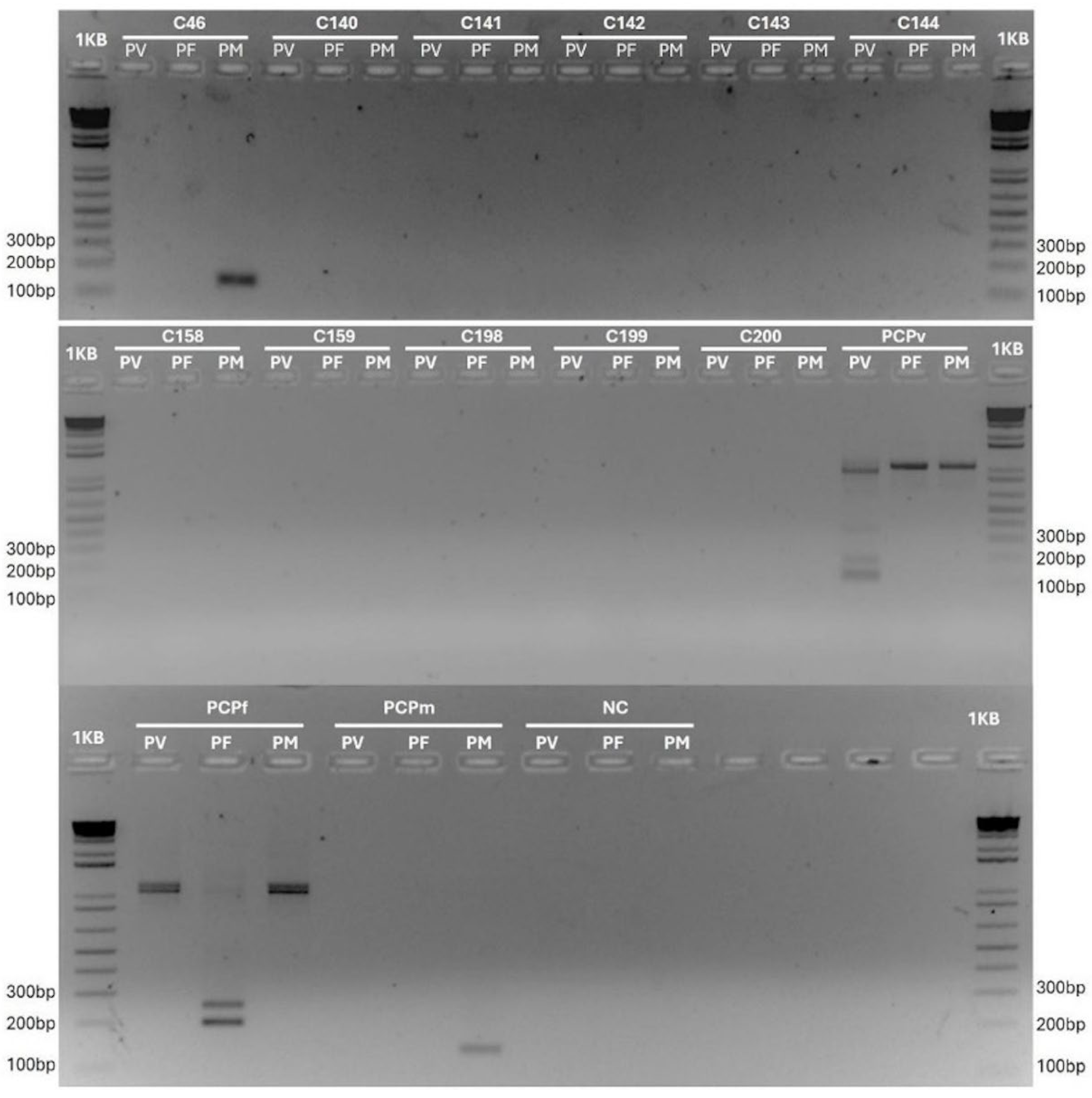
A total of 298 clot/whole blood or liver samples from Platyrrhini primates, collected across 47 municipalities in Minas Gerais (MG) and Bahia (BA), were analyzed (Table S1). The amplification of the mammalian cytochrome b gene was successful in all samples, confirming DNA integrity and suitability for molecular analysis. The molecular analysis revealed an overall Plasmodium infection rate of 0.34% (1/298; CI 95%: 0.01–0.99%) (Table 1, Figure 2). The positive sample showed the amplification of both the 18S small subunit ribosomal RNA (SSU rRNA) and cytochrome oxidase subunit I (coxI) genes from Plasmodium. All blood smears were negative for Plasmodium upon microscopic examination.
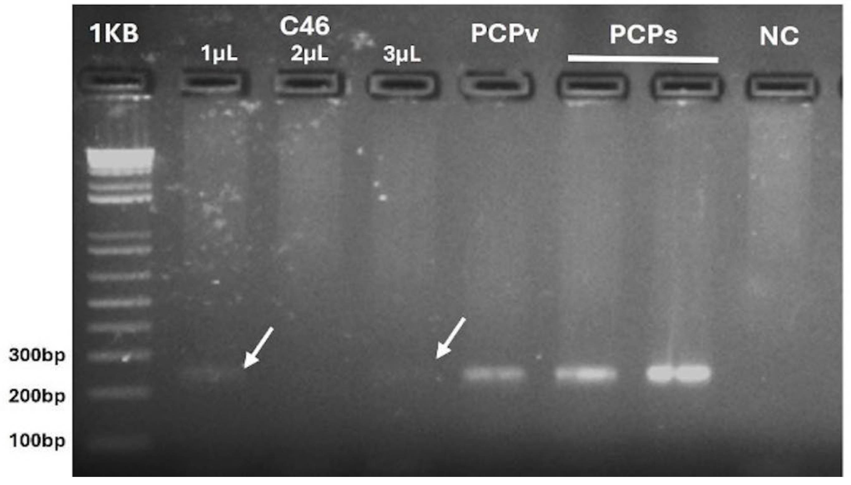
While the 18S amplicon fragment failed to produce a viable sequence, the coxI amplification product was re-amplified under the same conditions using 1, 2, or 3 µL of their initial reaction (Figure 3).
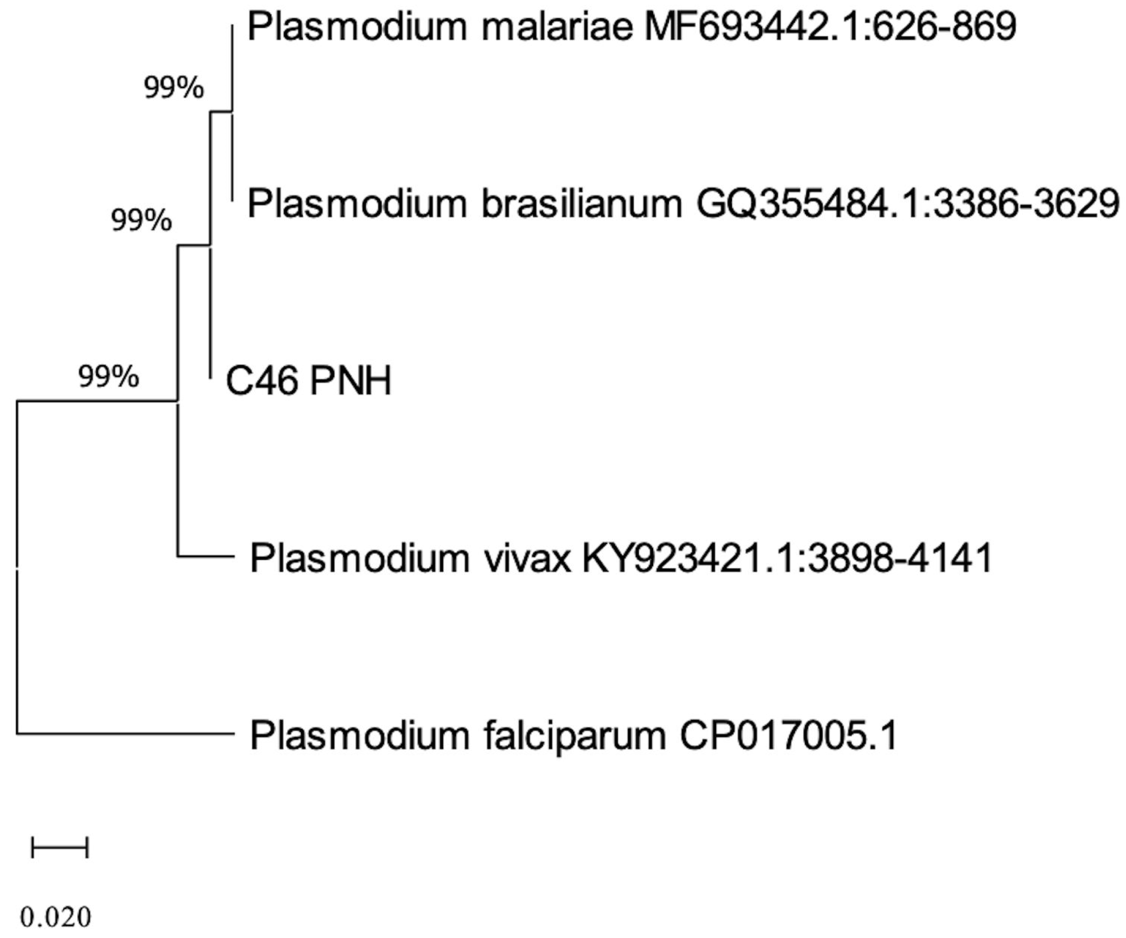
The resulting product was successfully sequenced, yielding a high-quality 244 bp sequence. This sequence exhibited 98% identity with Plasmodium malariae at sequences available in the GenBank database. Phylogenetic alignment with sequences from various Plasmodium species further supported this identification (Figure 4).
Specific PCR followed by genetic sequencing confirmed the presence of P. brasilianum/malariae. The infected animal was identified as a free-living adult male Callithrix geoffroyi, captured in a peri-urban area, surrounding an Atlantic Forest fragment, of the municipality of Porto Seguro, Bahia.

4. Discussion

This study represents the largest sampling of NHPs in Minas Gerais and Bahia states for Plasmodium infection research and provides the first report of P. malariae in free-living C. geoffroyi. The low prevalence observed may be influenced by the high number of individuals from the family Callitrichidae, as members of this family generally exhibit a low frequency of Plasmodium infections [14,15,25,26,27,28,29]. This low prevalence may be attributed to the natural resistance of Callitrichidae to Plasmodium infection [29], as well as their behavior. These primates are highly mobile and have a habit of sleeping in tree hollows from dusk to dawn, which reduces their exposure to mosquito vectors [30]. Plasmodium spp. are more frequently detected in genera such as Alouatta, Ateles, Brachyteles, Cacajao, Cebus, and Saimiri [31].
Despite this, our sampling included 26 specimens from the genus Alouatta (family Atelidae), comprising twenty-five A. caraya and one A. guariba clamitans, all of which tested negative for Plasmodium. These specimens were collected from relatively dry regions of the Cerrado or transitional areas between the Atlantic Forest and Cerrado, where bromeliads are scarce. The limited availability of bromeliads likely reduces the presence of the vector Anopheles (Kerteszia) spp., which may explain the absence of Plasmodium in these howler monkeys. Deane [25] previously reported a P. brasilianum infection rate of 10.3% (4/39) in A. caraya, but all infected individuals were collected in the Amazon-influenced states of Tocantins and Mato Grosso, where anopheline mosquitoes are more prevalent, and also, the human cases are more frequent [32]. Similarly, P. brasilianum infection rates ranging from 26% to 70% have been reported in A. guariba clamitans [25,33,34] from humid regions of the Atlantic Forest. These regions contrast with the drier collection sites in the present study, where climate and vegetation may not favor the presence of the mosquito vector and, consequently, the parasite.
In this study, Plasmodium brasilianum/P. malariae DNA was detected in a single C. geoffroyi individual—an overweight adult male in good physical condition—captured in an AF area in the district of Trancoso, municipality of Porto Seguro, southern Bahia. This represents the first detection of P. brasilianum/P. malariae DNA in C. geoffroyi in Bahia. Previously, Alvarenga et al. [27] reported the presence of P. brasilianum/P. malariae in captive C. geoffroyi in Rio de Janeiro, also in the AF biome. It is important to emphasize that great care was taken to prevent cross-contamination during all PCR amplifications. In Figure 3, the observation that the 1 µL band was more intense than the 3 µL band, while the 2 µL band was absent, may indicate the presence of inhibitors. The absence of parasites in blood smears suggests low parasitemia, as previously described for Callitrichidae [27].
Porto Seguro is located in the AF biome along the coast of Bahia, a region experiencing significant tourism and rapid urban expansion. The district of Trancoso, historically a fishing village, has undergone a recent population growth and urbanization, leading to deforestation and associated socio-environmental challenges [35]. These environmental changes have broad ecological consequences, impacting both biotic and abiotic factors within the local ecosystem. Anopheles (Kerteszia) cruzii, the primary vector of Plasmodium in the AF, thrives in areas with a high bromeliad density [36]. The presence of bromeliads in the region where Callitrichidae were captured suggests favorable conditions for vector proliferation. Furthermore, the Entomological Bulletin of the State of Bahia [37] reported Anopheles spp. in 89.4% (373/417) of the municipalities, including Porto Seguro, with the subgenus Kerteszia confirmed in the region. There was an outbreak of human malaria in this region in 2021, with fifty-eight confirmed cases of P. vivax, seven of them in Porto Seguro [38]. One infected person came from the Amazon, and the pathogen spread in the region.
These findings support the hypothesis that Plasmodium circulates in the habitat of the studied primates. Further epidemiological investigations are needed to monitor these animals and clarify the significance of this finding. The presence of infected NHPs in Porto Seguro raises concerns regarding the potential for zoonotic malaria transmission. This underscores the need for enhanced surveillance and public health measures, particularly in tourist areas, to raise awareness among local health authorities and the general public, including residents and visitors, about the potential risk of transmission.
It is still controversial whether P. brasilianum and P. malariae are genetically indistinguishable. In 2022, Bajic et al. [39] sequenced, for the first time, the complete genome of P. brasilianum. Plasmodium malariae and P. brasilianum shared 98.4% of nucleotides with a 97.3% pairwise identity. They suggested that both are strains of the same species. However, to understand the degree of similarity between P. brasilianum and P. malariae, a comprehensive analysis of whole-genome sequencing data using many more parasites from different hosts and different geographic regions is still required.
This study has some limitations that should be considered. First, the sample size, although the largest for NHPs in Minas Gerais and Bahia, may still not fully represent the diversity of NHPs in these regions, as well as the ecoregions that are not included equally, which is a sampling bias. Additionally, the detection method used relies on molecular techniques, which, while highly sensitive, may not detect very low parasitemia levels effectively. The limit of detection for the nested PCR assays used in this study, as described by Snounou et al., 1993 [22] and Alvarenga et al., 2018 [20], is estimated to be between 1 and 5 parasites/µL of blood under optimal conditions. However, this sensitivity can be affected by sample quality, presence of inhibitors, and DNA degradation, particularly in field-collected or stored samples. Therefore, the failure to obtain a sequence from the nested PCR product of the 18S rRNA gene may be attributed to the presence of inhibitors or to co-infection with multiple Plasmodium clones, which can result in overlapping signals and unreadable chromatograms. Future studies with a broader geographical scope, larger sample sizes, and a more integrated approach—including vector surveillance and serological analyses—are necessary to deepen our understanding of Plasmodium circulation in NHPs and its potential implications for human health.

5. Conclusions

Plasmodium brasilianum/P. malariae DNA was detected in a blood sample from neotropical NHPs in the state of Bahia, specifically in the AF biome, marking the first report of this parasite in Callithrix geoffroyi in the region. The relatively dry climate of the Cerrado appears to limit the transmission of Plasmodium spp. among neotropical primates in the studied area, likely due to unfavorable conditions for the vector’s survival and propagation. These findings highlight the importance of continued surveillance to better understand the epidemiology of Plasmodium in different ecosystems and its potential implications for both wildlife and human health.

Supplementary Materials

The following supporting information can be downloaded at https://www.mdpi.com/article/10.3390/pathogens14080757/s1: Table S1: Overview of Species, Biological Data, and nPCR Results for Plasmodium spp. in Neotropical Primates.

Author Contributions

Conceptualization, F.S.C., D.S.-T., G.R.A., F.V.S.A., B.M.R., P.M.R. and A.P.S.; methodology, L.K.N.S.S.S., S.M.A.-T., S.B.-V., B.S.D., M.N., A.R.C.S., D.A.M.A., L.B.S., R.S.O., I.A.C., C.H.O., M.E.G.-d.-S. and T.J.T.; software, D.A.M.A., C.F.A.B. and A.P.S.; validation, C.F.A.B. and D.A.M.A.; formal analysis, C.F.A.B., D.A.M.A. and I.A.C.; investigation, F.S.C., D.S.-T., G.R.A., F.V.S.A., C.F.A.B. and A.P.S.; resources, P.M.R. and D.S.-T.; data curation, D.A.M.A., C.F.A.B., L.K.N.S.S.S., S.M.A.-T., F.V.S.A. and G.R.A.; writing—original draft preparation, L.K.N.S.S.S., G.R.A., F.V.S.A. and C.F.A.B.; writing—review and editing, F.S.C., D.S.-T., G.R.A., F.V.S.A., S.B.-V., C.F.A.B. and A.P.S.; visualization, G.R.A.; supervision, G.R.A., F.V.S.A. and C.F.A.B.; project administration, F.S.C.; funding acquisition, P.M.R. and D.S.-T.; analysis and interpretation of data, B.S.D., M.N., A.R.C.S. and L.B.S.; analysis, drafting of the work, and reviewing of the work, R.S.O.; conception of the work, interpretation of data, C.H.O.; analysis, interpretation of data, and reviewing of the work, M.E.G.-d.-S.; analysis, interpretation of data, and reviewing of the work, T.J.T.; conception of the work, interpretation of data, and reviewing of the work, B.M.R. All authors have read and agreed to the published version of the manuscript.

Funding

This research was funded by the National Council of Scientific Development (Conselho Nacional de Desenvolvimento Científico e Tecnológico—CNPq, chamada CNPq/MS-SCTIE-Decit No 22/2019, process number 443215/2019-7 to PMR), Brazilian Office to Coordinate Improvement of Higher Education Personnel (Coordenação de Aperfeiçoamento de Pessoal de Nível Superior—CAPES, grant number: 001 to SBV), and the Santa Cruz State University (process number 073.6762.2020.0006539–71 to DST).

Institutional Review Board Statement

The animal study protocol was approved by the Biodiversity Authorization and Information System (SISBIO) of the Brazilian Institute of Environment and Natural Resources (ICMBIO) (Nos. 71714-6 and 75734-6) and approved by the Ethics Committee for the Use of Animals of the State University of Santa Cruz (CEUA/UESC) (No. 023/21) and the Ethics Committee of the Federal Institute of Northern Minas Gerais (CEUA/IFNMG) (No. 014/19).

Informed Consent Statement

No studies involving humans were conducted in this research.

Data Availability Statement

The data that support the findings of this work are available from the corresponding author upon reasonable request and in Table S1. The sequenced amplicons are available on NCBI.

Acknowledgments

We want to thank the Coordenação de Aperfeiçoamento de Pessoal de Nível Superior (CAPES) (S.B.V., A.V.V.R., R.P.L.), Conselho Nacional de Desenvolvimento Científico e Tecnológico (CNPq) (C.H.O., B.S.D, C.F.A.B.), and Fundação de Amparo à Pesquisa do Estado da Bahia (FAPESB) (L.K.N.S.S.S). This study was part of the multicentric projects: (1) “Use of genetic, mathematical and technological tools to assess the risk of transmission and strengthen surveillance of the yellow fever virus in the five Brazilian regions”, project No. 443215/2019-7, referring to Chamada 22/2019–CNPq/MS, P.M.R. (2) “Evaluation of the circulation of the Yellow Fever virus in non-human primates and vectors in the state of Bahia”, at the Universidade Estadual de Santa Cruz, UESC, No. 073.6762.2020.0006539–71, D.S.T. We would also like to extend our appreciation to Almada Mata Atlântica Project (AMAP) and BioBrasil Project along with the Center for Research and Conservation of the Royal Zoological Society of Antwerp–Belgium, for their data, capture logistics and procedures. B.M.R. (304223/2021-2), F.S.C. (309626/2021-8), G.R.A. (31263/2021-0), and P.M.R. (309024/2020-0) are CNPq research fellows.

Conflicts of Interest

The authors declare no conflicts of interest. The funders had no role in the design of this study; in the collection, analyses, or interpretation of the data; in the writing of this manuscript; or in the decision to publish the results.

Abbreviations

The following abbreviations are used in this manuscript:
AFAtlantic Forest
SISBIOBiodiversity Authorization and Information System
ICMBIOBrazilian Institute of Environment and Natural Resources
BABahia
MGMinas Gerais

References

  1. Galinski, M.R. Systems Biology of Malaria Explored with Nonhuman Primates. Malar. J. 2022, 21, 177. [Google Scholar] [CrossRef] [PubMed]
  2. Ministério da Saúde do Brasil. Guia de Vigilância Em Saúde: 6a Edição; Departamento de Ações Estratégicas de Epidemiologia e Vigilância em Saúde e Ambiente: Brasília, Brasil, 2024; Volume 3, ISBN 9786559935031. [Google Scholar]
  3. Deane, L.M.; Ferreira Neto, J.A.; Lima, M.M. The Vertical Dispersion of Anopheles (Kerteszia) cruzi in a Forest in Southern Brazil Suggests That Human Cases of Malaria of Simian Origin Might Be Expected. Mem. Inst. Oswaldo Cruz 1984, 79, 461–463. [Google Scholar] [CrossRef] [PubMed]
  4. Guimarães, A.E.; Arlé, M.; Machado, R.N.M. Mosquitos No Parque Nacional da Serra dos Órgãos, Estado Do Rio de Janeiro, Brasil. II. Distribuição Vertical. Mem. Inst. Oswaldo Cruz 1985, 80, 171–185. [Google Scholar] [CrossRef]
  5. Ashley, E.A.; Pyae Phyo, A.; Woodrow, C.J. Malaria. The Lancet 2018, 391, 1608–1621. [Google Scholar] [CrossRef] [PubMed]
  6. Davidson, G.; Chua, T.H.; Cook, A.; Speldewinde, P.; Weinstein, P. Defining the Ecological and Evolutionary Drivers of Plasmodium knowlesi Transmission Within a Multi-Scale Framework. Malar. J. 2019, 18, 18–66. [Google Scholar] [CrossRef] [PubMed]
  7. Deane, L.M.; Deane, M.P.; Ferreira Neto, J. Studies on Transmission of Simian Malaria and on a Natural Infection of Man with Plasmodium simium in Brazil. Bull. World Health Organ. 1966, 35, 805–808. [Google Scholar] [PubMed]
  8. Lalremruata, A.; Magris, M.; Vivas-Martínez, S.; Koehler, M.; Esen, M.; Kempaiah, P.; Jeyaraj, S.; Perkins, D.J.; Mordmüller, B.; Metzger, W.G. Natural Infection of Plasmodium brasilianum in Humans: Man and Monkey Share Quartan Malaria Parasites in the Venezuelan Amazon. EBioMedicine 2015, 2, 1186–1192. [Google Scholar] [CrossRef] [PubMed]
  9. Brasil, P.; Zalis, M.G.; de Pina-Costa, A.; Siqueira, A.M.; Júnior, C.B.; Silva, S.; Areas, A.L.L.; Pelajo-Machado, M.; de Alvarenga, D.A.M.; da Silva Santelli, A.C.F.; et al. Outbreak of Human Malaria Caused by Plasmodium simium in the Atlantic Forest in Rio de Janeiro: A Molecular Epidemiological Investigation. Lancet Glob. Health 2017, 5, e1038–e1046. [Google Scholar] [CrossRef] [PubMed]
  10. Fuehrer, H.P.; Campino, S.; Sutherland, C.J. The Primate Malaria Parasites Plasmodium malariae, Plasmodium brasilianum and Plasmodium ovale Genomic Insights into Distribution, Dispersal and Host Transitions. Malar. J. 2022, 21, 21–138. [Google Scholar] [CrossRef] [PubMed]
  11. Tazi, L.; Ayala, F.J. Unresolved Direction of Host Transfer of Plasmodium vivax v. P. simium and P. malariae v. P. brasilianum. Infect. Genet. Evol. 2011, 11, 209–221. [Google Scholar] [CrossRef]
  12. Lal, A.A.; De la Cruz, V.F.; Collins, W.E.; Campbell, G.H.; Procell, P.M.; McCutchan, T.F. Circumsporozoite Protein Gene from Plasmodium brasilianum. Animal Reservoirs for Human Malaria Parasites? J. Biol. Chem. 1988, 263, 5495–5498. [Google Scholar] [CrossRef] [PubMed]
  13. Guimarães, L.O.; Bajay, M.M.; Wunderlich, G.; Bueno, M.G.; Röhe, F.; Catão-Dias, J.L.; Neves, A.; Malafronte, R.S.; Curado, I.; Kirchgatter, K. The Genetic Diversity of Plasmodium malariae and Plasmodium brasilianum from Human, Simian and Mosquito Hosts in Brazil. Acta Trop. 2012, 124, 27–32. [Google Scholar] [CrossRef] [PubMed]
  14. Fandeur, T.; Volney, B.; Peneau, C.; De Thoisy, B. Monkeys of the Rainforest in French Guiana Are Natural Reservoirs for P. brasilianum/P. malariae Malaria. Parasitology 2000, 120, 11–21. [Google Scholar] [CrossRef] [PubMed]
  15. Duarte, A.M.R.d.C.; Malafronte, R.d.S.; Cerutti, C., Jr.; Curado, I.; de Paiva, B.R.; Maeda, A.Y.; Yamasaki, T.; Summa, M.E.L.; Neves, D.D.V.D.d.A.; de Oliveira, S.G.; et al. Natural Plasmodium Infections in Brazilian Wild Monkeys: Reservoirs for Human Infections? Acta Trop. 2008, 107, 179–185. [Google Scholar] [CrossRef] [PubMed]
  16. Fuentes-Ramírez, A.; Jiménez-Soto, M.; Castro, R.; Romero-Zuñiga, J.J.; Dolz, G. Molecular Detection of Plasmodium malariae/Plasmodium brasilianum in Non-Human Primates in Captivity in Costa Rica. PLoS One 2017, 12, e0170704. [Google Scholar] [CrossRef] [PubMed]
  17. Ministério da Saúde do Brasil. Guia de Vigilância de Epizootias em Primatas Não Humanos e Entomologia Aplicada à Vigilância da Febre Amarela, 2nd ed.; Ministério da Saúde do Brasil: São Paulo, Brazil, 2014; ISBN 9788533421028. [Google Scholar]
  18. Teixeira, D.S.; Bernal-Valle, S.; Ramos, A.V.V.; de Santos, L.K.N.S.S.; de Abreu, F.V.S.; dos Santos, E.; de Bandeira, J.C.; de Almeida, R.M. Capture and Collection of Biological Samples from Free-Living Neotropical Primates. Primate Conserv. 2022, 2022, 91–102. [Google Scholar]
  19. Gamble, K.C. Primates. In Exotic Animal Formulary; Carpenter, J.W., Marion, C.J., Eds.; Elsevier Saunders: Maryland Heights, MO, USA, 2023; pp. 575–615. ISBN 9780323444507. [Google Scholar]
  20. De Alvarenga, D.A.M.; Culleton, R.; De Pina-Costa, A.; Rodrigues, D.F.; Bianco, C.; Silva, S.; Nunes, A.J.D.; De Souza, J.C.; Hirano, Z.M.B.; Moreira, S.B.; et al. An Assay for the Identification of Plasmodium simium Infection for Diagnosis of Zoonotic Malaria in the Brazilian Atlantic Forest. Sci. Rep. 2018, 8, 86. [Google Scholar] [CrossRef] [PubMed]
  21. Kocher, T.D.; Thomas, W.K.; Meyer, A.; Edwards, S.V.; Paabo, S.; Villablanca, F.X.; Wilson, A.C. Dynamics of Mitochondrial DNA Evolution in Animals: Amplification and Sequencing with Conserved Primers. Proc. Natl. Acad. Sci. U S A 1989, 86, 6196–6200. [Google Scholar] [CrossRef] [PubMed]
  22. Snounou, G.; Viriyakosol, S.; Jarra, W.; Thaithong, S.; Brown, K.N. Identification of the Four Human Malaria Parasite Species in Field Samples by the Polymerase Chain Reaction and Detection of a High Prevalence of Mixed Infections. Mol. Biochem. Parasitol. 1993, 58, 283–292. [Google Scholar] [CrossRef] [PubMed]
  23. Kumar, S.; Stecher, G.; Li, M.; Knyaz, C.; Tamura, K. MEGA X: Molecular Evolutionary Genetics Analysis across Computing Platforms. Mol. Biol. Evol. 2018, 35, 1547–1549. [Google Scholar] [CrossRef] [PubMed]
  24. Tamura, K.; Nei, M. Estimation of the Number of Nucleotide Substitutions in the Control Region of Mitochondrial DNA in Humans and Chimpanzees. Mol. Biol. Evol. 1993, 10, 512–526. [Google Scholar] [CrossRef] [PubMed]
  25. Deane, L.M. Simian Malaria in Brazil. Mem. Inst. Oswaldo Cruz 1992, 87, 1–20. [Google Scholar] [CrossRef] [PubMed]
  26. Araújo, M.S.; Messias, M.R.; Figueiró, M.R.; Gil, L.H.S.; Probst, C.M.; Vidal, N.M.; Katsuragawa, T.H.; Krieger, M.A.; Pereira Da Silva, L.H.; Ozaki, L.S. Natural Plasmodium Infection in Monkeys in the State of Rondônia (Brazilian Western Amazon). Malar. J. 2013, 12, 180. [Google Scholar] [CrossRef] [PubMed]
  27. Alvarenga, D.A.M.; Pina-Costa, A.; Bianco, C.; Moreira, S.B.; Brasil, P.; Pissinatti, A.; Daniel-Ribeiro, C.T.; Brito, C.F.A. New Potential Plasmodium brasilianum Hosts: Tamarin and Marmoset Monkeys (Family Callitrichidae). Malar. J. 2017, 16, 16–71. [Google Scholar] [CrossRef] [PubMed]
  28. Lourenço-de-Oliveira, R.; Deane, L.M. Simian Malaria at Two Sites in the Brazilian Amazon. I--The Infection Rates of Plasmodium brasilianum in Non-Human Primates. Mem. Inst. Oswaldo Cruz 1995, 90, 331–339. [Google Scholar] [CrossRef] [PubMed]
  29. Aitken, E.H.; Bueno, M.G.; Dos Santos Ortolan, L.; Alvaréz, J.M.; Pissinatti, A.; Kierulff, M.C.M.; Catão-Dias, J.L.; Epiphanio, S. Survey of Plasmodium in the Golden-Headed Lion Tamarin (Leontopithecus chrysomelas) Living in Urban Atlantic Forest in Rio de Janeiro, Brazil. Malar. J. 2016, 15, 93. [Google Scholar] [CrossRef] [PubMed]
  30. Rylands, A.B. Marmosets and Tamarins: Systematics, Behavior, and Ecology.; Oxford University Press: Oxford, UK; New York, NY, USA, 1993. [Google Scholar]
  31. Kindlovits, L.M.; Kindlovits, A. Febre Amarela. Clínica e Terapêutica Em Primatas Tropicais. Livros, L.F., Ed.; 2a ed.SciELO—Scientific Electronic Library Online: Rio de Janeiro, Brazil, 2009. [Google Scholar]
  32. Ministério da Saúde do Brasil. Malária. Available online: https://www.gov.br/saude/pt-br/composicao/svsa/cnie/painel-malaria (accessed on 10 December 2024).
  33. de Abreu, F.V.S.; dos Santos, E.; Gomes, M.Q.; Vargas, W.P.; de Oliveira Passos, P.H.; Nunes e Silva, C.; Araújo, P.C.; Pires, J.R.; Romano, A.P.M.; Teixeira, D.S.; et al. Capture of Alouatta guariba clamitans for the Surveillance of Sylvatic Yellow Fever and Zoonotic Malaria: Which Is the Best Strategy in the Tropical Atlantic Forest? Am. J. Primatol. 2019, 81, e23000. [Google Scholar] [CrossRef] [PubMed]
  34. Nunes, A.J.D.; de Alvarenga, D.A.M.; de Souza Junior, J.C.; Peruchi, A.R.; Gonçalves, G.H.P.; Hirano, Z.M.B.; de Brito, C.F.A.; Cremer, M.J. Plasmodium Infection and Its Association with Biochemical and Haematological Parameters in Free-Living Alouatta guariba clamitans (Cabrera, 1940) (Primates: Atelidae) in Southern Brazil. Mem. Inst. Oswaldo Cruz 2019, 114, e190210. [Google Scholar] [CrossRef] [PubMed]
  35. Neto, S.P.G.d.C.; Da Silva, L.T. Turismo e Desenvolvimento: Transformações No Território da Região Do Extremo Sul Da Bahia. Caminhos Geogr. 2015, 16, 74–88. [Google Scholar] [CrossRef]
  36. Multini, L.C.; Wilke, A.B.B.; Marrelli, M.T. Neotropical Anopheles (Kerteszia) Mosquitoes Associated with Bromeliad-Malaria Transmission in a Changing World. Acta Trop. 2020, 205, 105413. [Google Scholar] [CrossRef] [PubMed]
  37. Secretaria da Saúde do Estado da Bahia (SESAB). Carta Anofélica da Bahia, Brasil (Diptera: Culicidae). Boletim Entomológico; Laboratório Central de Saúde Pública, Ed.; Secretaria Estadual de Saúde da Bahia: Salvador-Bahia, 2016; Volume 3. [Google Scholar]
  38. Secretaria da Saúde do Estado da Bahia. Malária—Confirmed Cases Reported in the Notification Disease Information System (According to Year 1st Symptom(s) and to Municipality of Infection). Available online: https://www.gov.br/saude/pt-br/assuntos/saude-de-a-a-z/m/malaria (accessed on 10 December 2024).
  39. Bajic, M.; Ravishankar, S.; Sheth, M.; Rowe, L.A.; Pacheco, M.A.; Patel, D.S.; Batra, D.; Loparev, V.; Olsen, C.; Escalante, A.A.; et al. The First Complete Genome of the Simian Malaria Parasite Plasmodium brasilianum. Sci. Rep. 2022, 12, 19802. [Google Scholar] [CrossRef] [PubMed]
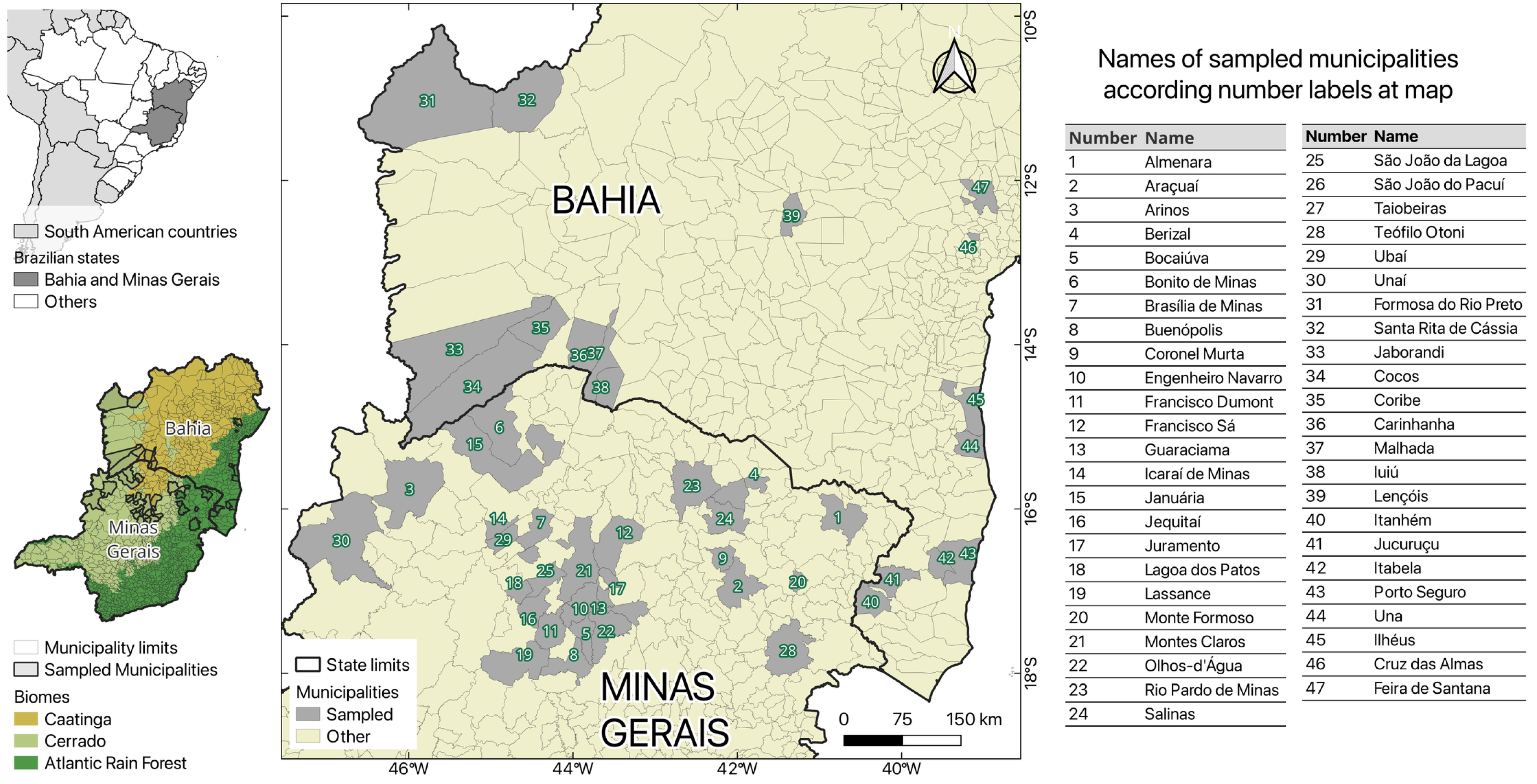
Figure 1. Map with studied areas, in Brazilian states Bahia and Minas Gerais, with their spatial relationship to the biomes.
Pathogens 14 00757 g001
Figure 2. The 2% agarose gel stained with ethidium bromide, showing the amplification of 18S SSU rRNA from a subset of NHP samples (C46, C140-C144, C158, C159, C198, to C200; see Table S1 for details) using primers for Plasmodium vivax (Pv), Plasmodium falciparum (Pf), and Plasmodium malariae (Pm). The lanes include a 1 kb Molecular Weight Standard Plus DNA Ladder (Invitrogen); positive controls for P. vivax (PCPv), P. falciparum (PCPf), and P. malariae (PCPm); as well as a negative control (NC) with no DNA.
Pathogens 14 00757 g002
Figure 3. The 2% agarose gel stained with ethidium bromide, showing the amplification of coxI [23] from the positive NHP sample (C46). Different volumes of the initial amplification product were used for the reamplification under the same conditions. The gel includes a 1 kb Plus DNA Ladder Molecular Weight Standard (Invitrogen), positive controls for Plasmodium vivax (PCPv) and Plasmodium simium (PCPs), and a negative control (NC) without DNA.
Pathogens 14 00757 g003
Figure 4. The evolutionary history was inferred by using the Maximum Likelihood method based on the Tamura–Nei model. The tree with the highest log likelihood (-498.95) is shown. Bootstrap support values (1000 replicates) are indicated next to each internal branch. Branch lengths are drawn to scale and represent the number of substitutions per site. The analysis included five nucleotide sequences and 245 aligned positions after complete deletion of gaps and missing data.
Pathogens 14 00757 g004
Table 1. Molecular detection of Plasmodium spp. in non-human primates in Bahia and Minas Gerais, Brazil.
Plasmodium PositivesPlasmodium Negatives
VariableCategoryn(%)n(%)Total
GenderFemale00.0142100.0144
Male10.715599.3156
Living conditionFree-living10.329199.7292
Captive00.06100.06
Age rangeBaby00.012100.012
Juvenile 00.065100.065
Adult10.422099.6221
SpeciesAlouatta caraya00.025100.025
Alouatta guariba clamitans00.01100.01
Callicebus melanochir00.01100.01
Callithrix spp.00.015100.015
Callithrix geoffroyi13.62796.428
Callithrix jacchus00.04100.04
Calllithrix khulii00.031100.031
Callithrix penicillata00.0175100.0175
Leontopithecus chrysomelas00.016100.016
Sapajus robustus00.01100.01
Sapajus xanthosthernos00.01100.01
Total10 species10.329799.7298
Disclaimer/Publisher’s Note: The statements, opinions and data contained in all publications are solely those of the individual author(s) and contributor(s) and not of MDPI and/or the editor(s). MDPI and/or the editor(s) disclaim responsibility for any injury to people or property resulting from any ideas, methods, instructions or products referred to in the content.