Article

Pediococcus pentosaceus MZF16 Probiotic Strain Prevents In Vitro Cytotoxic Effects of Pseudomonas aeruginosa H103 and Prolongs the Lifespan of Caenorhabditis elegans

1
CBSA UR 4312, Université de Rouen Normandie, Université de Caen Normandie, Normandie Université, F-76000 Rouen, France
2
Unité de Protéomique Fonctionnelle et Potentiel Nutraceutique de la Biodiversité de Tunisie, Institut Supérieur des Sciences Biologiques Appliquées de Tunis, Université Tunis El-Manar, Tunis 1006, Tunisia
*
Author to whom correspondence should be addressed.
Pathogens 2025, 14(3), 244; https://doi.org/10.3390/pathogens14030244
Submission received: 7 January 2025 / Revised: 21 February 2025 / Accepted: 24 February 2025 / Published: 3 March 2025

Abstract

:
Pseudomonas aeruginosa is an opportunistic pathogenic bacterium, responsible for several life-threatening infections due to its multiple virulence factors and problematic multi-drug resistance, hence the necessity to find alternatives such as competitive probiotics. Pediococcus pentosaceus MZF16 is an LAB strain, isolated from traditional dried meat “Ossban”, with high probiotic potential. Our study investigated the capacity of P. pentosaceus MZF16 to counteract P. aeruginosa H103 using several tests on intestinal cells (analysis of cytotoxicity, inflammation, adhesion/invasion) and on the in vivo Caenorhabditis elegans model. The effect of MZF16 on the quorum sensing of the pathogen was also examined. We found that P. pentosaceus MZF16 was able to reduce H103 cytotoxicity and inflammatory activity and prevented pathogen colonization and translocation across Caco-2/TC7 cells. MZF16 also exerted an anti-virulence effect by attenuating quorum-sensing (QS) molecules and pyoverdine production and extended C. elegans lifespan. The obtained results highlight the potential of P. pentosaceus MZF16 probiotic strain as an anti-Pseudomonas aeruginosa alternative and establish a basis for elucidating the mechanisms of P. pentosaceus MZF16 involved in countering P. aeruginosa virulence.

1. Introduction

Fermented foods are produced through controlled microbial growth and enzymatic conversion of food components. Historically, food fermentation was a common preservation method, used to maintain food integrity and reduce the risk of contamination with pathogenic microorganisms, likely due to the production of antimicrobial metabolites such as organic acids, ethanol and bacteriocins [1]. In recent years, fermented foods have surged in popularity, because of their health-promoting properties, especially for gastrointestinal health. These benefits are attributed to various mechanisms, including bioactive peptides and phenolic compound conversion to biologically active compounds, as well as the beneficial effects of constituent microorganisms [2]. Fermented foods such as dairy products, fermented meats and vegetables contain relatively resilient and stable microbial ecosystems, predominantly composed of lactic acid bacteria (LAB) and their primary metabolites including lactic acid [3]. Some LAB strains have long been recognized as probiotics due to their historical use and classification as beneficial bacteria with Generally Recognized As Safe (GRAS) status. Emerging probiotics are also being studied to better understand their potential health benefits. Among GRAS probiotics, the most well-known and studied bacteria belong to the Lactobacilli, recently reclassified into 23 genera [4] and various species including, for example, Lacticaseibacillus casei, Lactobacillus acidophilus, Lactobacillus johnsonii, and Lactiplantibacillus plantarum. These bacteria have demonstrated various health benefits, anti-carcinogenic, anti-diabetic, antioxidant, and anti-inflammatory potential, and also antimicrobial effects against food-borne pathogens [5].
Despite their frequent presence in many fermented products (i.e., meat, fish, dairy, beer, wine, fruits, vegetables, and beverages) [6], probiotics from the genus Pediococcus have been less studied. This genus consists of several species, including P. acidilactici, P. pentosaceus, P. damnosus, P. parvulus, P. inopinatus, P. halophilus, P. dextrinicus, and P. urinaeequi [7]. Among these, P. acidilactici and P. pentosaceus probiotic strains are notable for their high production of bacteriocins (pediocins) and bacteriocin-like substances (BLSs) [8]. Indeed, pediocins and other postbiotic metabolites (e.g., lactic acid, biosurfactants) from some Pediococcus species have exerted significant antibacterial and anti-virulence activities against several food-borne/food spoilage pathogens, such as Listeria monocytogenes and Salmonella enterica and also against the multi-drug-resistant (MDR) pathogen Pseudomonas aeruginosa [9,10,11,12,13,14].
P. aeruginosa is known to be a potential cause of the spoilage of various foods, notably those with high water content and nutrient-rich foods, such as milk, meats, and some fruits and vegetables [15,16]. It is also an opportunistic Gram-negative pathogen, widespread in the environment and responsible for healthcare-associated infections and food-borne diseases [17]. In some cases, this germ has been associated with diarrheal disease, systemic infections, enterocolitis and digestive cancers [18,19]. Its ability to cause severe acute and chronic life-threatening infections is attributed to its multiple virulence factors [20]. Moreover, the high versatility and adaptability of this pathogen are supported by its large genome (6 to 8 Mb), with over 8% dedicated to regulatory genes, enabling it to evade a broad spectrum of antimicrobial agents [21]. Thereby, alternative treatments, such as competitive probiotics, are urgently needed [22].
Some studies have explored the effects of postbiotic LAB metabolites, such as lactic acid, bacteriocins and BLSs produced by various Pediococcus species (P. pentosaceus and P. acidilactici, etc.) on P. aeruginosa [11,12,14]. However, interactions between these two species and also with the human host remain poorly characterized.
The P. pentosaceus MZF16 strain has been previously isolated in our laboratory from the artisanal Tunisian meat, “Dried Ossban”, and investigated for its probiotic potential [23,24]. The aim of the present study was to examine the interactions between P. pentosaceus MZF16 and the pathogen P. aeruginosa H103, with a particular focus on the MZF16 strain’s ability to modulate the virulence of the pathogen and its interactions with the host.

2. Materials and Methods

2.1. Bacterial Strains and Culture Conditions

The bacterial strains used in this study were the pathogen P. aeruginosa H103, a prototroph derivative of PAO1 strain [25], and the potential probiotic P. pentosaceus MZF16 [26]. They were, respectively, grown at 37 °C under rotary shaking (180 rpm) in Luria–Bertani (LB) medium and in static conditions in Man Rogosa Sharpe (MRS) medium.

2.2. P. aeruginosa H103-gfp and P. pentosaceus MZF16-mCherry Construction

P. aeruginosa H103 was grown in LB medium until reaching an OD600 nm of 0.5, then centrifuged at 8000× g for 10 min at 4 °C. The pellet was washed and resuspended in 300 mM sucrose buffer at a concentration of 1010 CFU/mL. A 100 µL aliquot of electrocompetent P. aeruginosa was mixed with 50 ng of plasmid pHC60 (gfp, tetR) [27] and electroporated at 1.8 kV in a 0.2 cm cuvette. Immediately after electroporation, 1 mL of LB medium was added, and the mixture was incubated for 2 h at 37 °C with shaking at 180 rpm P. aeruginosa H103-gfp transformants were selected on LB agar supplemented with Tetracycline (100 µg/mL).
For P. pentosaceus MZF16-mCherry construction, electrotransformation was performed using plasmid pRCR12 (mCherry, CmR), provided by Pr Paloma Lopez (Madrid, Spain), and following the method of Pérez-Ramos et al. (2018) [28]. Briefly, the bacterial pellet was washed and resuspended in 1 mL of lysozyme (2000 U/ mL) and incubated for 20 min at 37 °C. Subsequently, 50 μL of electrocompetent cells was mixed with 5 μL of pRCR12 plasmid (0.5 μg) and electroporated at 2.5 kV in a 0.2 cm cuvette. After electroporation, 500 µL of MRS medium was immediately added, and the cells were incubated for 2 h at 37 °C. P. pentosaceus MZF16-mCherry transformants were selected on MRS-agar supplemented with Chloramphenicol (10 μg/mL).
All bacterial strains used in this study are listed below (Table 1).

2.3. Caco-2/TC7 Cell Culture and Infection

Human colon adenocarcinoma Caco-2/TC7 cells provided by Chantret et al. (1994) [29] were cultured in Dulbecco’s Modified Eagle Medium (DMEM, Lonza, Basel, Switzerland) supplemented with 20% heat-inactivated fetal bovine serum (FBS, Sigma Aldrich) and penicillin/streptomycin (100 µg/mL). The cells were cultivated and incubated at 37 °C in 5% CO2 atmosphere, and the medium was regularly changed. Cells were passed after reaching approximately 90% of confluence [30].
For adhesion, invasion, cytotoxicity assays and IL-8 quantification, cells were seeded at a concentration of 1.105/mL, approximatively, in 24-well plates treated for tissue culture and grown to confluence. For bacterial translocation assay and transepithelial resistance (TER) measurements, cells were seeded on inserts (6.4 mm diameter, 3 μm pore size, Falcon) until full differentiation (19–21 days).
Bacterial suspensions of P. aeruginosa H103 and its mixture with P. pentosaceus MZF16 (1:100) were prepared in DMEM without FBS and antibiotics. These suspensions were added to confluent or differentiated Caco-2/TC7 cells and incubated at 37 °C in 5% CO2 for 3 h for adhesion and invasion assays, and overnight for the other experiments.

2.4. Cytotoxicity Assay and Interleukin-8 Quantification

Cytotoxicity and inflammation assays were performed on confluent Caco-2/TC7 cells grown in 24-well culture plates. Following overnight infection with bacterial suspensions, the supernatants were collected from Caco-2/TC7 monolayers [31]. Then, cytotoxicity was assessed by measuring lactate dehydrogenase (LDH), a cytoplasmic enzyme released upon cell death, using an enzymatic assay (Cyquant Kit, Invitrogen, Waltham, MA, USA). The amount of IL-8 cytokine in the supernatants was quantified using the IL-8 Human ELISA Kit (Invitrogen), a solid-phase sandwich Enzyme-Linked Immunosorbent (ELISA) Assay. The concentration of IL-8 (pg/mL) was defined using a correlation of a standard curve and the optical density OD450 nm.

2.5. In Vitro Adhesion and Invasion Assay

Bacterial adhesion and invasion into intestinal Caco-2/TC7 cells were assessed following Xu et al.’s method [32], with slight modifications. For the adhesion assay, Caco-2/TC7 cells were infected with suspensions of P. aeruginosa H103 and mixture with P. pentosaceus MZF16 (1:100 ratio) for 3 h. After incubation, cells were washed twice with phosphate-buffer saline (PBS) to remove non-associated bacteria and then lysed with 0.1% Triton X100. The lysates were serially diluted and plated on Cetrimide agar to quantify adherent pathogenic bacteria.
For the invasion assay, after 3 h of infection, adherent and non-internalized bacteria were eliminated by treating the cells with 300 μg/mL of gentamicin for 1 h. Following treatment, the cells were lysed, and internalized bacteria were quantified by plating.
For the visualization of bacterial adhesion, Caco-2/TC7 cells were seeded on glass-bottom 24-well plates (Sensoplate, Greiner) and infected with fluorescent strains P. aeruginosa H103-gfp and P. pentosaceus MZF16-mCherry for 3 h. Afterward, Caco-2/TC7 cells were stained with DAPI (4′,6-diamidino-2-phenylindole) and visualized using confocal laser scanning microscopy (CLSM). GFP, mCherry and DAPI were excited at 488 nm, 543 nm and 405 nm, respectively, and fluorescence emissions were detected between 484 and 549 nm, 539 and 599 nm, and 404 and 478 nm.

2.6. Bacterial Translocation and Transepithelial Electrical Resistance (TER) Measurement

After an overnight infection of Caco-2/TC7 differentiated cells cultured on inserts, aliquots from the basolateral compartment were collected [33]. The number of translocated P. aeruginosa that crossed the epithelial monolayers was determined by serial dilution and plating on Cetrimide agar.
To assess the integrity of the epithelial barrier, the transepithelial electrical resistance (TER) of infected and non-infected monolayers was measured using the Millicell Electrical Resistance System (Millipore Corp, Bedford, MA, USA).

2.7. Antimicrobial Activity Test

The antagonistic activity of P. pentosaceus MZF16 against P. aeruginosa H103 was assessed using the disk diffusion method. Overnight cultures of both strains were centrifuged at 7000 g for 5 min. Supernatants of P. pentosaceus MZF16 were either pH-adjusted (pH = 7) or not before being filtered (0.22 µm membrane).
A suspension of P. aeruginosa H103 at a concentration of 106 CFU/mL was spread on Muller Hinton Agar plate. Disks impregnated with cell-free supernatants (CFSs) of MZF16 (10 µL) were placed on the agar surface, and the plates were incubated at 37 °C for 24 h.

2.8. Bacterial Aggregation Assay

Auto- and coaggregation assays were performed as described previously [34] with minor modifications. Briefly, overnight cultures of H103 and MZF16 were centrifuged at 10,000× g for 10 min. The pellets were washed twice and resuspended in sterile phosphate-buffer saline (PBS). The bacterial suspensions were adjusted to an OD600 nm of approximately 0.3.
For the coaggregation assay, equal volumes of the standardized bacterial suspensions were mixed. Both individual and mixed suspensions were aliquoted into hemolysis tubes (4 mL) and incubated at 37 °C without agitation.
The OD600 nm of the suspensions was measured at the start of the experiment (A0) and after 3 h of incubation at 37 °C (At=3h). The autoaggregation percentage was calculated as follows:
A u t o a g g r e g a t i o n   % = 1 A t = 3 h A 0   ×   100
The coaggregation percentage was determined using the following formula:
C o a g g r e g a t i o n   % = A H 103 + M Z F 16 ( A m i x ) ( A H 103 + M Z F 16 )   ×   100
where AH103 and AMZF16 are the OD600 nm values of the mixed bacterial suspension at time 0, and Amix is the OD600 nm of the mixed suspension at t = 3 h.

2.9. Pyoverdine Production Measurement

Pyoverdine is a key siderophore and virulence factor of P. aeruginosa. Its production was quantified by spectrophotometry at A405 nm [35]. Bacterial suspensions of P. aeruginosa H103 and the mix with the probiotic P. pentosaceus MZF16 were grown in colorless DMEM medium supplemented with 20% fetal bovine serum (FBS). After incubation, pyoverdine production was measured from collected supernatants. Results are expressed as the relative pyoverdine levels (mean ± SEM) compared to the control, P. aeruginosa H103 culture.

2.10. Extraction and Quantification of AHL and HAQ Molecules

N-acyl-homoserine lactones (AHLs) and 2-alkyl-4-quinolones (HAQs, PQS system) signaling molecules were extracted from P. aeruginosa H103 cultures and cocultures with P. pentosaceus MZF16 (1:100), grown in DMEM medium, following the protocol described by Fletcher et al. (2007) [36]. For quantification, Escherichia coli-carrying plasmid pSB401 (luxRI′::luxCDABE) and P. aeruginosa PAO1 ΔpqsA CTX-lux::pqsA strain were used as biosensors for AHLs and HAQs, respectively. The assay was performed using a combined spectrophotometer/luminometer approach. Briefly, overnight cultures of the biosensor strains were adjusted to an OD580 nm = 1 and then diluted to 1:50 and 1:100 ratios. Crude extracts of quorum-sensing (QS) molecules were diluted in LB medium and added to a 1:50 dilution of the biosensors. The 3-oxo-C12-HSL, C4-HSL, HHQ, and PQS synthetic standards (Sigma-Aldrich, Saint-Louis, MO, USA) at final concentrations of 5 μM were added to a 1:100 dilution of the biosensor strains as positive controls.
The assay was performed in 96-well microtiter plates (white-sided, clear-bottom). Bioluminescence (relative light units, RLU) and absorbance (A580 nm) were measured every 15 min for 24 h at 37 °C using a Spark 20 M multimode microplate reader (Tecan, Männedorf, Switzerland). The bioluminescence data were normalized to the A580 nm values.

2.11. In Vivo Caenorhabditis elegans Killing Assay

The C. elegans slow killing assay was performed as previously described by Tan et al. [37]. C. elegans wild-type Bristol strain N2 worms were grown at 22 °C on nematode growth medium (NGM) agar plates using Escherichia coli OP50 strain as the nutrient. P. aeruginosa culture and its coculture with P. pentosaceus MZF16 were spread onto 35 mm NGM solidified agar plates before an overnight incubation at 37°. The plates were cooled to room temperature for 4 h. Then, 20 to 30 L4-synchronized worms prepared as described by Blier et al. [38] were plated and incubated at 22 °C in a humid environment to prevent plate drying. Worm survival was scored daily for 16 days using an optical microscope (Leica CME, Glattbrugg, Switzerland). The worms were considered dead when they remained static without grinder movements for 20 s or did not respond to light flashes. Results are expressed as a percentage of living worms. The results are the average of at least three independent assays. The nematode survival was calculated using the Kaplan–Meier method, and survival differences were tested for significance using the log–rank test (GraphPad Prism version 4.0, Dotmatics, San Diego, CA, USA).

2.12. Statistical Analysis

Statistical analysis of the data was performed using GraphPad Prism software version 10.2.2 with two-sample unpaired tests. All tests were conducted at least three times, and t-tests were used for all data analysis; statistical significance was considered at p < 0.05, p < 0.01 and p < 0.001.

3. Results

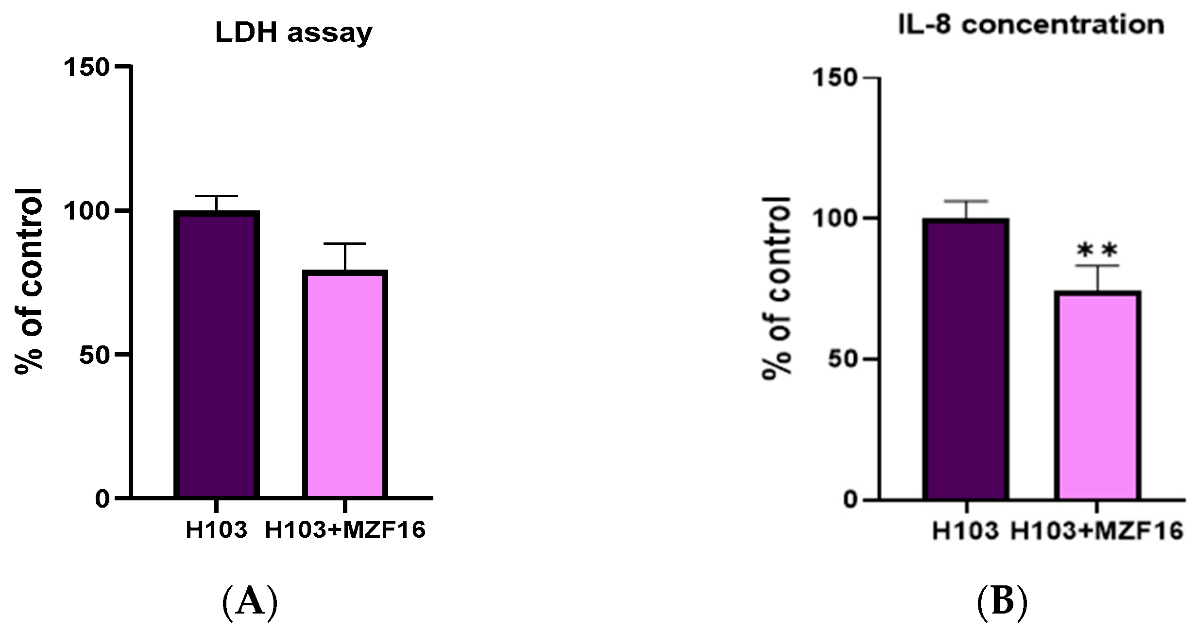
3.1. Cytotoxicity Assay and Interleukin-8 Quantification

The cytotoxicity and pro-inflammatory activity of P. aeruginosa H103 towards the intestinal Caco-2/TC7 cells were evaluated after overnight incubation in the presence or absence of P. pentosaceus MZF16. The results demonstrated that MZF16 slightly decreased the level of lactate dehydrogenase (LDH) produced by the eukaryotic cells in response to the cytotoxic effect of the pathogen, with a reduction of about 20% (Figure 1A). In addition, MZF16 significantly reduced the level of IL-8 by more than 25% (Figure 1B).

3.2. Adhesion and Invasion

The competitive exclusion of pathogen adhesion and invasion to Caco-2 cells by P. pentosaceus MZF16 was evaluated over 3 h by spreading cell lysates on Cetrimide agar. The simultaneous addition of P. pentosaceus MZF16 with P. aeruginosa H103 to intestinal Caco-2/TC7 cells resulted in a decrease of 39 ± 8% in the number of adhering P. aeruginosa H103 (Figure 2A), and a reduction of 85 ± 4% in invasive bacteria number (Figure 2B). Visualization of bacterial adhesion to Caco-2 cells using confocal laser scanning microscopy (CLSM) showed that MZF16-mCherry formed a layer on the cells. In contrast, H103-gfp appeared more scattered in the different stacks, likely due to its mobility and invasive ability (Figure 2C).

3.3. Transepithelial Electric Resistance (TER) and Translocation

The transepithelial resistance (TER) and the bacterial translocation of P. aeruginosa H103 were evaluated on differentiated Caco-2/TC7 cells after an overnight infection. Results showed that P. pentosaceus MZF16 prevented the reduction in TER provoked by P. aeruginosa H103, which was about 30% (Figure 3A). Additionally, P. pentosaceus MZF16 significantly reduced the translocation of the pathogen across the Caco-2/TC7 cells by three times (Figure 3B).

3.4. Autoaggregation and Coaggregation

The autoaggregation and coaggregation abilities of P. pentosaceus MZF16 and P. aeruginosa H103 were measured in PBS after 3 h incubation at 37 °C. Both strains H103 and MZF16 demonstrated a high capacity of autoaggregation of about 43.8 ± 7.8 and 44.7% ± 2.6, respectively. Similarly, their coaggregation ability was about 45.7% ± 3.6.

3.5. Antimicrobial Activity Test

The cell-free supernatant of P. pentosaceus MZF16 exhibited no antibacterial activity against P. aeruginosa H103 in our conditions, suggesting the absence of an anti-Pseudomonas bacteriocin- or bacteriocin-like metabolite in our strain.

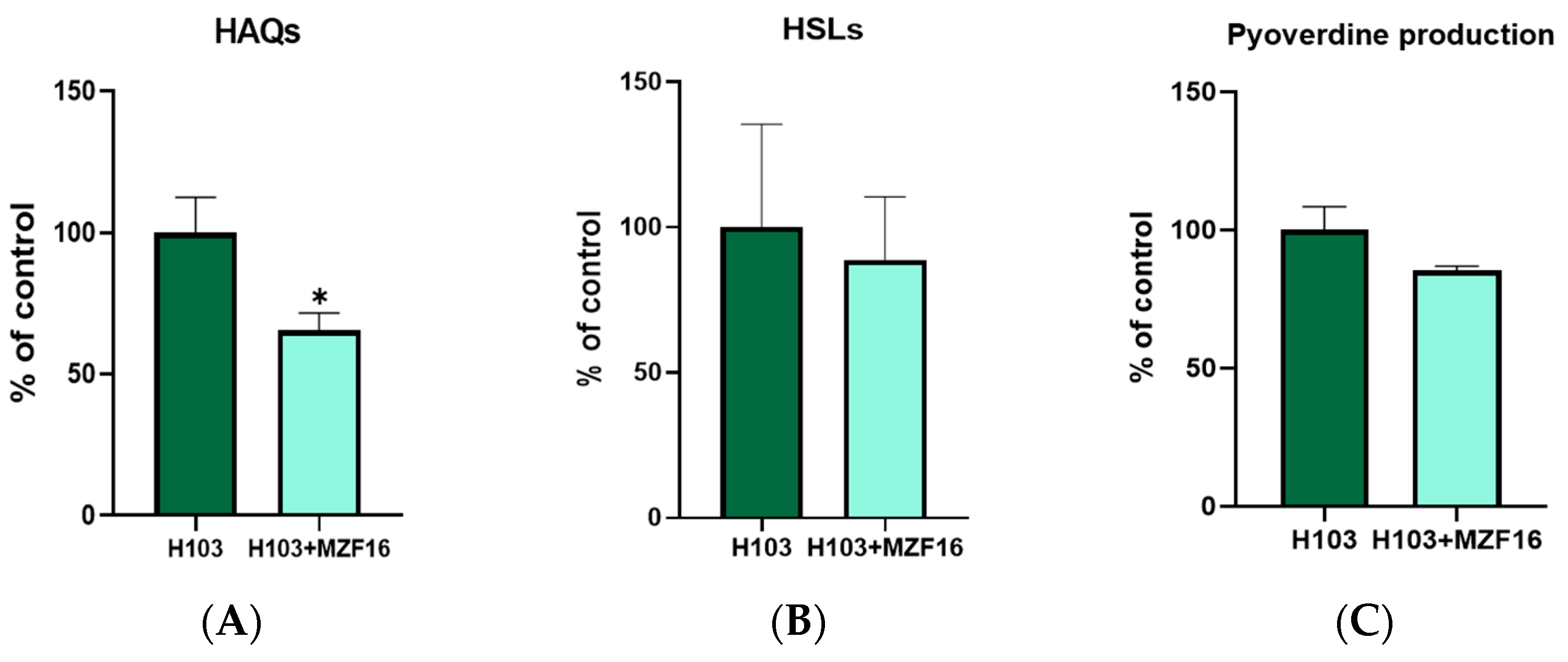
3.6. Quorum Sensing and Pyoverdine Production

N-acyl-homoserine lactones (AHLs) production was assessed using the biosensor strain E. coli harboring the plasmid pSB401 (luxRI′::luxCDABE), able to detect short (C4) and long (C12) HSL chains specifically produced by P. aeruginosa. The production of 2-alkyl-4-quinolones (HAQs) signaling molecules (HHQ and PQS) was assessed using the P. aeruginosa PAO1 ΔpqsA CTX-lux::pqsA biosensor strain, which detects HAQ derivatives produced by P. aeruginosa. Both molecules’ production was examined in the presence and absence of the MZF16 strain. Results showed that P. pentosaceus MZF16 significantly reduced (of about 35%) the production of HAQ molecules by P. aeruginosa H103 (Figure 4A), but had no effect on HSL (Figure 4B) and pyoverdine production (Figure 4C).

3.7. In Vivo Virulence Test on C. elegans

A slow killing assay was monitored over 16 days (Figure 5). The results showed that in the presence of P. pentosaceus MZF16, the lifespan of C. elegans was significantly prolonged compared to when the worms were exposed only to the pathogen P. aeruginosa H103. Specifically, on day 8 of the test, only 10% of the worms infected by H103 survived, while in the presence of P. pentosaceus MZF16, the survival rate of C. elegans infected by H103 was about 40%.

4. Discussion

P. aeruginosa is a major opportunistic pathogen responsible for both acute and chronic infections, especially in immunocompromised individuals [39]. The rise in multi-drug-resistant P. aeruginosa strains has led to an urgent need for alternative treatments to antibiotics, such as probiotics.
P. pentosaceus MZF16 strain, isolated from Tunisian dried meat (Ossban), exhibits probiotic properties, including resistance to the harsh gastrointestinal conditions and the high ability of adhesion to intestinal cells, without any risks for human health [23]. These traits underscore its potential in fighting pathogens.
In the present work, we evaluated the capacity of P. pentosaceus MZF16 to compete with P. aeruginosa H103 and reduce its virulence. For this, undifferentiated intestinal Caco-2/TC7 cells were treated simultaneously with both bacteria and the cytotoxicity, inflammatory potential, and adhesion/invasion of H103 have been investigated. Our study demonstrated that P. pentosaceus MZF16 can mitigate P. aeruginosa H103-induced cytotoxicity and inflammation in intestinal Caco-2/TC7 cells. Specifically, MZF16 reduced both LDH release (necrosis marker) and IL-8 secretion. These findings align with previous research showing similar protective effects of probiotics against various pathogens. For example, P. acidilactici SPM138 increased the human intestinal epithelial cells HT-29 cells’ survival rate from 20% to 98% and reduced by more than 60% IL-8 levels released upon Clostridium difficile infection [40]. Similarly, P. acidilactici TMAB26 demonstrated immunomodulatory effects by alleviating Klebsiella pneumoniae endotoxin-induced gut inflammation [12].
P. pentosaceus MZF16 also reduced the adhesion, invasion and translocation of P. aeruginosa H103 across undifferentiated/differentiated Caco-2/TC7 cells, preserving epithelial barrier integrity as evidenced by TEER measurements. Similar anti-adherence effects have been reported for P. acidilactici IAH-5 against S. Typhimurium [41]. In another study, P. pentosaceus L1 isolated from pickled radish also enabled the decrease in adhesion of an E. coli enterotoxigenic strain to porcine intestinal epithelial cells [42]. Likewise, P. pentosaceus 2–5 strain, isolated from chicken intestine, showed a strong ability to inhibit the adhesion of two enteropathogenic bacteria E. coli ATCC 25922 and S. Typhimurium ATCC 13311 to the epithelial Caco-2 cells, with the three anti-adhesion tested methods (competition, exclusion, substitution) [43].
The observed reduction in adhesion/invasion and cytotoxicity may result from direct probiotic–pathogen interaction or a postbiotic effect mediated by secreted factors. In fact, autoaggregation is stated to be a prerequisite for probiotic bacteria adhesion to intestinal epithelium, whereas their coaggregation abilities with pathogens enable forming an effective barrier, preventing epithelium colonization by harmful bacteria. Our strain P. pentosaceus MZF16 exhibited high autoaggregation (44.7%) and coaggregation abilities with H103 (45.7%), supporting its role in forming a protective barrier. This coaggregation capacity aligns with previous findings for P. pentosaceus DHR005 which showed lower autoaggregation (28.6%) and coaggregation (22%) against Salmonella Typhimurium PTCC1609 [41]. In a recent study, moderate to strong coaggregation abilities of two strains of P. pentosaceus H11 and H13, isolated from fermented bean curd, have been demonstrated with P. aeruginosa PAO1 [44], supporting our finding.
Since P. pentosaceus MZF16 is able to produce lactic acid and coagulin bacteriocin [23], we investigated if the protective effect of the probiotic was related to one of these aspects. Interestingly, despite the ability of P. pentosaceus MZF16 to produce antibacterial metabolites, no direct antimicrobial activity was detected against P. aeruginosa H103 under our experimental conditions. In contrast, Li et al. (2023) demonstrated that two strains P. pentosaceus H11 and H13 exhibited a different antibacterial effect on P. aeruginosa, suggesting that this antimicrobial ability was strain-specific [44]. In another studies, bacteriocins from other Pediococcus species; P. acidilactici and P. inopinatus strains exerted important antimicrobial activities against P. aeruginosa [10,14].
This lack of activity may be due to culture conditions, as bacteriocin production can vary based on time of exposition and media composition, notably the pH. Indeed, a comparative study has shown that no antibacterial activity against P. aeruginosa has been detected from pH-adjusted CFS of P. pentosaceus ENM104 comparing to the crude CFS, indicating that the production of antibacterial organic acids or bacteriocins may be active only under acidic conditions [45].
Nonetheless, we have demonstrated by spreading DMEM cocultured strains, that MZF16 does not inhibit H103 growth (Table S1). The ability of MZF16 to coexist with H103 suggests that in our experimental conditions, the protective effect of MZF16 was not directly linked to the production of lactic acid and/or the bacteriocin, but it may be related to a competitive mechanism for nutrients or epithelial binding sites [46].
Furthermore, other studies have shown that some Pediococcus species are able to produce exopolysaccharides and biosurfactants with a high ability to disrupt P. aeruginosa biofilms and display antimicrobial, anti-adhesive and anti-virulence properties [11,28].
The pathogenicity of P. aeruginosa involves the production of several virulence factors [47] such as pyoverdine and pyocyanin, regulated by the cell density-dependent signaling mechanism «quorum sensing (QS)» [48]. In P. aeruginosa, two main QS systems are defined, the system mediated by N-acylhomoserine lactones (AHLs) known as Las and Rhl, which are responsible for the production of homoserine lactone signal molecules, and the Pqs system depending on 2-alkyl-4-quinolones (HAQ) and which is interlinked with the Las and Rhl systems. These systems are involved in the pathogenesis of P. aeruginosa and the synthesis of some virulence factors such as rhamnolipid, pyocyanin, pyoverdine and the production of enzymes like proteases and lipases and biofilm formation [49,50].
The inhibition of this signalization can attenuate QS-related bacterial virulence [51,52]. Thus, anti-QS may be a promising strategy to fight against some pathogens including P. aeruginosa. In this context, we aimed to evaluate the effect of MZF16 on P. aeruginosa H103 QS. Interestingly, we found that the production of HSL molecules and PQS level were notably reduced in the presence of MZF16. Pyoverdine is a siderophore and an important virulence factor in P. aeruginosa, enabling iron sequestration from the host to improve the bacterial establishment during the infection. Our results showed that P. pentosaceus MZF16 decreased the pyoverdine level released in the coculture media, suggesting that P. pentosaceus MZF16 may be able to modulate the QS of P. aeruginosa H103. In agreement with our findings, P. acidilactici M7 have shown a quorum quenching activity, inhibiting short-chain HSL production, motility, and virulence factor production (elastase, protease, pyocyanin, and biofilm production) of P. aeruginosa isolates [53]. Another study on P. acidilactici HW01 bacteriocin showed a decrease in the production of virulence factors such as QS-regulated pyocyanin, protease, and rhamnolipid [10].
Due to the high homology of genes between nematodes and mammals and because nematodes have a relatively complete digestive system, C. elegans has been widely used as a host–pathogen interaction model in scientific research. In our study, the slow killing assay on C. elegans confirmed the anti-virulence effect of P. pentosaceus MZF16 against P. aeruginosa H103. Indeed, the presence of MZF16 has significantly enhanced the survival of C. elegans infected with P. aeruginosa H103. This result is consistent with studies showing that probiotics like P. acidilatici DM9 and P. pentosaceus SDL1409 remarkably extended C. elegans lifespan during P. aeruginosa PA14 infection [9].

5. Conclusions

This study highlights the potential of Pediococcus pentosaceus MZF16 as an effective probiotic against Pseudomonas aeruginosa H103. MZF16 is able to mitigate in vivo and in vitro cytotoxicity, decreases inflammation levels and pathogen adhesion and invasion in intestinal models, preserves epithelial cell integrity, and modulates quorum-sensing pathways, reducing virulence factor production. These findings underscore the potential of using probiotics as adjunct therapies to combat multi-drug-resistant pathogens. Further research is needed to decipher molecular mechanisms involved in these bacterial interactions.

Supplementary Materials

The following supporting information can be downloaded at: https://www.mdpi.com/article/10.3390/pathogens14030244/s1, Table S1: Coexistence of P. aeruginosa in coculture with P. pentosaceus MZF16.

Author Contributions

Conceptualization, N.C.; methodology, M.B., M.Z., M.F., O.L., A.T. and A.M.B.; validation, A.T. and N.C.; writing—original draft preparation, M.Z. and M.B.; writing—review and editing, M.B., O.L., A.T., A.M.B. and N.C.; supervision, N.C.; project administration, N.C. All authors have read and agreed to the published version of the manuscript.

Funding

This research received no external funding.

Institutional Review Board Statement

Not applicable.

Informed Consent Statement

Not applicable.

Data Availability Statement

Data are contained within the article.

Conflicts of Interest

The authors declare no conflicts of interest.

References

  1. Dimidi, E.; Cox, S.R.; Rossi, M.; Whelan, K. Fermented Foods: Definitions and Characteristics, Impact on the Gut Microbiota and Effects on Gastrointestinal Health and Disease. Nutrients 2019, 11, 1806. [Google Scholar] [CrossRef]
  2. Marco, M.L.; Heeney, D.; Binda, S.; Cifelli, C.J.; Cotter, P.D.; Foligné, B.; Gänzle, M.; Kort, R.; Pasin, G.; Pihlanto, A.; et al. Health Benefits of Fermented Foods: Microbiota and Beyond. Curr. Opin. Biotechnol. 2017, 44, 94–102. [Google Scholar] [CrossRef]
  3. Stiemsma, L.T.; Nakamura, R.E.; Nguyen, J.G.; Michels, K.B. Does Consumption of Fermented Foods Modify the Human Gut Microbiota? J. Nutr. 2020, 150, 1680–1692. [Google Scholar] [CrossRef]
  4. Zheng, J.; Wittouck, S.; Salvetti, E.; Franz, C.M.A.P.; Harris, H.M.B.; Mattarelli, P.; O’Toole, P.W.; Pot, B.; Vandamme, P.; Walter, J.; et al. A Taxonomic Note on the Genus Lactobacillus: Description of 23 Novel Genera, Emended Description of the Genus Lactobacillus Beijerinck 1901, and Union of Lactobacillaceae and Leuconostocaceae. Int. J. Syst. Evol. Microbiol. 2020, 70, 2782–2858. [Google Scholar] [CrossRef]
  5. Díaz, M.A.; González, S.N.; Alberto, M.R.; Arena, M.E. Human Probiotic Bacteria Attenuate Pseudomonas aeruginosa Biofilm and Virulence by Quorum-Sensing Inhibition. Biofouling 2020, 36, 597–609. [Google Scholar] [CrossRef]
  6. Todorov, S.D.; Dioso, C.M.; Liong, M.-T.; Nero, L.A.; Khosravi-Darani, K.; Ivanova, I.V. Beneficial Features of Pediococcus: From Starter Cultures and Inhibitory Activities to Probiotic Benefits. World J. Microbiol. Biotechnol. 2022, 39, 4. [Google Scholar] [CrossRef]
  7. Papagianni, M.; Anastasiadou, S. Pediocins: The Bacteriocins of Pediococci. Sources, Production, Properties and Applications. Microb. Cell Factories 2009, 8, 3. [Google Scholar] [CrossRef]
  8. Fugaban, J.I.I.; Vazquez Bucheli, J.E.; Park, Y.J.; Suh, D.H.; Jung, E.S.; de Melo Franco, B.D.G.; Ivanova, I.V.; Holzapfel, W.H.; Todorov, S.D. Antimicrobial Properties of Pediococcus acidilactici and Pediococcus pentosaceus Isolated from Silage. J. Appl. Microbiol. 2022, 132, 311–330. [Google Scholar] [CrossRef]
  9. Chelliah, R.; Choi, J.-G.; Hwang, S.; Park, B.-J.; Daliri, E.B.-M.; Kim, S.-H.; Wei, S.; Ramakrishnan, S.R.; Oh, D.-H. In Vitro and in Vivo Defensive Effect of Probiotic LAB against Pseudomonas aeruginosa Using Caenorhabditis elegans Model. Virulence 2018, 9, 1489–1507. [Google Scholar] [CrossRef]
  10. Lee, D.-H.; Kim, B.S.; Kang, S.-S. Bacteriocin of Pediococcus acidilactici HW01 Inhibits Biofilm Formation and Virulence Factor Production by Pseudomonas aeruginosa. Probiotics Antimicrob. Proteins 2020, 12, 73–81. [Google Scholar] [CrossRef]
  11. Adnan, M.; Siddiqui, A.J.; Hamadou, W.S.; Ashraf, S.A.; Hassan, M.I.; Snoussi, M.; Badraoui, R.; Jamal, A.; Bardakci, F.; Awadelkareem, A.M.; et al. Functional and Structural Characterization of Pediococcus pentosaceus-Derived Biosurfactant and Its Biomedical Potential against Bacterial Adhesion, Quorum Sensing, and Biofilm Formation. Antibiotics 2021, 10, 1371. [Google Scholar] [CrossRef]
  12. Barigela, A.; Bhukya, B. Probiotic Pediococcus acidilactici Strain from Tomato Pickle Displays Anti-Cancer Activity and Alleviates Gut Inflammation in-Vitro. 3 Biotech 2021, 11, 23. [Google Scholar] [CrossRef]
  13. Keeratikunakorn, K.; Kaewchomphunuch, T.; Kaeoket, K.; Ngamwongsatit, N. Antimicrobial Activity of Cell Free Supernatants from Probiotics Inhibits against Pathogenic Bacteria Isolated from Fresh Boar Semen. Sci. Rep. 2023, 13, 5995. [Google Scholar] [CrossRef]
  14. Yi, E.-J.; Kim, A.-J. Antimicrobial and Antibiofilm Effect of Bacteriocin-Producing Pediococcus inopinatus K35 Isolated from Kimchi against Multidrug-Resistant Pseudomonas aeruginosa. Antibiotics 2023, 12, 676. [Google Scholar] [CrossRef]
  15. Marcelli, V.; Osimani, A.; Aquilanti, L. Research Progress in the Use of Lactic Acid Bacteria as Natural Biopreservatives against Pseudomonas spp. in Meat and Meat Products: A Review. Food Res. Int. 2024, 196, 115129. [Google Scholar] [CrossRef]
  16. Li, X.; Gu, N.; Huang, T.Y.; Zhong, F.; Peng, G. Pseudomonas aeruginosa: A Typical Biofilm Forming Pathogen and an Emerging but Underestimated Pathogen in Food Processing. Front. Microbiol. 2023, 13, 1114199. [Google Scholar] [CrossRef]
  17. Allydice-Francis, K.; Brown, P.D. Diversity of Antimicrobial Resistance and Virulence Determinants in Pseudomonas aeruginosa Associated with Fresh Vegetables. Int. J. Microbiol. 2012, 2012, 426241. [Google Scholar] [CrossRef]
  18. Ohara, T.; Itoh, K. Significance of Pseudomonas aeruginosa Colonization of the Gastrointestinal Tract. Intern. Med. 2003, 42, 1072–1076. [Google Scholar] [CrossRef]
  19. Markou, P.; Apidianakis, Y. Pathogenesis of Intestinal Pseudomonas aeruginosa Infection in Patients with Cancer. Front. Cell. Infect. Microbiol. 2014, 3, 115. [Google Scholar] [CrossRef]
  20. Rodríguez-Rojas, A.; Oliver, A.; Blázquez, J. Intrinsic and Environmental Mutagenesis Drive Diversification and Persistence of Pseudomonas aeruginosa in Chronic Lung Infections. J. Infect. Dis. 2012, 205, 121–127. [Google Scholar] [CrossRef]
  21. Gellatly, S.L.; Hancock, R.E. Pseudomonas aeruginosa: New Insights into Pathogenesis and Host Defenses. Pathog. Dis. 2013, 67, 159–173. [Google Scholar] [CrossRef]
  22. Chatterjee, M.; Anju, C.P.; Biswas, L.; Anil Kumar, V.; Gopi Mohan, C.; Biswas, R. Antibiotic Resistance in Pseudomonas aeruginosa and Alternative Therapeutic Options. Int. J. Med. Microbiol. 2016, 306, 48–58. [Google Scholar] [CrossRef]
  23. Zommiti, M.; Cambronel, M.; Maillot, O.; Barreau, M.; Sebei, K.; Feuilloley, M.; Ferchichi, M.; Connil, N. Evaluation of Probiotic Properties and Safety of Enterococcus faecium Isolated From Artisanal Tunisian Meat “Dried Ossban”. Front. Microbiol. 2018, 9, 1685. [Google Scholar] [CrossRef]
  24. Zommiti, M.; Boukerb, A.M.; Feuilloley, M.G.J.; Ferchichi, M.; Connil, N. Draft Genome Sequence of Pediococcus pentosaceus MZF16, a Bacteriocinogenic Probiotic Strain Isolated from Dried Ossban in Tunisia. Microbiol. Resour. Announc. 2019, 8, e00285-19. [Google Scholar] [CrossRef]
  25. Hancock, R.E.; Carey, A.M. Outer Membrane of Pseudomonas aeruginosa: Heat-2-Mercaptoethanol-Modifiable Proteins. J. Bacteriol. 1979, 140, 902–910. [Google Scholar] [CrossRef]
  26. Zommiti, M.; Bouffartigues, E.; Maillot, O.; Barreau, M.; Szunerits, S.; Sebei, K.; Feuilloley, M.; Connil, N.; Ferchichi, M. In Vitro Assessment of the Probiotic Properties and Bacteriocinogenic Potential of Pediococcus pentosaceus MZF16 Isolated From Artisanal Tunisian Meat “Dried Ossban”. Front. Microbiol. 2018, 9, 2607. [Google Scholar] [CrossRef]
  27. Cheng, H.P.; Walker, G.C. Succinoglycan Is Required for Initiation and Elongation of Infection Threads during Nodulation of Alfalfa by Rhizobium meliloti. J. Bacteriol. 1998, 180, 5183–5191. [Google Scholar] [CrossRef]
  28. Pérez-Ramos, A.; Mohedano, M.L.; Pardo, M.Á.; López, P. β-Glucan-Producing Pediococcus parvulus 2.6: Test of Probiotic and Immunomodulatory Properties in Zebrafish Models. Front. Microbiol. 2018, 9, 1684. [Google Scholar] [CrossRef]
  29. Chantret, I.; Rodolosse, A.; Barbat, A.; Dussaulx, E.; Brot-Laroche, E.; Zweibaum, A.; Rousset, M. Differential Expression of Sucrase-Isomaltase in Clones Isolated from Early and Late Passages of the Cell Line Caco-2: Evidence for Glucose-Dependent Negative Regulation. J. Cell Sci. 1994, 107, 213–225. [Google Scholar] [CrossRef]
  30. Salvini, S.; Charbonnier, M.; Defoort, C.; Alquier, C.; Lairon, D. Functional Characterization of Three Clones of the Human Intestinal Caco-2 Cell Line for Dietary Lipid Processing. Br. J. Nutr. 2002, 87, 211–217. [Google Scholar] [CrossRef]
  31. Madi, A.; Lakhdari, O.; Blottière, H.M.; Guyard-Nicodème, M.; Le Roux, K.; Groboillot, A.; Svinareff, P.; Doré, J.; Orange, N.; Feuilloley, M.G.; et al. The Clinical Pseudomonas fluorescens MFN1032 Strain Exerts a Cytotoxic Effect on Epithelial Intestinal Cells and Induces Interleukin-8 via the AP-1 Signaling Pathway. BMC Microbiol. 2010, 10, 215. [Google Scholar] [CrossRef]
  32. Xu, H.; Jeong, H.S.; Lee, H.Y.; Ahn, J. Assessment of Cell Surface Properties and Adhesion Potential of Selected Probiotic Strains. Lett. Appl. Microbiol. 2009, 49, 434–442. [Google Scholar] [CrossRef]
  33. Madi, A.; Svinareff, P.; Orange, N.; Feuilloley, M.G.; Connil, N. Pseudomonas fluorescens Alters Epithelial Permeability and Translocates across Caco-2/TC7 Intestinal Cells. Gut Pathog. 2010, 2, 16. [Google Scholar] [CrossRef]
  34. Collado, M.C.; Meriluoto, J.; Salminen, S. Adhesion and Aggregation Properties of Probiotic and Pathogen Strains. Eur. Food Res. Technol. 2008, 226, 1065–1073. [Google Scholar] [CrossRef]
  35. Hoegy, F.; Mislin, G.L.A.; Schalk, I.J. Pyoverdine and Pyochelin Measurements. Methods Mol. Biol. 2014, 1149, 293–301. [Google Scholar] [CrossRef]
  36. Fletcher, M.P.; Diggle, S.P.; Cámara, M.; Williams, P. Biosensor-Based Assays for PQS, HHQ and Related 2-Alkyl-4-Quinolone Quorum sensing Signal Molecules. Nat Protoc 2007, 2, 1254–1262. [Google Scholar] [CrossRef]
  37. Tan, M.W.; Mahajan-Miklos, S.; Ausubel, F.M. Killing of Caenorhabditis elegans by Pseudomonas aeruginosa Used to Model Mammalian Bacterial Pathogenesis. Proc. Natl. Acad. Sci. USA 1999, 96, 715–720. [Google Scholar] [CrossRef]
  38. Blier, A.-S.; Veron, W.; Bazire, A.; Gerault, E.; Taupin, L.; Vieillard, J.; Rehel, K.; Dufour, A.; Le Derf, F.; Orange, N.; et al. C-Type Natriuretic Peptide Modulates Quorum sensing Molecule and Toxin Production in Pseudomonas aeruginosa. Microbiology 2011, 157, 1929–1944. [Google Scholar] [CrossRef]
  39. Pachori, P.; Gothalwal, R.; Gandhi, P. Emergence of Antibiotic Resistance Pseudomonas aeruginosa in Intensive Care Unit; a Critical Review. Genes Dis. 2019, 6, 109–119. [Google Scholar] [CrossRef]
  40. Choi, M.K.; Choi, S. In Vitro and in Vivo Anti-Clostridial Activity of Newly Isolated Pediococcus acidilactici SPM138 against Clostridium difficile. Anaerobe 2020, 61, 102146. [Google Scholar] [CrossRef]
  41. Vasiee, A.; Falah, F.; Behbahani, B.A.; Tabatabaee-yazdi, F. Probiotic Characterization of Pediococcus Strains Isolated from Iranian Cereal-Dairy Fermented Product: Interaction with Pathogenic Bacteria and the Enteric Cell Line Caco-2. J. Biosci. Bioeng. 2020, 130, 471–479. [Google Scholar] [CrossRef]
  42. Yin, H.; Ye, P.; Lei, Q.; Cheng, Y.; Yu, H.; Du, J.; Pan, H.; Cao, Z. In Vitro Probiotic Properties of Pediococcus pentosaceus L1 and Its Effects on Enterotoxigenic Escherichia coli-Induced Inflammatory Responses in Porcine Intestinal Epithelial Cells. Microb. Pathog. 2020, 144, 104163. [Google Scholar] [CrossRef]
  43. Wang, P.; Chen, S.; Liao, C.; Jia, Y.; Li, J.; Shang, K.; Chen, J.; Cao, P.; Li, W.; Li, Y.; et al. Probiotic Properties of Chicken-Derived Highly Adherent Lactic Acid Bacteria and Inhibition of Enteropathogenic Bacteria in Caco-2 Cells. Microorganisms 2022, 10, 2515. [Google Scholar] [CrossRef]
  44. Li, J.; Chen, X.; Xie, Z.; Liang, L.; Li, A.; Zhao, C.; Wen, Y.; Lou, Z. Screening and Metabolomic Analysis of Lactic Acid Bacteria-Antagonizing Pseudomonas aeruginosa. Foods 2023, 12, 2799. [Google Scholar] [CrossRef]
  45. Kompramool, S.; Singkhamanan, K.; Pomwised, R.; Chaichana, N.; Suwannasin, S.; Wonglapsuwan, M.; Jitpakdee, J.; Kantachote, D.; Yaikhan, T.; Surachat, K. Genomic Insights into Pediococcus pentosaceus ENM104: A Probiotic with Potential Antimicrobial and Cholesterol-Reducing Properties. Antibiotics 2024, 13, 813. [Google Scholar] [CrossRef]
  46. Barnett, A.M.; Roy, N.C.; Cookson, A.L.; McNabb, W.C. Metabolism of Caprine Milk Carbohydrates by Probiotic Bacteria and Caco-2:HT29MTX Epithelial Co-Cultures and Their Impact on Intestinal Barrier Integrity. Nutrients 2018, 10, 949. [Google Scholar] [CrossRef]
  47. Pang, Z.; Raudonis, R.; Glick, B.R.; Lin, T.-J.; Cheng, Z. Antibiotic Resistance in Pseudomonas aeruginosa: Mechanisms and Alternative Therapeutic Strategies. Biotechnol. Adv. 2019, 37, 177–192. [Google Scholar] [CrossRef]
  48. Eickhoff, M.J.; Bassler, B.L. SnapShot: Bacterial Quorum sensing. Cell 2018, 174, 1328-1328.e1. [Google Scholar] [CrossRef]
  49. Wade, D.S.; Calfee, M.W.; Rocha, E.R.; Ling, E.A.; Engstrom, E.; Coleman, J.P.; Pesci, E.C. Regulation of Pseudomonas Quinolone Signal Synthesis in Pseudomonas aeruginosa. J. Bacteriol. 2005, 187, 4372–4380. [Google Scholar] [CrossRef]
  50. Díaz-Pérez, S.P.; Solis, C.S.; López-Bucio, J.S.; Valdez Alarcón, J.J.; Villegas, J.; Reyes-De la Cruz, H.; Campos-Garcia, J. Pathogenesis in Pseudomonas aeruginosa PAO1 Biofilm-Associated Is Dependent on the Pyoverdine and Pyocyanin Siderophores by Quorum Sensing Modulation. Microb. Ecol. 2023, 86, 727–741. [Google Scholar] [CrossRef]
  51. Ghanei-Motlagh, R.; Mohammadian, T.; Gharibi, D.; Menanteau-Ledouble, S.; Mahmoudi, E.; Khosravi, M.; Zarea, M.; El-Matbouli, M. Quorum quenching Properties and Probiotic Potentials of Intestinal Associated Bacteria in Asian Sea Bass Lates Calcarifer. Mar. Drugs 2020, 18, 23. [Google Scholar] [CrossRef] [PubMed]
  52. Devi, S.; Chhibber, S.; Harjai, K. Optimization of Cultural Conditions for Enhancement of Anti-Quorum Sensing Potential in the Probiotic Strain Lactobacillus rhamnosus GG against Pseudomonas aeruginosa. 3 Biotech 2022, 12, 133. [Google Scholar] [CrossRef] [PubMed]
  53. Kiymaci, M.E.; Altanlar, N.; Gumustas, M.; Ozkan, S.A.; Akin, A. Quorum Sensing Signals and Related Virulence Inhibition of Pseudomonas aeruginosa by a Potential Probiotic Strain’s Organic Acid. Microb. Pathog. 2018, 121, 190–197. [Google Scholar] [CrossRef]
Figure 1. Effect of P. pentosaceus MZF16 on the cytotoxic activity of P. aeruginosa H103 toward Caco-2/TC7 cells (A) and on its pro-inflammatory potential and interleukin-8 (IL-8) secretion (B). Cytotoxicity was evaluated by measurement of lactate dehydrogenase (LDH) released from damaged cells after an overnight infection (** p < 0.01).
Pathogens 14 00244 g001
Figure 2. Inhibition of P. aeruginosa adhesion (A) and invasion (B) to Caco-2/TC7 cells by P. pentosaceus MZF16. The adhesion of the pathogen into Caco-2/TC7 cells in presence of P. pentosaceus MZF16 (1:100) was examined by plating cell lysates, after 3 h of contact, on Cetrimide agar, while invasive bacteria was selected by gentamicin (300 µg/mL) treatment for 1 h before plating the lysate. Each experiment was assayed at least four times (* p < 0.05, ** p < 0.01). (C) Three-dimensional CLSM visualization of H103-gfp and MZF16-mCherry adhesion to DAPI-colored Caco-2/TC7 cells. Observation was made using a 40× oil immersion objective.
Pathogens 14 00244 g002
Figure 3. P. pentosaceus MZF16 effect on bacterial translocation of P. aeruginosa (A) and on Caco-2/TC7 differentiated cells transepithelial resistance (B). P. aeruginosa translocation was assessed by plating the bottom of the well on Cetrimide medium after an overnight infection of Caco-2/TC7 with the pathogen H103 strain and its coculture with P. pentosaceus MZF16 (1:100) (* p < 0.05, *** p < 0.001).
Pathogens 14 00244 g003
Figure 4. QS signaling molecules and virulence factors production. PQS molecules’ (HAQs) (A) and HSLs’ (B) production were quantified, in presence of P. pentosaceus MZF16 cells, comparing to the basic P. aeruginosa H103 production in DMEM medium. Bioluminescence measurements (± the SEM) normalized with A600 nm along the bacterial growth of the AHL bioreporter strain, E. coli pSB401, and of the HAQ bioreporter strain, P. aeruginosa PAO1 ΔpqsA pqsA::lux. Histograms are a representation of both conditions at the peak of bioluminescence. The relative quantifications (± the SEM) of pyoverdine production (C) were determined by supernatants’ absorbance measurements at 405 nm. Each experiment was assayed at least four times independently. Statistics were determined by t-tests (* p < 0.05).
Pathogens 14 00244 g004
Figure 5. C. elegans nematode lifespan in contact with P. aeruginosa H103 (blue curve), and cocultures with P. pentosaceus MZF16 (inoculated in 1:100) (pink curve) ratio (*** p < 0.001). Untreated nematodes were used as control of basic survival (green curve). Experiment was conducted at least three times independently.
Pathogens 14 00244 g005
Table 1. Bacterial strains used in this study.
StrainsGenotypeReferences
Pseudomonas aeruginosa PAO1 (H103)Wild Type[25]
Pediococcus pentosaceus MZF16Wild Type[26]
P. aeruginosa H103-gfppHC60 (gfp, tetR)This study
P. pentosaceus MZF16-mCherrypRCR12 (mCherry, CmR)This study
Disclaimer/Publisher’s Note: The statements, opinions and data contained in all publications are solely those of the individual author(s) and contributor(s) and not of MDPI and/or the editor(s). MDPI and/or the editor(s) disclaim responsibility for any injury to people or property resulting from any ideas, methods, instructions or products referred to in the content.

