Review

Role of Metabolic Adaptation of Streptococcus suis to Host Niches in Bacterial Fitness and Virulence

by
Muriel Dresen
*,
Peter Valentin-Weigand
and
Yenehiwot Berhanu Weldearegay
Institute for Microbiology, University of Veterinary Medicine Hannover, 30173 Hannover, Germany
*
Author to whom correspondence should be addressed.
Pathogens 2023, 12(4), 541; https://doi.org/10.3390/pathogens12040541
Submission received: 3 March 2023 / Revised: 24 March 2023 / Accepted: 27 March 2023 / Published: 31 March 2023
(This article belongs to the Special Issue The Biology of Streptococcus and Streptococcal Infection)

Abstract

:
Streptococcus suis, both a common colonizer of the porcine upper respiratory tract and an invasive pig pathogen, successfully adapts to different host environments encountered during infection. Whereas the initial infection mainly occurs via the respiratory tract, in a second step, the pathogen can breach the epithelial barrier and disseminate within the whole body. Thereby, the pathogen reaches other organs such as the heart, the joints, or the brain. In this review, we focus on the role of S. suis metabolism for adaptation to these different in vivo host niches to encounter changes in nutrient availability, host defense mechanisms and competing microbiota. Furthermore, we highlight the close link between S. suis metabolism and virulence. Mutants deficient in metabolic regulators often show an attenuation in infection experiments possibly due to downregulation of virulence factors, reduced resistance to nutritive or oxidative stress and to phagocytic activity. Finally, metabolic pathways as potential targets for new therapeutic strategies are discussed. As antimicrobial resistance in S. suis isolates has increased over the last years, the development of new antibiotics is of utmost importance to successfully fight infections in the future.

1. Introduction

S. suis is a common colonizer of the upper porcine respiratory tract [1]. As almost 100% of pig farms all over the word are seropositive for S. suis [2], it poses a major threat to the pig industry. Especially the tonsils of healthy pigs are regarded as the natural niche of S. suis where the pathogen can survive and hide from the immune system [3]. S. suis is often considered as a commensal. However, it can also breach the epithelial barrier leading to invasive disease in its natural host. Symptoms of the disease include pneumonia, arthritis, meningitis, as well as septicemia. Moreover, subclinically infected pigs suffer from reduced weight gain leading to high economic losses in the pig industry worldwide [2,4]. The classical infection of pigs primarily occurs via the respiratory tract, either oro-nasally or via contaminated particles [5].
Importantly, S. suis also causes infections in humans with symptoms such as meningitis and septicemia, including streptococcal toxic shock-like syndrome [6,7]. To establish invasive infection, S. suis crosses the respiratory epithelium. Subsequently, S. suis enters the blood and disseminates within the host. Finally, S. suis crosses the blood–brain barrier to reach the brain and cause meningitis [3,8]. Several virulence and virulence-associated factors contribute to the pathogenicity of S. suis [1]. Important virulence factors comprise the capsule, the muramidase-released protein, the extracellular factor, the pore-forming toxin suilysin (SLY) as well as different adhesins and enzymes [3]. Moreover, some of them also contribute to survival in different host environments [4]. Host niches and infection sites of S. suis in the pig are illustrated in Figure 1.

1.1. S. suis Metabolism

During colonization, bacteria encounter different environmental conditions. They are faced with changes in nutrient availability and a variety of host defense mechanisms. Additionally, bacteria have to compete with residual microorganisms for resources [11]. Therefore, regulation of metabolism is key for establishing colonization and infection. Often environmental stimuli are used to control bacterial metabolism as well as pathogenicity [11]. Since nutrient-acquisition is required for successful host colonization [12], metabolic regulators were also shown to take part in virulence of different bacteria by linking environmental conditions to changes in (virulence) gene expression [12,13,14].
In addition, appropriate metabolic activity contributes to pathogen survival and infection [15,16]. During colonization and infection S. suis encounters host niches, such as saliva, the tonsils, the airway epithelium, the intestinal epithelium, the genital tract, joints, blood, or cerebrospinal fluid (CSF) [1,3,8,17]. The adaptation to these different body parts is accompanied by variation of metabolic gene expression [18].
The genus Streptococcus comprises a small genome with a size of about 2 Mbp. It is characterized by a homofermentative metabolism and mainly uses glycolysis to produce energy [19,20,21]. During the homofermentative metabolism, S. suis reduces pyruvate into lactate [22]. However, in the presence of glycogen heterofermentative growth with mixed acid fermentation is induced producing formate, acetate, and ethanol [23]. The annotated genome of S. suis suggests that it comprises several components of carbon metabolism including glycolysis as well as genes for the pentose phosphate pathway (PPP) or the Leloir pathway. Genes encoding the Entner–Doudoroff (ED) pathway are missing [20]. S. suis does not possess a complete tricarboxylic acid cycle (TCA) [24]. Similarly, nearly all oral streptococci lack a complete TCA and therefore, a respiratory metabolism [25]. In S. suis glucose is primarily metabolized via the Embden–Meyerhof–Parnas (EMP) pathway to pyruvate [20]. Moreover, phosphoenolpyruvate (PEP) plays a central role in glucose catabolism as it enables oxaloacetate synthesis by carboxylation. Oxaloacetate is an important precursor of different amino acids and other metabolites [20].
S. suis grows on a vast number of different carbohydrates including glucose, mannose, trehalose or raffinose as wells as maltotriose or glycogen [20,26]. To import these sugars S. suis mainly uses the phosphotransferase system (PTS) or ATP-binding cassette (ABC) transporters [27]. In relation to their genome size, streptococci possess a high density of carbohydrate uptake systems [28]. Subsequently, intracellular kinases phosphorylate sugars imported by ABC transporters. Thus, they can be catabolized via the EMP pathway [27].
To successfully colonize the host, it is important to keep replication “costs” as low as possible. Thus, auxotrophic bacteria may persist longer in the host as the biosynthesis of amino acid is very costly [29]. Notably, S. suis is auxotrophic for several amino acids in chemically defined medium (CDM), including arginine, glutamine/glutamic acid, histidine, leucine, and tryptophan [20]. For example, blood, a host site used by S. suis for its dissemination, is rich in glucose and free amino acids. Therefore, the auxotrophies of S. suis might be an evolutionary adaptation to the host environments encountered during infection [27]. Nevertheless, aromatic amino acids were shown to be crucial for S. suis virulence [30].
Functional groups linked to e.g., fatty acid metabolism are conserved within S. suis and among different streptococcal species [31]. However, functional groups linked to the biosynthesis of amino acids or nutrient uptake are less conserved representing the adaptation of different streptococci to their specific host environments [31].

1.2. The Respiratory Habitat of S. suis

The porcine respiratory tract represents the natural habitat of S. suis. Thereof especially the tonsils of healthy pigs are regarded as the main reservoir for S. suis [3]. Often several serotypes/genotypes of S. suis are present in an individual animal [32]. However, this biological niche is also inhabited by other microorganisms leading to a competitive environment [33,34]. The porcine respiratory microbiota differs between the lower and the upper respiratory tract (LRT/URT) [33]. In the URT Proteobacteria and Firmicutes predominate the natural flora. However, the genus distribution differs in the nasal and oropharyngeal cavity [35]. In the oropharyngeal cavity the most prevalent genera comprise Streptococcus, Lactobacillus, Actinobacillus, Bergeyella, Escherichia-Shigella, Bacteroides, and Prevotella [36]. The presence of certain streptococcal species is linked to different sites in the URT. Streptococcus thoraltensis, Streptococcus pluranimalium and Streptococcus acidominimus were mainly found in the nostrils, whereas S. suis, Streptococcus porci and Streptococcus hyointestinalis were primarily isolated from tonsil samples [33]. To survive and integrate themselves in these mixed microbial communities, streptococci generate different extracellular factors [37]. In addition, these communities are highly competitive for space and nutrition. Therefore, streptococci must cope with inhibitory molecules of other bacteria such as bacteriocins or toxins, compete for adhesion sites and additionally evade the host defense system [38]. As these bacteria-rich environments on the respiratory epithelium differ substantially from sterile body parts, e.g., the blood stream or the brain, streptococci need to rapidly adjust their metabolism upon the initiation of infection [27,39]. S. suis has evolved different mechanisms to assert itself in the respiratory tract. The multidrug-resistant strain WUSS351 comprises different antimicrobial systems that the pathogen also uses for bacteriocin production and release [40]. These bacteriocins constitute an important strategy to combat other microorganisms [41,42,43]. The bacteriocin Lcn351 of strain WUSS351 is especially active against two different serotypes of S. suis but was also able to reduce the growth of Bacillus subtilis [40].
Respiratory disease poses an economic threat to the pig industry worldwide. Due to its multifactorial character, pneumonia in pigs is often termed porcine respiratory disease complex (PRDC) [44]. Factors contributing to the PRDC are environmental conditions such as overcrowding, bad air quality and poor hygiene, animal specific factors like age or immune suppression as well as infection with different viral and bacterial pathogens. These pathogens can be categorized into primary pathogens leading to prior damage of the respiratory epithelium and secondary or opportunistic agents benefiting from these lesions. S. suis is regarded as a classical secondary pathogen [44]. Apart from S. suis, Actinobacillus pleuropneumoniae plays a notable role in the PRDC [45]. S. suis and A. pleuropneumoniae were shown to form mixed biofilms in vitro [46]. Biofilm formation represents an important survival strategy of bacteria to combat the host immune system or antibiotic treatment [47]. Both S. suis and A. pleuropneumoniae exhibited a higher resistance towards antibiotics as well as an upregulation of virulence related genes. However, growth of S suis was negatively affected in the presence of A. pleuropneumoniae, whereas growth of A. pleuropneumoniae was not affected in the presence of S. suis. All in all, co-culture in mixed biofilms seems to induce cooperative behavior of the pathogens which might help to establish and maintain infection in vivo [46].
Additionally, commensal bacteria in a microbiome or biofilm can benefit from their neighbor microorganisms. Whereas anaerobic bacteria grow in the core of a bacterial community, aerobic, and facultative aerobic taxa live in the periphery. Furthermore, these communities share nutrients and different metabolites as consumers and producers stay close to each other [48]. Knowledge of specific effects on S. suis metabolism in microbial communities is scarce and needs to be further investigated in the future.

2. Transcriptomic Response of S. suis to Host Environments

Streptococcus suis infection can lead to septicemia, endocarditis, pneumonia, arthritis, peritonitis, and meningitis both in pigs and humans [2,4,6,7]. Adaptation mechanisms can be revealed by analyzing the transcriptomic and proteomic responses of S. suis in the various host niches. Transcriptomic analysis of S. suis has been conducted in various models including in vivo, ex vivo in blood and spinal cord, in primary and immortalized cell lines and modified cell culture systems such as blood–cerebrospinal fluid model systems. The results from these studies pointed out that there are specific adaptations of S. suis to the different in vivo niches, which influence virulence and/or survival [18,49,50,51]. In addition, host responses, mainly of inflammatory cytokine responses, have been reported [52,53].
Furthermore, comparative transcriptomic analysis of S. suis epidemic strains revealed that genes linked to methionine biosynthesis and uptake as well as genes related to adhesion and immune evasion contributed to the increased pathogenicity of epidemic isolates. The upregulation of amino acid metabolism seems to be crucial, especially for the early interaction of epidemic strains with the host [54]. The infection with epidemic strains is characterized by a strong immune response of the host resulting in excessive inflammation [55].

2.1. Transcriptional Response of S. suis after In Vivo and In Vitro Infection

Experimental infection of piglets with S. suis revealed significant differential expression of putative regulator genes from bacteria isolated from the infection materials compared to those grown in vitro in Todd–Hewitt broth (THB) medium [50]. Most of the identified regulators in this study, including CodY and CiaR are predicted to participate in metabolism and transport as well as pathogenesis and virulence. Previous studies with mutants of codY (in a mouse infection model [56]) and ciaR (in macrophage, mice and pig infection models [57]) have indicated attenuation of mutant strains, strengthening the speculation that in vivo induction of these regulators enhances the resistance against phagocytosis and antimicrobial peptides promoting survival of the pathogen in the blood, the crossing of the blood–brain barrier as well as colonization of the meninges [50]. Furthermore, covS, a gene coding for one protein in the CovRS regulatory system of the global repressor of virulence and colonization in many pathogenic bacteria [58,59], were down-regulated in vivo compared to in vitro in THB. Taken together, the differential regulation of these and, other repertoires of regulators are predicted to be used by S. suis for adaptation in different in vivo niches [50,60].
In a recent study, where transcriptional analysis was conducted in S. suis after intranasal infection of piglets showed that virulence of S. suis plays a role in the host immune response in different in vivo niches [61]. In this study, they compared innate immune responses after intra-nasal infection of colostrum deprived piglets by S. suis serotype 2, virulent strain 10 (S10) and avirulent T15. Accordingly, they observed slight changes in the expression of genes coding for antibacterial innate immune response in blood, with S10 having an earlier response compared to T15, a more sustained transcription of inflammation related genes such as interleukin 1 beta (IL1B), IL1A, and interferon regulatory factor 7 (IRF7) in the nasal swabs of S10 infected piglets. However, most of the differential gene expression in trachea, lung and associated lymph nodes was observed in piglets infected with the non-virulent T15 strain. Therefore, the authors concluded that the sustained immune response at the lymph nodes during infection with the less virulent T15 strain might have contributed to the rapid control at the site of infection. On the contrary, the virulent strain prevented robust lymph node response thereby maintaining the bacterium at the site of infection, which continues to elicit inflammatory mediators [61]. The clinical outcomes could be influenced by several factors including environment, host or bacterial virulence.
S. suis is involved in the infection of the central nervous system, with lesions on the choroid plexus [62,63,64]. Research has been conducted on the transcriptomic analysis involving the cells of the central nervous system, such as the choroid plexus cells [51,65,66,67]. Accordingly, choroid plexus cells of porcine and human origin, challenged with S. suis respond via cytokine and chemokine gene expression and protein secretion. Moreover, transcriptomic analysis of porcine alveolar macrophages, primary porcine choroid plexus epithelial cells (PCPEC) and THP-1 monocytes infected with S. suis revealed overrepresentation of genes involved in the host immune response, apoptosis or programmed cell death, as well as signal transduction pathways [51,52,53].

2.2. Transcriptomic Analysis of S. suis in Blood and Cerebrospinal Fluid

As S. suis has a wide range of serotypes and strains, establishment of disease requires virulence and metabolic activity of the involved strain in the respective host environment [18]. To reveal the adaptation mechanisms of S. suis to different host niches, Koczula et al. performed transcriptomic analysis of S. suis grown in blood and CSF and compared their results with the bacterium grown in THB medium. Surprisingly, distinct differences were observed in gene ontologies of the differentially expressed genes in these in vivo niches. Genes coding for carbohydrate transport and metabolism were differentially expressed in S. suis grown in blood suggesting a lack of glucose as the main sugar source in the bloodstream, whereas genes involved in the production of branched-chain and aromatic amino acids were differentially expressed in CSF to fight low amino acid concentrations. Many amino acids were reduced ten-fold in porcine CSF in comparison to serum [18]. Although the central carbon metabolism is conserved in CSF and blood, biosynthesis of amino acids varies, e.g., the production of isoleucine is increased in CSF [20].
In another study by Wu et al., it was reported that genes associated with the synthesis of capsular polysaccharide (CPS) were significantly upregulated in contrast to downregulation of these genes in CSF [49]. This shows a mechanism of how S. suis evades the host defense as well as its adherence and invasion mechanisms in the different host niches.
Many sRNAs have been identified in different streptococcal species, such as Streptococcus pyogenes [68,69], Streptococcus pneumoniae [70,71], Streptococcus mutans [72], and Streptococcus agalactiae [73]. Interestingly, small RNAs (sRNAs) as regulators of virulence in S. suis were identified for the first time using transcriptomic approaches in blood and CSF [49]. In that study, 29 sRNAs were identified in S. suis, of which some are involved in the regulation of polysaccharide capsule synthesis.
The close link between metabolism and virulence is not only mirrored in the transcriptional data of S. suis but is also reflected in the genome organization of this pathogen. The pathogenicity of bacteria is often associated with a reduction in genome size. Accordingly, S. suis disease isolates comprise a smaller genome than commensal strains [74]. Endosymbionts or mutualists often lose their metabolic genes as they highly rely on the host nutrient supply [75,76]. However, in S. suis the reduced genome size in virulent strains is not associated with a loss in metabolic genes. In contrast, pathogenic S. suis strains even had more metabolic genes than commensal ones [74].

3. S. suis Metabolism, Biological Fitness and Virulence

Metabolism regulation is key for pathogen survival and virulence [11,12,13,14]. In the following chapter we focus on the role of metabolism in different biological processes. Firstly, we focus on metabolism and its relevance for biological fitness as well as the close link between metabolism regulation and virulence gene expression. Secondly, we will focus on the effects of co-infections with other pathogens in the respiratory tract. Finally, we describe the role of S. suis metabolism in antibiotic resistance.

3.1. Catabolite Control Protein A (CcpA)

Nutrient-acquisition is the main goal for all living organisms, including pathogenic and commensal bacteria [77], as well as a prerequisite for successful colonization and infection [12]. Where available, glucose is the preferred carbon source of S. suis and it is essential for replication and survival in the host [27]. In general, blood contains high levels of glucose compared to other body parts [27,78]. One of the main regulators of glucose metabolism in S. suis is the catabolite control protein A, (CcpA). Furthermore, CcpA also contributes to bacterial fitness of S. suis. Similarly, the amylopullulanase (ApuA) and the arginine deiminase system (ADS) contribute to this. In the following, these regulatory systems are explained in more detail.
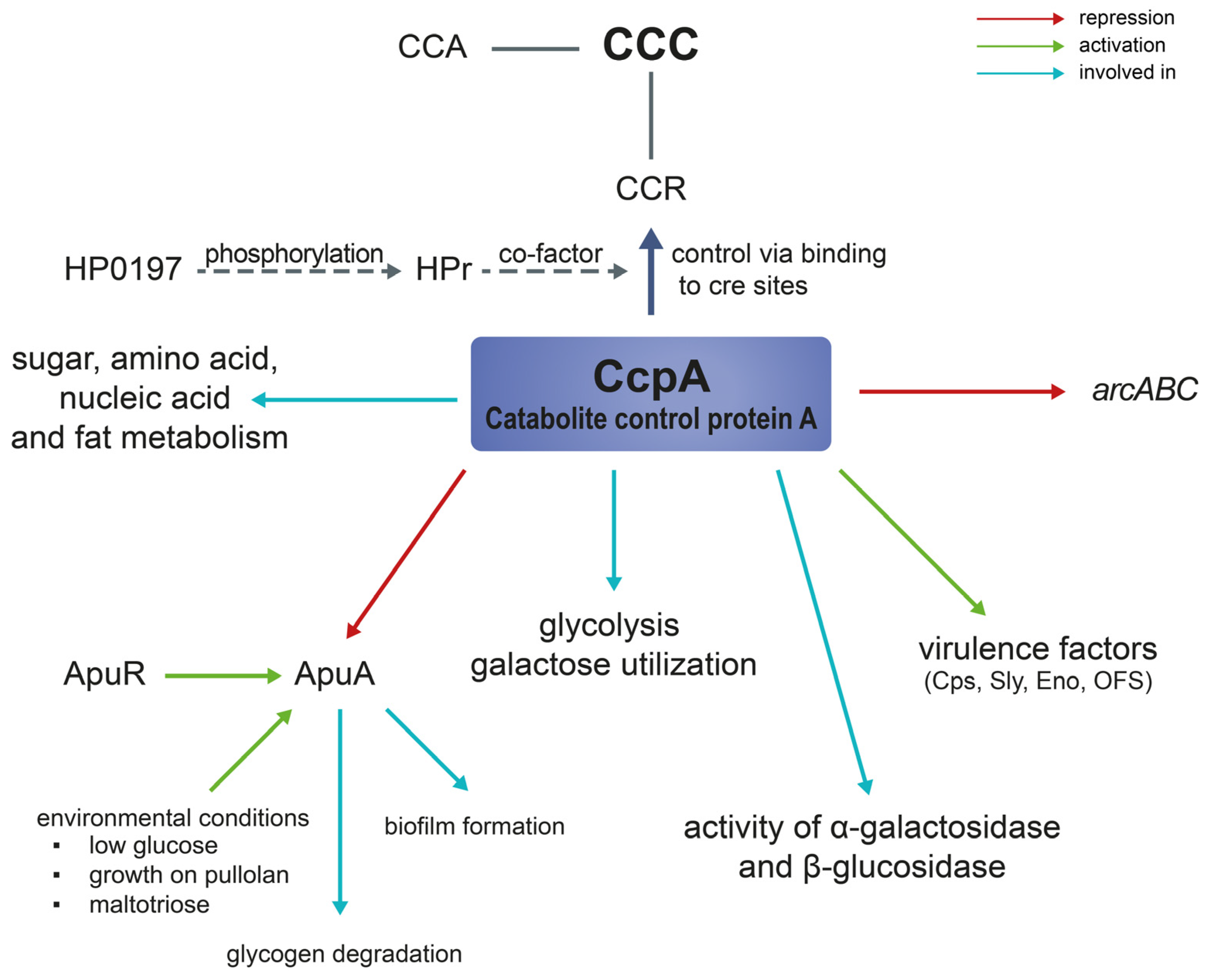
To ensure the uptake of the preferred carbon source, bacteria make use of a regulatory mechanism called Carbon Catabolite Control (CCC) [22,77]. This process can be divided into Carbon Catabolite Repression (CCR) and Carbon Catabolite Activation [77,79]. When the preferred carbon source is available, CCR downregulates the expression or activity of genes involved in the usage of secondary carbon sources [77]. Thereby, the bacteria can optimally utilize the available nutrients, thus competing successfully with other microorganisms [80]. CcpA controls CCR by binding to specific motifs in the promotor region, so-called cis-acting catabolite response element (cre) sites [81,82]. CcpA plays a key role in the metabolic adaptation of gram-positive bacteria including many pathogenic streptococci, e.g., S. suis or S. pneumoniae [24,83]. In S. suis ccpA expression is constitutive [13]. CCR can be classified as a carbon source intake mechanism contributing to carbon metabolism in general.
Willenborg et al. investigated the role of CcpA during growth with glucose consumption by analyzing the transcriptome of S. suis serotype 2 strain 10 and its ccpA-deficient mutant. Most of the differentially expressed genes encoded for transcriptional regulation, metabolism, and other unknown functions. Some of the affected genes seemed to be part of CCR regulation, as their expression increased in the ccpA-deficient mutant. These were mainly related to carbohydrate metabolism or carbohydrate and amino acid transport, e.g., the arginine deiminase system (ADS, arcABC) or the glycogen synthase cluster (glgCAB) [24]. CcpA plays a major role in glycolysis and takes part in galactose utilization. However, it is not involved in the PPP [24]. Many affected genes were not directly controlled by ccpA. Therefore, their regulation in vivo might also include the activity of cofactors [24].
Tang et al. also investigated the effect of ccpA on CCR [84]. Their study showed that ccpA is involved in the repression of α-galactosidase and β-glucosidase activities. The deletion of ccpA reduced the repression of these enzymes but did not alter their sugar utilization pattern. However, the activity of the α-glucosidase was not significantly affected in the ccpA-deficient mutant, suggesting the contribution of other factors to CCR e.g., potential phosphotransferase systems [84].
The group of Lang et al., applied gene expression profile analysis, metabolomics, as well as proteomics, to investigate the role of ccpA in S. suis. Their studies underlined an involvement of CcpA in sugar, amino acid, nucleic acid and fat metabolism as ccpA activity alters the concentration of certain metabolites [85,86]. A decrease in succinic, aspartic, and citric acid concentrations changed glucose availability and therefore, affected S. suis metabolism regulation [87].
As already mentioned, there is a close relationship between metabolism and virulence in bacteria. Pathogens often concatenate/combine regulation of expression of metabolic with virulence genes as it saves energy [88]. In gram-positive bacteria this is achieved by three main global regulators, CcpA, CodY and Rex [88]. CcpA and CodY were shown to be involved in S. suis capsule expression and virulence [13,22,56]. Many genes associated with virulence were downregulated in the ccpA-deficient mutant such as, suilysin, opacity factor, surface antigen one or the capsule synthesis cluster [13,84]. Interestingly, the expression of the virulence factor arcB was higher in the knockout strain [13]; ArcB encodes an ornithine carbamoyltransferase and is part of the ADS involved in pathogen survival and fitness [89,90]. The ADS system is described in more detail in Section 3.3.
The capsule of S. suis protects the pathogen from being phagocytosed [91,92]. Depending on the host environment, capsule synthesis is either enhanced to act as a protection against the host immune system, e.g., in the bloodstream, or decreased to facilitate adherence to epithelial barriers and subsequent invasion of the tissue [8]. In host environments containing high glucose levels, capsule expression depends on ccpA. Gene expression analysis of the ccpA-deficient mutant revealed downregulation of capsule and sialic acid synthesis [13,84]. The ccpA-deficient mutant of S. suis showed an attenuated phenotype with reduced survival in a phagocytic assay which might be linked to the lower capsule expression [13]. Former studies have already shown that an unencapsulated S. suis mutant showed reduced colonization capacity and resistance to phagocytosis as well as attenuated virulence in a mouse infection model [91,93]. As the capsule is one of the main factors for mediating colonization, invasion, and resistance to host defense mechanisms, its regulation is of utmost importance for establishing infection [3]. Therefore, ccpA plays an important role for the evasion of host immunity by contributing to phagocytosis resistance via capsule expression [13].
Accordingly, the deletion of the global regulator CodY also resulted in a reduced resistance to phagocytosis [56]. A codY-deletion mutant displayed a reduced capsule thickness as well as an inhibited expression of sialic acid genes. This was also reflected in the altered capsule composition showing a reduced amount of sialic acid content [56]. CodY regulation activity is linked to amino acid availability as well as stress [50,94]. Its expression was upregulated in S. suis isolated from the bloodstream or the brain. Therefore, the authors suggested that CodY supports resistance to phagocytosis as well as antimicrobial peptides which is a prerequisite for survival and colonization of these host environments [50]. Furthermore, both the adhesion and invasion capacity to and into endothelial cells as well as the virulence in a mouse infection model decreased in the ccpA-deficient mutant [84]. Thus, CcpA itself is regarded as a virulence factor [13].
Interestingly, Zhang et al. observed a comparable phenotype in an hp0197-knockout mutant of S. suis serotype 2 strain 05ZY [95]. HP0197 is a surface protective antigen without sequence homology to other proteins [96,97]. The hp0197-deficient strain showed an attenuated phenotype in mice and pig infection experiments, a decreased resistance to phagocytosis as well as a similar pattern of differentially expressed genes as a ccpA-deficient strain in other studies [13,95]. However, ccpA was not downregulated in the hp0197-deficient mutant. Phosphorylated HPr, a phosphocarrier protein, is an important co-effector for CcpA binding to cre sites [98]. HPr isolated from Δhp0197 exhibited a weaker binding in combination with CcpA in comparison to wild-type HPr indicating reduced phosphorylation of HPr in the mutant [95]. Therefore, these studies underline the importance of CcpA for bacterial virulence. Similar findings were observed in other pathogenic streptococci. CcpA-deficient mutants showed an attenuated phenotype in mice infection experiments [83,99]. Wen and Burne showed that in S. mutans CcpA is essential for the formation of biofilms [100]. Furthermore, CcpA is involved in the colonization and survival of S. pneumoniae on the respiratory epithelium [83]. In S. pyogenes ccpA activates the transcriptional regulator Mga which is involved in the expression of virulence genes [101]. Additionally, the expression of virulence factors regulated by ccpA is dependent on the nutrient availability in the environment [77].
To evade the host immunity and to overcome nutrient starvation, S. suis is able to form protective biofilms [102]. However, biofilm formation has also the disadvantage of reduced pathogenicity reflected in decreased metabolism and virulence gene expression as well as inhibition of the efficacy of bacterial toxins as they are trapped in the extracellular matrix [103]. Recently, Bulock et al. investigated the effects of codY and ccpA deletion in Staphylococcus aureus on biofilm formation [104]. The ccpA-deficient mutant showed impaired biofilm formation, whereas the codY-deficient mutant formed a robust biofilm structure. In the ccpA-codY-double knock-out strain, the overall biofilm mass was reduced indicating a linkage between central metabolism and biofilm formation [104]. Similarly, ccpA deletion in Streptoccocus gordonii led to impaired biofilm formation [105]. To the best of our knowledge, the effect of ccpA deletion on biofilm formation in S. suis has not been investigated so far. However, based on results published for other Streptococci [104,105] a similar effect is likely.
Figure 2 illustrates the role and functions of ccpA in S. suis metabolism as well as virulence. In summary, ccpA acts as a carbon catabolite repressor in S. suis but is also involved in many other cellular functions and can indirectly influence transcription [24].

3.2. Amylopullulanase (apuA)

Although glucose is the preferred carbon source of S. suis [27], the pathogen needs to adapt to varying sugar availability in different in vivo situations. On the one hand, glucose is present in the oropharyngeal cavity, but at varying concentrations, which are affected, e.g., by uptake of food. After feeding, glucose concentrations drop due to the direct use of this carbon source either by the host or the resident microflora [22,106]. On the other hand, the concentration of starch α-glucans in the oral cavity is much more stable and a substantial part of animal feed [22,107]. Ferrando et al. compared metabolism and virulence gene expression during S. suis growth on either glucose or α-glucan starch (pullulan). They showed that both the expression of an amylopullulanase, apuA, and the pore-forming toxin suilysin (sly) increased upon growth on pullulan.
Suilysin (SLY) is a member of the group of cholesterol-dependent cytolysins (CDC) which can be found in many gram-positive bacteria [108]. SLY is considered a virulence-associated factor due to its essential contribution to the pathogenesis of S. suis [109,110]. SLY is cytotoxic to a variety of different cell types including epithelial cells, endothelial cells, phagocytes, red blood cells or more complex cell culture models, e.g., air–liquid interface cultures or precision-cut lung slices (PCLS) [108,111,112,113,114,115]. In addition, SLY enhances both adherence and colonization in the PCLS system [114]. Therefore, SLY participates in both the pathogenic as well as the commensal phase of S. suis.
Both apuA and sly contain a conserved cre motif in their promoter region suggesting repressed transcription during growth on glucose. The authors also identified a potential cre site in many other virulence genes differentially expressed in pullulan compared to glucose, encompassing the capsular polysaccharide [22]. The direct binding of CcpA to both the sly promotor and the capsule synthesis cluster was confirmed by Willenborg et al. [24]. Moreover, the expression of apuA was induced by maltotriose. The authors suggested that apuA might be activated by the putative transcriptional regulator ApuR and repressed by CCR mediated via ccpA [22]. Accordingly, the transcription of apuA and sly was higher in body sites containing less glucose than the blood, e.g., the brain, heart and joints facilitating colonization, invasion, and the use of alternative carbohydrates. Though, these findings have yet not been proven in vivo, they may contribute to new therapeutic strategies, e.g., adaptation of feed composition or blocking of certain enzymes needed for starch degradation [22]. The induction of the expression of the ADS and sly at low glucose levels underlines the importance of nutrient availability for S. suis pathogenicity [13]. At low glucose concentrations sly might be relieved from ccpA regulation explaining the increased expression during growth on pullulan [22]. Furthermore, the authors revealed that the adherence and invasion capacity of S. suis to newborn pig tracheal (NPTr) cells increased upon growth in DMEM supplemented with pullulan instead of glucose. The elevated expression of sly might contribute to this observation, as the toxin has already been shown to promote invasion to host cells [93].
Additionally, the increase in sly expression resulted in a higher hemolytic activity of bacteria when grown in pullulan. Although, the increased expression of sly probably resulted from the missing CCR rather than a starch/pullulan specific effect [22].
Furthermore, Tan et al. investigated the effects of exogenous glycogen utilization on S. suis pathogenicity. For this, the authors constructed an apuA-deficient mutant and compared its growth on glycogen with the wild-type strain. Inactivation of apuA led to a switch from homofermentative to heterofermentative metabolism inducing mixed-acid fermentation [23]. Supplementation of the media with glycogen resulted in an increased hemolytic activity of the pathogen, which is in accordance with the induced expression of sly. However, the deletion of apuA decreased sly production. Furthermore, the presence of glycogen induced a higher adhesion and invasion capacity whereas the deletion of apuA had the opposite effect. Finally, biofilm formation was reduced in the apuA-deficient mutant and in the presence of glycogen. The authors concluded that ApuA can be regarded as an important virulence factor of S. suis promoting hemolysin activity, adherence, and invasion as well as biofilm formation [23].

3.3. Arginine Deiminase System (ADS)

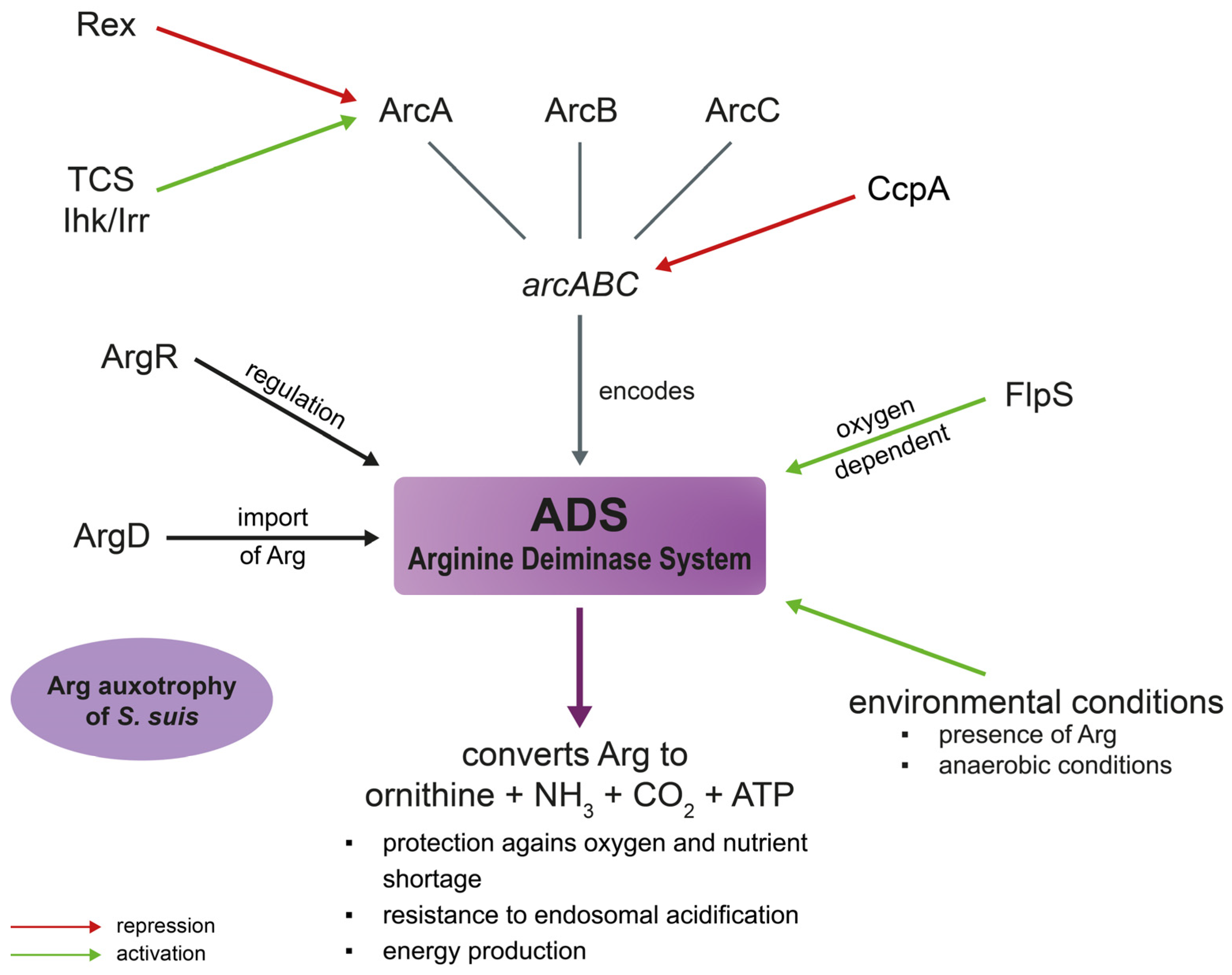
The arcABC operon in S. suis encodes the ADS and allows the pathogen to grow under acidic conditions by neutralizing acidification via production of ammonium [116]. The ADS consists of the arginine deiminase (ArcA), the ornithine carbamoyltransferase (ArcB) and the carbamate kinase (ArcC) [89]. Catalyzing the conversion of arginine to ornithine, ammonia as well as CO2, the ADS generates energy by the production of ATP [117]. Thereby, it protects bacteria from oxygen and nutrient shortage [118]. S. suis is auxotrophic for arginine in CDM [20,119]. Therefore, its survival is dependent on arginine import. The arginine–ornithine antiporter (ArcD) plays an important role in this process as it provides arginine for the ADS which, in turn, is important for S. suis fitness and pathogenicity [119]. In S. suis the ADS is regulated by the system specific transcriptional regulator ArgR [116]. This contrasts with other bacteria where argR regulates genes related to both arginine anabolism and catabolism [120,121,122]. Moreover, the FNR-like protein FlpS of S. suis takes part in ADS activation. In an flps-deficient mutant the expression of arcABC was significantly decreased. Flps was shown to be involved in regulating the central carbon and nucleotide metabolism. Oxygen dependent flps-mediated activation of arcABC underlines the role of FlpS for important adaptation mechanisms to specific in vivo host niches linked to redox conditions [123]. In accordance with these results, the ADS is activated by environmental conditions such as the presence of arginine or glucose and anaerobic conditions [89]. Furthermore, the two-component system (TCS) Ihk/Irr also seems to be involved in ADS regulation. In an iKR-deficient mutant strain, ADS was downregulated leading to reduced adherence capacity and stress resistance under acidic conditions [124]. Similarly, in S. pyogenes it was also shown that the ADS participates in host cell adhesion [124]. The arginine–ornithine antiporter ArcD provides the ADS of S. suis with arginine [119]. This is especially important for intracellular survival of the bacteria, as the generation of ammonium via the ADS can prevent pH drops. Thereby, S. suis can resist endosomal acidification inside the host cell. An arcD-deficient, an arcR-deficient and an arcABC-mutant all showed reduced survival inside Hep-2 cells [116,119]. This effect was reduced in Hep-2 cells treated with bafilomycin which prevents endosomal acidification. For that reason, the decreased survival of the mutants is due to a reduced acidic stress resistance [116,119].
Rex is a redox-sensing regulator involved in metabolism as well as virulence in different bacterial species. A Rex orthologue in S. suis was shown to be important for its pathogenicity and stress competence as its absence resulted in reduced virulence and stress resistance [125]. Metabolic pathways associated with central metabolism were altered in the rex-deficient mutant. ArcA expression was significantly upregulated in the mutant strain. The authors could show that rSsrex was able to directly interact with the arcA promoter suggesting a possible function as a transcription repressor of this gene [125]. Figure 3 summarizes the role of the ADS in S. suis virulence and metabolism.

3.4. Amino Acid Metabolism

Amino acid availability is important for bacteria to synthesize various cell components or for utilization in metabolic pathways [126]. As S. suis is auxotroph for different amino acids including tryptophan [20], uptake of these amino acids is of utmost importance. Therefore, e.g., tryptophan transporters are crucial for pathogenesis. Without the substrate-binding protein TrpX of its tryptophan ABC transporter TrpXYZ, S. suis is not able to survive in tryptophan-limited conditions [127] or in porcine blood [50]. Therefore, TrpX significantly contributes to nutrient acquisition and bacterial growth during infection. Underlining the important role of ABC transporters in nutrient acquisition they are regarded as important fitness factors that also contribute to bacterial virulence [12].
Another important enzyme for the central carbon catabolism is the PEP carboxylase (ppc) which is needed for the biosynthesis of oxaloacetate. Oxaloacetate serves as a precursor for the amino acids aspartic acid and threonine [20]. A ppc-deficient mutant of S. suis serotype 2 strain 10 showed impaired growth in porcine blood as well as CSF. Although the mutant survived for the tested time, adequate nutrient uptake for normal growth of the pathogen seemed to be missing. Therefore, ppc is essential for S. suis fitness in the host [20].

4. S. suis Metabolism and Co-Infections

Respiratory disease is a common problem in the pig industry worldwide. Often pneumonia is not caused by a single pathogen but by an interplay of different viruses and/or bacteria [44].
To the best of our knowledge, a direct study of the metabolic adaptation of S. suis to different in vivo niches, co-infected with other pathogens has not been conducted, as S. suis is also a pathobiont in the respiratory tract of healthy pigs [4]. However, the host immune response as an indirect measure of S. suis strategies to establish itself in the host during co-infection with other respiratory pathogens, for example swine influenza virus (SIV) [128,129], porcine reproductive and respiratory syndrome virus (PRRSV) [130,131], and porcine circovirus (PCV) [132] have been investigated and it was found that pre-infection of host cells with the aforementioned pathogens paves the way for S. suis infection. Co-infections resulted in higher induction of genes involved in inflammatory response [128,129,130,131,132]. Moreover, co-infection of host cells with SIV was found to facilitate S. suis adherence to the virus haemagglutinin protein and further invasion of S. suis to the bloodstream due to the structural component of S. suis capsule, sialic acid, which allows it as a bacterial virus receptor. The capsular sialic acid of S. suis protects capsulated S. suis from phagocytosis, and enables the pathogen to invade the respiratory epithelium, spread and induce systemic infection [114,128]. Therefore, we can hypothesize that structural components of S. suis in co-infections can indirectly facilitate bacterial metabolic adaptation to certain in vivo niches, making it a dynamic pathogen, capable of a thriving successful infection, with or without co-infecting pathogens.
In addition, co-infections of S. suis with SIV, PRRSV or Bordetella bronchiseptica were shown to not only promote adherence and invasion but also the cytotoxicity of S suis to NPTr cells, porcine alveolar macrophages (PAMs), PCLS or in vivo [114,131,133,134]. On the contrary, an in vitro co-infection experiment with Glaesserella parasuis and S. suis showed similar adhesion levels of the pathogens in single and co-infection trials [135].
A study by Wang et al. investigated the effect of a co-infection of PCV and S. suis in swine tracheal epithelial cells on reactive oxygen species (ROS). The authors showed that the coinfection decreased the activity of NADPH oxidase compared to S. suis infection alone [136]. NADPH oxidase is an important ROS generator [137,138]. Therefore, reduced NADPH activity also led to lower ROS concentrations and thereby to an increased intracellular survival of S. suis [136]. Changes in enzyme expression and ROS metabolism of the host may also have potential effects on the metabolism of the infecting pathogens.
In conclusion, most of the co-infection studies showed a positive effect on S. suis virulence and survival. However, many of these studies have been performed in vitro. Therefore, effects of the host immune system, the residual microflora or environmental factors could not be taken into account. The effects of co-infections on the regulation of metabolic genes need to be addressed in future studies.

5. S. suis Metabolism and Antibiotic Resistance

Since antibiotic-resistant S. suis isolates have increased over the past years, there is an urgent need for new therapeutics. S. suis plays an important role as a reservoir for resistance genes [139,140]. Especially macrolides that are often used to treat S. suis infections. Therefore, many isolates are resistant against this class of antibiotic [141]. Advances in science and modern technologies like omics approaches demonstrated a link between drug resistance and changes in bacterial metabolism [142,143]. A recent study by Wu et al. investigated the effect of L-serine supplementation on a macrolide resistant S. suis isolate [144]. L-serine addition led to both an increased susceptibility to macrolides and a decreased biofilm formation capacity in the resistant strain. Moreover, the authors showed that L-serine supplementation in combination with tylosin administration resulted in an increased level of ROS inside the bacteria leading to enhanced DNA damage [144]. The authors suggest that macrolide resistance in S. suis is conferred by an alteration of the serine metabolic pathway along with an inhibition of ROS production [144].
A link between metabolism and antibiotic resistance has also been shown in other bacterial species. Therapeutic efficacy of β-lactam antibiotics was increased in an MRSA S. aureus strain by supplementation with d-serine [145]. Exogenous concentrations of alanine or glucose enabled killing of resistant Edwardsiella tarda by kanamycin [146].
In S. suis it was shown that the ABC transporter SatAb was involved in fluoroquinolone resistance by extruding the antibiotics norfloxacin and ciprofloxacin. Although the exact function of this ABC transporter is still unclear, the genetic environment suggests a role in basic metabolism [147].
In other bacteria, e.g., Streptoccocus gordonii or S. aureus CcpA has been shown to be involved in antibiotic resistance. The knock-out of ccpA resulted in reduced tolerance towards β-lactam or glycopeptide antibiotics [148,149]. However, whether CcpA also plays a role in antibiotic resistance in S. suis needs to be investigated in future studies. In conclusion, all these studies demonstrate that antibiotic resistance mechanisms are closely linked to bacterial metabolism. However, future studies are needed to investigate the role of host metabolites as well as microenvironment specific metabolite generation of the bacteria itself [150]. It needs to be considered that bacterial metabolites can affect each other in the same biological niche [151].

6. Summary and Conclusions

In conclusion, metabolic adaptation of S. suis to its in vivo niches is a prerequisite for successful survival and establishment of infection. Each niche constitutes a microenvironment with differences in nutrient availability, host defense mechanisms and competing microbiota. S. suis adaptation to these varying conditions is reflected in the transcriptomic data obtained in different studies [18,20,49,50]. The analysis of gene transcriptional levels represents a powerful tool to investigate the adaptations and metabolic changes of S. suis during infection. In addition, it can reveal valuable insights about the host immune response.
Metabolism regulation also plays an important role in virulence. Mutants deficient in metabolic regulators often showed an attenuated phenotype in infection experiments or survival assays [13,84]. In addition, expression of many virulence factors including sly and the capsule of S. suis is closely linked to the expression of catabolite control protein A (CcpA) which is involved in sugar metabolism [13,22]. The capsule represents an important protection of S. suis against the host immune system [91,92], whereas suilysin(SLY) exhibits cytotoxic effects on various cell types and facilitates invasion [108]. Therefore, they are needed at different stages of infection. Capsule expression is mainly upregulated in the bloodstream to inhibit phagocytosis [8], whereas sly expression is induced in different organs, such as the brain and the heart, enhancing colonization and invasion. These host niches represent environments with low glucose concentrations highlighting the link between nutrient availability and virulence [22].
Co-infections of S. suis with other pathogens are very common in the pig population [44,152]. They have been shown to promote S. suis adhesion, invasion, and virulence to different cell types [114,131,133,134,136]. However, knowledge about the effects on metabolic adaptations of the different pathogens is scarce and needs to be further investigated in future studies. In addition, the role of the existing microbiota during infection needs to be taken into consideration.
Antibiotic resistance (AMR) is a major challenge for human and animal health. The Organisation for Economic Co-operation and Development (OECD) estimates the costs of fighting AMR at up to USD 2.9 trillion by 2050 compared to an AMR-free world [153]. Accordingly, the number of S. suis isolates carrying AMR genes has increased over the past decades [139,140]. Importantly, S. suis may also spread these genes to other streptococcal species including human pathogens, thereby serving as an AMR reservoir [139]. As metabolism regulation is key for bacteria to survive in the host, it constitutes an interesting target for new therapeutic strategies. Furthermore, metabolism represents an important factor for bacterial persistence [154]. Still today, most of the modes of action of antibiotics concentrate on the synthesis of proteins, folate and the cell envelope or DNA replication [155]. However, different metabolic pathways such as fatty acid metabolism or iron metabolism were also shown to be promising targets for the design of new antibiotics [156,157]. Effects of antibiotics on metabolic pathways can be divided into three main parts: antibiotics can change the metabolism of bacteria resulting in death or growth inhibition; the metabolic state of the pathogen can affect its susceptibility; and the efficacy of antibiotic treatment can be promoted by influencing the bacterial metabolic state [158]. When investigating new targets for antibiotic treatment it is of utmost importance to use targets that do not have a human counterpart or use different catalytic pathways. In addition, bacterial energy metabolism represents a promising target as it differs from usually used AMR sites [159]. Furthermore, ABC transporters which are closely involved in different metabolic processes [160], but also regulatory elements such as T-box riboswitches are regarded as auspicious targets for novel antimicrobials [161,162].
Taken together, metabolic activity of S. suis is crucial for its role as a pathobiont in the porcine respiratory tract and, thus also contributes to virulence, Furthermore, mechanisms of metabolic adaptation of S. suis in its host should be considered in approaches for new therapeutic strategies.

Author Contributions

Conceptualization, M.D. and Y.B.W.; Writing—Original Draft Preparation, M.D. and Y.B.W.; Review and Editing, M.D., Y.B.W. and P.V.-W. All authors have read and agreed to the published version of the manuscript.

Funding

This project received funding to P.V.-W. from the European Union’s Horizon 2020 research and innovation program under grant agreement no. 727966.

Institutional Review Board Statement

Not applicable.

Data Availability Statement

Not applicable.

Acknowledgments

Figure 1 was created with BioRender.com. Figure 2 and Figure 3 were created with Adobe Illustrator software 27.2.

Conflicts of Interest

The authors declare no conflict of interest.

References

  1. Segura, M.; Calzas, C.; Grenier, D.; Gottschalk, M. Initial steps of the pathogenesis of the infection caused by Streptococcus suis: Fighting against nonspecific defenses. FEBS Lett. 2016, 590, 3772–3799. [Google Scholar] [CrossRef] [PubMed] [Green Version]
  2. Goyette-Desjardins, G.; Auger, J.P.; Xu, J.; Segura, M.; Gottschalk, M. Streptococcus suis, an important pig pathogen and emerging zoonotic agent-an update on the worldwide distribution based on serotyping and sequence typing. Emerg. Microbes. Infect. 2014, 3, e45. [Google Scholar] [CrossRef] [PubMed]
  3. Fittipaldi, N.; Segura, M.; Grenier, D.; Gottschalk, M. Virulence factors involved in the pathogenesis of the infection caused by the swine pathogen and zoonotic agent Streptococcus suis. Future Microbiol. 2012, 7, 259–279. [Google Scholar] [CrossRef]
  4. Votsch, D.; Willenborg, M.; Weldearegay, Y.B.; Valentin-Weigand, P. Streptococcus suis—The “Two Faces” of a Pathobiont in the Porcine Respiratory Tract. Front. Microbiol. 2018, 9, 480. [Google Scholar] [CrossRef] [PubMed] [Green Version]
  5. Dekker, N.; Bouma, A.; Daemen, I.; Klinkenberg, D.; van Leengoed, L.; Wagenaar, J.A.; Stegeman, A. Effect of spatial separation of pigs on spread of Streptococcus suis serotype 9. PLoS ONE 2013, 8, e61339. [Google Scholar] [CrossRef] [Green Version]
  6. Tang, J.; Wang, C.; Feng, Y.; Yang, W.; Song, H.; Chen, Z.; Yu, H.; Pan, X.; Zhou, X.; Wang, H.; et al. Streptococcal toxic shock syndrome caused by Streptococcus suis serotype 2. PLoS Med. 2006, 3, e151. [Google Scholar] [CrossRef] [Green Version]
  7. Gottschalk, M.; Xu, J.; Calzas, C.; Segura, M. Streptococcus suis: A new emerging or an old neglected zoonotic pathogen? Future Microbiol. 2010, 5, 371–391. [Google Scholar] [CrossRef]
  8. Gottschalk, M.; Segura, M. The pathogenesis of the meningitis caused by Streptococcus suis: The unresolved questions. Vet. Microbiol. 2000, 76, 259–272. [Google Scholar] [CrossRef]
  9. Madsen, L.W.; Bak, H.; Nielsen, B.; Jensen, H.E.; Aalbaek, B.; Riising, H.J. Bacterial colonization and invasion in pigs experimentally exposed to Streptococcus suis serotype 2 in aerosol. J. Vet. Med. Infect. Dis. Vet. Public Health 2002, 49, 211–215. [Google Scholar] [CrossRef]
  10. Arends, J.P.; Hartwig, N.; Rudolphy, M.; Zanen, H.C. Carrier rate of Streptococcus suis capsular type 2 in palatine tonsils of slaughtered pigs. J. Clin. Microbiol. 1984, 20, 945–947. [Google Scholar] [CrossRef] [Green Version]
  11. Rohmer, L.; Hocquet, D.; Miller, S.I. Are pathogenic bacteria just looking for food? Metabolism and microbial pathogenesis. Trends Microbiol. 2011, 19, 341–348. [Google Scholar] [CrossRef] [PubMed] [Green Version]
  12. Tanaka, K.J.; Song, S.; Mason, K.; Pinkett, H.W. Selective substrate uptake: The role of ATP-binding cassette (ABC) importers in pathogenesis. Biochim. Biophys. Acta Biomembr. 2018, 1860, 868–877. [Google Scholar] [CrossRef] [PubMed]
  13. Willenborg, J.; Fulde, M.; de Greeff, A.; Rohde, M.; Smith, H.E.; Valentin-Weigand, P.; Goethe, R. Role of glucose and CcpA in capsule expression and virulence of Streptococcus suis. Microbiol. (Read. Engl.) 2011, 157, 1823–1833. [Google Scholar] [CrossRef] [PubMed] [Green Version]
  14. Hondorp, E.R.; McIver, K.S. The Mga virulence regulon: Infection where the grass is greener. Mol. Microbiol. 2007, 66, 1056–1065. [Google Scholar] [CrossRef] [PubMed]
  15. Graham, M.R.; Virtaneva, K.; Porcella, S.F.; Barry, W.T.; Gowen, B.B.; Johnson, C.R.; Wright, F.A.; Musser, J.M. Group A Streptococcus transcriptome dynamics during growth in human blood reveals bacterial adaptive and survival strategies. Am. J. Pathol. 2005, 166, 455–465. [Google Scholar] [CrossRef] [PubMed] [Green Version]
  16. Richards, V.P.; Choi, S.C.; Pavinski Bitar, P.D.; Gurjar, A.A.; Stanhope, M.J. Transcriptomic and genomic evidence for Streptococcus agalactiae adaptation to the bovine environment. BMC Genom. 2013, 14, 920. [Google Scholar] [CrossRef] [PubMed] [Green Version]
  17. Murase, K.; Watanabe, T.; Arai, S.; Kim, H.; Tohya, M.; Ishida-Kuroki, K.; Võ, T.H.; Nguyễn, T.P.B.; Nakagawa, I.; Osawa, R.; et al. Characterization of pig saliva as the major natural habitat of Streptococcus suis by analyzing oral, fecal, vaginal, and environmental microbiota. PLoS ONE 2019, 14, e0215983. [Google Scholar] [CrossRef]
  18. Koczula, A.; Jarek, M.; Visscher, C.; Valentin-Weigand, P.; Goethe, R.; Willenborg, J. Transcriptomic Analysis Reveals Selective Metabolic Adaptation of Streptococcus suis to Porcine Blood and Cerebrospinal Fluid. Pathogens 2017, 6, 7. [Google Scholar] [CrossRef] [Green Version]
  19. Van der Putten, B.C.L.; Roodsant, T.J.; Haagmans, M.A.; Schultsz, C.; van der Ark, K.C.H. Five Complete Genome Sequences Spanning the Dutch Streptococcus suis Serotype 2 and Serotype 9 Populations. Microbiol. Resour. Announc. 2020, 9, e01439-19. [Google Scholar] [CrossRef] [Green Version]
  20. Willenborg, J.; Huber, C.; Koczula, A.; Lange, B.; Eisenreich, W.; Valentin-Weigand, P.; Goethe, R. Characterization of the pivotal carbon metabolism of Streptococcus suis serotype 2 under ex vivo and chemically defined in vitro conditions by isotopologue profiling. J. Biol. Chem. 2015, 290, 5840–5854. [Google Scholar] [CrossRef] [Green Version]
  21. Hoskins, J.; Alborn, W.E., Jr.; Arnold, J.; Blaszczak, L.C.; Burgett, S.; DeHoff, B.S.; Estrem, S.T.; Fritz, L.; Fu, D.J.; Fuller, W.; et al. Genome of the bacterium Streptococcus pneumoniae strain R6. J. Bacteriol. 2001, 183, 5709–5717. [Google Scholar] [CrossRef] [PubMed] [Green Version]
  22. Ferrando, M.L.; van Baarlen, P.; Orrù, G.; Piga, R.; Bongers, R.S.; Wels, M.; De Greeff, A.; Smith, H.E.; Wells, J.M. Carbohydrate availability regulates virulence gene expression in Streptococcus suis. PLoS ONE 2014, 9, e89334. [Google Scholar] [CrossRef] [PubMed] [Green Version]
  23. Tan, M.F.; Tan, J.; Zhang, F.F.; Li, H.Q.; Ji, H.Y.; Fang, S.P.; Wu, C.C.; Rao, Y.L.; Zeng, Y.B.; Yang, Q. Exogenous glycogen utilization effects the transcriptome and pathogenicity of Streptococcus suis serotype 2. Front. Cell. Infect. Microbiol. 2022, 12, 938286. [Google Scholar] [CrossRef] [PubMed]
  24. Willenborg, J.; de Greeff, A.; Jarek, M.; Valentin-Weigand, P.; Goethe, R. The CcpA regulon of Streptococcus suis reveals novel insights into the regulation of the streptococcal central carbon metabolism by binding of CcpA to two distinct binding motifs. Mol. Microbiol. 2014, 92, 61–83. [Google Scholar] [CrossRef]
  25. Abranches, J.; Zeng, L.; Kajfasz, J.K.; Palmer, S.R.; Chakraborty, B.; Wen, Z.T.; Richards, V.P.; Brady, L.J.; Lemos, J.A. Biology of Oral Streptococci. Microbiol. Spectr. 2018, 6. [Google Scholar] [CrossRef]
  26. Ferrando, M.L.; Fuentes, S.; de Greeff, A.; Smith, H.; Wells, J.M. ApuA, a multifunctional alpha-glucan-degrading enzyme of Streptococcus suis, mediates adhesion to porcine epithelium and mucus. Microbiol. (Read. Engl.) 2010, 156, 2818–2828. [Google Scholar] [CrossRef] [Green Version]
  27. Willenborg, J.; Goethe, R. Metabolic traits of pathogenic streptococci. FEBS Lett. 2016, 590, 3905–3919. [Google Scholar] [CrossRef] [Green Version]
  28. Bidossi, A.; Mulas, L.; Decorosi, F.; Colomba, L.; Ricci, S.; Pozzi, G.; Deutscher, J.; Viti, C.; Oggioni, M.R. A functional genomics approach to establish the complement of carbohydrate transporters in Streptococcus pneumoniae. PLoS ONE 2012, 7, e33320. [Google Scholar] [CrossRef] [Green Version]
  29. Juliao, P.C.; Marrs, C.F.; Xie, J.; Gilsdorf, J.R. Histidine auxotrophy in commensal and disease-causing nontypeable Haemophilus influenzae. J. Bacteriol. 2007, 189, 4994–5001. [Google Scholar] [CrossRef] [Green Version]
  30. Fittipaldi, N.; Harel, J.; D’Amours, B.; Lacouture, S.; Kobisch, M.; Gottschalk, M. Potential use of an unencapsulated and aromatic amino acid-auxotrophic Streptococcus suis mutant as a live attenuated vaccine in swine. Vaccine 2007, 25, 3524–3535. [Google Scholar] [CrossRef]
  31. Holden, M.T.; Hauser, H.; Sanders, M.; Ngo, T.H.; Cherevach, I.; Cronin, A.; Goodhead, I.; Mungall, K.; Quail, M.A.; Price, C.; et al. Rapid evolution of virulence and drug resistance in the emerging zoonotic pathogen Streptococcus suis. PLoS ONE 2009, 4, e6072. [Google Scholar] [CrossRef] [Green Version]
  32. Arndt, E.R.; Farzan, A.; Slavic, D.; MacInnes, J.I.; Friendship, R.M. An epidemiological study of Streptococcus suis serotypes of pigs in Ontario determined by a multiplex polymerase chain reaction. Can. Vet. J. 2018, 59, 997–1000. [Google Scholar] [PubMed]
  33. Pirolo, M.; Espinosa-Gongora, C.; Alberdi, A.; Eisenhofer, R.; Soverini, M.; Eriksen, E.; Pedersen, K.S.; Guardabassi, L. Bacterial topography of the upper and lower respiratory tract in pigs. Anim. Microbiome 2023, 5, 5. [Google Scholar] [CrossRef] [PubMed]
  34. Fredriksen, S.; Guan, X.; Boekhorst, J.; Molist, F.; van Baarlen, P.; Wells, J.M. Environmental and maternal factors shaping tonsillar microbiota development in piglets. BMC Microbiol. 2022, 22, 224. [Google Scholar] [CrossRef] [PubMed]
  35. Pirolo, M.; Espinosa-Gongora, C.; Bogaert, D.; Guardabassi, L. The porcine respiratory microbiome: Recent insights and future challenges. Anim. Microbiome 2021, 3, 9. [Google Scholar] [CrossRef] [PubMed]
  36. Wang, Q.; Cai, R.; Huang, A.; Wang, X.; Qu, W.; Shi, L.; Li, C.; Yan, H. Comparison of Oropharyngeal Microbiota in Healthy Piglets and Piglets with Respiratory Disease. Front. Microbiol. 2018, 9, 3218. [Google Scholar] [CrossRef] [PubMed] [Green Version]
  37. Jakubovics, N.S.; Yassin, S.A.; Rickard, A.H. Community interactions of oral streptococci. Adv. Appl. Microbiol. 2014, 87, 43–110. [Google Scholar] [CrossRef]
  38. Nobbs, A.H.; Jenkinson, H.F.; Everett, D.B. Generic determinants of Streptococcus colonization and infection. Infect. Genet. Evol. 2015, 33, 361–370. [Google Scholar] [CrossRef] [Green Version]
  39. Nitsche-Schmitz, D.P.; Rohde, M.; Chhatwal, G.S. Invasion mechanisms of Gram-positive pathogenic cocci. Thromb. Haemost. 2007, 98, 488–496. [Google Scholar] [CrossRef] [Green Version]
  40. Liang, Z.; Wu, H.; Bian, C.; Chen, H.; Shen, Y.; Gao, X.; Ma, J.; Yao, H.; Wang, L.; Wu, Z. The antimicrobial systems of Streptococcus suis promote niche competition in pig tonsils. Virulence 2022, 13, 781–793. [Google Scholar] [CrossRef]
  41. Cotter, P.D.; Hill, C.; Ross, R.P. Bacteriocins: Developing innate immunity for food. Nat. Rev. Microbiol. 2005, 3, 777–788. [Google Scholar] [CrossRef] [PubMed]
  42. Jack, R.W.; Tagg, J.R.; Ray, B. Bacteriocins of gram-positive bacteria. Microbiol. Rev. 1995, 59, 171–200. [Google Scholar] [CrossRef] [PubMed]
  43. Watanabe, A.; Kawada-Matsuo, M.; Le, M.N.; Hisatsune, J.; Oogai, Y.; Nakano, Y.; Nakata, M.; Miyawaki, S.; Sugai, M.; Komatsuzawa, H. Comprehensive analysis of bacteriocins in Streptococcus mutans. Sci. Rep. 2021, 11, 12963. [Google Scholar] [CrossRef] [PubMed]
  44. Opriessnig, T.; Giménez-Lirola, L.G.; Halbur, P.G. Polymicrobial respiratory disease in pigs. Anim. Health Res. Rev. 2011, 12, 133–148. [Google Scholar] [CrossRef] [PubMed]
  45. Saade, G.; Deblanc, C.; Bougon, J.; Marois-Créhan, C.; Fablet, C.; Auray, G.; Belloc, C.; Leblanc-Maridor, M.; Gagnon, C.A.; Zhu, J.; et al. Coinfections and their molecular consequences in the porcine respiratory tract. Vet. Res. 2020, 51, 80. [Google Scholar] [CrossRef]
  46. Wang, Y.; Gong, S.; Dong, X.; Li, J.; Grenier, D.; Yi, L. In vitro Mixed Biofilm of Streptococcus suis and Actinobacillus pleuropneumoniae Impacts Antibiotic Susceptibility and Modulates Virulence Factor Gene Expression. Front. Microbiol. 2020, 11, 507. [Google Scholar] [CrossRef]
  47. Donlan, R.M.; Costerton, J.W. Biofilms: Survival mechanisms of clinically relevant microorganisms. Clin. Microbiol. Rev. 2002, 15, 167–193. [Google Scholar] [CrossRef] [Green Version]
  48. Mark Welch, J.L.; Rossetti, B.J.; Rieken, C.W.; Dewhirst, F.E.; Borisy, G.G. Biogeography of a human oral microbiome at the micron scale. Proc. Natl. Acad. Sci. USA 2016, 113, E791–E800. [Google Scholar] [CrossRef] [Green Version]
  49. Wu, Z.; Wu, C.; Shao, J.; Zhu, Z.; Wang, W.; Zhang, W.; Tang, M.; Pei, N.; Fan, H.; Li, J.; et al. The Streptococcus suis transcriptional landscape reveals adaptation mechanisms in pig blood and cerebrospinal fluid. RNA 2014, 20, 882–898. [Google Scholar] [CrossRef] [Green Version]
  50. Arenas, J.; Bossers-de Vries, R.; Harders-Westerveen, J.; Buys, H.; Ruuls-van Stalle, L.M.F.; Stockhofe-Zurwieden, N.; Zaccaria, E.; Tommassen, J.; Wells, J.M.; Smith, H.E.; et al. In vivo transcriptomes of Streptococcus suis reveal genes required for niche-specific adaptation and pathogenesis. Virulence 2019, 10, 334–351. [Google Scholar] [CrossRef] [Green Version]
  51. Schwerk, C.; Adam, R.; Borkowski, J.; Schneider, H.; Klenk, M.; Zink, S.; Quednau, N.; Schmidt, N.; Stump, C.; Sagar, A.; et al. In vitro transcriptome analysis of porcine choroid plexus epithelial cells in response to Streptococcus suis: Release of pro-inflammatory cytokines and chemokines. Microbes. Infect. 2011, 13, 953–962. [Google Scholar] [CrossRef] [PubMed]
  52. De Greeff, A.; Benga, L.; Wichgers Schreur, P.J.; Valentin-Weigand, P.; Rebel, J.M.; Smith, H.E. Involvement of NF-kappaB and MAP-kinases in the transcriptional response of alveolar macrophages to Streptococcus suis. Vet. Microbiol. 2010, 141, 59–67. [Google Scholar] [CrossRef] [PubMed]
  53. Liu, M.; Tan, C.; Fang, L.; Xiao, S.; Chen, H. Microarray analyses of THP-1 cells infected with Streptococcus suis serotype 2. Vet. Microbiol. 2011, 150, 126–131. [Google Scholar] [CrossRef] [PubMed]
  54. Wang, J.; Liang, P.; Sun, H.; Wu, Z.; Gottschalk, M.; Qi, K.; Zheng, H. Comparative transcriptomic analysis reveal genes involved in the pathogenicity increase of Streptococcus suis epidemic strains. Virulence 2022, 13, 1455–1470. [Google Scholar] [CrossRef] [PubMed]
  55. Ye, C.; Zheng, H.; Zhang, J.; Jing, H.; Wang, L.; Xiong, Y.; Wang, W.; Zhou, Z.; Sun, Q.; Luo, X.; et al. Clinical, experimental, and genomic differences between intermediately pathogenic, highly pathogenic, and epidemic Streptococcus suis. J. Infect. Dis. 2009, 199, 97–107. [Google Scholar] [CrossRef] [Green Version]
  56. Feng, L.; Zhu, J.; Chang, H.; Gao, X.; Gao, C.; Wei, X.; Yuan, F.; Bei, W. The CodY regulator is essential for virulence in Streptococcus suis serotype 2. Sci. Rep. 2016, 6, 21241. [Google Scholar] [CrossRef] [Green Version]
  57. Li, J.; Tan, C.; Zhou, Y.; Fu, S.; Hu, L.; Hu, J.; Chen, H.; Bei, W. The two-component regulatory system CiaRH contributes to the virulence of Streptococcus suis 2. Vet. Microbiol. 2011, 148, 99–104. [Google Scholar] [CrossRef]
  58. Federle, M.J.; McIver, K.S.; Scott, J.R. A response regulator that represses transcription of several virulence operons in the group A streptococcus. J. Bacteriol. 1999, 181, 3649–3657. [Google Scholar] [CrossRef] [Green Version]
  59. Lee, S.F.; Delaney, G.D.; Elkhateeb, M. A two-component covRS regulatory system regulates expression of fructosyltransferase and a novel extracellular carbohydrate in Streptococcus mutans. Infect. Immun. 2004, 72, 3968–3973. [Google Scholar] [CrossRef] [Green Version]
  60. Zheng, C.; Xu, J.; Li, J.; Hu, L.; Xia, J.; Fan, J.; Guo, W.; Chen, H.; Bei, W. Two Spx regulators modulate stress tolerance and virulence in Streptococcus suis serotype 2. PLoS ONE 2014, 9, e108197. [Google Scholar] [CrossRef]
  61. Neila-Ibanez, C.; Brogaard, L.; Pailler-Garcia, L.; Martinez, J.; Segales, J.; Segura, M.; Heegaard, P.M.H.; Aragon, V. Piglet innate immune response to Streptococcus suis colonization is modulated by the virulence of the strain. Vet. Res. 2021, 52, 145. [Google Scholar] [CrossRef] [PubMed]
  62. Sanford, S.E. Gross and histopathological findings in unusual lesions caused by Streptococcus suis in pigs. II. Central nervous system lesions. Can. J. Vet. Res. = Rev. Can. De Rech. Vet. 1987, 51, 486–489. [Google Scholar]
  63. Williams, A.E.; Blakemore, W.F. Pathogenesis of meningitis caused by Streptococcus suis type 2. J. Infect. Dis. 1990, 162, 474–481. [Google Scholar] [CrossRef]
  64. Beineke, A.; Bennecke, K.; Neis, C.; Schroder, C.; Waldmann, K.H.; Baumgartner, W.; Valentin-Weigand, P.; Baums, C.G. Comparative evaluation of virulence and pathology of Streptococcus suis serotypes 2 and 9 in experimentally infected growers. Vet. Microbiol. 2008, 128, 423–430. [Google Scholar] [CrossRef] [PubMed]
  65. Lauer, A.N.; Scholtysik, R.; Beineke, A.; Baums, C.G.; Klose, K.; Valentin-Weigand, P.; Ishikawa, H.; Schroten, H.; Klein-Hitpass, L.; Schwerk, C. A Comparative Transcriptome Analysis of Human and Porcine Choroid Plexus Cells in Response to Streptococcus suis Serotype 2 Infection Points to a Role of Hypoxia. Front. Cell. Infect. Microbiol. 2021, 11, 639620. [Google Scholar] [CrossRef]
  66. Tenenbaum, T.; Matalon, D.; Adam, R.; Seibt, A.; Wewer, C.; Schwerk, C.; Galla, H.J.; Schroten, H. Dexamethasone prevents alteration of tight junction-associated proteins and barrier function in porcine choroid plexus epithelial cells after infection with Streptococcus suis in vitro. Brain Res. 2008, 1229, 1–17. [Google Scholar] [CrossRef]
  67. Schwerk, C.; Papandreou, T.; Schuhmann, D.; Nickol, L.; Borkowski, J.; Steinmann, U.; Quednau, N.; Stump, C.; Weiss, C.; Berger, J.; et al. Polar invasion and translocation of Neisseria meningitidis and Streptococcus suis in a novel human model of the blood-cerebrospinal fluid barrier. PLoS ONE 2012, 7, e30069. [Google Scholar] [CrossRef]
  68. Tesorero, R.A.; Yu, N.; Wright, J.O.; Svencionis, J.P.; Cheng, Q.; Kim, J.H.; Cho, K.H. Novel regulatory small RNAs in Streptococcus pyogenes. PLoS ONE 2013, 8, e64021. [Google Scholar] [CrossRef] [Green Version]
  69. Deltcheva, E.; Chylinski, K.; Sharma, C.M.; Gonzales, K.; Chao, Y.; Pirzada, Z.A.; Eckert, M.R.; Vogel, J.; Charpentier, E. CRISPR RNA maturation by trans-encoded small RNA and host factor RNase III. Nature 2011, 471, 602–607. [Google Scholar] [CrossRef] [Green Version]
  70. Mann, B.; van Opijnen, T.; Wang, J.; Obert, C.; Wang, Y.D.; Carter, R.; McGoldrick, D.J.; Ridout, G.; Camilli, A.; Tuomanen, E.I.; et al. Control of virulence by small RNAs in Streptococcus pneumoniae. PLoS Pathog. 2012, 8, e1002788. [Google Scholar] [CrossRef]
  71. Kumar, R.; Shah, P.; Swiatlo, E.; Burgess, S.C.; Lawrence, M.L.; Nanduri, B. Identification of novel non-coding small RNAs from Streptococcus pneumoniae TIGR4 using high-resolution genome tiling arrays. BMC Genom. 2010, 11, 350. [Google Scholar] [CrossRef] [PubMed] [Green Version]
  72. Lee, H.J.; Hong, S.H. Analysis of microRNA-size, small RNAs in Streptococcus mutans by deep sequencing. FEMS Microbiol. Lett. 2012, 326, 131–136. [Google Scholar] [CrossRef] [PubMed]
  73. Pichon, C.; du Merle, L.; Caliot, M.E.; Trieu-Cuot, P.; Le Bouguenec, C. An in silico model for identification of small RNAs in whole bacterial genomes: Characterization of antisense RNAs in pathogenic Escherichia coli and Streptococcus agalactiae strains. Nucleic Acids Res. 2012, 40, 2846–2861. [Google Scholar] [CrossRef] [PubMed] [Green Version]
  74. Murray, G.G.R.; Charlesworth, J.; Miller, E.L.; Casey, M.J.; Lloyd, C.T.; Gottschalk, M.; Tucker, A.W.D.; Welch, J.J.; Weinert, L.A. Genome Reduction Is Associated with Bacterial Pathogenicity across Different Scales of Temporal and Ecological Divergence. Mol. Biol. Evol. 2021, 38, 1570–1579. [Google Scholar] [CrossRef]
  75. Moran, N.A. Microbial minimalism: Genome reduction in bacterial pathogens. Cell 2002, 108, 583–586. [Google Scholar] [CrossRef] [Green Version]
  76. Zientz, E.; Dandekar, T.; Gross, R. Metabolic interdependence of obligate intracellular bacteria and their insect hosts. Microbiol. Mol. Biol. Rev. 2004, 68, 745–770. [Google Scholar] [CrossRef] [Green Version]
  77. Görke, B.; Stülke, J. Carbon catabolite repression in bacteria: Many ways to make the most out of nutrients. Nat. Rev. Microbiol. 2008, 6, 613–624. [Google Scholar] [CrossRef]
  78. Shelburne, S.A.; Davenport, M.T.; Keith, D.B.; Musser, J.M. The role of complex carbohydrate catabolism in the pathogenesis of invasive streptococci. Trends Microbiol. 2008, 16, 318–325. [Google Scholar] [CrossRef] [Green Version]
  79. Poncet, S.; Milohanic, E.; Mazé, A.; Abdallah, J.N.; Aké, F.; Larribe, M.; Deghmane, A.E.; Taha, M.K.; Dozot, M.; De Bolle, X.; et al. Correlations between carbon metabolism and virulence in bacteria. Contrib. Microbiol. 2009, 16, 88–102. [Google Scholar] [CrossRef]
  80. Nair, A.; Sarma, S.J. The impact of carbon and nitrogen catabolite repression in microorganisms. Microbiol. Res. 2021, 251, 126831. [Google Scholar] [CrossRef]
  81. Moreno, M.S.; Schneider, B.L.; Maile, R.R.; Weyler, W.; Saier, M.H., Jr. Catabolite repression mediated by the CcpA protein in Bacillus subtilis: Novel modes of regulation revealed by whole-genome analyses. Mol. Microbiol. 2001, 39, 1366–1381. [Google Scholar] [CrossRef] [PubMed]
  82. Deutscher, J.; Francke, C.; Postma, P.W. How phosphotransferase system-related protein phosphorylation regulates carbohydrate metabolism in bacteria. Microbiol. Mol. Biol. Rev. 2006, 70, 939–1031. [Google Scholar] [CrossRef] [PubMed] [Green Version]
  83. Iyer, R.; Baliga, N.S.; Camilli, A. Catabolite control protein A (CcpA) contributes to virulence and regulation of sugar metabolism in Streptococcus pneumoniae. J. Bacteriol. 2005, 187, 8340–8349. [Google Scholar] [CrossRef] [Green Version]
  84. Tang, Y.; Wu, W.; Zhang, X.; Lu, Z.; Chen, J.; Fang, W. Catabolite control protein A of Streptococcus suis type 2 contributes to sugar metabolism and virulence. J. Microbiol. 2012, 50, 994–1002. [Google Scholar] [CrossRef] [PubMed]
  85. Lang, X.; Wan, Z.; Bu, Z.; Wang, X.; Wang, X.; Zhu, L.; Wan, J.; Sun, Y.; Wang, X. Catabolite control protein A is an important regulator of metabolism in Streptococcus suis type 2. Biomed. Rep. 2014, 2, 709–712. [Google Scholar] [CrossRef] [PubMed] [Green Version]
  86. Lang, X.; Wan, Z.; Pan, Y.; Bu, Z.; Wang, X.; Wang, X.; Ji, X.; Zhu, L.; Wan, J.; Sun, Y.; et al. Catabolite control protein A has an important role in the metabolic regulation of Streptococcus suis type 2 according to iTRAQ-based quantitative proteomic analysis. Mol. Med. Rep. 2015, 12, 5967–5972. [Google Scholar] [CrossRef] [Green Version]
  87. Lang, X.; Wan, Z.; Pan, Y.; Wang, X.; Wang, X.; Bu, Z.; Qian, J.; Zeng, H.; Wang, X. Investigation into the role of catabolite control protein A in the metabolic regulation of Streptococcus suis serotype 2 using gene expression profile analysis. Exp. Ther. Med. 2015, 10, 127–132. [Google Scholar] [CrossRef] [Green Version]
  88. Richardson, A.R.; Somerville, G.A.; Sonenshein, A.L. Regulating the Intersection of Metabolism and Pathogenesis in Gram-positive Bacteria. Microbiol. Spectr. 2015, 3, 129–165. [Google Scholar] [CrossRef] [Green Version]
  89. Gruening, P.; Fulde, M.; Valentin-Weigand, P.; Goethe, R. Structure, regulation, and putative function of the arginine deiminase system of Streptococcus suis. J. Bacteriol. 2006, 188, 361–369. [Google Scholar] [CrossRef] [Green Version]
  90. Winterhoff, N.; Goethe, R.; Gruening, P.; Rohde, M.; Kalisz, H.; Smith, H.E.; Valentin-Weigand, P. Identification and characterization of two temperature-induced surface-associated proteins of Streptococcus suis with high homologies to members of the Arginine Deiminase system of Streptococcus pyogenes. J. Bacteriol. 2002, 184, 6768–6776. [Google Scholar] [CrossRef] [Green Version]
  91. Benga, L.; Fulde, M.; Neis, C.; Goethe, R.; Valentin-Weigand, P. Polysaccharide capsule and suilysin contribute to extracellular survival of Streptococcus suis co-cultivated with primary porcine phagocytes. Vet. Microbiol. 2008, 132, 211–219. [Google Scholar] [CrossRef] [PubMed]
  92. Smith, H.E.; Damman, M.; van der Velde, J.; Wagenaar, F.; Wisselink, H.J.; Stockhofe-Zurwieden, N.; Smits, M.A. Identification and characterization of the cps locus of Streptococcus suis serotype 2: The capsule protects against phagocytosis and is an important virulence factor. Infect. Immun. 1999, 67, 1750–1756. [Google Scholar] [CrossRef] [PubMed]
  93. Seitz, M.; Baums, C.G.; Neis, C.; Benga, L.; Fulde, M.; Rohde, M.; Goethe, R.; Valentin-Weigand, P. Subcytolytic effects of suilysin on interaction of Streptococcus suis with epithelial cells. Vet. Microbiol. 2013, 167, 584–591. [Google Scholar] [CrossRef] [PubMed]
  94. Brinsmade, S.R. CodY, a master integrator of metabolism and virulence in Gram-positive bacteria. Curr. Genet. 2017, 63, 417–425. [Google Scholar] [CrossRef]
  95. Zhang, A.; Chen, B.; Yuan, Z.; Li, R.; Liu, C.; Zhou, H.; Chen, H.; Jin, M. HP0197 contributes to CPS synthesis and the virulence of Streptococcus suis via CcpA. PLoS ONE 2012, 7, e50987. [Google Scholar] [CrossRef]
  96. Zhang, A.; Xie, C.; Chen, H.; Jin, M. Identification of immunogenic cell wall-associated proteins of Streptococcus suis serotype 2. Proteomics 2008, 8, 3506–3515. [Google Scholar] [CrossRef]
  97. Zhang, A.; Chen, B.; Li, R.; Mu, X.; Han, L.; Zhou, H.; Chen, H.; Meilin, J. Identification of a surface protective antigen, HP0197 of Streptococcus suis serotype 2. Vaccine 2009, 27, 5209–5213. [Google Scholar] [CrossRef]
  98. Schumacher, M.A.; Allen, G.S.; Diel, M.; Seidel, G.; Hillen, W.; Brennan, R.G. Structural basis for allosteric control of the transcription regulator CcpA by the phosphoprotein HPr-Ser46-P. Cell 2004, 118, 731–741. [Google Scholar] [CrossRef] [Green Version]
  99. Shelburne, S.A., 3rd; Keith, D.; Horstmann, N.; Sumby, P.; Davenport, M.T.; Graviss, E.A.; Brennan, R.G.; Musser, J.M. A direct link between carbohydrate utilization and virulence in the major human pathogen group A Streptococcus. Proc. Natl. Acad. Sci. USA 2008, 105, 1698–1703. [Google Scholar] [CrossRef] [Green Version]
  100. Wen, Z.T.; Burne, R.A. Functional genomics approach to identifying genes required for biofilm development by Streptococcus mutans. Appl. Env. Microbiol. 2002, 68, 1196–1203. [Google Scholar] [CrossRef] [Green Version]
  101. Almengor, A.C.; Kinkel, T.L.; Day, S.J.; McIver, K.S. The catabolite control protein CcpA binds to Pmga and influences expression of the virulence regulator Mga in the Group A streptococcus. J. Bacteriol. 2007, 189, 8405–8416. [Google Scholar] [CrossRef] [PubMed] [Green Version]
  102. Grenier, D.; Grignon, L.; Gottschalk, M. Characterisation of biofilm formation by a Streptococcus suis meningitis isolate. Vet. J. 2009, 179, 292–295. [Google Scholar] [CrossRef] [PubMed]
  103. Wang, Y.; Zhang, W.; Wu, Z.; Lu, C. Reduced virulence is an important characteristic of biofilm infection of Streptococcus suis. FEMS Microbiol. Lett. 2011, 316, 36–43. [Google Scholar] [CrossRef] [PubMed] [Green Version]
  104. Bulock, L.L.; Ahn, J.; Shinde, D.; Pandey, S.; Sarmiento, C.; Thomas, V.C.; Guda, C.; Bayles, K.W.; Sadykov, M.R. Interplay of CodY and CcpA in Regulating Central Metabolism and Biofilm Formation in Staphylococcus aureus. J. Bacteriol. 2022, 204, e0061721. [Google Scholar] [CrossRef] [PubMed]
  105. Zheng, L.; Chen, Z.; Itzek, A.; Herzberg, M.C.; Kreth, J. CcpA regulates biofilm formation and competence in Streptococcus gordonii. Mol. Oral Microbiol. 2012, 27, 83–94. [Google Scholar] [CrossRef]
  106. Gough, H.; Luke, G.A.; Beeley, J.A.; Geddes, D.A. Human salivary glucose analysis by high-performance ion-exchange chromatography and pulsed amperometric detection. Arch. Oral Biol. 1996, 41, 141–145. [Google Scholar] [CrossRef] [PubMed]
  107. Navarro, D.; Abelilla, J.J.; Stein, H.H. Structures and characteristics of carbohydrates in diets fed to pigs: A review. J. Anim. Sci. Biotechnol. 2019, 10, 39. [Google Scholar] [CrossRef]
  108. Tenenbaum, T.; Asmat, T.M.; Seitz, M.; Schroten, H.; Schwerk, C. Biological activities of suilysin: Role in Streptococcus suis pathogenesis. Future Microbiol. 2016, 11, 941–954. [Google Scholar] [CrossRef]
  109. Jacobs, A.A.; Loeffen, P.L.; van den Berg, A.J.; Storm, P.K. Identification, purification, and characterization of a thiol-activated hemolysin (suilysin) of Streptococcus suis. Infect. Immun. 1994, 62, 1742–1748. [Google Scholar] [CrossRef] [Green Version]
  110. Takeuchi, D.; Akeda, Y.; Nakayama, T.; Kerdsin, A.; Sano, Y.; Kanda, T.; Hamada, S.; Dejsirilert, S.; Oishi, K. The contribution of suilysin to the pathogenesis of Streptococcus suis meningitis. J. Infect. Dis. 2014, 209, 1509–1519. [Google Scholar] [CrossRef] [Green Version]
  111. Lalonde, M.; Segura, M.; Lacouture, S.; Gottschalk, M. Interactions between Streptococcus suis serotype 2 and different epithelial cell lines. Microbiol. (Read. Engl.) 2000, 146 Pt 8, 1913–1921. [Google Scholar] [CrossRef] [PubMed] [Green Version]
  112. Norton, P.M.; Rolph, C.; Ward, P.N.; Bentley, R.W.; Leigh, J.A. Epithelial invasion and cell lysis by virulent strains of Streptococcus suis is enhanced by the presence of suilysin. FEMS Immunol. Med. Microbiol. 1999, 26, 25–35. [Google Scholar] [CrossRef] [PubMed] [Green Version]
  113. Charland, N.; Nizet, V.; Rubens, C.E.; Kim, K.S.; Lacouture, S.; Gottschalk, M. Streptococcus suis serotype 2 interactions with human brain microvascular endothelial cells. Infect. Immun. 2000, 68, 637–643. [Google Scholar] [CrossRef] [PubMed] [Green Version]
  114. Meng, F.; Wu, N.H.; Nerlich, A.; Herrler, G.; Valentin-Weigand, P.; Seitz, M. Dynamic Virus-Bacterium Interactions in a Porcine Precision-Cut Lung Slice Coinfection Model: Swine Influenza Virus Paves the Way for Streptococcus suis Infection in a Two-Step Process. Infect. Immun. 2015, 83, 2806–2815. [Google Scholar] [CrossRef] [Green Version]
  115. Meng, F.; Wu, N.H.; Seitz, M.; Herrler, G.; Valentin-Weigand, P. Efficient suilysin-mediated invasion and apoptosis in porcine respiratory epithelial cells after streptococcal infection under air-liquid interface conditions. Sci. Rep. 2016, 6, 26748. [Google Scholar] [CrossRef] [Green Version]
  116. Fulde, M.; Willenborg, J.; de Greeff, A.; Benga, L.; Smith, H.E.; Valentin-Weigand, P.; Goethe, R. ArgR is an essential local transcriptional regulator of the arcABC operon in Streptococcus suis and is crucial for biological fitness in an acidic environment. Microbiol. (Read. Engl.) 2011, 157, 572–582. [Google Scholar] [CrossRef] [Green Version]
  117. Zúñiga, M.; Pérez, G.; González-Candelas, F. Evolution of arginine deiminase (ADI) pathway genes. Mol. Phylogenet. Evol. 2002, 25, 429–444. [Google Scholar] [CrossRef]
  118. Champomier Vergès, M.C.; Zuñiga, M.; Morel-Deville, F.; Pérez-Martínez, G.; Zagorec, M.; Ehrlich, S.D. Relationships between arginine degradation, pH and survival in Lactobacillus sakei. FEMS Microbiol. Lett. 1999, 180, 297–304. [Google Scholar] [CrossRef]
  119. Fulde, M.; Willenborg, J.; Huber, C.; Hitzmann, A.; Willms, D.; Seitz, M.; Eisenreich, W.; Valentin-Weigand, P.; Goethe, R. The arginine-ornithine antiporter ArcD contributes to biological fitness of Streptococcus suis. Front. Cell. Infect. Microbiol. 2014, 4, 107. [Google Scholar] [CrossRef] [Green Version]
  120. Hashim, S.; Kwon, D.H.; Abdelal, A.; Lu, C.D. The arginine regulatory protein mediates repression by arginine of the operons encoding glutamate synthase and anabolic glutamate dehydrogenase in Pseudomonas aeruginosa. J. Bacteriol. 2004, 186, 3848–3854. [Google Scholar] [CrossRef] [Green Version]
  121. Larsen, R.; Kok, J.; Kuipers, O.P. Interaction between ArgR and AhrC controls regulation of arginine metabolism in Lactococcus lactis. J. Biol. Chem. 2005, 280, 19319–19330. [Google Scholar] [CrossRef] [PubMed] [Green Version]
  122. Park, S.M.; Lu, C.D.; Abdelal, A.T. Cloning and characterization of argR, a gene that participates in regulation of arginine biosynthesis and catabolism in Pseudomonas aeruginosa PAO1. J. Bacteriol. 1997, 179, 5300–5308. [Google Scholar] [CrossRef] [PubMed] [Green Version]
  123. Willenborg, J.; Koczula, A.; Fulde, M.; de Greeff, A.; Beineke, A.; Eisenreich, W.; Huber, C.; Seitz, M.; Valentin-Weigand, P.; Goethe, R. FlpS, the FNR-Like Protein of Streptococcus suis Is an Essential, Oxygen-Sensing Activator of the Arginine Deiminase System. Pathogens 2016, 5, 51. [Google Scholar] [CrossRef] [PubMed] [Green Version]
  124. Han, H.; Liu, C.; Wang, Q.; Xuan, C.; Zheng, B.; Tang, J.; Yan, J.; Zhang, J.; Li, M.; Cheng, H.; et al. The two-component system Ihk/Irr contributes to the virulence of Streptococcus suis serotype 2 strain 05ZYH33 through alteration of the bacterial cell metabolism. Microbiol. (Read. Engl.) 2012, 158, 1852–1866. [Google Scholar] [CrossRef] [Green Version]
  125. Zhu, H.; Wang, Y.; Ni, Y.; Zhou, J.; Han, L.; Yu, Z.; Mao, A.; Wang, D.; Fan, H.; He, K. The Redox-Sensing Regulator Rex Contributes to the Virulence and Oxidative Stress Response of Streptococcus suis Serotype 2. Front. Cell. Infect. Microbiol. 2018, 8, 317. [Google Scholar] [CrossRef] [Green Version]
  126. Neis, E.P.; Dejong, C.H.; Rensen, S.S. The role of microbial amino acid metabolism in host metabolism. Nutrients 2015, 7, 2930–2946. [Google Scholar] [CrossRef] [PubMed] [Green Version]
  127. Dresen, M.; Schaaf, D.; Arenas, J.; de Greeff, A.; Valentin-Weigand, P.; Nerlich, A. Streptococcus suis TrpX is part of a tryptophan uptake system, and its expression is regulated by a T-box regulatory element. Sci. Rep. 2022, 12, 13920. [Google Scholar] [CrossRef] [PubMed]
  128. Wang, Y.; Gagnon, C.A.; Savard, C.; Music, N.; Srednik, M.; Segura, M.; Lachance, C.; Bellehumeur, C.; Gottschalk, M. Capsular sialic acid of Streptococcus suis serotype 2 binds to swine influenza virus and enhances bacterial interactions with virus-infected tracheal epithelial cells. Infect. Immun. 2013, 81, 4498–4508. [Google Scholar] [CrossRef] [Green Version]
  129. Dang, Y.; Lachance, C.; Wang, Y.; Gagnon, C.A.; Savard, C.; Segura, M.; Grenier, D.; Gottschalk, M. Transcriptional approach to study porcine tracheal epithelial cells individually or dually infected with swine influenza virus and Streptococcus suis. BMC Vet. Res. 2014, 10, 86. [Google Scholar] [CrossRef] [PubMed] [Green Version]
  130. Auray, G.; Lachance, C.; Wang, Y.; Gagnon, C.A.; Segura, M.; Gottschalk, M. Transcriptional Analysis of PRRSV-Infected Porcine Dendritic Cell Response to Streptococcus suis Infection Reveals Up-Regulation of Inflammatory-Related Genes Expression. PLoS ONE 2016, 11, e0156019. [Google Scholar] [CrossRef] [PubMed] [Green Version]
  131. Li, J.; Wang, J.; Liu, Y.; Yang, J.; Guo, L.; Ren, S.; Chen, Z.; Liu, Z.; Zhang, Y.; Qiu, W.; et al. Porcine reproductive and respiratory syndrome virus NADC30-like strain accelerates Streptococcus suis serotype 2 infection in vivo and in vitro. Transbound Emerg. Dis. 2019, 66, 729–742. [Google Scholar] [CrossRef] [PubMed]
  132. Wang, Q.; Zhou, H.; Hao, Q.; Li, M.; Liu, J.; Fan, H. Coinfection with porcine circovirus type 2 and Streptococcus suis serotype 2 enhances pathogenicity by dysregulation of the immune responses in piglets. Vet. Microbiol. 2020, 243, 108653. [Google Scholar] [CrossRef]
  133. Wu, N.H.; Meng, F.; Seitz, M.; Valentin-Weigand, P.; Herrler, G. Sialic acid-dependent interactions between influenza viruses and Streptococcus suis affect the infection of porcine tracheal cells. J. Gen. Virol. 2015, 96, 2557–2568. [Google Scholar] [CrossRef] [PubMed]
  134. Vötsch, D.; Willenborg, M.; Baumgärtner, W.; Rohde, M.; Valentin-Weigand, P. Bordetella bronchiseptica promotes adherence, colonization, and cytotoxicity of Streptococcus suis in a porcine precision-cut lung slice model. Virulence 2021, 12, 84–95. [Google Scholar] [CrossRef]
  135. Mathieu-Denoncourt, A.; Letendre, C.; Auger, J.P.; Segura, M.; Aragon, V.; Lacouture, S.; Gottschalk, M. Limited Interactions between Streptococcus Suis and Haemophilus Parasuis in In Vitro Co-Infection Studies. Pathogens 2018, 7, 7. [Google Scholar] [CrossRef] [PubMed] [Green Version]
  136. Wang, Q.; Zhou, H.; Fan, H.; Wang, X. Coinfection with Porcine Circovirus Type 2 (PCV2) and Streptococcus suis Serotype 2 (SS2) Enhances the Survival of SS2 in Swine Tracheal Epithelial Cells by Decreasing Reactive Oxygen Species Production. Infect. Immun. 2020, 88, e00537-20. [Google Scholar] [CrossRef] [PubMed]
  137. Krause, K.H. Aging: A revisited theory based on free radicals generated by NOX family NADPH oxidases. Exp. Gerontol. 2007, 42, 256–262. [Google Scholar] [CrossRef]
  138. Brieger, K.; Schiavone, S.; Miller, F.J., Jr.; Krause, K.H. Reactive oxygen species: From health to disease. Swiss Med. Wkly 2012, 142, w13659. [Google Scholar] [CrossRef]
  139. Palmieri, C.; Varaldo, P.E.; Facinelli, B. Streptococcus suis, an Emerging Drug-Resistant Animal and Human Pathogen. Front. Microbiol. 2011, 2, 235. [Google Scholar] [CrossRef] [PubMed] [Green Version]
  140. Tan, M.F.; Tan, J.; Zeng, Y.B.; Li, H.Q.; Yang, Q.; Zhou, R. Antimicrobial resistance phenotypes and genotypes of Streptococcus suis isolated from clinically healthy pigs from 2017 to 2019 in Jiangxi Province, China. J. Appl. Microbiol. 2021, 130, 797–806. [Google Scholar] [CrossRef]
  141. Uruén, C.; García, C.; Fraile, L.; Tommassen, J.; Arenas, J. How Streptococcus suis escapes antibiotic treatments. Vet. Res. 2022, 53, 91. [Google Scholar] [CrossRef] [PubMed]
  142. Cohen, A.; Bont, L.; Engelhard, D.; Moore, E.; Fernández, D.; Kreisberg-Greenblatt, R.; Oved, K.; Eden, E.; Hays, J.P. A multifaceted ‘omics’ approach for addressing the challenge of antimicrobial resistance. Future Microbiol. 2015, 10, 365–376. [Google Scholar] [CrossRef] [Green Version]
  143. Kuang, S.F.; Chen, Y.T.; Chen, J.J.; Peng, X.X.; Chen, Z.G.; Li, H. Synergy of alanine and gentamicin to reduce nitric oxide for elevating killing efficacy to antibiotic-resistant Vibrio alginolyticus. Virulence 2021, 12, 1737–1753. [Google Scholar] [CrossRef]
  144. Wu, T.; Wang, X.; Dong, Y.; Xing, C.; Chen, X.; Li, L.; Dong, C.; Li, Y. Effects of l-Serine on Macrolide Resistance in Streptococcus suis. Microbiol. Spectr. 2022, 10, e0068922. [Google Scholar] [CrossRef]
  145. Wang, Q.; Lv, Y.; Pang, J.; Li, X.; Lu, X.; Wang, X.; Hu, X.; Nie, T.; Yang, X.; Xiong, Y.Q.; et al. In vitro and in vivo activity of d-serine in combination with β-lactam antibiotics against methicillin-resistant Staphylococcus aureus. Acta Pharm Sin. 2019, 9, 496–504. [Google Scholar] [CrossRef]
  146. Peng, B.; Su, Y.B.; Li, H.; Han, Y.; Guo, C.; Tian, Y.M.; Peng, X.X. Exogenous alanine and/or glucose plus kanamycin kills antibiotic-resistant bacteria. Cell Metab. 2015, 21, 249–262. [Google Scholar] [CrossRef] [Green Version]
  147. Escudero, J.A.; San Millan, A.; Gutierrez, B.; Hidalgo, L.; La Ragione, R.M.; AbuOun, M.; Galimand, M.; Ferrándiz, M.J.; Domínguez, L.; de la Campa, A.G.; et al. Fluoroquinolone efflux in Streptococcus suis is mediated by SatAB and not by SmrA. Antimicrob. Agents Chemother. 2011, 55, 5850–5860. [Google Scholar] [CrossRef] [Green Version]
  148. Bizzini, A.; Entenza, J.M.; Moreillon, P. Loss of penicillin tolerance by inactivating the carbon catabolite repression determinant CcpA in Streptococcus gordonii. J. Antimicrob. Chemother. 2007, 59, 607–615. [Google Scholar] [CrossRef] [Green Version]
  149. Seidl, K.; Stucki, M.; Ruegg, M.; Goerke, C.; Wolz, C.; Harris, L.; Berger-Bächi, B.; Bischoff, M. Staphylococcus aureus CcpA affects virulence determinant production and antibiotic resistance. Antimicrob. Agents Chemother. 2006, 50, 1183–1194. [Google Scholar] [CrossRef] [PubMed] [Green Version]
  150. Bhargava, P.; Collins, J.J. Boosting bacterial metabolism to combat antibiotic resistance. Cell Metab. 2015, 21, 154–155. [Google Scholar] [CrossRef] [Green Version]
  151. Vega, N.M.; Allison, K.R.; Samuels, A.N.; Klempner, M.S.; Collins, J.J. Salmonella typhimurium intercepts Escherichia coli signaling to enhance antibiotic tolerance. Proc. Natl. Acad. Sci. USA 2013, 110, 14420–14425. [Google Scholar] [CrossRef] [PubMed] [Green Version]
  152. Obradovic, M.R.; Segura, M.; Segalés, J.; Gottschalk, M. Review of the speculative role of co-infections in Streptococcus suis-associated diseases in pigs. Vet. Res. 2021, 52, 49. [Google Scholar] [CrossRef]
  153. Antimicrobial Resistance in G7 Countries and beyond: Economic Issues, Policies and Options for Action. Available online: https://www.oecd.org/els/health-systems/Antimicrobial-Resistance-in-G7-Countries-and-Beyond.pdf (accessed on 9 February 2023).
  154. Amato, S.M.; Fazen, C.H.; Henry, T.C.; Mok, W.W.; Orman, M.A.; Sandvik, E.L.; Volzing, K.G.; Brynildsen, M.P. The role of metabolism in bacterial persistence. Front. Microbiol. 2014, 5, 70. [Google Scholar] [CrossRef] [Green Version]
  155. Walsh, C. Where will new antibiotics come from? Nat. Rev. Microbiol. 2003, 1, 65–70. [Google Scholar] [CrossRef] [PubMed]
  156. Yao, J.; Rock, C.O. Bacterial fatty acid metabolism in modern antibiotic discovery. Biochim. Biophys Acta Mol. Cell. Biol. Lipids 2017, 1862, 1300–1309. [Google Scholar] [CrossRef]
  157. Ballouche, M.; Cornelis, P.; Baysse, C. Iron metabolism: A promising target for antibacterial strategies. Recent Pat. Anti-Infect. Drug Discov. 2009, 4, 190–205. [Google Scholar] [CrossRef]
  158. Stokes, J.M.; Lopatkin, A.J.; Lobritz, M.A.; Collins, J.J. Bacterial Metabolism and Antibiotic Efficacy. Cell Metab. 2019, 30, 251–259. [Google Scholar] [CrossRef]
  159. Haag, N.L.; Velk, K.K.; Wu, C. Potential Antibacterial Targets in Bacterial Central Metabolism. Int. J. Adv. Life Sci. 2012, 4, 21–32. [Google Scholar] [PubMed]
  160. Garmory, H.S.; Titball, R.W. ATP-binding cassette transporters are targets for the development of antibacterial vaccines and therapies. Infect. Immun. 2004, 72, 6757–6763. [Google Scholar] [CrossRef] [PubMed] [Green Version]
  161. Mehdizadeh Aghdam, E.; Hejazi, M.S.; Barzegar, A. Riboswitches: From living biosensors to novel targets of antibiotics. Gene 2016, 592, 244–259. [Google Scholar] [CrossRef]
  162. Anupam, R.; Nayek, A.; Green, N.J.; Grundy, F.J.; Henkin, T.M.; Means, J.A.; Bergmeier, S.C.; Hines, J.V. 4,5-Disubstituted oxazolidinones: High affinity molecular effectors of RNA function. Bioorg. Med. Chem. Lett. 2008, 18, 3541–3544. [Google Scholar] [CrossRef] [PubMed] [Green Version]
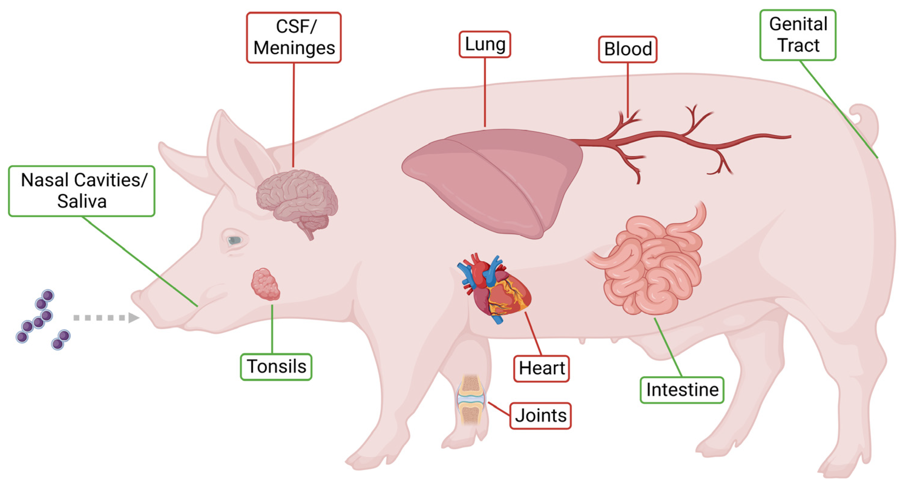
Figure 1. Host niches and infection sites of S. suis in the pig. Host niches colonized (marked in green) or infected (marked in red) by S. suis. Healthy colonized animals are classified as asymptomatic carriers [9,10]. Created with BioRender.com.
Pathogens 12 00541 g001
Figure 2. Carbon catabolite repression and the role of catabolite control protein A (CcpA) in S. suis. CcpA controls the CCR in S. suis via binding to cre sites [22,24]. HPr an important co-factor of this binding is phosphorylated by the protein HP0197 [95]. CcpA is involved in diverse metabolic pathways, the expression of virulence factors and certain enzymes such as the α-galactosidase [13,24,84,85,87]. In addition, it takes part in glycolysis and galactose utilization. CcpA represses the arcABC operon [24] and the expression of apuA. ApuA is regulated by ApuR and primarily induced during growth on pullulan or under glucose-deprived conditions. It plays a role in glycogen degradation as well as biofilm formation [22,23].
Pathogens 12 00541 g002
Figure 3. Role of the arginine deiminase system (ADS) in S. suis virulence and metabolism. The ADS is regulated by argR and encoded by the arcABC operon. ArcA can be downregulated by the transcription repressor rex [125] and induced by the two-component system Ihk/Irr [124]. The ADS converts arginine to ornithine, ammonia, carbon dioxide and ATP [116,117]. Thereby the ADS contributes to the protection against oxygen and nutrient shortage, energy production as well as resistance to endosomal acidification [116,119]. As S. suis is auxotroph for arginine, the transporter ArgD plays an important role in the supply of this amino acid [20,119]. Furthermore, the ADS can be activated by environmental stimuli such as the presence of arginine or anaerobic conditions [89]. Additionally, FlpS can activate the ADS in an oxygen dependent manner [123].
Pathogens 12 00541 g003
Disclaimer/Publisher’s Note: The statements, opinions and data contained in all publications are solely those of the individual author(s) and contributor(s) and not of MDPI and/or the editor(s). MDPI and/or the editor(s) disclaim responsibility for any injury to people or property resulting from any ideas, methods, instructions or products referred to in the content.