Review

Staphylococcus aureus and Cystic Fibrosis—A Close Relationship. What Can We Learn from Sequencing Studies?

Institute of Medical Microbiology, University Hospital Münster, 48149 Münster, Germany
*
Author to whom correspondence should be addressed.
Both authors contributed equally.
Pathogens 2021, 10(9), 1177; https://doi.org/10.3390/pathogens10091177
Submission received: 30 July 2021 / Revised: 7 September 2021 / Accepted: 8 September 2021 / Published: 13 September 2021
(This article belongs to the Special Issue Respiratory Pathogens in Cystic Fibrosis)

Abstract

:
Staphylococcus aureus is next to Pseudomonas aeruginosa the most isolated pathogen from the airways of cystic fibrosis (CF) patients, who are often infected by a dominant S. aureus clone for extended periods. To be able to persist, the pathogen has to adapt to the hostile niche of the airways to counteract host defence, antibiotic therapy and the competition with coinfecting pathogens. S. aureus is equipped with many virulence factors including adhesins, toxins that are localized on the chromosome, on plasmids or are phage-related. S. aureus is especially versatile and adaptation and evolution of the pathogen occurs by the acquisition of new genes by horizontal gene transfer (HGT), changes in nucleotides (single nucleotide variations, SNVs) that can cause a selective advantage for the bacteria and become fixed in subpopulations. Methicillin-resistant S. aureus are a special threat to CF patients due to the more severe lung disease occurring in infected patients. Today, with decreasing costs for sequencing, more and more studies using S. aureus isolates cultured from CF patients are being published, which use whole genome sequencing (WGS), multilocus sequence typing (MLST) or spa-sequence typing (spa-typing) to follow the population dynamics of S. aureus, elucidate the underlying mechanisms of phenotypic variants, newly acquired resistance or adaptation to the host response in this particular niche. In the first part of this review, an introduction to the genetic make-up and the pathogenesis of S. aureus with respect to CF is provided. The second part presents an overview of recent studies and their findings using genotypic methods such as single or multilocus sequencing and whole genome sequencing, which identify factors contributing to the adaptation of S. aureus and its evolution in the airways of individuals with CF.

1. Pathogenesis of S. aureus with Respect to Cystic Fibrosis

The genetic disease cystic fibrosis (CF) is caused by mutations in an important chloride channel called the CF transmembrane conductance regulator (CFTR). The resulting dysfunction alters the ion transport of cell membranes [1]. Therefore, viscous mucus is formed in many organs, including the lungs, making them particularly susceptible to infection [2]. Staphylococcus aureus is one of the first pathogens detectable in the airways of young CF patients and the bacterium with the highest prevalence at an early age. Already among children younger than two years of age its prevalence exceeds 50%, reaching its peak value in the early teenage years at a prevalence of almost 80% [3,4] (Figure 1). In many patients, long term colonization with individual S. aureus clones is observed [5,6,7,8].
In 2018, the prevalence of Methicillin-resistant S. aureus (MRSA) and Methicillin-sensitive S. aureus (MSSA) in individuals with CF in the United States was 25% and 55.3%, respectively [3,9]. In Europe, the prevalence of MRSA is below 10% in most countries, while the prevalence of MSSA is also high, except in the UK, where continuous anti-staphylococcal treatment is part of the regular therapy regimen from the beginning of diagnosis [10]. Patients infected by MRSA are characterized by worse lung function, higher mortality, and increased hospitalization, and therefore MRSA pose a particular threat to CF patients, while there is so far no consensus on optimal treatment strategies for either MRSA or for MSSA [11,12,13,14].
Pathogens such as S. aureus are exposed to many hostile environmental conditions in the airways of CF patients [15]. Ecological influences such as oxygen level and nutrients, host immune defence, antibiotics and competition with other pathogens, such as Pseudomonas aeruginosa, cause a great selective pressure. To be able to colonize the airways and to survive in such harsh surroundings, S. aureus must adapt to and evolve in such unfavourable environments. How do these stressors cause adaptations, what are the evolutionary traits and what are the consequences? To address these questions, phenotypic variations need to be considered in relation to genotypic changes and the clinical impact of the adaptation processes needs to be elucidated. Once the underlying mechanisms are deciphered, targeted preventive measures can be taken and treatment options can be chosen accordingly.
This review will provide an overview about recent studies and their findings using genotypic methods such as single or multilocus sequencing (spa-typing or MLST) and whole genome sequencing (WGS) to identify factors contributing to the adaptation of S. aureus and its evolution in the airways of individuals with CF.

1.1. Adaptation to CF Airways

Persistent S. aureus clones tend to form a genetically (and especially phenotypically) heterogeneous population [15]. The genetic diversity is promoted by mutations and horizontal gene transfer (HGT). Events of S. aureus genome alteration have been shown to occur at significantly higher rates in isolates of CF patients than in isolates of healthy individuals [16]. The phenotypical heterogeneity of S. aureus populations in CF patients is intensified by differences in gene expression, which is mediated by regulatory mechanisms responding to specific milieu conditions (Figure 2) [15].
This view is supported by the finding stating that identical S. aureus clones are translated into differing phenotypes in distinct hosts: this suggests that gene expression is indeed influenced by the surrounding conditions, and, consequently from the latter, that individual host-factors relevantly shape the S. aureus adaptation process [18,19,20]. By analogy with higher mutation rates, more different S. aureus phenotypes are found in the airways of CF patients than in healthy subjects [21,22,23]. Genotypic as well as phenotypic changes occur time-dependently and show a longitudinal accumulation over the course of the disease [24]. The overall trend of the adaptation process seems to be directed towards a persistent and colonizing rather than infecting lifestyle and towards the establishment of a heterogeneous population, which fit into different niches in CF airways and are comparable with the “morphotypes” of P. aeruginosa, which describe different phenotypes [25,26,27]. This is also reflected by the fact that, even in case of an exacerbation, the presence of S. aureus is restricted to the airways and systemic disease is rarely observed [28,29]. The preponderance of colonizing clones results from the suppression of virulence genes for the sake of an intensified expression of adhesins and biofilm formation, granting protection against antibiotic therapy and immune-response [15,30]. This is, at least partially, due to the inactivity of the agr quorum-sensing system [31]. A special S. aureus phenotype highly trimmed towards persistence and regularly observed in CF patients are the so-called small colony variants (SCVs), which show slow growth rates, are able to persist intracellularly, feature increased resistance against antibiotics and are associated with more severe lung disease [18,32,33,34], Figure 3.

1.2. Clinical Impact

In young patients with CF, the presence of S. aureus is associated with increased inflammatory activity in the airways and a worse nutritional status [21,35,36,37]. However, there is still controversy concerning the different therapeutic approaches targeted at S. aureus, namely: prophylaxis, eradication or only treatment of exacerbations. This situation is reflected in the absence of internationally accepted therapeutic guidelines addressing S. aureus treatment in CF patients and inconsistent guidelines at national levels [38]. Prophylaxis is recommended in the UK and Australia, but not clinical practice in the US and continental Europe [39]. The main reason for ambiguity and differing approaches in therapy, which arise over the importance of antibiotic S. aureus prophylaxis in young children, is the shortage in valid studies and reliable data in this respect [40]. Only a few studies on this topic have been published and several of them are of poor quality [41]. Clearly, the open questions about risks and benefits of prophylaxis should be addressed in randomized controlled trials. In recent years, the need to gain knowledge in this sphere has become even more pressing: The increase in neonatal CF screening has considerably raised the number of very young patients with minimal lung disease, a condition for which effective and evidence-based treatment options are necessary [3,4]. For the status quo, Smyth et al. propose an antibiotic prophylaxis until the age of 3 years. During this period, prophylaxis seems to reduce S. aureus colonization rates without increasing the prevalence of P. aeruginosa [40]. The replacement of S. aureus by P. aeruginosa is a common process, regularly witnessed in the course of the disease (Figure 1). Nevertheless, in adult CF patients S. aureus prevalence still remains at about 40% and has been declining in recent years due to early antibiotic eradication regimens [3,4]. The role of S. aureus during the later stages of the disease is not well understood by now. Commonly, a distinction is made between S. aureus colonization and infection. The former mostly does not cause immediate clinical consequences and usually there is no need for therapeutic intervention, whereas the latter can lead to pulmonary exacerbations requiring treatment [42]. Until now, no bacterial characteristics have been identified in S. aureus that are clearly associated with infection and worse lung function [42]. However, if known, these characteristics could be of great clinical importance; they might be used to differentiate between colonizing and infecting S. aureus clones and thus might facilitate therapeutic decision-making.

1.3. S. aureus—Fundamentals of Pathogenicity

The population structure of S. aureus is clonal. Of all S. aureus strains which have been isolated from humans, 10 dominant strains (referred to as “clonal complexes” (CC)), which can be subdivided into numerous substrains, colonize, and infect humans [43,44]. Confrontation with this pathogen results in a spectrum of conditions ranging from subclinical colonization to fulminant or even lethal infections. The clinical fate is not determined by the pathogen per se, as each of the 10 clonal complexes is able to act as a harmless colonialist on the one hand and as a life-threatening pathogen on the other hand [44,45]. However, certain clones tend to be more aggressive than others [46], which raises questions about differences in the otherwise shared genetic bases accounting for S. aureus’ differentially pronounced pathogenic potential.
The S. aureus genome has a size of about 26.000 kb: 75% of S. aureus genes are found in more than 95% of the strains and represent the bacteria’s core genome (Figure 4).
These genes are located on the chromosome and mainly encode proteins with housekeeping function, but also some virulence proteins (spa, hlg, clf, fnb) [47,48]. About 25% of S. aureus genes can be assigned to the variable genome, which is further divided into core variable genes and mobile genetic elements (MGEs).

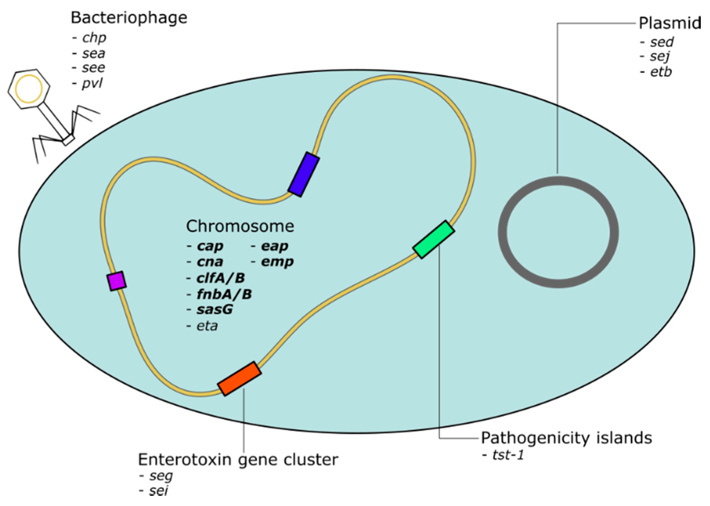
1.4. S. aureus Virulence Genome

S. aureus exhibits an extensive exoproteome, featuring a broad repertoire of virulence genes, mainly located in the variable genome. This virulence factor pool can be roughly divided into adhesins on the one hand and toxins on the other hand (Figure 5). The expression of these proteins is controlled by a complex framework of gene regulatory mechanisms, which in turn reacts to the surrounding conditions (Figure 2) [49,50]. Cell-wall-associated adhesins are predominantly expressed during the exponential growth phase, whereas toxins are often secreted in the post-logarithmic phase [51]. This sequence of expression is considered to reflect the process of colonization, invasion and subsequent persistence. During colonization and invasion, recognition of and adhesion to surface molecules is vital, whereas in the phase of consecutive persistence a continuous nutrition supply, provided by secreted enzymes and toxins, is necessary [51]. However, this time-dependent sequence of virulence gene expression must merely be considered as a general tendency and varies between different S. aureus clones, diseases and hosts. Furthermore, it is still unclear if this sequence occurs during S. aureus colonization and infection of CF airways.

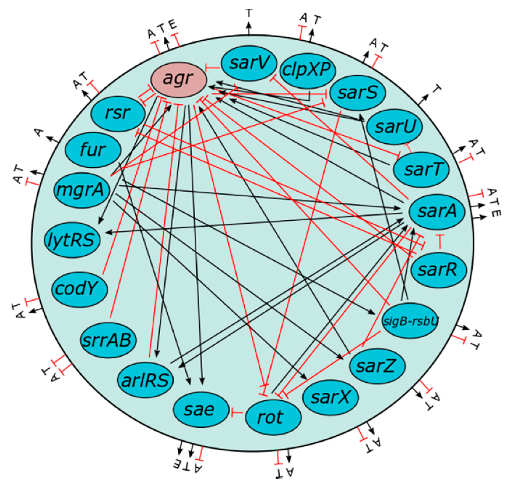
1.5. Role of Agr in Virulence Gene Regulation Via Quorum Sensing

Quorum sensing describes a process of cell-to-cell communication in bacteria, allowing a transfer of information concerning environmental conditions, such as bacterial density [52]. The underlying principle is the bacterial production of autoinducer peptides (AIPs). If the bacterial population reaches a certain density level, the concentration of AIPs rises and causes receptor-mediated changes in gene expression [53]. The expression of adhesins and toxins, as well as the formation of biofilms are regulated in this way by one of the most important and best understood S. aureus regulator-systems, the accessory-gene-regulator system (agr) [51]. The agr pathway results in a downregulation of cell wall anchored virulence factors, mainly adhesins and an induction of secreted virulence factors, mainly toxins [53]. The agr systems contain a variable region in AgrC, which allows the differentiation of four different agr types, which correlate with certain, toxin-mediated diseases; for instance, agr-type 3 associates with toxic shock syndrome (TSS) and agr-type 4 with staphylococcal scalded skin syndrome (SSSS) [54]. However, it remains unclear whether differing activities of the agr-types are the main causative factor determining this association or whether different agr-types are merely an indicator of a certain clonal background, that for its part accounts for the occurrence of the respective syndromes [45,54]. Furthermore, some S. aureus clones bear an inactive agr-system, a state referred to as agr-dysfunction. Corresponding to the toxin-inducing and adhesin reducing effect of agr, agr-dysfunction causes a more adherent and less toxic phenotype [55]. The downregulation of agr has been associated with improved persistence of S. aureus and therefore seems to facilitate chronic, localized infections [31,56]. In accordance with those correlations, it has been found that the agr-system is often inactive in CF airways, which provides a perfect example of the above-mentioned chronic, localized infection [31,57].

1.6. Role of Horizontal Gene Transfer in the Spread of Virulence Genes

Due to their haploid set of chromosomes, bacteria show a more direct genotype–phenotype correlation than diploid eukaryotes. This characteristic, coupled with the high pace of asexual (vertical) bacterial reproduction and mechanisms of horizontal gene transfer, allows a rapid spread of successful alleles within a bacterial population [58]. Peacock et al. observed high rates of HGT; interestingly, chromosomally localized genes also showed high transmissibility [59]. Restriction mechanisms impede DNA transfer between alien S. aureus strains and pose an important barrier to the emergence of highly virulent S. aureus clones [60]. Fortunately, due to HGT restriction no S. aureus strains featuring all major virulence, resistance and adaptation genes have been observed so far [61]. Whether a successfully transferred gene eventually persists in a bacterial population depends on the relation of advantages to fitness costs the gene offers in the current surroundings [62]. Considering that bacteria have a vast coding density of approximately 95%, it can be assumed that the majority of HGT events will interfere with pre-existing genes and thus will likely have adverse effects on the recipient bacterium [62]. For this reason, the process of HGT must be regulated by means of restriction mechanisms. In addition, the high likelihood of interference gives a plausible explanation for the preponderance of HGT events between related strains: in this case integration into functional networks is more probable, resulting in a relatively low risk of host-toxicity caused by the transferred gene [62]. Hence, the genetic background into which a gene is integrated decisively determines its value to the recipient [58].
Bacteriophage-mediated transduction is the most important mechanism of HGT in S. aureus [47,61]. β-hemolysin converting bacteriophages encoding immune evasion proteins (staphylococcal complement inhibitor (SCIN), chemotaxis inhibitory protein of S. aureus (CHIPS), staphylokinase (SAK)) are widespread among human S. aureus clones and appear to be of pathogenetic importance [63,64]. Since bacteriophages carrying immune evasion genes were found to be more prevalent in S. aureus isolates collected from the human host compared to isolates from animals, a role in human colonization can be assumed [65,66]. When integrating into the bacterial genome, bacteriophages interrupt the hlb gene and in turn provide the genes of the immune evasion cluster [64]. As a result, virulence traits can change drastically, and the bacterial population divides into various subgroups featuring different virulence gene profiles [66]. The intensity of phage-mediated transduction within a bacterial population has shown to be highly milieu-dependent. In case of environmental stress, such as antibiotic therapy and the host immune-response, it sharply increases in frequency [16,67,68]. Environmental stress does not only induce bacteriophage propagation within the population, but also reinforces the expression of bacteriophage-linked (virulence) genes [67]. Consequently, under certain conditions, the intensity of transduction can significantly influence bacterial virulence [69]. Indeed, this has been found to be the case in several toxin-mediated diseases, such as the haemolytic–uremic syndrome (HUS), toxic shock syndrome (TSS) and staphylococcal scalded skin syndrome (SSSS), in which the causing toxin-genes are phage-associated [69]. The course of these diseases can be aggravated iatrogenically by the administration of antibiotics inducing transduction, while not interfering with toxin gene expression.
In accordance with these findings, Goerke et al. demonstrated that S. aureus isolates obtained from CF lungs exhibit higher rates of genome plasticity than carrier isolates populating the nostrils of healthy subjects [22,57]. High HGT rates in turn are associated with increased virulence; however, it is unclear whether HGT is the cause or the consequence of virulence [62]. Without doubt, especially under selection pressure, there are HGT events which are highly beneficial for the recipient, e.g., in case of a transfer of resistance genes [62]. However, Knöppel et al. were able to show that the majority of successfully transferred genes show significant advantages neither on the level of pathogenicity and persistence, nor disadvantageous impacts on bacterial fitness, and are mostly not incorporated into the recipient’s genome on the long term [62]. This observation is referred to as “neutral diversity” and implies that a considerable number, if not the vast majority, of HGT events has no immediate impact on the recipient’s fitness. It is presumed that HGT caused “neutral diversity” reflects the possibility that some MGEs represent “selfish DNA”, as pointed out by the evolutionary biologist Dawkins, who hypothesized that the spread of MGEs lie in their own interest, but not necessarily in the interest of the recipient [70]. Nevertheless, even if “neutral diversity” lacks benefits to the single bacterium, according to the “insurance hypothesis” there might be one for the bacterial population; a heterogeneous population is more likely to successfully cope with environmental stress than a homogenous one [22,71].

1.7. Impact of Varying Genotypes and Single Genes on S. aureus Pathogenicity

The fact that almost all human-specific S. aureus isolates can cause life-threatening systemic diseases raises questions concerning the impact of the considerable amount of genetic variation and the high intensity of HGT among S. aureus clones on their pathogenic potential [44,46]. The current state of the research in this regard remains ambiguous. The evaluation of the influence of single virulence genes on bacterial pathogenicity is particularly challenging since single genes are embedded into a genetic background with a framework of often not fully understood expression regulation mechanisms that decisively affect gene expression (Figure 2) [57,71]. To introduce a universally accepted definition of virulence genes, Falkow et al. formulated the so called “Molecular Koch’s postulates” as follows [72]:
  • The considered virulence gene should be associated with pathogenic clones or strains;
  • The inactivation of the gene should measurably decrease virulence, and
  • The reactivation of the gene should restore pathogenicity.
Nevertheless, until now the greatest achievement in examining the impact of single virulence genes on pathogenicity was the identification of several toxin genes as a necessary requirement for certain toxin-mediated diseases. The presence of those toxin genes, however, is neither a sufficient condition for the occurrence of these diseases, nor are these genes associated with an increased overall pathogenicity [73]. On the contrary, invasive isolates often show comparatively low levels of toxin production [73]. As a matter of fact, focusing on single virulence genes as markers for overall pathogenicity can be misleading. The research addressing the role of Panton–Valentine leukocidin (pvl) serves as an excellent example: the pore-forming toxin has been considered a major pathogenic driver of life-threatening invasive disease, such as necrotizing pneumonia [74]. Recent research, however, revealed that there is no clear association between infections with pvl-positive strains and poor clinical outcomes [75]. This illustrates why it is not valid to use single virulence genes when screening for strains with high pathogenic potential, or to take eradication or treatment decisions based on the presence of these genes. These recent findings have led to the assumption that the critical parameter defining S. aureus pathogenicity cannot be the single virulence gene, but rather the clone or strain itself that is characterized by a unique set of virulence genes [59]. As mentioned before, according to the concept of “neutral diversity” many virulence gene encoding MGEs can be considered “selfish DNA”, for whose spread the main preconditions are a functional transfer-vehicle, such as bacteriophages, and the absence of disadvantageous fitness effects on the host. Being of advantage to the host decisively fosters a rapid propagation of a virulence gene in a population, but particularly concerning pathogenicity, it has to be considered to what extent virulence benefits a foremost commensal bacterium such as S. aureus in the long run. High pathogenicity, associated with fulminant disease, rapidly leads to an evolutionary dead end due and to host death [73]. In this context it must also be considered the possibility that human-pathogenic S. aureus strains might have a common foundation of pathogenicity located on the core genome that is only differently shaped by varying sets of core variable genes and MGEs. If this scenario holds true, intrahost conditions rather than special sets of virulence genes represent the crucial factor deciding whether colonization or infection takes place [44].

2. Genotyping of S. aureus in Cystic Fibrosis

Increasing numbers of studies are looking for answers at the genomic level in order to associate molecular alterations with changes in the clinical outcome. The characterization of S. aureus isolates using whole genome sequencing (WGS) enables the identification of predominant strains, allows conclusions to be drawn on occurrence, evolution, adaptation and spread and helps to control outbreaks [76,77].
First generation sequencing—or Sanger sequencing—has been used for more than 30 years to perform genetic studies in research and diagnostics and to uncover genetic variants [78]. However, the study of larger regions of the genome was not made possible until 2004 with next generation sequencing (NGS), which meant that many genes, the whole exome (WES), or the entire genome (WGS) could be sequenced [77,79]. Nonetheless, methods from both generations provide insights into the genetic basis of S. aureus infections and can help researchers to answer questions about population structure, evolution, pathogenicity, and virulence considering the unique circumstances in CF airways. So far, the most commonly used typing methods in studies investigating the clonal nature of S. aureus in cystic fibrosis are spa-typing [80,81,82], MLST [83,84] and, in recent times, WGS [76].
Protein A (SpA) is a ubiquitous S. aureus surface protein that belongs to the group of MSCRAMMs (microbial surface components recognizing adhesive matrix molecules), which mediate interaction with the extracellular matrix) which is anchored to the cell wall. Differences in the variable repeat region X are used for spa-sequence typing [85]. The composition of the polymorphic region (variable number of tandem repeats, VNTR) in the 3′ coding region of spa is analysed and the particular spa-type is deduced from the sequence and order of the specific repeats [86]. Individual spa-types can in turn be grouped into clonal complexes based on their similarity by BURP analysis (based upon repeat pattern) [82]. This can be used to trace the evolutionary lineage and degree of relatedness of strains to each other, and for identifying a common ancestor.
MLST analyses sequence fragments of seven housekeeping genes, where each strain is characterized by a seven allele profile and assigned to different sequence types (ST) accordingly [83]. The similarities between different sequence types are grouped into clonal complexes [87].
Whole genome sequencing allows the determination of differences on a single nucleotide basis and therefore enables an even higher resolution typing of strains, prediction of antimicrobial resistance and discovery of virulence factors [76,77,79]. Single nucleotide polymorphism (SNP) analysis examines the occurrence of single nucleotide changes, whereas core genome (cg) MLST looks at allelic changes in specific conserved genes (n = 1861) throughout the genome [88,89]. Whole genome (wg) MLST includes additional selected accessory loci in the analysis [90].
Genotyping of S. aureus has been used both in studies tracing a particular change observed in a single CF patient over time and in large groups of participants for gathering more general information on the molecular background in individuals with CF. Multicenter, longitudinal studies offer the opportunity to collect comprehensive data and associate disease progression to genotypic and phenotypic changes in S. aureus isolates and therefore combine both aspects. Table 1 gives an overview of recent studies using different sequencing approaches to determine the molecular basis of S. aureus population dynamics, transmission, acquisition of resistance and adaptation during the chronic airway infection of CF patients.

2.1. Adaptation of S. aureus towards Persistence in Cystic Fibrosis Using WGS

McAdam et al. were the first to investigate persistent S. aureus strains from CF patients by WGS [91], Table 1. They compared three isolates from one patient cultured over a period of 26 months. One of their results was that the genomes differed in phage content. The loss of the β-converting phage, which carries immune evasion proteins such as CHIPS [64], leads to the activity of ß-toxin visible as a double haemolysis zone on Columbia blood agar as depicted in Figure 3, which acts as a sphingomyelinase and is toxic to many human cells such as monocytes, neutrophils, keratinocytes and lymphocytes [92]. The authors also showed that genetic polymorphisms occurred in genes which have an impact on antibiotic resistance and global regulation of virulence. Most changes were isolate-specific suggesting the existence of a heterogeneous infection population, which evolved from a single infecting strain of S. aureus. Interestingly, there was a high frequency of mutations in the alternate transcription factor SigB. To investigate, if SigB is also effected in other persisting S. aureus strains, the authors compared DNA sequences of SigB-associated loci, such as rsbU, and others from 16 long-persisting S. aureus strains from four other CF patients [7]. However, they could not confirm elevated changes in the other strains. From these results, the authors concluded that the elevated mutation rate in SigB associated loci is related to the genetic background of the strain or to patient specific selective pressures.
Long et al. conducted a longitudinal multicentre study and examined 1382 collected respiratory S. aureus isolates of 246 children with CF treated at 5 US CF centres over a mean period of 2.2 years using WGS with 5.6 S. aureus isolates per patient (range 2–22), [93]. With their analysis, the authors intended to investigate the population dynamics of S. aureus and to figure out if there are special genes prone to adaptation. WGS revealed that in their cohort of patients, 45.5% of patients carried multiple coexisting lineages with different resistance profiles. It would have been interesting to know if any of the coexisting lineages persisted during the study period or if they were lost, but this was not reported by the authors. However, more than 50% of isolates belonged to 3 MLST-types (ST5, 8 and 30), with ST5 representing a well-known healthcare-associated MRSA lineage and ST8 a community-associated MRSA lineage. Only few strains were occasionally shared between individuals belonging to the same household or due to clinical exposure. Some MLST-types were distributed throughout all centres and included MRSA as well as MSSA, which resembles the S. aureus carriage profile in the study of Westphal et al. described as “prevalent” [94]. In addition to the conserved core genes, the authors analysed the variable accessory gene content. Interestingly, 655 core genes were present in more than 99% of isolates and 1142 in more than 95% of isolates, respectively. Many unique genes (n = 21,358) were identified in the pangenome for the entire isolate collection. From the number of new genes which were identified per genome, the authors concluded that S. aureus has an open pangenome with an ongoing flow of new genetic elements coming into the S. aureus populations. The in vivo mutation rate for the investigated isolates (including SCVs) was comparable to that of general S. aureus populations, contrary to their assumptions and the observations by Besier et al. [95]. An increase in mutation rates was only observed for isolates with mutS or mutL non-synonymous mutations. Strains with such mutations are often cultured during persistent infection in CF and can lead to a hypermutator phenotype, as shown for P. aeruginosa in particular, but also for S. aureus, which is associated with trimethoprim-sulfamethoxazole and macrolide resistance [95,96,97,98]. Furthermore, it could be shown that the reason for the genetic diversity in patients was mainly due to the simultaneous infection with multiple S. aureus lineages, which has also been observed by Ankrum et al. and Westphal et al., described here as the “dominant” carriage profile [94,99]. An excess in non-synonymous mutations was detected in nine genes indicating a positive selection during adaptation to the CF airway environment. Five of the genes (thyA, rpoB, rspH, fmtB, and walK) were associated with antibiotic resistance. Consistent with agr-dysfunction, isolates with mutations in the virulence regulators agrA and rsbU, displayed changes in virulence factor expression such as haemolysis, pigmentation and protease activity. The authors argued that such a positive selection with the loss of these regulatory functions in these important regulators favours a phenotypic change from acute to chronic infection.
Lange et al. investigated isolates obtained during a longitudinal, multicentre study, which included 183 CF patients from 16 centres in Germany and one centre in Austria during a period of 21 months. They collected 3893 S. aureus isolates, which they characterised by spa-typing, with a mean number of 21 isolates per patient (range 1–83) [94]. Furthermore, PCR analysis of 25 important virulence genes (cap 5/8, chp, cna, clf A/B, sdrC/D/E, fnbA/B, sasG/H, eap, emp, sea–sej, eta/etb, tst-1, pvl, hlg and the four agr-specificity groups I to IV) was performed [100]—see Table 1. No association could be established between specific virulence genes and the clinical status, but agr-type IV was associated with poorer disease outcome. The authors hypothesized a connection between the phylogenetic background of a clone and the agr-system, in which the latter might play an indicator role, as has also been shown by Jarraud et al. [54]. This was supported by the observation that the prevalence count of specific virulence genes could be unambiguously assigned to an agr-type, as could each predominant spa-type. Additionally, this analysis showed that there was a time-dependent loss of virulence genes, which the authors concluded was most likely due to S. aureus adaptation towards persistence and colonization, as virulence factors are potential targets for immune response. In this study, environmental stress, which was defined by antibiotic therapy and/or immune response as indicated by an increase in IL-6, most likely favoured HGT, as the rate of virulence plasticity increased under these conditions, indicating that the S. aureus virulence genome changes due to an impact of iatrogenic and host-specific factors.
Azarian et al. [101] analysed 85 MRSA strains from four CF patients with long-term persistence. An average of 21 isolates per patient (range 19–23) with a mean persistence of 3.8 years (range 2.5–4.6 years) were subjected to WGS to study intrahost genomic diversity, population structure, evolution history, evidence of switched intergenic regions (IGR) and signatures of adaptation. The authors show that a dominant lineage of S. aureus persisted together with transient additional lineages, which is in line with the studies by Long et al. [93] and Westphal et al. [94]. There was considerable intrahost genomic diversity and also variations in antibiotic resistance, even in isolates collected on the same day. Within-host nucleotide diversity varied among patients, but were in the range of other reports between 10−7 to 10−5 SNPs per site per year. By diversifying the selection of genes across clonal intrahost populations, 11 genes were identified in three of the four studied individuals. Some of the altered genes (odhA, mprF, clpX) have already been associated with persistence (e.g., biofilm formation) and attenuated virulence. Only one gene, spa, was found to have evidence of genewide diversification selection in all patients. The authors show that intrahost adaptation is a continuous process despite purifying selective pressure. However, a limitation of the study is the fact that three of the studied patients were infected by relatively closely related ST5 populations, which could have led to an underestimation of evolutionary events.
Langhanki et al. investigated 21 S. aureus isolates from the respiratory tract of a single CF patient over the course of 13 years [102] by WGS, which separated 7 different lineages, which is in line by other studies [93,99,101]. Importantly, the isolates collected during long-term persistence allowed them to study two successfully persisting lineages, which belonged to spa-types t012 and t021. WGS revealed that these isolates were closely related, suggesting that the t021-lineage evolved from the t012-lineage. This hypothesis was corroborated by a nearly identical set of mobile genetic elements detected in both lineages. During the transformation from t012 to t021, an increase in genomic changes, including HGT from other S. aureus lineages, occurred. Such events are in line with the observations of Long et al., both in terms of the presence of several S. aureus lineages and also due to the finding that S. aureus has an open pangenome, with new genes coming into existing S. aureus populations [93].
In their study, Herzog et al. investigated the interaction of S. aureus with neutrophils regarding their potential to escape from neutrophil extracellular traps (NETs) [103], which kill bacteria by antimicrobial peptides residing on expelled DNA [104]. However, S. aureus secretes nuclease, which can degrade NETs, thereby facilitating escape from NET-killing [105]. Herzog et al. hypothesized that during the persistence of S. aureus, the bacteria would adapt to NET-mediated killing. To study this, the authors analysed 111 S. aureus isolates from one CF patient over a period of 14 years regarding nuclease activity using a nuclease FRET assay [106]. After 11 years of persistence, isolates with increased nuclease activity were selected, which survived significantly better during NETosis compared to isolates with unchanged nuclease activity [103]. WGS of seven isolates of this patient was used to confirm the relatedness of strains and to analyse the underlying mechanism for increased nuclease activity. While in laboratory strains, nuclease has been shown to be regulated by the two-component system SaeRS [107,108], this regulator was not affected in the clinical isolates. However, isolates with high nuclease expression carried two non-synonymous SNPs in agrA. In line with this, an inverse association of agr transcription, as assessed by the transcription of its effector molecule, RNAIII and high nuclease expression was established, indicating agr-dependent regulation of nuclease.
Tan et al. sequenced 3 pairs of early/late S. aureus isolates from CF-patients with long-term persistence (mean 6 years, range 3 and 9 years) by WGS [6]. In addition, the authors tested biofilm formation, intracellular persistence, proteomic and metabolic profiles. For example, increased biofilm formation and intracellular persistence of late isolates was most likely due to mutations in fakA, fatty acid kinase A, which has been shown to be important for biofilm formation in MRSA [109]. Additionally, mutations in agrC are responsible for the downregulation of toxins and upregulation of adhesins leading to increased intracellular persistence, while mutations in thyA conferred resistance to trimethoprim-sulfamethoxazole and the thymidine-dependent SCV phenotype [110,111]. Some such mutations have also been shown in the study of Long et al., as genes that show a convergent evolution in CF-S. aureus isolates, which is also supported by the data of Tan et al. [93].
In a recent study, Haim et al. investigated the intra- and interhost diversity of 20 MRSA isolates collected from four CF patients within two short periods [112]. Genetic alterations were found both in the core and the accessory genomes, with independent mutations in thyA, htrA, rspJ and gyrA. The authors concluded that mutations in these genes can be associated with benefits for survival, and adaptation, namely antimicrobial resistance, virulence, iron scavenging, and oxidative stress.

2.2. S. aureus Population Dynamics in CF

Ankrum et al. used WGS of 301 S. aureus isolates recovered from 115 CF patients from a single centre during a period of 22 months to explore the population dynamics and relatedness of strains, to uncover transmission events and to determine a genomic molecular clock [99]. The number of differences in single nucleotide polymorphisms between pairs of isolates was used to rank the degree of relatedness and to reveal transmission events between siblings. In addition, the prediction tool PPFS2 was largely successfully used to predict MRSA phenotypes, which was mainly attributed to the gain of mecA. It was found that the phylogenetic order did not match the chronological order in which the isolates were collected, suggesting copopulation of different strains at the same point in time.
Westphal et al. investigated the dynamics of S. aureus in individuals with CF persistently colonized with S. aureus by spa-typing 3893 isolates from 183 patients from 17 centres throughout Germany and one centre from Austria during a 21-month period [94]. Four S. aureus carriage profiles could be observed: 1. Patients were considered to have prevalent spa-types (n = 68) if isolates with the respective spa-types were isolated from at least 10 other patients from the same or other centres and isolates with the prevalent spa-type were in at least 50% of respiratory specimens of the patient; 2. Patients were considered to have one spa-type (n = 64), if the isolates mostly belonged to only one spa-type. To be assigned to this group, patients were allowed to carry isolates of only one other respiratory specimen belonging to another spa-type. 3. Patients were considered to have a dominant spa-type (n = 65) if one spa-type was traceable throughout all visits but isolates belonging to other spa-types also occurred. 4. Patients we considered to have related spa-types (n = 33) if the repeat region of the spa-type indicated that mutations including insertions, deletions or point-mutations occurred. The carriage profiles of patients were associated with age, antibiotic treatment and lung function. While antibiotic treatment was associated with patients belonging to the “one spa-type” carriage profile, patients with “dominant” or “related” spa-types were significantly older [94]. The clonal relatedness of the isolates of individual patients with related spa-types was confirmed by WGS. Twenty-four isolates from nine patients were sequenced by WGS and compared by using their cgMLST to corroborate the close relationship of isolates with related spa-types for every patient. Deletions in the VNTR region are ultimately beneficial for the persistence of S. aureus since SpA induces the inflammatory response. It has been shown by Garofalo et al. that a shorter VNTR region is associated with a lower degree of immune response [113]. Additionally, the increasing predominance of only “one spa-type” in CF patients with higher antibiotic treatment burden indicates the selection of a specific clone and the prevention of the acquisition of other, new clones in response to anti-staphylococcal treatment.
Ormerod et al. took another approach and used WGS to compare the genomics of non-pseudomonal bacterial species present in cystic fibrosis patients, including six S. aureus isolates from three CF patients from broncho-alveolar lavage [114]. They sequenced four sequential isolates from patient A and one isolate from patients B and C, respectively. All isolates belonged to different MLSTs, except isolates three to five from patient A, which were closely related suggesting persistence for one year. All isolates carried the blaZ/blaI/blaRI operon conferring penicillin-resistance, but were susceptible to a wide range of antibiotics. Only strains three to five from patient A carried the CHIPS gene chp, while strain one from patient A contained more enterotoxin genes and had a deletion in the capsule gene, capH.

2.3. MRSA in Cystic Fibrosis—Antibiotic Resistance, Persistence and Patho-Adaptation

Methicillin resistance is attributed to the acquisition of the mecA resistance cassette, which encodes an altered penicillin-binding protein (PBP2a) and results in resistance to all β-lactam antibiotics available [115]. The staphylococcal cassette chromosome mec (SCCmec) is a mobile genetic element and horizontal gene transfer between different species is suspected due to its conserved nature [116]. Variability within the mec gene complex suggests frequent genetic exchange and, to date, at least 12 types of SCC elements have been described [115,116]. Hospital-acquired (HA-) MRSA tend to carry additional resistance genes on SCCmec and are often type I, whereas community-acquired (CA) MRSA, usually type IV or V, usually have only one additional resistance but are frequently Panton-Valentine leukocidin (PVL) positive [117]. Additionally, livestock-associated MRSA have recently been reported to infect CF patients [118,119]. LA-MRSA have been reported in food-producing animals and in humans exposed to such animals [65], but rarely in CF patients.
Porterfield et al. investigated 97 MRSA isolates from 74 individuals with CF from 2017 and determined that MLST types showing ST5 (n = 55), ST8 (n = 15) and ST105 (n = 14) were the most prevalent [120]. Closely related MRSA strains were only shared by family members and rarely between unrelated individuals. The analysis of the accessory genome content of all isolates allowed them to distinguish three different clusters, which differed in age and clonal distribution. Cluster A was present in all age groups, while cluster B was more prevalent in patients younger 20 years. Patients with isolates belonging to cluster A had lower lung function and higher sputum biomass compared to patients infected by cluster B isolates.
Several studies used WGS to unravel the mechanism of linezolid or tedizolid resistance:
Antonelli et al. used WGS to sequence a linezolid-resistant MRSA from a CF patient and analyse the resistome, revealing that the strain carried the cfr gene, which is responsible for linezolid resistance [121], and other resistance genes being consistent with the phenotypic resistance profile [122]. Obviously, the cfr gene must have been acquired by the MRSA strain recently since earlier MRSA isolates were linezolid susceptible. Unfortunately, earlier MRSA strains were not available for further analysis.
Rouard et al. identified the underlying mechanism of linezolid resistance in MRSA isolates of a patient infected by MRSA for 15 years [123]. The authors sequenced 13 MRSA isolates of this patient collected over 5 years by WGS. The patients was repeatedly treated with linezolid which resulted in rapid acquisition of high-level resistance. Sequencing revealed a specific mutation (G2567T) in all five copies of the 23S RNA gene (rrl), which was associated with a strong increase in generation time. In addition, varying numbers of altered rrl copies matching the course of treatment were found and positively linked to the MIC.
Boudet et al. studied the emergence of linezolid-resistance in two CF siblings with persistent MRSA infection by analysing 17 isolates by WGS [124]. Both patients had previously been treated with more than 10 courses of linezolid therapy over 15 days. After the first emergence of a linezolid-resistant strain, in all subsequent samples linezolid resistance appeared. However, if up to five morphotypes per sample were investigated, linezolid-susceptible strains were also, observed documenting intrasample heterogeneity of susceptibility. Four linezolid-resistant strains were subjected to WGS, which revealed a unique mutation (G2576T substitution) that was not present in the susceptible isolates. SNP-based analysis revealed highly diversified strains which, according to the classification by Ankrum et al. [99], were clonally related.
Roch et al. studied the susceptibility to tedizolid, a new oxazolidinone with substantial lung penetration, of 330 S. aureus isolates from three CF centres [125] and detected two isolates from two different centres with values above the FDA breakpoints. Both isolates were sequenced and revealed mutations in the domain V region of the 23S rRNA gene, rrl, a G2576T mutation, which has been also found by Boudet et al. in a linezolid-resistant strain [124]. Additional mutations in other ribosomal protein-associated genes, rplA and rpsQ, were found, which have not been characterized in terms of functionality.
Rolain et al. sequenced one MRSA isolate of a clone named CF-Marseille, because this clone has been shown to spread among CF-patients [126]. In their retrospective analysis, 25 of 80 patients infected by MRSA showed the special phenotype of this clone, which grows on Cepacia agar with intense orange pigmentation, has a thickened cell wall in electron-microscopy and displays a specific susceptibility profile with resistance to tobramycin, ciprofloxacin, cotrimoxazole and heterointermediate resistance to vancomycin. WGS of one isolate of this clone revealed the presence of a new mobile genetic element, a new SCCmec type IV mosaic cassette that has become integrated into a plasmid and a new phage.
López-Collazo et al. investigated the evolution of MRSA during long-term persistence and the interaction of early and late MRSA isolates with the host [127], Table 1. The authors analysed the genetic evolution of a single clone of an individual CF patient by sequencing an early and a later isolate recovered 13 years apart, which resulted in both reduction of the bacterial chromosome and decrease in the immune response of the patient, as determined by challenge of the patient’s monocytes with the two strains. From these results, the authors concluded that the attenuation of virulence with decreased immune response establishes an equilibrium and facilitates the long-term persistence of S. aureus.
Long et al. elucidated the underlying molecular mechanism in a MRSA isolate with high ceftaroline resistance [128], which was isolated from the blood of one individual CF patient. Using WGS, the authors identified two SNPs in the adjacent codons of the gene encoding PBP2a. The mutations resulted in the change of two amino acids in the ceftacaroline binding pocket, leading to the development of resistance. The authors confirmed the function of this mutation by construction of a mutant, which carried this mutation. Furthermore, mutations in thyA were discovered that supposedly resulted in the formation of the observed SCV phenotype [110].

2.4. Biofilm Formation—An Adaptive Mechanism of S. aureus in Cystic Fibrosis

Biofilm formation has been described as an important virulence trait and mechanism for the adaptation of S. aureus in CF airways as it facilitates immune evasion and helps to protect the pathogen from antibiotic treatment [129,130]. Similarly, mucoidity and biofilm forming in P. aeruginosa strains have been associated with long-term persistence and worse disease outcomes [131].
Schwartbeck et al. [132], Table 1, investigated the underlying mechanism for mucoid isolates, which were isolated from some CF patients during long-term periods. The authors determined the prevalence of mucoid isolates retrospectively from the microbiological database (8 out of 313 CF-patients, 2.5%). Mucoidity was linked to strong biofilm formation in a microtiter plate assay and was associated on the genomic level with a 5 bp deletion in the intergenic region between icaR and icaA. After years of persistence of mucoid isolates, non-mucoid isolates with a 5bp deletion emerged, which carried compensatory mutations in other ica genes [132]. WGS of the sequential isolates of two patients with long-persisting mucoid isolates showed that the isolates within the individual patients clustered closely together, while the isolates between the patients did not.
Gabryszewski et al. analysed 12 MRSA isolates collected within a two to four-year time period from three CF patients by WGS [133]. The authors showed that the adaption process is accompanied by the acquisition of mutations in the genes that affect the staphylococcal metabolism. Mutations in dagA and gpdP, which are involved in the synthesis and hydrolysis of cyclic-di-adenosine monophosphate (c-di-AMP), have been associated with an increase in biofilm formation. Alterations in the cell wall structure were attributed to mutations in ltaS, which encodes a lipoteichoic acid synthase, resulting in susceptibility to Congo Red, which inhibits the activity of ltaS. Furthermore, mutations in genes involved in the tricarboxylic acid (TCA) cycle, most significantly fumC, which resulted in the use of different carbon sources have been shown to positively impact the biofilm formation.
Bernardy et al. examined in an in-depth study 64 S. aureus isolates from 50 CF patients for their differences at the genomic level as assessed by WGS [134] and their phenotypes in terms of haemolysis and growth on Congo Red agar, as well as the interaction with mucoid and non-mucoid P. aeruginosa [135]. While the toxin genes hla and hlb were present in all isolates, the haemolytic phenotype did not entirely correspond. The authors defined a mucoid phenotype solely by evaluation of colonies on Congo Red agar and inferred from this a polysaccharide production for 69.2% of the isolates. However, the authors did not find the 5-bp deletion in the intergenic region as demonstrated by our group for real mucoid isolates, which were present in much smaller percentage [132,136] and displayed another growth on Congo Red agar, as shown in Figure 3. Therefore, it is not clear if the isolates of the study by Bernardy et al. are really PIA overproducers. This should be validated for example using microtiter biofilm experiments. Furthermore, the authors characterized the isolates in terms of their interaction with non-mucoid and mucoid P. aeruginosa laboratory strains, which allowed them to differentiate three groups of interaction: group 1, isolates killed by non-mucoid P. aeruginosa; group 2, isolates killed by non-mucoid and mucoid P. aeruginosa and group 3, not killed at all. However, there was no genetic relationship of the different interaction groups. The authors suggested that the group 1 isolates may come from initial infection, because these isolates were mainly cultured from younger patients. The group 2 isolates were non-haemolytic, but were more resistant to antibiotics, indicating that these isolates cause more chronic infection due to the loss of expression of virulence genes. The group 3 isolates were from patients being coinfected with P. aeruginosa indicating that these isolates acquired defence strategies that allow them to coexist with P. aeruginosa.
Tomlinson et al. investigated the immunometabolic response of S. aureus [137]. Their data showed that S. aureus induced the production of the immunoregulatory metabolite itaconate in airway immune cells by stimulating mitochondrial oxidative stress. Itaconate, in turn, inhibited S. aureus glycolysis and growth and directed carbon flux through bacterial metabolic pathways, thereby increasing biofilm formation. These results of itaconate-induced metabolic changes were mirrored in longitudinal isolates of an individual patient being infected by S. aureus for 14 years. WGS of these isolates revealed several SNPs and mutations in various genes responsible for metabolism, virulence factors and regulators including agrA.
Table 1. Recent studies using sequencing approaches.
Study AuthorsNumber of PatientsLength
of Study [Years]
Number of
S. aureus Isolates
for WGS
Sequencing TechniquesS. aureus
Methicillin
Resistance
[%]
Most Important Outcome
Antonelli et al.,
2016 [122]
1N/A1WGS
SCCmec-typing
100- the MRSA isolate was linezolid resistant and
carried a cfr gene. The strain belonged to the
ST5-MRSA-II lineage
spa-type t306
Ankrum et al.,
2017 [99]
1151.8301- WGS
- cgMLST
-SCCmec typing
70- definition of relatedness of strains by WGS:
same strains: less than 71 SNPs
very closely related strains: between 72 and </= 123
SNPs
closely related strains: between 124 and </=156 SNPs
distantly related strains: between 157 and </= 1514
SNPs
unrelated strains: between 5957 and 21,644 SNPs- prediction tool PPFS2 was successful in predicting
MRSA phenotype- Sequential isolates of patients did not follow a
molecular clock (multiple strains at the same time
that were not isolated?)
Azarian et al.,
2019 [101]
43.885- WGS
- cgMLST
- spa-typing
100- peristence of a dominant lineage- intrahost diversity: variation in antibiotic resistance,
mutations in eleven genes of three patients, clpX,
odHA, fmtC, mfd among others- rates of molecular evolution between 2.21 and 8.64
nucleotide polymorphisms per year- gene-wide diversifying selection of spa
Bernardy et al.,
2019 [134]
50N/A65- WGS - sequences are provided online
- no further analysis performed
Bernardy et al.,
2020 [135]
50N/A64- WGS
- MLST
- spa-types
- agr-types
61- variety of genotypes, not a specific “CF-clone”, CC5
and CC8 most common
- CC8 clones carried PVL
- between patients no transmission
- all isolates carried alpha- and beta-toxins and many
other virulence genes
- isolates group together in terms of polysaccharide
production
- genetic association based on the status of coinfection
with P. aeruginosa- close relatedness of longitudinal isolates sharing
similar phenotypes
Boudet et al.,
2021 [124]
2717- WGS100- highly diversified clonally related populations
- resistance to linezolid due to a G2576T
substitution in a variable number of 23S rRNA
gene copies
- intrasample heterogeneity of linezolid
susceptible/resistant strains
Gabryszewski et al., 2019 [133]3412- WGS100- association of persistent MRSA infection with
staphyloccocal metabolic change- altered carbon metabolism: adaption by
selective use of tricarboxylic acid cycle and
biofilm formation- increased transcription of specific metabolic
genes, most significantly fumC associated
with decreased induction of proinflammatory
cytokines
Haim et al.,
2021 [112]
40.720- spa-typing (Sanger)
- MLST (Sanger)
- WGS
100- intrahost diversity- genetic differences in sequential isolates in both
core and accessory genomes - independent mutations in thyA, htrA, rspJ and
gyrA- non-synonymous mutations in genes
associated with antimicrobial resistance,
virulence, iron scavenging and
oxidative stress resistance
Herzog et al.,
2019 [103]
1147- spa-typing (Sanger)
- WGS/cgMLST
- inverse expression of nucleases and agr
during long-term adaption- high nuclease activity facilitates survival with
NETs- mutation in agr most likely responsible for
increased nuclease
Lange et al.,
2020 [100]
1951.75-- spa-typing (Sanger)
(3180)
- multiplex PCR for
adhesin genes:
clfA/B,cna, eap,
emp,fnbA/B, sasG/H,
sdrC/D/E toxin genes:
eta,etb, hlg, pvl,
sea-j,tst
immune
evasion
genes:
chip, cap5/8
- association of agr-type 1 and 4 with clinical
status- virulence gene count varies with agr-type- loss of virulence genes during persistence- increased virulence gene plasticity in the
presence of environmental stress
(antibiotic therapy/inflammation)
Langhanki et al., 2018 [102]11375- spa-typing (Sanger)
- MLST
- WGS
- several lineages were present, but only two
lineages co-existed for extended periods
- genomic elements are transferred between
lineages- increase in genomic changes - adaptation of successful lineages and
elimination of inferior lineages
Long et al.,
2014 [128]
10.46- WGS100- 2 SNPs in PBP2a leading to ceftaroline
resistance- mutation in thyA leading to the SCV
phenotype
Long et al.,
2021 [93]
2462.21382- WGS
-wgMLST
- SCCmec-typing
(WGS)
28- frequent co-infection by multiple, genetically
distinct S. aureus strains often differing in
antibiotic susceptibility- identification of common set of S. aureus
genes frequently mutated during long-term
adaptation (rpoB,fmtB, agrA, rpsJ, rsbU, thyA,
set9, walK, ebh)- contribution to antimicrobial resistance (rpsJ)
and bacterial persistence (agrA, rsbU, ebh)
or both (rpoB,thyA)
- open pangenome with incoming new genes
Lopez-Collazo et al., 2015 [127]1132- WGS
SCCmec-typing

(Sanger)
- spa-typing (Sanger)
100- reduction of bacterial chromosome
- loss of one of three phages in the late isolate
compared to the early isolate - decrease in immune response
McAdam et al.,
2011 [91]
12.23- WGS - variation in phage content
- genetic polymorphism in genes, which impact
antibiotic resistance and global regulation of
virulence
- genetic variations correlated with
phenotypic changes in terms of hemolytic
activity, antibiotic sensitivity, growth rate
Ormerod et al.,
2015 [114]
316- WGS
- MLST
0- all patients carried different strains
- persistent infection in one patient
- different virulence factor profiles in strains
Porterfield et al.,
2021 [120]
74N/A97- WGS100- most prevalent ST5 (HA-MRSA) and ST8
(CA-MRSA)
- closely related MRSA strains only shared by
family members
- accessory gene content discriminates 3
clusters
Cluster A: including ST5 and ST105, highly
prevalent at all ages
Cluster B: including ST8, younger patients
Cluster C: only 6 distantly related strains
- patients infected by cluster A had lower lung
function and higher sputum mass compared
to age matched patients infected by cluster B
Roch et al.
2019 [125]
2N/A2- WGS - underlying mutations for tedizolid resistance
identified
- G2576T mutation in rrl and new mutations in
rplA and rspQ
- functional experimental validation of the new
mutations necessary
Rolain et al.
2009 [126]
1N/A1- WGS100- CF-Marseille clone with a hetero-glycopeptide
Intermediate resistance
- discovery of a new SCCmec type IV mosaic
cassette and a new phage
Rouard et al.,
2018 [123]
157- spa typing (Sanger)
- MLST (Sanger)
- wgMLST
100- G2576T mutation in all 5 rrl copies leading to
linezolid resistance
- correlation of MIC and mutated copies of rrl
Schwartbeck et al., 2016 [132]21.212- WGS
- cgMLST
- 2.5% of patients with mucoid S. aureus isolates - closely related strains within an individual
patient, but not between patients
- 5bp deletion in the intergenic region of the
ica operon responsible for the mucoid
phenotype
Tan et al.
2019 [6]
366- WGS
- MLST
- long-term persistence of the same clone in
patients
- identification of underlying genetic
modifications that induce protein expression
profiles and metabolic changes of late
isolates compared to early isolates (biofilm,
SCV, intracellular persistence)
- changes were identified in e.g., agrC, saeR,
thyA, gyrB, fakA
Tomlinson et al.
2021 [136]
1147- WGS - S. aureus induces an itaconate-dominated
immunometabolic response in immune cells
- itaconate inhibited S. aureus glycolysis and
redirected carbon flux to the formation of
polysaccharide biofilm
- longitudinal isolates revealed numerous
SNPs in virulence genes and regulators and
genes involved in metabolism
Westphal et al.,
2020 [94]
81.7524- spa-typing (Sanger)
(3893 isolates
183 patients)
- no association of spa-type and lung function
- association of antibiotic therapy and age on
carriage profile of spa-types during
persistence- antibiotic treatment most likely prevents
acquisition of new S. aureus clones
- probability of dominant or related
spa-types increases with age
- related spa-types are mainly caused by
deletions in the VNTR region
- isolates with related spa-types belong to a
clonal lineage as confirmed by WGS

3. Summary and Conclusions

In our review, we aimed to summarize the results of all the available studies that used WGS of S. aureus CF isolates to address a variety of research questions such as determining population dynamics, confirming the persistence of a single S. aureus lineage during chronic infection, elucidating the patho-adaptation during persistence, identifying the underlying mechanisms of metabolic or phenotypic changes, or new resistance mechanisms.
The results of the studies provide results which are in agreement on some points but differ on others:
  • There is not “a” particular CF clone, but CF patients are colonized and infected by various S. aureus clones, which are also common in both healthy and diseased humans.
  • CF patients are infected by coexisting lineages; however, a dominant clone largely persists.
  • There is limited transmission of clones within the CF population.
  • The sequencing of long-term persistent sequential isolates reveals evolution and adaptation of isolates, some of them being convergent, especially as mutations in important virulence regulators such as agr and sigB, were observed. Additionally, mutations in thyA were often present especially in centres where patients are treated with trimethoprim-sulfamethoxazole.
  • During persistence, S. aureus changes from an acute virulent to a chronic non-virulent isolate.
  • S. aureus has an open pangenome with new incoming genes from coinfecting lineages.
Since S. aureus is nowadays the most commonly isolated pathogen occurring the airways of CF patients in many countries, it is expected that more sequencing studies will be performed. For these upcoming studies, it will be important to optimize sequencing in terms of investigating several isolates from single visits and also from long-term persisting and sequential isolates. This will allow us to understand the diversity of S. aureus and their evolution during long-term infection for the development of more effective treatment, which might lead to personalized treatments depending on the observed adaptation of S. aureus lineages.

Author Contributions

Conceptualization, C.R., J.L. and B.C.K.; writing—original draft preparation, C.R., J.L. and B.C.K.; writing—review and editing, C.R., J.L., B.S. and B.C.K.; visualization, J.L. and B.S.; supervision, B.C.K.; funding acquisition, B.S. and B.C.K. All authors have read and agreed to the published version of the manuscript.

Funding

This research was funded by the Mukoviszidose e.V. to B.C.K. grant number 1806 and by the Deutsche Forschungsgemeinschaft KA2249/5-1 and of the Innovative Medical Research Fund (IMF) to B. Sch., grant number SC 11 19 11. We acknowledge support from the Open Access Publication Fund of the University of Muenster.

Institutional Review Board Statement

Not applicable, since all reviewed studies were already approved by the respective Institutional Review Boards if required.

Informed Consent Statement

Not applicable, since all reviewed studies included this information, where appropriate.

Data Availability Statement

Not applicable, since all reviewed studies provided this statement, if necessary.

Conflicts of Interest

The authors declare no conflict of interest.

References

  1. O’Sullivan, B.P.; Freedman, S.D. Cystic fibrosis. Lancet 2009, 373, 1891–1904. [Google Scholar] [CrossRef]
  2. Elborn, J.S. Cystic fibrosis. Lancet 2016, 388, 2519–2531. [Google Scholar] [CrossRef]
  3. Cystic Fibrosis Foundation Patient Registry 2019 Annual Data Report, Bethesda, Maryland, 2020, Cystuc Fibrosis Foundation. Available online: https://www.cff.org/Research/Researcher-Resources/Patient-Registry/2019-Patient-Registry-Annual-Data-Report (accessed on 11 September 2021).
  4. Zolin, A.; Orenti, A.; Naehrlich, L.; Jung, A.; van Rens, J. European Cystic Fibrosis Society Patient Registry Annual Report 2018. 2020. Available online: https://www.ecfs.eu/sites/default/files/general-content-files/working-groups/ecfs-patient-registry/ECFSPR_Report_2018_v1.4 (accessed on 11 September 2021).
  5. Schwerdt, M.; Neumann, C.; Schwartbeck, B.; Kampmeier, S.; Herzog, S.; Görlich, D.; Dübbers, A.; Große-Onnebrink, J.; Kessler, C.; Küster, P.; et al. Staphylococcus aureus in the airways of cystic fibrosis patients—A retrospective long-term study. Int. J. Med. Microbiol. 2018, 308, 631–639. [Google Scholar] [CrossRef] [PubMed]
  6. Tan, X.; Coureuil, M.; Ramond, E.; Euphrasie, D.; Dupuis, M.; Tros, F.; Meyer, J.; Nemazanyy, I.; Chhuon, C.; Guerrera, I.C.; et al. Chronic Staphylococcus aureus Lung Infection Correlates with Proteogenomic and Metabolic Adaptations Leading to an Increased Intracellular Persistence. Clin. Infect. Dis. 2019, 69, 1937–1945. [Google Scholar] [CrossRef] [PubMed]
  7. Branger, C.; Gardye, C.; Lambert-Zechovsky, N. Persistence of Staphylococcus aureus strains among cystic fibrosis patients over extended periods of time. J. Med. Microbiol. 1996, 45, 294–301. [Google Scholar] [CrossRef] [PubMed]
  8. Kahl, B.C.; Duebbers, A.; Lubritz, G.; Haeberle, J.; Koch, H.G.; Ritzerfeld, B.; Reilly, M.; Harms, E.; Proctor, R.A.; Herrmann, M.; et al. Population Dynamics of Persistent Staphylococcus aureus Isolated from the Airways of Cystic Fibrosis Patients during a 6-Year Prospective Study. J. Clin. Microbiol. 2003, 41, 4424–4427. [Google Scholar] [CrossRef] [PubMed] [Green Version]
  9. Delaisi, B.; Grosskopf, C.; Reignault, E.; Goehrs, J.M.; Navarro, J. International registry on mucoviscidosis: Comparison of the French data with the European data for 1995. Arch. Pediatrie Organe Off. Soc. Fr. Pediatrie 1998, 5, 384–388. [Google Scholar]
  10. Ahmed, M.I.; Mukherjee, S. Treatment for chronic methicillin-sensitive Staphylococcus aureus pulmonary infection in people with cystic fibrosis. Cochrane Database Syst. Rev. 2018, 7, CD011581. [Google Scholar] [CrossRef]
  11. Dasenbrook, E.C. Update on methicillin-resistant Staphylococcus aureus in cystic fibrosis. Curr. Opin. Pulm. Med. 2011, 17, 437–441. [Google Scholar] [CrossRef]
  12. Chmiel, J.F.; Aksamit, T.R.; Chotirmall, S.H.; Dasenbrook, E.C.; Elborn, J.S.; LiPuma, J.J.; Ranganathan, S.C.; Waters, V.J.; Ratjen, F.A. Antibiotic management of lung infections in cystic fibrosis. I. The microbiome, methicillin-resistant Staphylococcus aureus, gram-negative bacteria, and multiple infections. Ann. Am. Thorac. Soc. 2014, 11, 1120–1129. [Google Scholar] [CrossRef] [Green Version]
  13. Zobell, J.T.; Epps, K.L.; Young, D.C.; Montague, M.; Olson, J.; Ampofo, K.; Chin, M.J.; Marshall, B.C.; Dasenbrook, E. Utilization of antibiotics for methicillin-resistant Staphylococcus aureus infection in cystic fibrosis. Pediatr. Pulmonol. 2015, 50, 552–559. [Google Scholar] [CrossRef] [PubMed]
  14. Esposito, S.; Pennoni, G.; Mencarini, V.; Palladino, N.; Peccini, L.; Principi, N. Antimicrobial Treatment of Staphylococcus aureus in Patients With Cystic Fibrosis. Front. Pharmacol. 2019, 10, 849. [Google Scholar] [CrossRef] [PubMed]
  15. Goerke, C.; Wolz, C. Adaptation of Staphylococcus aureus to the cystic fibrosis lung. Int. J. Med. Microbiol. 2010, 300, 520–525. [Google Scholar] [CrossRef] [PubMed]
  16. Goerke, C.; Papenberg, S.M.; Dasbach, S.; Dietz, K.; Ziebach, R.; Kahl, B.C.; Wolz, C. Increased frequency of genomic alterations in Staphylococcus aureus during chronic infection is in part due to phage mobilization. J. Infect. Dis. 2004, 189, 724–734. [Google Scholar] [CrossRef] [PubMed] [Green Version]
  17. Priest, N.K.; Rudkin, J.K.; Feil, E.J.; Van Den Elsen, J.M.; Cheung, A.; Peacock, S.J.; Laabei, M.; Lucks, D.A.; Recker, M.; Massey, R.C. From genotype to phenotype: Can systems biology be used to predict Staphylococcus aureus virulence? Nat. Rev. Microbiol. 2012, 10, 791–797. [Google Scholar] [CrossRef] [PubMed]
  18. Kahl, B.; Herrmann, M.; Everding, A.S.; Koch, H.G.; Becker, K.; Harms, E.; Proctor, R.A.; Peters, G. Persistent Infection with Small Colony Variant Strains of Staphylococcus aureus in Patients with Cystic Fibrosis. J. Infect. Dis. 1998, 177, 1023–1029. [Google Scholar] [CrossRef] [PubMed]
  19. Ziebandt, A.-K.; Kusch, H.; Degner, M.; Jaglitz, S.; Sibbald, M.J.J.B.; Arends, J.P.; Chlebowicz, M.A.; Albrecht, D.; Pantucek, R.; Doškar, J.; et al. Proteomics uncovers extreme heterogeneity in the Staphylococcus aureus exoproteome due to genomic plasticity and variant gene regulation. Proteomics 2010, 10, 1634–1644. [Google Scholar] [CrossRef] [PubMed]
  20. Liew, Y.K.; Hamat, R.A.; Van Belkum, A.; Chong, P.P.; Neela, V. Comparative Exoproteomics and Host Inflammatory Response in Staphylococcus aureus Skin and Soft Tissue Infections, Bacteremia, and Subclinical Colonization. Clin. Vaccine Immunol. 2015, 22, 593–603. [Google Scholar] [CrossRef]
  21. Goss, C.H.; Muhlebach, M.S. Review: Staphylococcus aureus and MRSA in cystic fibrosis. J. Cyst. Fibros. 2011, 10, 298–306. [Google Scholar] [CrossRef] [Green Version]
  22. Goerke, C.; Gressinger, M.; Endler, K.; Breitkopf, C.; Wardecki, K.; Stern, M.; Wolz, C.; Kahl, B.C. High phenotypic diversity in infecting but not in colonizing Staphylococcus aureus populations. Environ. Microbiol. 2007, 9, 3134–3142. [Google Scholar] [CrossRef]
  23. Besier, S.; Zander, J.; Siegel, E.; Saum, S.H.; Hunfeld, K.-P.; Ehrhart, A.; Brade, V.; Wichelhaus, T.A. Thymidine-Dependent Staphylococcus aureus Small-Colony Variants: Human Pathogens That Are Relevant Not Only in Cases of Cystic Fibrosis Lung Disease. J. Clin. Microbiol. 2008, 46, 3829–3832. [Google Scholar] [CrossRef] [PubMed] [Green Version]
  24. Hirschhausen, N.; Block, D.; Bianconi, I.; Bragonzi, A.; Birtel, J.; Lee, J.; Dübbers, A.; Küster, P.; Kahl, J.; Peters, G.; et al. Extended Staphylococcus aureus persistence in cystic fibrosis is associated with bacterial adaptation. Int. J. Med. Microbiol. 2013, 303, 685–692. [Google Scholar] [CrossRef] [PubMed]
  25. Hogardt, M.; Schubert, S.; Adler, K.; Götzfried, M.; Heesemann, J. Sequence variability and functional analysis of MutS of hypermutable Pseudomonas aeruginosa cystic fibrosis isolates. Int. J. Med. Microbiol. 2006, 296, 313–320. [Google Scholar] [CrossRef]
  26. Bragonzi, A.; Paroni, M.; Nonis, A.; Cramer, N.; Montanari, S.; Rejman, J.; DI Serio, M.S.; Döring, G.; Tümmler, B. Pseudomonas aeruginosa Microevolution during Cystic Fibrosis Lung Infection Establishes Clones with Adapted Virulence. Am. J. Respir. Crit. Care Med. 2009, 180, 138–145. [Google Scholar] [CrossRef]
  27. Wieneke, M.K.; Dach, F.; Neumann, C.; Görlich, D.; Kaese, L.; Thißen, T.; Dübbers, A.; Kessler, C.; Große-Onnebrink, J.; Küster, P.; et al. Association of Diverse Staphylococcus aureus Populations with Pseudomonas aeruginosa Coinfection and Inflammation in Cystic Fibrosis Airway Infection. mSphere 2021, 6, e0035821. [Google Scholar] [CrossRef]
  28. de Souza, D.C.; Cogo, L.L.; Palmeiro, J.K.; Dalla-Costa, L.M.; de Oliveira Tomaz, A.P.; Riedi, C.A.; Rosario Filho, N.A. Thymidine-auxotrophic Staphylococcus aureus small-colony variant bacteremia in a patient with cystic fibrosis. Pediatric Pulmonol. 2020, 55, 1388–1393. [Google Scholar] [CrossRef] [PubMed]
  29. McCarthy, M.M.; Rourk, M.H.; Spock, A. Bacteremia in Patients with Cystic Fibrosis. Clin. Pediatr. 1980, 19, 746–748. [Google Scholar] [CrossRef]
  30. Kahl, B.C. Impact of Staphylococcus aureus on the pathogenesis of chronic cystic fibrosis lung disease. Int. J. Med. Microbiol. 2010, 300, 514–519. [Google Scholar] [CrossRef] [PubMed]
  31. Goerke, C.; Campana, S.; Bayer, M.G.; Döring, G.; Botzenhart, K.; Wolz, C. Direct Quantitative Transcript Analysis of the agr Regulon of Staphylococcus aureus during Human Infection in Comparison to the Expression Profile In Vitro. Infect. Immun. 2000, 68, 1304–1311. [Google Scholar] [CrossRef] [Green Version]
  32. Wolter, D.J.; Emerson, J.C.; McNamara, S.; Buccat, A.M.; Qin, X.; Cochrane, E.; Houston, L.S.; Rogers, G.B.; Marsh, P.; Prehar, K.; et al. Staphylococcus aureus small-colony variants are independently associated with worse lung disease in children with cystic fibrosis. Clin. Infect. Dis. 2013, 57, 384–391. [Google Scholar] [CrossRef]
  33. Besier, S.; Smaczny, C.; von Mallinckrodt, C.; Krahl, A.; Ackermann, H.; Brade, V.; Wichelhaus, T.A. Prevalence and Clinical Significance of Staphylococcus aureus Small-Colony Variants in Cystic Fibrosis Lung Disease. J. Clin. Microbiol. 2007, 45, 168–172. [Google Scholar] [CrossRef] [Green Version]
  34. Wolter, D.J.; Onchiri, F.M.; Emerson, J.; Precit, M.R.; Lee, M.; McNamara, S.; Nay, L.; Blackledge, M.; Uluer, A.; Orenstein, D.M.; et al. Prevalence and clinical associations of Staphylococcus aureus small-colony variant respiratory infection in children with cystic fibrosis (SCVSA): A multicentre, observational study. Lancet Respir. Med. 2019, 7, 1027–1038. [Google Scholar] [CrossRef]
  35. Gangell, C.; Gard, S.; Douglas, T.; Park, J.; de Klerk, N.; Keil, T.; Brennan, S.; Ranganathan, S.; Robins-Browne, R.; Sly, P.D.; et al. Inflammatory Responses to Individual Microorganisms in the Lungs of Children With Cystic Fibrosis. Clin. Infect. Dis. 2011, 53, 425–432. [Google Scholar] [CrossRef] [PubMed] [Green Version]
  36. Sagel, S.D.; Gibson, R.L.; Emerson, J.; McNamara, S.; Burns, J.L.; Wagener, J.S.; Ramsey, B.W. Impact of Pseudomonas and Staphylococcus Infection on Inflammation and Clinical Status in Young Children with Cystic Fibrosis. J. Pediatr. 2009, 154, 183–188.e3. [Google Scholar] [CrossRef] [PubMed] [Green Version]
  37. Armstrong, D.S.; Hook, S.; Jamsen, K.M.; Nixon, G.M.; Carzino, R.; Carlin, J.; Robertson, C.F.; Grimwood, K. Lower Airway Inflammation in Infants with Cystic Fibrosis Detected by Newborn Screening. Pediatr. Pulmonol. 2005, 40, 500–510. [Google Scholar] [CrossRef] [PubMed]
  38. Smyth, A.R.; Walters, S. Prophylactic anti-staphylococcal antibiotics for cystic fibrosis. Cochrane Database Syst Rev. 2003, CD001912. [Google Scholar] [CrossRef]
  39. Smyth, A.R.; Bell, S.; Bojcin, S.; Bryon, M.; Duff, A.; Flume, P.; Kashirskaya, N.; Munck, A.; Ratjen, F.; Schwarzenberg, S.J.; et al. European Cystic Fibrosis Society Standards of Care: Best Practice guidelines. J. Cyst. Fibros. 2014, 13, S23–S42. [Google Scholar] [CrossRef] [Green Version]
  40. Smyth, A. Prophylactic Antibiotics in Cystic Fibrosis: A Conviction without Evidence? Pediatr. Pulmonol. 2005, 40, 471–476. [Google Scholar] [CrossRef]
  41. Stone, A.; Saiman, L. Update on the epidemiology and management of Staphylococcus aureus, including methicillin-resistant Staphylococcus aureus, in patients with cystic fibrosis. Curr. Opin. Pulm. Med. 2007, 13, 515–521. [Google Scholar] [CrossRef]
  42. Junge, S.; Görlich, D.; Reijer, M.D.; Wiedemann, B.; Tümmler, B.; Ellemunter, H.; Dübbers, A.; Küster, P.; Ballmann, M.; Koerner-Rettberg, C.; et al. Factors Associated with Worse Lung Function in Cystic Fibrosis Patients with Persistent Staphylococcus aureus. PLoS ONE 2016, 11, e0166220. [Google Scholar] [CrossRef]
  43. Lindsay, J.A. Staphylococci: Evolving Genomes. Microbiol. Spectr. 2019, 7, 485–498. [Google Scholar] [CrossRef] [PubMed]
  44. Lindsay, J.; Moore, C.; Day, N.P.; Peacock, S.J.; Witney, A.A.; Stabler, R.; Husain, S.E.; Butcher, P.D.; Hinds, J. Microarrays Reveal that Each of the Ten Dominant Lineages of Staphylococcus aureus Has a Unique Combination of Surface-Associated and Regulatory Genes. J. Bacteriol. 2006, 188, 669–676. [Google Scholar] [CrossRef] [PubMed] [Green Version]
  45. Lindsay, J.A. Genomic variation and evolution of Staphylococcus aureus. Int. J. Med. Microbiol. 2010, 300, 98–103. [Google Scholar] [CrossRef]
  46. Melles, D.C.; Gorkink, R.F.J.; Boelens, H.A.M.; Snijders, S.V.; Peeters, J.K.; Moorhouse, M.J.; Van Der Spek, P.J.; Van Leeuwen, W.B.; Simons, G.; Verbrugh, H.A.; et al. Natural population dynamics and expansion of pathogenic clones of Staphylococcus aureus. J. Clin. Investig. 2004, 114, 1732–1740. [Google Scholar] [CrossRef] [Green Version]
  47. Grumann, D.; Nübel, U.; Bröker, B.M. Staphylococcus aureus toxins—Their functions and genetics. Infect. Genet. Evol. 2014, 21, 583–592. [Google Scholar] [CrossRef] [Green Version]
  48. Kusch, H.; Engelmann, S. Secrets of the secretome in Staphylococcus aureus. Int. J. Med. Microbiol. 2014, 304, 133–141. [Google Scholar] [CrossRef] [PubMed]
  49. Recsei, P.; Kreiswirth, B.; O’Reilly, M.; Schlievert, P.; Gruss, A.; Novick, R. Regulation of exoprotein gene expression in Staphylococcus aureus by agr. Mol. Genet. Genom. 1986, 202, 58–61. [Google Scholar] [CrossRef] [PubMed]
  50. Foster, T.J.; Geoghegan, J.A.; Ganesh, V.K.; Höök, M. Adhesion, invasion and evasion: The many functions of the surface proteins of Staphylococcus aureus. Nat. Rev. Genet. 2013, 12, 49–62. [Google Scholar] [CrossRef] [Green Version]
  51. Smeltzer, M.; Lee, C.Y.; Harik, N.; Hart, M.E. Molecular Basis of Pathogenicity. In Staphylococci in Human Disease, 2nd ed.; Crossley, K.B., Jefferson, K.K., Archer, G.I., Fowler, V.G., Jr., Eds.; John Wiley & Sons: Hoboken, NJ, USA, 2009. [Google Scholar]
  52. Mukherjee, S.; Bassler, B.L. Bacterial quorum sensing in complex and dynamically changing environments. Nat. Rev. Genet. 2019, 17, 371–382. [Google Scholar] [CrossRef]
  53. Novick, R.P.; Ross, H.F.; Projan, S.J.; Kornblum, J.; Kreiswirth, B.; Moghazeh, S. Synthesis of staphylococcal virulence factors is controlled by a regulatory RNA molecule. EMBO J. 1993, 12, 3967–3975. [Google Scholar] [CrossRef]
  54. Jarraud, S.; Mougel, C.; Thioulouse, J.; Lina, G.; Meugnier, H.; Forey, F.; Nesme, X.; Etienne, J.; Vandenesch, F. Relationships between Staphylococcus aureus Genetic Background, Virulence Factors, agr Groups (Alleles), and Human Disease. Infect. Immun. 2002, 70, 631–641. [Google Scholar] [CrossRef] [Green Version]
  55. Traber, K.; Lee, E.; Benson, S.; Corrigan, R.; Cantera, M.; Shopsin, B.; Novick, R.P. agr function in clinical Staphylococcus aureus isolates. Microbiology 2008, 154, 2265–2274. [Google Scholar] [CrossRef] [PubMed] [Green Version]
  56. Goerke, C.; Kümmel, M.; Dietz, K.; Wolz, C. Evaluation of Intraspecies Interference Due to agr Polymorphism in Staphylococcus aureus during Infection and Colonization. J. Infect. Dis. 2003, 188, 250–256. [Google Scholar] [CrossRef] [PubMed] [Green Version]
  57. Goerke, C.; Wolz, C. Regulatory and genomic plasticity of Staphylococcus aureus during persistent colonization and infection. Int. J. Med. Microbiol. 2004, 294, 195–202. [Google Scholar] [CrossRef] [PubMed]
  58. Lindsay, J.; Holden, M.T.G. Understanding the rise of the superbug: Investigation of the evolution and genomic variation of Staphylococcus aureus. Funct. Integr. Genom. 2006, 6, 186–201. [Google Scholar] [CrossRef]
  59. Peacock, S.J.; Moore, C.; Justice, A.; Kantzanou, M.; Story, L.; Mackie, K.; O’Neill, G.; Day, N.P.J. Virulent Combinations of Adhesin and Toxin Genes in Natural Populations of Staphylococcus aureus. Infect. Immun. 2002, 70, 4987–4996. [Google Scholar] [CrossRef] [Green Version]
  60. McCarthy, A.J.; Lindsay, J.A. The distribution of plasmids that carry virulence and resistance genes in Staphylococcus aureus is lineage associated. BMC Microbiol. 2012, 12, 104. [Google Scholar] [CrossRef] [Green Version]
  61. Lindsay, J.A. Staphylococcus aureus genomics and the impact of horizontal gene transfer. Int. J. Med. Microbiol. 2014, 304, 103–109. [Google Scholar] [CrossRef]
  62. Knöppel, A.; Lind, P.; Lustig, U.; Näsvall, J.; Andersson, D.I. Minor Fitness Costs in an Experimental Model of Horizontal Gene Transfer in Bacteria. Mol. Biol. Evol. 2014, 31, 1220–1227. [Google Scholar] [CrossRef] [PubMed] [Green Version]
  63. de Haas, C.J.; Veldkamp, K.E.; Peschel, A.; Weerkamp, F.; Van Wamel, W.J.; Heezius, E.C.; Poppelier, M.J.; Van Kessel, K.P.; Van Strijp, J.A. Chemotaxis inhibitory protein of Staphylococcus aureus, a bacterial antiinflammatory agent. J. Exp. Med. 2004, 199, 687–695. [Google Scholar] [CrossRef]
  64. van Wamel, W.J.; Rooijakkers, S.H.; Ruyken, M.; van Kessel, K.P.; van Strijp, J.A. The innate immune modulators staphylococcal complement inhibitor and chemotaxis inhibitory protein of Staphylococcus aureus are located on beta-hemolysin-converting bacteriophages. J. Bacteriol. 2006, 188, 1310–1315. [Google Scholar] [CrossRef] [Green Version]
  65. Ye, X.; Wang, X.; Fan, Y.; Peng, Y.; Li, L.; Li, S.; Huang, J.; Yao, Z.; Chen, S. Genotypic and Phenotypic Markers of Livestock-Associated Methicillin-Resistant Staphylococcus aureus CC9 in Humans. Appl. Environ. Microbiol. 2016, 82, 3892–3899. [Google Scholar] [CrossRef] [PubMed] [Green Version]
  66. Sung, J.M.-L.; Lloyd, D.H.; Lindsay, J.A. Staphylococcus aureus host specificity: Comparative genomics of human versus animal isolates by multi-strain microarray. Microbiology 2008, 154, 1949–1959. [Google Scholar] [CrossRef] [Green Version]
  67. Goerke, C.; Köller, J.; Wolz, C. Ciprofloxacin and Trimethoprim Cause Phage Induction and Virulence Modulation in Staphylococcus aureus. Antimicrob. Agents Chemother. 2006, 50, 171–177. [Google Scholar] [CrossRef] [PubMed] [Green Version]
  68. Ubeda, C.; Maiques, E.; Knecht, E.; Lasa, I.; Novick, R.; Penadés, J.R. Antibiotic-induced SOS response promotes horizontal dissemination of pathogenicity island-encoded virulence factors in staphylococci. Mol. Microbiol. 2005, 56, 836–844. [Google Scholar] [CrossRef] [PubMed]
  69. Wagner, P.L.; Waldor, M.K. Bacteriophage Control of Bacterial Virulence. Infect. Immun. 2002, 70, 3985–3993. [Google Scholar] [CrossRef] [PubMed] [Green Version]
  70. Zhou, Z.; McCann, A.; Litrup, E.; Murphy, R.; Cormican, M.; Fanning, S.; Brown, D.; Guttman, D.S.; Brisse, S.; Achtman, M. Neutral Genomic Microevolution of a Recently Emerged Pathogen, Salmonella enterica Serovar Agona. PLoS Genet. 2013, 9, e1003471. [Google Scholar] [CrossRef] [Green Version]
  71. Yachi, S.; Loreau, M. Biodiversity and ecosystem productivity in a fluctuating environment: The insurance hypothesis. Proc. Natl. Acad. Sci. USA 1999, 96, 1463–1468. [Google Scholar] [CrossRef] [PubMed] [Green Version]
  72. Falkow, S. Molecular Koch’s postulates applied to microbial pathogenicity. Rev. Infect. Dis. 1988, 10 (Suppl. 2), S274–S276. [Google Scholar] [CrossRef] [Green Version]
  73. Laabei, M.; Uhlemann, A.-C.; Lowy, F.D.; Austin, E.D.; Yokoyama, M.; Ouadi, K.; Feil, E.; Thorpe, H.A.; Williams, B.; Perkins, M.; et al. Evolutionary Trade-Offs Underlie the Multi-faceted Virulence of Staphylococcus aureus. PLoS Biol. 2015, 13, e1002229. [Google Scholar] [CrossRef] [Green Version]
  74. Gillet, Y.; Issartel, B.; Vanhems, P.; Fournet, J.-C.; Lina, G.; Bes, M.; Vandenesch, F.; Piémont, Y.; Brousse, N.; Floret, D.; et al. Association between Staphylococcus aureus strains carrying gene for Panton-Valentine leukocidin and highly lethal necrotising pneumonia in young immunocompetent patients. Lancet 2002, 359, 753–759. [Google Scholar] [CrossRef]
  75. Shallcross, L.J.; Fragaszy, E.; Johnson, A.M.; Hayward, A. The role of the Panton-Valentine leucocidin toxin in staphylococcal disease: A systematic review and meta-analysis. Lancet Infect. Dis. 2013, 13, 43–54. [Google Scholar] [CrossRef] [Green Version]
  76. Sharma, P.; Gupta, S.; Rolain, J.-M. Whole genome sequencing of bacteria in cystic fibrosis as a model for bacterial genome adaptation and evolution. Expert Rev. Anti-Infect. Ther. 2014, 12, 343–355. [Google Scholar] [CrossRef]
  77. Fournier, P.-E.; Drancourt, M.; Raoult, D. Bacterial genome sequencing and its use in infectious diseases. Lancet Infect. Dis. 2007, 7, 711–723. [Google Scholar] [CrossRef]
  78. Sanger, F.; Nicklen, S.; Coulson, A.R. DNA sequencing with chain-terminating inhibitors. Proc. Natl. Acad. Sci. USA 1977, 74, 5463–5467. [Google Scholar] [CrossRef] [PubMed] [Green Version]
  79. Price, J.; Gordon, N.C.; Crook, D.; Llewelyn, M.; Paul, J. The usefulness of whole genome sequencing in the management of Staphylococcus aureus infections. Clin. Microbiol. Infect. 2013, 19, 784–789. [Google Scholar] [CrossRef] [Green Version]
  80. Harmsen, D.; Claus, H.; Witte, W.; Rothgänger, J.; Claus, H.; Turnwald, D.; Vogel, U. Typing of Methicillin-Resistant Staphylococcus aureus in a University Hospital Setting by Using Novel Software for spa Repeat Determination and Database Management. J. Clin. Microbiol. 2003, 41, 5442–5448. [Google Scholar] [CrossRef] [Green Version]
  81. Kahl, B.C.; Mellmann, A.; Deiwick, S.; Peters, G.; Harmsen, D. Variation of the Polymorphic Region X of the Protein A Gene during Persistent Airway Infection of Cystic Fibrosis Patients Reflects Two Independent Mechanisms of Genetic Change in Staphylococcus aureus. J. Clin. Microbiol. 2005, 43, 502–505. [Google Scholar] [CrossRef] [PubMed] [Green Version]
  82. Mellmann, A.; Weniger, T.; Berssenbrügge, C.; Rothgänger, J.; Sammeth, M.; Stoye, J.; Harmsen, D. Based Upon Repeat Pattern (BURP): An algorithm to characterize the long-term evolution of Staphylococcus aureus populations based on spa polymorphisms. BMC Microbiol. 2007, 7, 98. [Google Scholar] [CrossRef] [PubMed] [Green Version]
  83. Enright, M.C.; Spratt, B.G. Multilocus sequence typing. Trends Microbiol. 1999, 7, 482–487. [Google Scholar] [CrossRef]
  84. Enright, M.; Day, N.P.J.; Davies, C.E.; Peacock, S.J.; Spratt, B.G. Multilocus Sequence Typing for Characterization of Methicillin-Resistant and Methicillin-Susceptible Clones of Staphylococcus aureus. J. Clin. Microbiol. 2000, 38. [Google Scholar] [CrossRef] [Green Version]
  85. Foster, T.J. The Staphylococcus aureus “superbug”. J. Clin. Investig. 2004, 114, 1693–1696. [Google Scholar] [CrossRef] [Green Version]
  86. Frénay, H.M.E.; Bunschoten, A.E.; Schouls, L.M.; Van Leeuwen, W.J.; Vandenbroucke-Grauls, C.M.J.E.; Verhoef, J.; Mooi, F.R. Molecular typing of methicillin-resistant Staphylococcus aureus on the basis of protein A gene polymorphism. Eur. J. Clin. Microbiol. Infect. Dis. 1996, 15, 60–64. [Google Scholar] [CrossRef]
  87. Robinson, D.; Enright, M. Multilocus sequence typing and the evolution of methicillin-resistant Staphylococcus aureus. Clin. Microbiol. Infect. 2004, 10, 92–97. [Google Scholar] [CrossRef] [Green Version]
  88. Mellmann, A.; Andersen, P.S.; Bletz, S.; Friedrich, A.W.; Kohl, T.A.; Lilje, B.; Niemann, S.; Prior, K.; Rossen, J.; Harmsen, D. High Interlaboratory Reproducibility and Accuracy of Next-Generation-Sequencing-Based Bacterial Genotyping in a Ring Trial. J. Clin. Microbiol. 2017, 55, 908–913. [Google Scholar] [CrossRef] [Green Version]
  89. Leopold, S.R.; Goering, R.V.; Witten, A.; Harmsen, D.; Mellmann, A. Bacterial whole-genome sequencing revisited: Portable, scalable, and standardized analysis for typing and detection of virulence and antibiotic resistance genes. J. Clin. Microbiol. 2014, 52, 2365–2370. [Google Scholar] [CrossRef] [PubMed] [Green Version]
  90. Blanc, D.S.; Magalhães, B.; Koenig, I.; Senn, L.; Grandbastien, B. Comparison of Whole Genome (wg-) and Core Genome (cg-) MLST (BioNumerics(TM)) Versus SNP Variant Calling for Epidemiological Investigation of Pseudomonas aeruginosa. Front. Microbiol. 2020, 11, 1729. [Google Scholar] [CrossRef] [PubMed]
  91. McAdam, P.R.; Holmes, A.; Templeton, K.; Fitzgerald, J.R. Adaptive Evolution of Staphylococcus aureus during Chronic Endobronchial Infection of a Cystic Fibrosis Patient. PLoS ONE 2011, 6, e24301. [Google Scholar] [CrossRef] [Green Version]
  92. Salgado-Pabón, W.; Herrera, A.; Vu, B.G.; Stach, C.S.; Merriman, J.A.; Spaulding, A.R.; Schlievert, P.M. Staphylococcus aureus beta-toxin production is common in strains with the beta-toxin gene inactivated by bacteriophage. J. Infect. Dis. 2014, 210, 784–792. [Google Scholar] [CrossRef] [PubMed] [Green Version]
  93. Long, D.R.; Wolter, D.J.; Lee, M.; Precit, M.; McLean, K.; Holmes, E.; Penewit, K.; Waalkes, A.; Hoffman, L.R.; Salipante, S.J. Polyclonality, Shared Strains, and Convergent Evolution in Chronic Cystic Fibrosis Staphylococcus aureus Airway Infection. Am. J. Respir. Crit. Care Med. 2021, 203, 1127–1137. [Google Scholar] [CrossRef]
  94. Westphal, C.; Görlich, D.; Kampmeier, S.; Herzog, S.; Braun, N.; Hitschke, C.; Mellmann, A.; Peters, G.; Kahl, B.C.; Staphylococcal CF Study Group; et al. Antibiotic Treatment and Age Are Associated With Staphylococcus aureus Carriage Profiles During Persistence in the Airways of Cystic Fibrosis Patients. Front. Microbiol. 2020, 11, 230. [Google Scholar] [CrossRef] [PubMed]
  95. Besier, S.; Zander, J.; Kahl, B.C.; Kraiczy, P.; Brade, V.; Wichelhaus, T.A. The Thymidine-Dependent Small-Colony-Variant Phenotype Is Associated with Hypermutability and Antibiotic Resistance in Clinical Staphylococcus aureus Isolates. Antimicrob. Agents Chemother. 2008, 52, 2183–2189. [Google Scholar] [CrossRef] [Green Version]
  96. Oliver, A.; Cantón, R.; Campo, P.; Baquero, F.; Blazquez, J. High Frequency of Hypermutable Pseudomonas aeruginosa in Cystic Fibrosis Lung Infection. Science 2000, 288, 1251–1253. [Google Scholar] [CrossRef] [PubMed]
  97. Prunier, A.; Malbruny, B.; Laurans, M.; Brouard, J.; Duhamel, J.; Leclercq, R. High Rate of Macrolide Resistance in Staphylococcus aureus Strains from Patients with Cystic Fibrosis Reveals High Proportions of Hypermutable Strains. J. Infect. Dis. 2003, 187, 1709–1716. [Google Scholar] [CrossRef] [PubMed] [Green Version]
  98. Tkadlec, J.; Vařeková, E.; Pantůček, R.; Doškař, J.; Růžičková, V.; Botka, T.; Fila, L.; Melter, O. Characterization of Staphylococcus aureus Strains Isolated from Czech Cystic Fibrosis Patients: High Rate of Ribosomal Mutation Conferring Resistance to MLS(B) Antibiotics as a Result of Long-Term and Low-Dose Azithromycin Treatment. Microb. Drug. Resist. 2015, 21, 416–423. [Google Scholar] [CrossRef]
  99. Ankrum, A.; Hall, B.G. Population Dynamics of Staphylococcus aureus in Cystic Fibrosis Patients To Determine Transmission Events by Use of Whole-Genome Sequencing. J. Clin. Microbiol. 2017, 55, 2143–2152. [Google Scholar] [CrossRef] [PubMed] [Green Version]
  100. Lange, J.; Heidenreich, K.; Higelin, K.; Dyck, K.; Marx, V.; Reichel, C.; Wamel, W.V.; Reijer, M.D.; Görlich, D.; Kahl, B.C. Staphylococcus aureus Pathogenicity in Cystic Fibrosis Patients-Results from an Observational Prospective Multicenter Study Concerning Virulence Genes, Phylogeny, and Gene Plasticity. Toxins 2020, 12, 279. [Google Scholar] [CrossRef]
  101. Azarian, T.; Ridgway, J.P.; Yin, Z.; David, M. Long-Term Intrahost Evolution of Methicillin Resistant Staphylococcus aureus Among Cystic Fibrosis Patients With Respiratory Carriage. Front. Genet. 2019, 10, 546. [Google Scholar] [CrossRef] [Green Version]
  102. Langhanki, L.; Berger, P.; Treffon, J.; Catania, F.; Kahl, B.C.; Mellmann, A. In vivo competition and horizontal gene transfer among distinct Staphylococcus aureus lineages as major drivers for adaptational changes during long-term persistence in humans. BMC Microbiol. 2018, 18, 152. [Google Scholar] [CrossRef]
  103. Herzog, S.; Dach, F.; De Buhr, N.; Niemann, S.; Schlagowski, J.; Chaves-Moreno, D.; Neumann, C.; Goretzko, J.; Schwierzeck, V.; Mellmann, A.; et al. High Nuclease Activity of Long Persisting Staphylococcus aureus Isolates Within the Airways of Cystic Fibrosis Patients Protects Against NET-Mediated Killing. Front. Immunol. 2019, 10, 2552. [Google Scholar] [CrossRef] [Green Version]
  104. Brinkmann, V.; Reichard, U.; Goosmann, C.; Fauler, B.; Uhlemann, Y.; Weiss, D.S.; Weinrauch, Y.; Zychlinsky, A. Neutrophil extracellular traps kill bacteria. Science 2004, 303, 1532–1535. [Google Scholar] [CrossRef]
  105. Berends, E.T.; Horswill, A.R.; Haste, N.M.; Monestier, M.; Nizet, V.; Von Köckritz-Blickwede, M. Nuclease Expression by Staphylococcus aureus Facilitates Escape from Neutrophil Extracellular Traps. J. Innate Immun. 2010, 2, 576–586. [Google Scholar] [CrossRef] [Green Version]
  106. Schilcher, K.; Andreoni, F.; Uchiyama, S.; Ogawa, T.; Schuepbach, R.; Zinkernagel, A.S. Increased Neutrophil Extracellular Trap–Mediated Staphylococcus aureus Clearance Through Inhibition of Nuclease Activity by Clindamycin and Immunoglobulin. J. Infect. Dis. 2014, 210, 473–482. [Google Scholar] [CrossRef] [Green Version]
  107. Giraudo, A.T.; Cheung, A.L.; Nagel, R. The sae locus of Staphylococcus aureus controls exoprotein synthesis at the transcriptional level. Arch. Microbiol. 1997, 168, 53–58. [Google Scholar] [CrossRef]
  108. Rogasch, K.; Rühmling, V.; Pané-Farré, J.; Hoöper, D.; Weinberg, C.; Fuchs, S.; Schmudde, M.; Bröker, B.; Wolz, C.; Hecker, M.; et al. Influence of the Two-Component System SaeRS on Global Gene Expression in Two Different Staphylococcus aureus Strains. J. Bacteriol. 2006, 188, 7742–7758. [Google Scholar] [CrossRef] [Green Version]
  109. Sabirova, J.S.; Hernalsteens, J.-P.; De Backer, S.; Xavier, B.B.; Moons, P.; Turlej-Rogacka, A.; De Greve, H.; Goossens, H.; Malhotra-Kumar, S. Fatty acid kinase A is an important determinant of biofilm formation in Staphylococcus aureus USA300. BMC Genom. 2015, 16, 861. [Google Scholar] [CrossRef] [PubMed] [Green Version]
  110. Chatterjee, I.; Kriegeskorte, A.; Fischer, A.; Deiwick, S.; Theimann, N.; Proctor, R.A.; Peters, G.; Herrmann, M.; Kahl, B.C. In vivo mutations of thymidylate synthase (thyA) are responsible for thymidine-dependency in clinical small colony variants (TD-SCVs) of Staphylococcus aureus. J. Bacteriol. 2008, 190, 834–842. [Google Scholar] [CrossRef] [PubMed] [Green Version]
  111. Besier, S.; Ludwig, A.; Ohlsen, K.; Brade, V.; Wichelhaus, T.A. Molecular analysis of the thymidine-auxotrophic small colony variant phenotype of Staphylococcus aureus. Int. J. Med. Microbiol. 2007, 297, 217–225. [Google Scholar] [CrossRef] [PubMed]
  112. Haim, M.S.; Zaheer, R.; Bharat, A.; Di Gregorio, S.; Di Conza, J.; Galanternik, L.; Lubovich, S.; Golding, G.R.; Graham, M.R.; Van Domselaar, G.; et al. Comparative genomics of ST5 and ST30 methicillin-resistant Staphylococcus aureus sequential isolates recovered from paediatric patients with cystic fibrosis. Microb. Genom. 2021, 7, 000510. [Google Scholar] [CrossRef]
  113. Garofalo, A.; Giai, C.; Lattar, S.; Gardella, N.; Mollerach, M.; Kahl, B.; Becker, K.; Prince, A.S.; Sordelli, D.O.; Gómez, M.I. The Length of the Staphylococcus aureus Protein A Polymorphic Region Regulates Inflammation: Impact on Acute and Chronic Infection. J. Infect. Dis. 2012, 206, 81–90. [Google Scholar] [CrossRef]
  114. Ormerod, K.L.; George, N.M.; Fraser, J.; Wainwright, C.; Hugenholtz, P. Comparative genomics of non-pseudomonal bacterial species colonising paediatric cystic fibrosis patients. PeerJ 2015, 3, e1223. [Google Scholar] [CrossRef]
  115. Lakhundi, S.; Zhang, K. Methicillin-Resistant Staphylococcus aureus: Molecular Characterization, Evolution, and Epidemiology. Clin. Microbiol. Rev. 2018, 31, e00020-18. [Google Scholar] [CrossRef] [Green Version]
  116. Hanssen, A.M.; Ericson Sollid, J.U. SCCmec in staphylococci: Genes on the move. FEMS Immunol. Med. Microbiol. 2006, 46, 8–20. [Google Scholar] [CrossRef] [Green Version]
  117. Vandenesch, F.; Naimi, T.; Enright, M.; Lina, G.; Nimmo, G.R.; Heffernan, H.; Liassine, N.; Bes, M.; Greenland, T.; Reverdy, M.-E.; et al. Community-Acquired Methicillin-Resistant Staphylococcus aureus Carrying Panton-Valentine Leukocidin Genes: Worldwide Emergence. Emerg. Infect. Dis. 2003, 9, 978–984. [Google Scholar] [CrossRef] [PubMed]
  118. Treffon, J.; Fotiadis, S.A.; Van Alen, S.; Becker, K.; Kahl, B.C. The Virulence Potential of Livestock-Associated Methicillin-Resistant Staphylococcus aureus Cultured from the Airways of Cystic Fibrosis Patients. Toxins 2020, 12, 360. [Google Scholar] [CrossRef] [PubMed]
  119. Dodémont, M.; Argudín, M.; Willekens, J.; Vanderhelst, E.; Piérard, D.; Deyi, V.M.; Hanssens, L.; Franckx, H.; Schelstraete, P.; Leroux-Roels, I.; et al. Emergence of livestock-associated MRSA isolated from cystic fibrosis patients: Result of a Belgian national survey. J. Cyst. Fibros. 2018, 18, 86–93. [Google Scholar] [CrossRef]
  120. Porterfield, H.S.; Maakestad, L.J.; LaMarche, M.M.; Thurman, A.L.; Kienenberger, Z.E.; Pitcher, N.J.; Hansen, A.R.; Zirbes, C.F.; Boyken, L.; Muyskens, B.L.; et al. MRSA strains with distinct accessory genes predominate at different ages in cystic fibrosis. Pediatr. Pulmonol. 2021, 56, 2868–2878. [Google Scholar] [CrossRef]
  121. Shen, J.; Wang, Y.; Schwarz, S. Presence and dissemination of the multiresistance gene cfr in Gram-positive and Gram-negative bacteria. J. Antimicrob. Chemother. 2013, 68, 1697–1706. [Google Scholar] [CrossRef]
  122. Antonelli, A.; D’Andrea, M.M.; Galano, A.; Borchi, B.; Brenciani, A.; Vaggelli, G.; Cavallo, A.; Bartoloni, A.; Giovanetti, E.; Rossolini, G.M. Linezolid-resistant cfr-positive MRSA, Italy. J. Antimicrob. Chemother. 2016, 71, 2349–2351. [Google Scholar] [CrossRef] [PubMed] [Green Version]
  123. Rouard, C.; Garnier, F.; Leraut, J.; Lepainteur, M.; Rahajamananav, L.; Languepin, J.; Ploy, M.-C.; Bourgeois-Nicolaos, N.; Doucet-Populaire, F. Emergence and Within-Host Genetic Evolution of Methicillin-Resistant Staphylococcus aureus Resistant to Linezolid in a Cystic Fibrosis Patient. Antimicrob. Agents Chemother. 2018, 62, e00720-18. [Google Scholar] [CrossRef] [PubMed] [Green Version]
  124. Boudet, A.; Jay, A.; Dunyach-Remy, C.; Chiron, R.; Lavigne, J.-P.; Marchandin, H. In-Host Emergence of Linezolid Resistance in a Complex Pattern of Toxic Shock Syndrome Toxin-1-Positive Methicillin-Resistant Staphylococcus aureus Colonization in Siblings with Cystic Fibrosis. Toxins 2021, 13, 317. [Google Scholar] [CrossRef] [PubMed]
  125. Roch, M.; Varela, M.C.; Taglialegna, A.; Rosato, A.E. Tedizolid is a promising antimicrobial option for the treatment of Staphylococcus aureus infections in cystic fibrosis patients. J. Antimicrob. Chemother. 2019, 75, 126–134. [Google Scholar] [CrossRef] [PubMed]
  126. Rolain, J.-M.; François, P.; Hernandez, D.; Bittar, F.; Richet, H.; Fournous, G.; Mattenberger, Y.; Bosdure, E.; Stremler, N.; Dubus, J.-C.; et al. Genomic analysis of an emerging multiresistant Staphylococcus aureus strain rapidly spreading in cystic fibrosis patients revealed the presence of an antibiotic inducible bacteriophage. Biol. Direct 2009, 4, 1. [Google Scholar] [CrossRef] [PubMed] [Green Version]
  127. López-Collazo, E.; Jurado, T.; de Dios Caballero, J.; Pérez-Vázquez, M.; Vindel, A.; Hernández-Jiménez, E.; Tamames, J.; Cubillos-Zapata, C.; Manrique, M.; Tobes, R.; et al. In vivo attenuation and genetic evolution of a ST247-SCCmecI MRSA clone after 13 years of pathogenic bronchopulmonary colonization in a patient with cystic fibrosis: Implications of the innate immune response. Mucosal. Immunol. 2015, 8, 362–371. [Google Scholar] [CrossRef]
  128. Long, S.W.; Olsen, R.J.; Mehta, S.C.; Palzkill, T.; Cernoch, P.L.; Perez, K.; Musick, W.L.; Rosato, A.E.; Musser, J.M. PBP2a Mutations Causing High-Level Ceftaroline Resistance in Clinical Methicillin-Resistant Staphylococcus aureus Isolates. Antimicrob. Agents Chemother. 2014, 58, 6668–6674. [Google Scholar] [CrossRef] [Green Version]
  129. Donlan, R.M.; Costerton, J.W. Biofilms: Survival Mechanisms of Clinically Relevant Microorganisms. Clin. Microbiol. Rev. 2002, 15, 167–193. [Google Scholar] [CrossRef] [Green Version]
  130. McKenney, D.; Pouliot, K.L.; Wang, Y.; Murthy, V.; Ulrich, M.; Döring, G.; Lee, J.C.; Goldmann, D.A.; Pier, G.B. Broadly Protective Vaccine for Staphylococcus aureus Based on an in Vivo-Expressed Antigen. Science 1999, 284, 1523–1527. [Google Scholar] [CrossRef]
  131. Li, Z.; Kosorok, M.R.; Farrell, P.M.; Laxova, A.; West, S.E.; Green, C.G.; Collins, J.; Rock, M.J.; Splaingard, M.L. Longitudinal development of mucoid Pseudomonas aeruginosa infection and lung disease progression in children with cystic fibrosis. JAMA 2005, 293, 581–588. [Google Scholar] [CrossRef] [PubMed] [Green Version]
  132. Schwartbeck, B.; Birtel, J.; Treffon, J.; Langhanki, L.; Mellmann, A.; Kale, D.; Kahl, J.; Hirschhausen, N.; Neumann, C.; Lee, J.; et al. Dynamic in vivo mutations within the ica operon during persistence of Staphylococcus aureus in the airways of cystic fibrosis patients. PLoS Pathog. 2016, 12, e1006024. [Google Scholar] [CrossRef]
  133. Gabryszewski, S.J.; Lung, T.W.F.; Annavajhala, M.; Tomlinson, K.; Riquelme, S.A.; Khan, I.; Noguera, L.; Wickersham, M.; Zhao, A.; Mulenos, A.M.; et al. Metabolic Adaptation in Methicillin-Resistant Staphylococcus aureus Pneumonia. Am. J. Respir. Cell Mol. Biol. 2019, 61, 185–197. [Google Scholar] [CrossRef]
  134. Bernardy, E.E.; Petit, R.A., III; Moller, A.G.; Blumenthal, J.A.; McAdam, A.J.; Priebe, G.P.; Chande, A.T.; Rishishwar, L.; Jordan, I.K.; Read, T.D.; et al. Whole-Genome Sequences of Staphylococcus aureus Isolates from Cystic Fibrosis Lung Infections. Microbiol. Resour. Announc. 2019, 8, e01564-18. [Google Scholar] [CrossRef] [PubMed] [Green Version]
  135. Bernardy, E.E.; Petit, R.A., III; Raghuram, V.; Alexander, A.M.; Read, T.D.; Goldberg, J.B. Genotypic and Phenotypic Diversity of Staphylococcus aureus Isolates from Cystic Fibrosis Patient Lung Infections and Their Interactions with Pseudomonas aeruginosa. mBio 2020, 11, e00735-20. [Google Scholar] [CrossRef]
  136. Lennartz, F.E.; Schwartbeck, B.; Dübbers, A.; Große-Onnebrink, J.; Kessler, C.; Küster, P.; Schültingkemper, H.; Peters, G.; Kahl, B.C. The prevalence of Staphylococcus aureus with mucoid phenotype in the airways of patients with cystic fibrosis—A prospective study. Int. J. Med Microbiol. 2019, 309, 283–287. [Google Scholar] [CrossRef] [PubMed]
  137. Tomlinson, K.L.; Lung, T.W.F.; Dach, F.; Annavajhala, M.K.; Gabryszewski, S.J.; Groves, R.A.; Drikic, M.; Francoeur, N.J.; Sridhar, S.H.; Smith, M.L.; et al. Staphylococcus aureus induces an itaconate-dominated immunometabolic response that drives biofilm formation. Nat. Commun. 2021, 12, 1399. [Google Scholar] [CrossRef] [PubMed]
Figure 1. Age-dependent prevalence of different bacteria in CF patients (adapted from Cystic Fibrosis Foundation “2019 Patient Registry Annual Data Report” based on the data of 31.199 patients [3]).
Pathogens 10 01177 g001
Figure 2. S. aureus gene regulatory network. S. aureus regulatory genes, their reciprocal interference and their impact on adhesiveness (A) toxicity (T) and evasiveness (E) (adapted from Priest et al. [17]). Gene expression in S. aureus is regulated by a highly complex and not fully understood network of genes which mutually influence their activity (black arrow: induction, red arrow: inhibition).
Pathogens 10 01177 g002
Figure 3. Images of different S. aureus phenotypes, which are often present in CF specimens. S. aureus strains were cultured on Columbia Blood agar and Congo Red agar (facilitates the discrimination of non-mucoid and mucoid isolates) plates. They are characterized by the following parameters: size, pigmentation, haemolysis, ß-toxin and mucoidy. Size: normal (n), small colony variant (SCV); pigmentation: yellow (y), grey (g), white (w); haemolysis: haemolytic (h), non-haemolytic (nh), ß-toxin: negative (−), positive (+); mucoidy: non-mucoid (nm), mucoid (m).
Pathogens 10 01177 g003
Figure 4. S. aureus genome overview: 75% of S. aureus genes are found in more than 95% of the strains and represent the bacteria’s “core genome”. The distribution of “core variable genes” differs between varying S. aureus strains. Mobile genetic elements (MGEs) such as bacteriophages, plasmids and pathogenicity islands, mainly encode for resistance proteins, superantigens and toxins. In addition to vertical transfer, they can be transmitted horizontally [44,45].
Pathogens 10 01177 g004
Figure 5. S. aureus virulence genes (selected) and their localization. Virulence factors of S. aureus are localized on different sites. While many of the adhesin genes (in bold italics) are located in the chromosome), toxins are often located on plasmids, pathogenicity islands or carried and transduced by phages.
Pathogens 10 01177 g005
Publisher’s Note: MDPI stays neutral with regard to jurisdictional claims in published maps and institutional affiliations.

