Article

High Diversity of Cryptosporidium Species and Subtypes Identified in Cryptosporidiosis Acquired in Sweden and Abroad

by
Marianne Lebbad
1,
Jadwiga Winiecka-Krusnell
1,
Christen Rune Stensvold
2 and
Jessica Beser
1,*
1
Department of Microbiology, Public Health Agency of Sweden, 171 82 Solna, Sweden
2
Department of Bacteria, Parasites and Fungi, Statens Serum Institut, DK-2300 Copenhagen S, Denmark
*
Author to whom correspondence should be addressed.
Pathogens 2021, 10(5), 523; https://doi.org/10.3390/pathogens10050523
Submission received: 9 April 2021 / Revised: 23 April 2021 / Accepted: 23 April 2021 / Published: 26 April 2021

Abstract

:
The intestinal protozoan parasite Cryptosporidium is an important cause of diarrheal disease worldwide. The aim of this study was to expand the knowledge on the molecular epidemiology of human cryptosporidiosis in Sweden to better understand transmission patterns and potential zoonotic sources. Cryptosporidium-positive fecal samples were collected between January 2013 and December 2014 from 12 regional clinical microbiology laboratories in Sweden. Species and subtype determination was achieved using small subunit ribosomal RNA and 60 kDa glycoprotein gene analysis. Samples were available for 398 patients, of whom 250 (63%) and 138 (35%) had acquired the infection in Sweden and abroad, respectively. Species identification was successful for 95% (379/398) of the samples, revealing 12 species/genotypes: Cryptosporidium parvum (n = 299), C. hominis (n = 49), C. meleagridis (n = 8), C. cuniculus (n = 5), Cryptosporidium chipmunk genotype I (n = 5), C. felis (n = 4), C. erinacei (n = 2), C. ubiquitum (n = 2), and one each of C. suis, C. viatorum, C. ditrichi, and Cryptosporidium horse genotype. One patient was co-infected with C. parvum and C. hominis. Subtyping was successful for all species/genotypes, except for C. ditrichi, and revealed large diversity, with 29 subtype families (including 4 novel ones: C. parvum IIr, IIs, IIt, and Cryptosporidium horse genotype VIc) and 81 different subtypes. The most common subtype families were IIa (n = 164) and IId (n = 118) for C. parvum and Ib (n = 26) and Ia (n = 12) for C. hominis. Infections caused by the zoonotic C. parvum subtype families IIa and IId dominated both in patients infected in Sweden and abroad, while most C. hominis cases were travel-related. Infections caused by non-hominis and non-parvum species were quite common (8%) and equally represented in cases infected in Sweden and abroad.

1. Introduction

Cryptosporidiosis is a global parasitic disease, which usually presents with self-limiting diarrhea. However, the disease can be severe, especially in immunocompromised and malnourished individuals [1]. The causative agent is the intestinal protozoan parasite Cryptosporidium, which infects a wide range of animals, including humans [2]. There are more than 40 recognized species described, but 2 in particular (Cryptosporidium hominis and Cryptosporidium parvum) account for most cases of human cryptosporidiosis [3]. In addition, around 20 other species/genotypes have also been observed in humans, including Cryptosporidium meleagridis, Cryptosporidium cuniculus, Cryptosporidium ubiquitum, Cryptosporidium canis, Cryptosporidium felis, Cryptosporidium viatorum, and Cryptosporidium chipmunk genotype I [3,4].
Cryptosporidiosis has been a notifiable disease in Sweden since 2004, with an increasing incidence from 69 (0.8 cases/100,000 inhabitants) cases annually in 2005 to 1088 (10.3/100,000 inhabitants) cases in 2019 [5]. There are several reasons for this increase, including better general knowledge of cryptosporidiosis, and the introduction of more sensitive diagnostic methods and, perhaps more importantly, multiple-agent diagnostic approaches, whereby clinicians do not need to specifically request testing for Cryptosporidium. Where this approach is used, the number of reported cases tends to increase [6,7]. Nevertheless, there is substantial local variation in the number of reported cases, which suggests that cryptosporidiosis is still underdiagnosed in Sweden [5].
Knowledge about the distribution of Cryptosporidium species and subtypes within a country is crucial for the management of cryptosporidiosis and to identify and understand transmission patterns and potential zoonotic sources [8]. The first larger molecular study of sporadic cryptosporidiosis cases in Sweden was conducted between April 2006 and November 2008. Patients from the Stockholm metropolitan area diagnosed with cryptosporidiosis were investigated, and 194 samples were successfully genotyped. A high occurrence of C. parvum was found; 111 cases vs. 65 of C. hominis. Only 17 (26%) of the C. hominis infections were acquired in Sweden, compared with 57 (51%) of the C. parvum infections. In addition, less common species such as C. meleagridis (n = 11), C. felis (n = 2), C. viatorum (n = 2), and Cryptosporidium chipmunk genotype I (n = 2) were found [9].
The largest known Cryptosporidium outbreaks in Sweden (and Europe) occurred in Östersund (Jämtland County) and Skellefteå (Västerbotten County) in 2010 and 2011. Both these outbreaks were caused by contamination of the municipal drinking water with C. hominis subtype IbA10G2, together affecting an estimated 45,500 persons [10,11]. Cryptosporidium parvum has been documented in water- and foodborne outbreaks as well as in outbreaks caused by animal contacts [12,13,14].
The primary aim of the present study was to identify if observations from the Stockholm metropolitan area (i.e., predominance of C. parvum but with relatively frequent detection of unusual species) reflected the situation in the rest of Sweden, and thereby to geographically expand the molecular investigation of cryptosporidiosis cases in Sweden. The second aim was to investigate if the C. hominis subtype IbA10G2 was established in the population following the large waterborne outbreaks in 2010 and 2011.

2. Materials and Methods

2.1. Invitation of Participating Laboratories

All clinical microbiology laboratories performing parasite diagnostic tests on human samples in Sweden were invited to participate in the study. The laboratories were asked to forward stool or fecal DNA from Cryptosporidium-positive cases to the Public Health Agency of Sweden (PHAS) for molecular species determination and subtyping, free of charge. Sample collection lasted for two years, from January 2013 to December 2014. All local departments of communicable disease control and prevention were also informed about the study, and the typing results were continuously submitted to the national mandatory notifications system (SmiNet).

2.2. Collection of Patient Data

Each submitted Cryptosporidium-positive sample was accompanied by information on the age, sex, and geographical location of the patient. Information concerning travel abroad within two weeks prior to onset of disease and, in some instances, assumed routes of transmission was retrieved from the referral and/or SmiNet and/or the local department of communicable disease control.

2.3. Laboratory Investigations

The original Cryptosporidium diagnoses were made at local clinical laboratories using modified Ziehl–Neelsen staining or real-time PCR. In total, samples from 398 patients from 12 different microbiological laboratories were forwarded to PHAS for molecular analysis (Table 1). In total, 70 of the stool samples had been fixed in sodium acetate–acetic acid–formalin (SAF), while 328 samples consisted of stool without preservative and/or DNA extracted from unpreserved stool. Most of the SAF-fixed samples had been washed with phosphate buffered saline at the local laboratory before shipment.
DNA was extracted directly from the stool specimens using a QIAamp DNA mini kit (Qiagen, Germany) according to the manufacturer’s recommendations. Prior to extraction, oocysts were disrupted using a bead-beater (Techtum, Sweden). DNA from external laboratories was extracted with local methods prior to submission to PHAS. To characterize Cryptosporidium species and subtypes, all samples were initially subjected to nested PCR of the small subunit rRNA (SSU rRNA) gene with subsequent restriction fragment length polymorphism (RFLP) and nested PCR of the 60 kDa glycoprotein (gp60) gene followed by sequencing [15,16,17]. In addition to RFLP analysis, bi-directional Sanger sequencing of the SSU rDNA amplicons was performed on (i) non-hominis and non-parvum isolates originally identified by RFLP, (ii) new or uncommon gp60 subtype families, (iii) isolates where no amplification product was obtained at the gp60 locus, and (iv) isolates where mixed species were suspected by RFLP. Species determination using a 70-kilo Dalton heat shock (hsp70) gene segment was performed on a limited number of samples where SSU rRNA PCR failed [18]. For the subtype determination of species not amplified with the Alves gp60 primers [17], partial sequences of the gp60 gene were amplified using different PCR protocols, depending on the investigated species, and products obtained by nested PCR were sequenced [19,20,21,22,23].
Cryptosporidium-specific actin and heat shock protein (hsp70) genes were amplified and sequenced in order to further characterize some uncommon species/genotypes [24,25].
Sequences were edited and analyzed using the BioEdit Sequence Alignment (version 7.0.9.0.) and compared with isolates in the GenBank database using the basic local alignment search tool (BLAST). All chromatograms were manually inspected for the presence of double peaks indicating mixed species/subtypes. In addition, gp60 chromatograms and fasta files were examined using our in-house gp60 sequence analyzer software CryptoTyper (unpublished).
Phylogenetic analysis was performed on SSU rDNA and gp60 DNA sequences generated in the present study as well as sequences from known Cryptosporidium species and subtypes. Phylogenetic trees were generated using the neighbor-joining method based on Kimura’s 2-parameter model [26]. To estimate robustness, bootstrap proportions were computed after 1000 replications. Evolutionary analyses were conducted in MEGA X (https://www.megasoftware.net/ accessed on 24 April 2021).
Unique and uncommon nucleotide sequences were deposited in GenBank under the following accession numbers: gp60—KU727289, KU852701-KU852740; SSU rRNA—KU892559-KU892561, KU892564-KU892566; actin—KU892568, KU892571 and KU892572; hsp70—KU892574 and KU892577.

3. Results

3.1. Participating Laboratories and Patient Demographics

Samples were provided from 12 of the 21 clinical laboratories performing Cryptosporidium diagnostics in Sweden at the time of the study (representing 11 of the 21 counties) (Table 1). Altogether, samples from 398 patients (124 collected in 2013 and 274 in 2014) were received, representing 63% (398/628) of all cases reported in Sweden during these two years [5] (Figure 1). Among the participants, 217 (55%) were women and 181 (45%) were men. The age range was 1 to 88 years (mean (standard deviation), 34.7 (18.09) years); see Figure 1. The majority of the 398 isolates came from sporadic cases (n = 369), while 22 were related to outbreaks and seven to family clusters.

3.2. Cryptosporidium Species Identified

Species identification was successful for 95% (379/398) of the samples, and 12 different species/genotypes were identified (Table 2). In total, 370 of the isolates were identified by RFLP and/or sequencing of the SSU rRNA gene, and 4 by sequencing of the hsp70 gene (3 C. parvum and 1 C. meleagridis). For five samples with negative SSU rRNA PCR, species determination was based on the positive gp60 result (4 C. parvum and 1 C. hominis). Of 19 samples that were PCR-negative and therefore not typeable (Table 1 and Table 2), 14 had been preserved in SAF. Cryptosporidium parvum was the species most commonly observed (79%; 299/379), followed in frequency by C. hominis (13%; 49/379), C. meleagridis (n = 8), C. cuniculus (n = 5), Cryptosporidium chipmunk genotype I (n = 5), C. felis (n = 4), Cryptosporidium erinacei (n = 2), C. ubiquitum (n = 2), and 1 each of Cryptosporidium suis, C. viatorum, Cryptosporidium ditrichi, and Cryptosporidium horse genotype. One patient was co-infected with C. parvum and C. hominis, as evidenced by RFLP analysis of the SSU RNA gene followed by Sanger sequencing of the same gene.

3.3. Origin of Infection

Out of the 398 patients, 250 (63%) were infected in Sweden, while 138 (35%) had acquired infection while traveling abroad to 55 different countries, representing all continents except Oceania and Antarctica, over the two weeks preceding disease onset. Ten cases had missing or uncertain data concerning origin of infection (Table 2). Cryptosporidium parvum was the most common species in cases of contracting infection in Sweden (84%; 211/250) and abroad (57%; 78/138); meanwhile, C. hominis was identified in 3% (8/250) of domestic cases and in 30% (41/138) of the cases infected abroad.
Cryptosporidium chipmunk genotype I (n = 5), C. ubiquitum (n = 2), and C. ditrichi (n = 1) were found only in cases infected in Sweden, while C. meleagridis (n = 8), as well as C. suis, C. viatorum, and Cryptosporidium horse genotype (n = 1 each), were exclusively found in travel-related cases. Cryptosporidium felis, C. erinacei, and C. cuniculus were diagnosed both in domestic and travel-related cases (Table 2).

3.4. Molecular Characterization of Cryptosporidium parvum

Subtyping using the gp60 protocol was successful for 99% (296/300) of the C. parvum-positive samples, including the sample with mixed C. hominis and C. parvum (Table 3). In total, nine subtype families (IIa, IIc, IId, IIe, IIl, IIn, IIr, IIs, and IIt) and 42 subtypes were identified (Table 3). The most common subtype family was IIa, which was observed in 164 patients. It was represented by 22 different subtypes, of which IIaA16G1R1 (n = 42), IIaA15G2R1 (n = 31), and IIaA17G1R1 (n = 20) were the most common. The remaining 19 IIa subtypes were found in 1–13 patients each. Subtype family IId (n = 118 patients) was the second-most common subtype family, represented by 11 different subtypes, of which IIdA22G1 (n = 37) and IIdA20G1 (n = 24) were the most frequent. The remaining nine IId subtypes were found in 1–16 patients each. Three patients were co-infected with two C. parvum subtypes: one with IIaA14G2R1 and IIaA15G2R1, and two with IIaA15G2R1 and IIdA19G1 (Table 3). Subtypes from subtype families IIc, IIe, IIl, IIn, IIr, IIs, and IIt were found in one or two patients each (Table 3).
Among the IIa and IId subtypes identified in the present study, several sequence variations were observed, either in the conserved non-repetitive part or in the repetitive area. In Table 3 and Table 4, all subtypes and subtype variants are referred to via a corresponding GenBank acc. no. with 100% identity. For instance, in subtype IIaA16G1R1, the most common variant is IIaA16G1R1b (EU647727), which was found in 39 patients, and was the most common subtype in patients infected in Sweden (n = 30). Subtype IIaA16G1R1b_variant (KT895368), wherein the TCG repeat is located at a different position in the repeat region compared to IIaA16G1R1b, was found in two patients. This type of sequence variation has been described by Alsmark et al. [14], and it was found in four IIa subtypes, but not in any other subtype family (Table 3). Subtype variations were also seen in IId subtypes, exemplified by IIdA22G1, where three variants were identified, differing by one single nucleotide polymorphism (SNP) in the conserved part, AY166806 (n = 16), FJ917374 (IIdA22G1c; n = 19), and KR349103 (n = 2). Another type of sequence variation was found in IIaA14G1R1r1, which has an interruption of the TCA repeats by an ACA. This subtype, which was found in six patients, was named according to the proposed nomenclature by Amer et al. [28], who described a similar sequence variation, IIaA14G1R1r1b, in a sample from a calf.
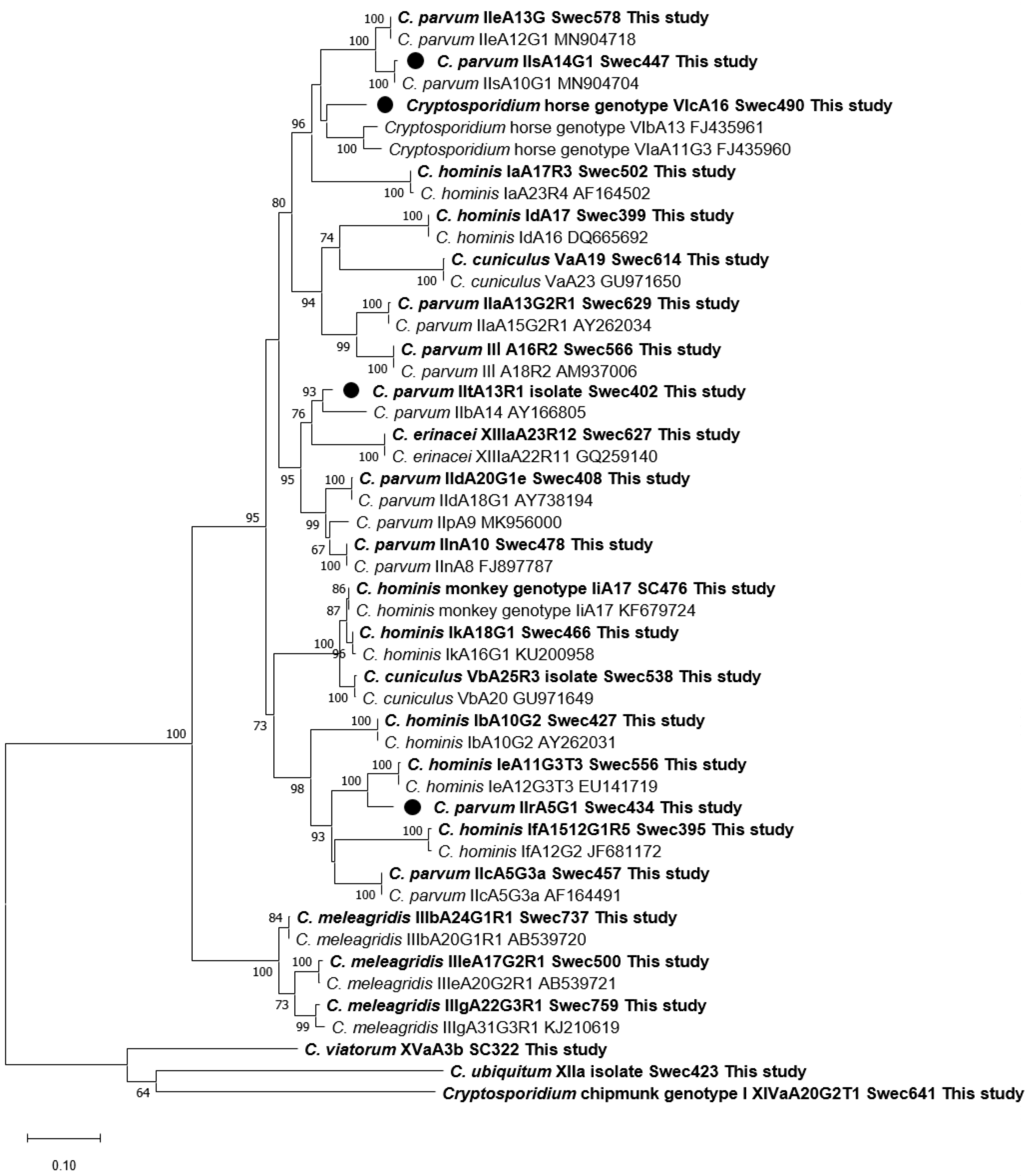
Three new C. parvum gp60 subtype families, IIr, IIs and IIt, were identified in one patient each. The sequences showed 100% identity with C. parvum at the SSU rRNA gene: IIr and IIt to AF164102 (gene copy A), and IIs to LC270281 (gene copy B). At the gp60 locus, subtype IItA13R1 (KU852718) was identified in a patient (Swec402) who had visited Tanzania prior to infection. The closest matches in GenBank were 93% to C. parvum subtype family IIb (AY166805) and 90% to IIp (MK956000). The other two patients (Swec447 and Swec434) were both infected in Sweden, the first with gp60 subtype IIsA14G1 (KU852720), where the closest match was 95% identity to subtype family IIe (KU852716), and the second with subtype IIrA5G1 (KU852719), which showed 92% similarity to the C. hominis subtype family Ie (AY738184). This observation prompted us to investigate two additional genes, actin and hsp70. As regards the actin locus, the isolate was 100% identical to C. parvum sequence MH542350, and at the hsp70 locus, where 1911 bp were successfully sequenced, it differed by eight SNPs compared with the IOWA C. parvum strain (XM625373) in the conserved part. These SNPs, however, did not display any changes in the amino acid sequence.

3.5. Molecular Characterization of Cryptosporidium hominis

Gp60 subtyping was successful for 49 out of 50 C. hominis isolates (including the sample with mixed species) (Table 4). Seven subtype families, Ia, Ib, Id, Ie, If, Ii, and Ik, and 17 subtypes were identified. The most common subtype was IbA10G2 (n = 15), wherein 13 cases had contracted infection while abroad; the remaining 2 reflected domestic infections. The second-most common subtype was IbA9G3 (n = 9), detected in patients who were infected in nine different countries, mainly in Africa (Table 4). In addition to the two cases with IbA10G2, another three subtypes were found in patients infected with C. hominis in Sweden: IaA18R3 (n = 2), IfA12G1R5 (n = 1), and IkA18G1 (n = 2). The cases with the uncommon subtype families Ii and Ik have been described in a separate article [29].

3.6. Molecular Characterization of C. hominis/C. parvum Mixed Infection

One patient, a 4-year-old girl from Syria, had a mixed C. hominis and C. parvum infection with subtypes IaA18R3 and IIaA16R1.

3.7. Outbreaks and Family Clusters

Four outbreaks, all caused by C. parvum, were identified during the study period (Table 5). The first outbreak occurred among veterinary students in 2013, and two different subtypes were found, IIaA16G1R1 and IIdA24G1 [27]. Outbreaks 2, 3 and 4 were all foodborne and involved subtypes IIaA16G2R1, IIaA17R1, and IIdA17R1, respectively. Three small family clusters were also detected; two related to traveling, and one where a contaminated water well was the suspected vehicle (Table 5).

3.8. Molecular Characterization of Additional Species

Of 30 isolates from additional species, 28 were successfully sequenced at the SSU rRNA locus. Subtyping with the gp60 protocol was successful for 29 isolates; the only exception was C. ditrichi, wherein no suitable primers were available (Table 6).
A recently adopted child from Lithuania was infected with C. suis. A gp60 sequence was achieved using the Cryptosporidium chipmunk primers [19], and by means of additional sequencing primers (data not shown) subtype XXVaR37 was obtained. This case and method will be described in a separate article.
Cases with Cryptosporidium chipmunk genotype I subtype XIVaA20G2T1 and C. ubiquitum subtype XIIa-1 were found in five and two patients, respectively, all infected in Sweden.
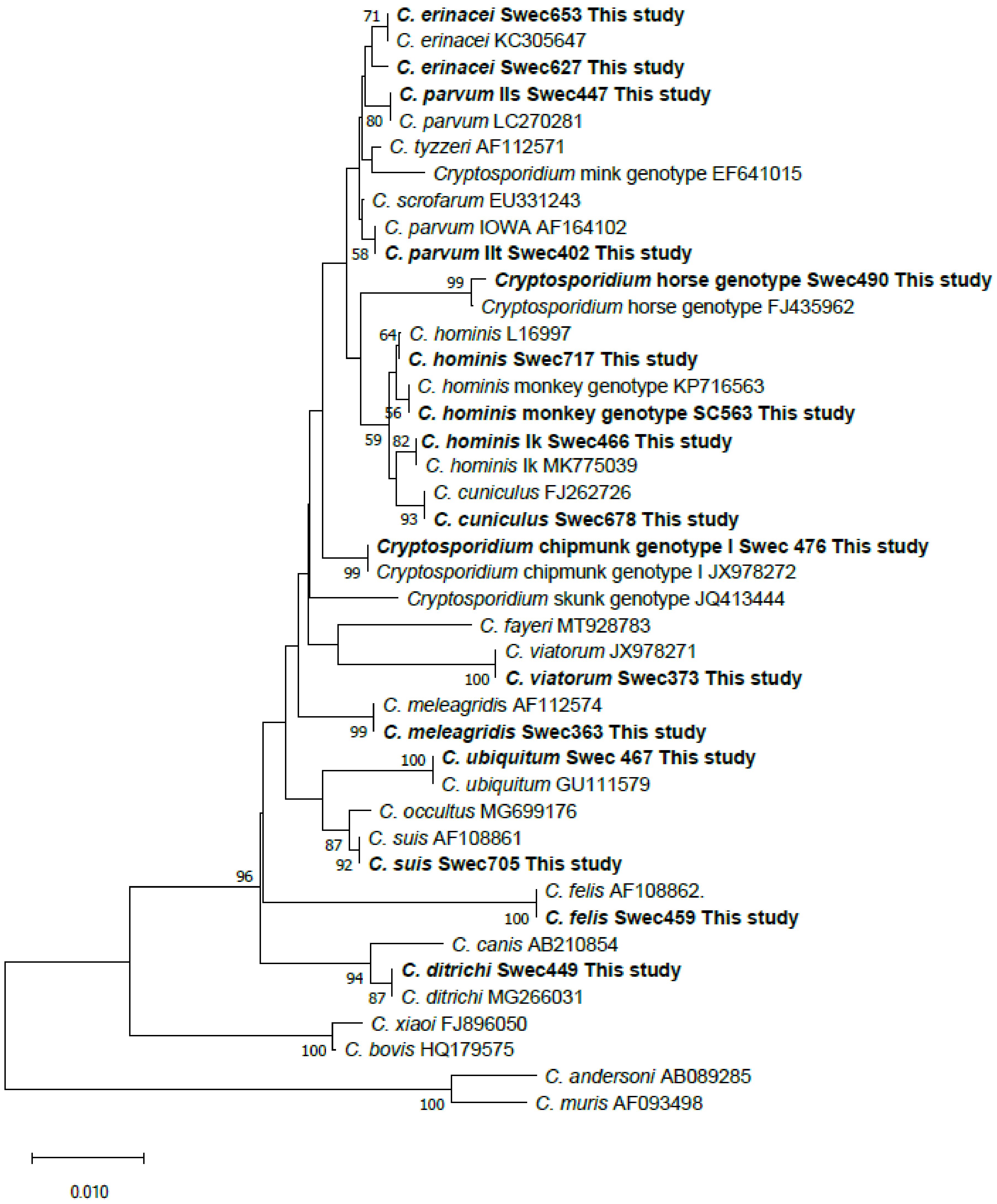
Two patients were infected with C. erinacei. The SSU rDNA sequences from their samples were not identical; the sequence from patient Swec627 (KU892565) infected in Sweden differed by two SNPs from the closest C. erinacei match in GenBank (KC3056047), while the sequence from patient Swec653 infected in Greece was 100% identical to KC3056047 (Figure 2). The actin DNA sequence obtained from isolate Swec627 was 100% identical to a C. erinacei isolate (MN237648) from a European badger in Poland (unpublished). Based on gp60 analysis, a new C. erinacei subtype, XIIIaA23R12, was identified in the first patient (Swec627), while subtype XIIIaA24R9 was found in the second patient (Swec653).
Four patients were diagnosed with C. felis; three in Sweden, and one in Indonesia. The recently described gp60 assay for C. felis [21] yielded four different sequence types.
One patient infected in Kenya with C. viatorum subtype XVaA3b was identified during the study period. Material from this case was used to develop a C. viatorum gp60 subtyping assay [22].
The eight patients diagnosed with C. meleagridis were all infected in Asia. Six of the samples were identified as genotype 1 at the SSU rRNA locus (AF112574), while sequencing of this gene failed for the remaining two isolates. Investigation of the gp60 gene was successful for all eight isolates and revealed three subtype families, IIIb, IIIe and IIIg, and six different subtypes (Table 6). Subtype IIIbA23G1R1c (KU852727) differed by eight SNPs in the post-repetitive part of the sequence from IIIbA23G1R1b (KJ210609), and was named IIIbA23G1R1c according to the proposed nomenclature [23].
Two subtype families of C. cuniculus, Va and Vb, and five different gp60 subtypes were observed. It was noted that the C. cuniculus Vb sequences published in GenBank carried 2–5 ACA repeats just after the TCA repeats (data not shown); thus, we followed the recommendation by Nolan et al. from 2010 [32] and included an R in the subtype designation, as in VbA29R4 for isolate Swe658, with 29 TCA repeats followed by 4 ACA repeats (KU852734). One patient infected in Greece carried a new subtype, VbA31R4. The sequence from patient Swec678 had the same numbers of TCA and ACA repeats as a VbA20R2 strain from the UK (GU971649), but differed by six SNPs and a 3 bp deletion in the post-repetitive part of the sequence, resulting in five amino acid changes. The subtype variant was designated VbA20R2b (KU852735), according to the proposed gp60 nomenclature [33].
A third subtype family of Cryptosporidium horse genotype, VIc, was identified in a patient (Swe490) who had visited Kenya. This subtype, referred to as VIcA16 (KU852738), showed 88% and 87% identity to subtype families VIa and VIb, respectively. Four ACA repeats followed just after the TCA repeats, a feature also observed in C. cuniculus subtype family Vb. An extended molecular investigation including the SSU rRNA, actin, and hsp70 genes was performed. The SSU rDNA sequence (KU892564) differed by one SNP from Cryptosporidium horse genotype sequences deposited in GenBank (FJ435962, MK775041). The closest match for the actin sequence (KU892571) was a sequence from Cryptosporidium tyzzeri (AF382343), from which it differed by eight SNPs. No actin sequences from the horse genotype were available in GenBank for comparison. The hsp70 sequence KU892577 (1895 bp) showed 100% identity with the four Cryptosporidium horse genotype sequences available in GenBank. However, none of these sequences (298–403 bp) were long enough to cover the area with the 12 bp segment repeats towards the 3′ end of the hsp70 gene. The horse genotype sequence from our study exhibited 10 repeats of a 12 bp segment with SNPs at the third and sixth bases—GG(C/T)GG(A/T)ATGCCA.

3.9. Phylogenetic Analyses

A phylogenetic tree, which contained representative SSU rDNA sequences from all 12 species and genotypes detected in the present study and published sequences from most Cryptosporidium species/genotypes hitherto detected in humans, was constructed (Figure 2). In the gp60 tree, one representative sequence from each subtype family detected in this study, except C. felis and C. suis, was included (n = 26). Sequences from established subtype families are clustered with sequences from this study. The new C. parvum subtype family IIt clustered with IIb, subtype family IIr with Ie, and subtype family IIs with IIe, while the new Cryptosporidium horse genotype subtype family VIc clustered with VIa and VIb (Figure 3).

4. Discussion

In the present study, performed between January 2013 and December 2014 and including samples from 398 patients with cryptosporidiosis, a high diversity of Cryptosporidium species and subtypes was identified. Cryptosporidium parvum was still the dominant species, and even fewer C. hominis cases were identified compared with a previous study performed in the Stockholm metropolitan area between April 2006 and November 2008. Meanwhile, the total number of patients infected with non-hominis and non-parvum species was quite high (8%), corroborating earlier observations [9].
Species determination was successful for 95% of the samples. Cryptosporidium parvum (79%) dominated both in patients infected in Sweden (84%) and abroad (57%), while C. hominis was much less common (13%) and identified in only 3% of the domestic cases and in 30% of the cases infected abroad. Mixed C. hominis and C. parvum infection was observed only in 1 patient, and for 10 of the patients infected with C. parvum, the origin of infection was unknown or uncertain. The high occurrence of C. parvum compared with C. hominis observed in Sweden is similar to the situation in other industrial countries, such as France, Ireland, and Canada [34,35,36], although a higher percentage of C. hominis has been observed in Spain and Australia [37,38]. Shifting trends have been seen over time in the Netherlands and New Zealand, showing the importance of longitudinal studies [39,40].
In terms of the analysis of cases according to age, the observed bimodal distribution (Figure 1) supports previous observations [9], with a relatively high number of cases observed among infants and toddlers and with a second peak—and the largest one—in the 30–44-year-olds. Quite similar age distributions have been observed in studies from Denmark, France, and Canada, but with the second peak in the 20–35-year-olds [34,36,41]. The bimodal age distribution may reflect transmission between parents and their children; however, no such family clusters were detected during the present study. There was a slight difference in the proportions of female and male cases: 55% and 45%, respectively. Interestingly, this difference in gender distribution has been seen in cryptosporidiosis cases in Sweden every year since 2005 (57% and 43% on average for women and men, respectively) [5]. A similar gender distribution was observed in neighboring Denmark in 2010–2014 [41]. Whether this difference in gender distribution reflects a higher awareness among females of the need to seek medical care, or something else, remains unknown.
A few C. parvum outbreaks were included in the present study (Table 5), but even if those patients (n = 22) are excluded from the dataset, C. parvum remains the dominant species in patients infected in Sweden, at least in the four areas that contributed most of the samples (Table 1). Differences in the geographical distribution of C. parvum and C. hominis, with more C. parvum in rural regions and more C. hominis in urban settings, have been described [42,43], and a similar tendency was seen in our study, where the rural region of Halland had relatively more cases of C. parvum and fewer cases of C. hominis compared with the metropolitan region of Stockholm (Table 1). These differences are often attributed to the closer contact with farm animals in rural areas and more traveling activity for people living in urban areas [44].
The subtyping of C. parvum showed a great variability; nine subtype families and 42 subtypes were observed. Two C. parvum subtype families, IIa (n = 164) and IId (n = 118), dominated both in patients infected in Sweden and abroad, while the other subtype families (IIc, IIe, IIl, IIn, IIr, IIs, IIt) were found only in one or two isolates each. The most common C. parvum subtype in the present study was IIaA16G1R1b (EU647727), observed in 39 patients. Detected in 24 sporadic cases and 4 cases related to an outbreak among veterinary students, it was also previously observed as the most common subtype amongst people infected in Sweden [9,27] (Table 5). This subtype is common in Swedish cattle and has been involved in earlier outbreaks and family clusters in Sweden [9,45], but was not reported amongst 48 subtyped C. parvum outbreaks between 2009 and 2017 in the UK [46].
Subtype IIaA15G2R1 is the dominating C. parvum subtype in many countries, both in cattle and in humans, and is responsible for numerous outbreaks of human cryptosporidiosis, probably due to its biological fitness and high virulence [41,46,47,48,49]. This subtype was found in 33 patients, of whom 16 were infected in Sweden; 13 were sporadic cases, and 3 cases were part of a family cluster (Table 5). This subtype has been detected in a few Swedish calves [50], but has not been associated with any known larger Cryptosporidium outbreak in Sweden.
Many of the IIa and IId subtype sequences in the present study exhibited examples of polymorphism in the non-repetitive part, which is sometimes indicated by a lower-case letter when sequences are reported to GenBank [33], but this is not always the case. To overcome this dilemma, all subtypes and subtype variants in Table 3 and Table 4 are referred to via a specific GenBank acc. no. As an example, the most common IId subtype, IIdA22G1 (n = 37), occurred in three variants in this study, and one of them (AY166806) (n = 15) was only detected in domestic cases, most of them in connection to a foodborne outbreak, in which parsley was the suspected vehicle (Table 5). Another variant of this subtype, IIdA22G1c (FJ917374), was identified in 19 cases, of which 15 represented sporadic cases infected in Sweden. Both these variants of IIdA22G1 have been seen in earlier Swedish studies of cattle and humans [9,50]. The third variant of this subtype, KR349104, was seen in only two cases; one infected in France and one in Sweden.
Subtype IIdA24G1 gained attention in 2011 in connection to a foodborne outbreak linking two Swedish cities, and it was also involved in an outbreak in 2013 among veterinary students occurring during the present study [12,27]. The same subtype, which is considered to be quite rare, was linked to 1 out of 48 C. parvum outbreaks in the UK where gp60 subtyping was performed [46].
The subtypes mentioned above, IIdA22G1c and IIdA24G1, were involved in two foodborne outbreaks, which occurred simultaneously in late 2019 in 10 and 12 Swedish counties, respectively. Subtype IIdA22G1c was identified in 122 cases and IIdA24G1 in 86 cases. Spinach juice was the suspected vehicle for subtype IIdA22G1c, while no specific food item could be identified as a source of infection for IIdA24G1 [51].
Two new C. parvum subtypes were identified within the subtype families IIe and IIn. Subtype IIeA13G1 (KU852716) was found in a patient infected in Sweden, and subtype IInA10 (KU852717) in two patients; one infected in Tanzania, and one with unclear origin of infection (either Tanzania or Sweden (secondary infection)). Cryptosporidium parvum IIe is a well-known anthroponotic subtype family, but since only two cases (both humans from India) have been reported from subtype family IIn [52], it is impossible to say whether this one might be an anthroponotic or zoonotic subtype family. The same is true for the 3 new C. parvum subtype families, IIr, IIs and IIt, that were added to the 16 families already described [48]. Subtype IIrA5G1 and IIsA14G1 were found in patients infected in Sweden. Interestingly, a patient with subtype IIsA10G1 was recently described in a publication from Zambia [53]. The patient with subtype IItA13R1 was infected in Tanzania.
One of the objectives of the present study was to investigate whether C. hominis subtype IbA10G2 had proliferated in Sweden after the large waterborne C. hominis outbreaks in Östersund and Skellefteå in 2010–2011, wherein an estimated 45,500 persons developed cryptosporidiosis. In the earlier study by Insulander et al. [9], performed from 2006 to 2008, 12 of the 17 C. hominis samples from patients infected in the Stockholm area carried subtype IbA10G2, while in the present study only two of the eight C. hominis patients infected in different parts of Sweden carried this subtype. The higher frequency of this subtype in the earlier study could be explained by the fact that several outbreaks at day care centers, with an index person infected abroad, appeared during that study period, while no such outbreaks were recorded in the current study. Few samples were submitted from the former outbreak areas; four from Jämtland County (Östersund) and four from Västerbotten County (Skellefteå), with all cases representing C. parvum (Table 1). The number of reported cases from these two counties during 2013 and 2014 was also low, at eight and five, respectively, and remained quite low during the following years up till now [5]. Therefore, even with these limitations in mind, we speculate that subtype IbA10G2 has failed to establish itself in Sweden following the large outbreaks.
In the US, where IbA10G2 used to be the most common outbreak-related subtype, a new C. hominis subtype, IaA28R4, emerged in 2007 as a major subtype in sporadic cases and waterborne outbreaks [54]. Since 2013, subtype IfA12G1R5 has emerged as the dominant C. hominis subtype in the US, as well as in Western Australia [37,55]. In the present study, IaA28R4 was observed in two patients, both of whom had recently visited the US, and subtype IfA12G1R5 in two patients, one of whom was infected in Sweden and one in Germany.
Cryptosporidium hominis is generally regarded as an anthroponotic species only occasionally infecting other animals. However, recent studies have shown that for a certain subtype family, Ik, equines are natural hosts [56]. During the present study, the first human cases infected with this subtype family, Ik, were detected [29] (Table 4). Two unrelated patients, both infected in Sweden, carried the same gp60 subtype, IkA18G1. This subtype was recently detected in a horse from China (MK770627)—information reinforcing the suspicion that zoonotic transmission might have occurred. Another uncommon C. hominis subtype family, Ii, was found in a father and son after returning from Thailand, where they visited a monkey farm. This C. hominis variant was previously described as C. hominis monkey genotype, and has rarely been detected in humans [29].
In the present study, 10 Cryptosporidium species/genotypes were found in addition to C. parvum and C. hominis, corresponding to 8% (30/379) of the total number of genotyped samples. This agrees with an earlier Swedish investigation performed on Cryptosporidium patients from the Stockholm metropolitan area, where 9% (17/194) of the genotyped samples represented species other than C. hominis and C. parvum. However, in the Stockholm study, the majority of these infections were acquired abroad; only three patients, two with Cryptosporidium chipmunk genotype I and one with C. felis, were infected in Sweden [9]. In the present study, an equal number of patients with species other than C. hominis or C. parvum were infected in Sweden (n = 15) and abroad (n = 15) (Table 1). This difference might reflect differences in study populations; in the first study, most patients originated from an urban area, while in the present study, many of the patients were from rural areas and probably more exposed to zoonotic Cryptosporidium species endemic to Sweden.
The most common cause of non-hominis and non-parvum infections acquired in Sweden was Cryptosporidium chipmunk genotype I, which was diagnosed in five adults; four women and one man. Cryptosporidiosis caused by the chipmunk genotype is considered an emerging infection in the US [19,43]. Meanwhile, in Europe, only one human case (from France) has been reported outside Sweden [19]. The first Swedish cases were diagnosed in September 2007 and August 2008, at a time when no gp60 subtype method was available for the chipmunk genotype I [57]. However, later analyses have shown that they carried the same subtype as the patients from the present study, XIVaA20G2T1, a subtype that recently was detected in red squirrels in Sweden (unpublished information). Red squirrels are consequently the most possible source of infection for our patients; the other described host animals—chipmunks, grey squirrels, and deer mice— are not native to the country [19,58]. Recent observations have shown a rising number of chipmunk genotype I infections acquired in Sweden, reflecting an emerging infection in this country (manuscript submitted).
Cryptosporidium erinacei was first described in European hedgehogs, but has also been identified in other animals, such as horses and rats [59,60,61]. Most human cases have been reported from New Zealand (n = 13), while reports from Europe are scarce, including one case from the Czech Republic and one from France [40,62,63]. The European hedgehog is considered an endangered species in Sweden and other parts of Europe, which might explain the low number of reports from Europe compared with New Zealand, where it has become an invasive species since its introduction about 150 years ago. The two cases found during the present study are the first human C. erinacei cases reported from Sweden. The patient infected in Sweden carried a unique gp60 subtype, XIIIaA23R12, while the patient infected in Greece carried subtype XIIIaA24R9, which has recently been reported in New Zealand [40].
Cryptosporidium cuniculus is a common species among rabbits worldwide, while human infection with this species has gained special attention in the UK, where, in addition to a documented outbreak, sporadic cases are quite common [64]. Reports of human infections from other parts of the world are increasing, and sporadic cases have been described in Nigeria, France, Spain, Australia, and New Zealand [40,48]. Two C. cuniculus subtype families have been described, Va and Vb. Four of the study patients were infected with Vb, which is the most commonly reported subtype family both in rabbits and humans, as well as in a few other animals, such as kangaroo and alpaca [65,66]. Subtype family Va, which was observed in one of the patients infected in Sweden, appears to be less frequent globally, and previous human infections were all reported in the UK [64]. These are the first documented human cases of C. cuniculus infection in Sweden.
One patient was infected with C. viatorum while visiting Kenya [22]. This species was initially thought to be anthroponotic because only human cases had been detected until 2018, when the first non-human host, an Australian swamp rat, was identified [67,68]. This swamp rat isolate was genetically quite different from all known human isolates, but recent studies from China focusing on different rat species have found C. viatorum isolates genetically similar to human isolates, and subtyping with gp60 has demonstrated the same subtype, XVaA3g, in wild rats from China and a human from Australia [37,69].
Another visitor to Kenya was diagnosed with Cryptosporidium horse genotype. All horse and donkey samples positive for this species when sequencing of the gp60 locus was performed carried subtype family VIa [70,71], which has also been found in one human case from Poland (MK784560) (unpublished). Only four cases with subtype family VIb have been described: two human cases, one in the UK and one in the US [54,72], and two cases in four-toed hedgehogs from Japan [73,74]. No reports of the VIb subtype family in equine hosts are available. The patient from our study carried a new gp60 subtype family (VIc) of Cryptosporidium horse genotype. Since no other reports of this subtype family have been published to date, we cannot speculate on how this patient contracted the infection.
Two children of 3 and 5 years of age, respectively, were infected in Sweden with C. ubiquitum, gp60 subtype XIIa-1. These cases were unrelated, and no probable source of infection was identified. Cryptosporidium ubiquitum has a wide host range and has been found in wild and domestic ruminants, rodents, and primates, including humans. In the UK, it is believed that most human infections with C. ubiquitum are related to exposure to sheep, which are common carriers of subtype XIIa, while infected humans in the US mainly carry the rodent-associated subtypes XIIb and XIIc [20]. The only published C. ubiquitum case amongst Swedish animals is from a calf [45], and the occurrence in other animals in Sweden, including sheep, remains unknown and should be investigated.
The natural hosts of C. suis are domesticated pigs and wild boars. Human infections are not frequently diagnosed; only around 12 cases were reported prior to our case, which was a two 2-year-old adopted child diagnosed with cryptosporidiosis upon arrival in Sweden from Lithuania [48,75].
Human infection with the feline parasite C. felis is not uncommon; indeed, it is regarded as one of the five most common Cryptosporidium species infecting humans world-wide [48]. During the study period, four C. felis cases were detected, with three patients being infected in Sweden and one in Indonesia. Two of the Swedish patients had known contact with cats, and analyses of fecal samples from cats and their owners using a newly described gp60 method showed that they shared gp60 subtypes; thus, zoonotic infection was confirmed [21,30].
One study patient was infected with C. ditrichi, a recently described Cryptosporidium species in Apodemus spp. in Europe [76]. This was the first time C. ditrichi was diagnosed in a human, and later on, two more Swedish cases were identified [31].
Eight patients were infected with C. meleagridis, the third most common Cryptosporidium species infecting humans [77]. Cryptosporidium meleagridis has a wide host range, ranging from various birds to humans, and in some countries (e.g., Thailand and Peru), this species is reportedly more common in humans than C. parvum [77]. All patients infected with C. meleagridis in the present study had acquired infection while traveling in different Asian countries. The same gp60 subtype was identified in two patients who had made the same journey to China, while the remaining six patients carried different subtypes (Table 6). Subtypes IIIeA17G2R1, IIIeA19G2R1 and IIIeA21G2R1 have been observed in chicken in China, and subtype IIIbA24G1R1 in poultry from Brazil, indicating zoonotic transmission as a possible route of infection [78,79]. Zoonotic transmission has been documented at a Swedish organic farm, where the infected person and the chickens carried the same hsp70 and gp60 subtypes [23,80].
When this study was initiated, the intention was to cover the entire country of Sweden, and all 21 clinical laboratories carrying out parasitology diagnostic tests in Sweden on a routine basis at the time of the study were invited to participate. One limitation is that by the end of the study period, only 12 of the regional laboratories (representing 11 different counties) had provided samples for the study. Our results, however, could very well be considered to reflect the reality, as nine counties only reported zero to one patient each with cryptosporidiosis during the study period (2013 and 2014) [5]. However, the number of referred samples varied considerably between the laboratories, with only four of them accounting for 95% of the submitted samples (Table 1). With this skewed distribution, we cannot conclude that the results of this study might be generalizable to the whole country. Another reason for this biased referral of samples could be the limited access to unfixed stool samples at some laboratories recommending that stool samples for parasitology be fixed in SAF. The intention at the beginning of the study was to avoid receiving samples fixed in SAF or formalin, which has a known negative effect on the PCR success rate [81], but in the second year, one laboratory with a high detection rate of Cryptosporidium and limited access to native (i.e., non-preserved) samples was invited to provide fixed specimens. In total, we received 70 samples fixed in SAF, 63 from this laboratory and 7 from other participating laboratories. The success rate of PCR for SAF-fixed samples was 80% (56/70), compared with 99% (323/328) for native stool samples. One recent study pointed out the limited availability of native stool material for the molecular analyses of stool parasites [34], but with the increased use of DNA-based diagnostics, the availability of unfixed material has increased, and the problem with fixed samples is in decline.
Another potential bias is that the clinical laboratories performing the primary diagnostics used different testing strategies and detection methods. Some diagnostic real-time PCR methods are designed to primarily detect C. parvum and C. hominis, and do not target some of the less common and genetically distinct species, which might have led to an underrepresentation of these in some of the counties [82].

5. Conclusions

Cryptosporidium parvum was the dominant species both in cases infected abroad and in domestic cases. The observed occurrence of C. hominis was generally low, and no indication of an expansion of the subtype IbA10G2 previously causing the vast Cryptosporidium outbreaks in Sweden was found. There was a high diversity of species and subtypes, with 8% of the cases reflecting species other than C. parvum and C. hominis, some of which were found for the first time in humans in Sweden (e.g., C. cuniculus, C. erinacei, and C. ubiquitum). Our study also identified humans as a new host of C. ditrichi. Overall, zoonotic species and subtypes plays a major role in human cryptosporidiosis in Sweden.

Author Contributions

Conceptualization, J.B. and J.W.-K.; methodology, J.B. and M.L.; software J.B. and M.L.; validation, J.B. and M.L.; formal analysis, J.B., J.W.-K. and M.L.; investigation, J.B., J.W.-K. and M.L.; resources, J.B.; data curation, J.B., J.W.-K. and M.L.; writing—original draft preparation, J.B., M.L. and C.R.S.; writing—review and editing, J.B., M.L. and C.R.S.; visualization, J.B., M.L. and C.R.S.; supervision, J.B.; project administration, J.B.; funding acquisition, J.B. All authors have read and agreed to the published version of the manuscript.

Funding

The study was funded by a grant from the Swedish Civil Contingencies Agency (Grant number: 2012-172).

Institutional Review Board Statement

Approval (registration number 2013/201-31/4) was obtained from the Ethical Review Board at Karolinska Institutet, Stockholm, Sweden, including approval to publish the data (date of approval: 27 February 2013).

Informed Consent Statement

Patient consent was waived since all samples were from patients with cryptosporidiosis, which is a notifiable disease in Sweden according to the Swedish Disease Act (2004:168) and included in the national surveillance of microbial pathogens. Species identification and genotyping of Cryptosporidium and communication of these data form part of the surveillance and do not require consent from the patient (https://www.folkhalsomyndigheten.se/the-public-health-agency-of-sweden/communicable-disease-control/surveillance-of-communicable-diseases/notifiable-diseases/ accessed on 24 April 2021).

Data Availability Statement

Data presented in this study are available on request from the corresponding author, Jessica Beser. Due to existing general data protection rules, the data are not publicly available.

Acknowledgments

The authors are grateful to all the clinical microbiological laboratories that provided samples to the study and the laboratory personnel at PHAS that performed analyses. The authors would also like to thank Margareta Löfdahl for providing epidemiological data and Erik Alm for the development of the software CryptoTyper.

Conflicts of Interest

The authors declare no conflict of interest.

References

  1. Kotloff, K.L.; Nataro, J.P.; Blackwelder, W.C.; Nasrin, D.; Farag, T.H.; Panchalingam, S.; Wu, Y.; Sow, S.O.; Sur, D.; Breiman, R.F.; et al. Burden and aetiology of diarrhoeal disease in infants and young children in developing countries (the Global Enteric Multicenter Study, GEMS): A prospective, case-control study. Lancet 2013, 382, 209–222. [Google Scholar] [CrossRef]
  2. Ryan, U.; Fayer, R.; Xiao, L. Cryptosporidium species in humans and animals: Current understanding and research needs. Parasitology 2014, 141, 1667–1685. [Google Scholar] [CrossRef] [Green Version]
  3. Feng, Y.; Ryan, U.M.; Xiao, L. Genetic diversity and population structure of Cryptosporidium. Trends Parasitol. 2018, 34, 997–1011. [Google Scholar] [CrossRef] [PubMed]
  4. Zahedi, A.; Ryan, U. Cryptosporidium—An update with an emphasis on foodborne and waterborne transmission. Res. Vet. Sci. 2020, 132, 500–512. [Google Scholar] [CrossRef] [PubMed]
  5. Public Health Agency of Sweden. Surveillance of Communicable Diseases Sweden. Available online: https://www.folkhalsomyndigheten.se/folkhalsorapportering-statistik/statistik-a-o/sjukdomsstatistik/cryptosporidiuminfektion/ (accessed on 24 April 2021).
  6. Harvala, H.; Ogren, J.; Boman, P.; Riedel, H.M.; Nilsson, P.; Winiecka-Krusnell, J.; Beser, J. Cryptosporidium infections in Sweden-understanding the regional differences in reported incidence. Clin. Microbiol. Infect. 2016, 22, 1012–1013. [Google Scholar] [CrossRef] [PubMed]
  7. Ogren, J.; Dienus, O.; Beser, J.; Henningsson, A.J.; Matussek, A. Protozoan infections are under-recognized in Swedish patients with gastrointestinal symptoms. Eur. J. Clin. Microbiol. Infect. Dis. 2020, 39, 2153–2160. [Google Scholar] [CrossRef]
  8. Waldron, L.S.; Dimeski, B.; Beggs, P.J.; Ferrari, B.C.; Power, M.L. Molecular epidemiology, spatiotemporal analysis, and ecology of sporadic human cryptosporidiosis in Australia. Appl. Environ. Microbiol. 2011, 77, 7757–7765. [Google Scholar] [CrossRef] [Green Version]
  9. Insulander, M.; Silverlås, C.; Lebbad, M.; Karlsson, L.; Mattsson, J.; Svenungsson, B. Molecular epidemiology and clinical manifestations of human cryptosporidiosis in Sweden. Epidemiol. Infect. 2013, 141, 1009–1020. [Google Scholar] [CrossRef] [PubMed] [Green Version]
  10. Bjelkmar, P.; Hansen, A.; Schönning, C.; Bergström, J.; Löfdahl, M.; Lebbad, M.; Wallensten, A.; Allestam, G.; Stenmark, S.; Lindh, J. Early outbreak detection by linking health advice line calls to water distribution areas retrospectively demonstrated in a large waterborne outbreak of cryptosporidiosis in Sweden. BMC Public Health 2017, 17, 328. [Google Scholar] [CrossRef] [PubMed]
  11. Widerström, M.; Schönning, C.; Lilja, M.; Lebbad, M.; Ljung, T.; Allestam, G.; Ferm, M.; Bjorkholm, B.; Hansen, A.; Hiltula, J.; et al. Large outbreak of Cryptosporidium hominis infection transmitted through the public water supply, Sweden. Emerg. Infect. Dis. 2014, 20, 581. [Google Scholar] [CrossRef] [PubMed]
  12. Gherasim, A.; Lebbad, M.; Insulander, M.; Decraene, V.; Kling, A.; Hjertqvist, M.; Wallensten, A. Two geographically separated food-borne outbreaks in Sweden linked by an unusual Cryptosporidium parvum subtype, October 2010. Eurosurveillance 2012, 17, 20318. [Google Scholar] [CrossRef] [PubMed] [Green Version]
  13. Insulander, M.; Lebbad, M.; Stenström, T.A.; Svenungsson, B. An outbreak of cryptosporidiosis associated with exposure to swimming pool water. Scand. J. Infect. Dis. 2005, 37, 354–360. [Google Scholar] [CrossRef]
  14. Alsmark, C.; Nolskog, P.; Angervall, A.L.; Toepfer, M.; Winiecka-Krusnell, J.; Bouwmeester, J.; Bjelkmar, P.; Troell, K.; Lahti, E.; Beser, J. Two outbreaks of cryptosporidiosis associated with cattle spring pasture events. Vet. Parasitol. Reg. Stud. Rep. 2018, 14, 71–74. [Google Scholar] [CrossRef]
  15. Xiao, L.; Escalante, L.; Yang, C.; Sulaiman, I.; Escalante, A.A.; Montali, R.J.; Fayer, R.; Lal, A.A. Phylogenetic analysis of Cryptosporidium parasites based on the small-subunit rRNA gene locus. Appl. Environ. Microbiol. 1999, 65, 1578–1583. [Google Scholar] [CrossRef] [Green Version]
  16. Xiao, L.; Bern, C.; Limor, J.; Sulaiman, I.; Roberts, J.; Checkley, W.; Cabrera, L.; Gilman, R.H.; Lal, A.A. Identification of 5 types of Cryptosporidium parasites in children in Lima, Peru. J. Infect. Dis. 2001, 183, 492–497. [Google Scholar] [CrossRef] [Green Version]
  17. Alves, M.; Xiao, L.; Sulaiman, I.; Lal, A.A.; Matos, O.; Antunes, F. Subgenotype analysis of Cryptosporidium isolates from humans, cattle, and zoo ruminants in Portugal. J. Clin. Microbiol. 2003, 41, 2744–2747. [Google Scholar] [CrossRef] [PubMed] [Green Version]
  18. Morgan, U.M.; Monis, P.T.; Xiao, L.; Limor, J.; Sulaiman, I.; Raidal, S.; O′Donoghue, P.; Gasser, R.; Murray, A.; Fayer, R.; et al. Molecular and phylogenetic characterisation of Cryptosporidium from birds. Int. J. Parasitol. 2001, 31, 289–296. [Google Scholar] [CrossRef]
  19. Guo, Y.; Cebelinski, E.; Matusevich, C.; Alderisio, K.A.; Lebbad, M.; McEvoy, J.; Roellig, D.M.; Yang, C.; Feng, Y.; Xiao, L. Subtyping novel zoonotic pathogen Cryptosporidium chipmunk genotype I. J. Clin. Microbiol. 2015, 53, 1648–1654. [Google Scholar] [CrossRef] [PubMed] [Green Version]
  20. Li, N.; Xiao, L.; Alderisio, K.; Elwin, K.; Cebelinski, E.; Chalmers, R.; Santin, M.; Fayer, R.; Kvac, M.; Ryan, U.; et al. Subtyping Cryptosporidium ubiquitum, a zoonotic pathogen emerging in humans. Emerg. Infect. Dis. 2014, 20, 217. [Google Scholar] [CrossRef] [PubMed]
  21. Rojas-Lopez, L.; Elwin, K.; Chalmers, R.M.; Enemark, H.L.; Beser, J.; Troell, K. Development of a gp60-subtyping method for Cryptosporidium felis. Parasites Vectors 2020, 13, 1–8. [Google Scholar] [CrossRef] [PubMed]
  22. Stensvold, C.; Elwin, K.; Winiecka-Krusnell, J.; Chalmers, R.; Xiao, L.; Lebbad, M. Development and application of a gp60-based typing assay for Cryptosporidium viatorum. J. Clin. Microbiol. 2015, 53, 1891–1897. [Google Scholar] [CrossRef] [PubMed] [Green Version]
  23. Stensvold, C.R.; Beser, J.; Axén, C.; Lebbad, M. High applicability of a novel method for gp60-based subtyping of Cryptosporidium meleagridis. J. Clin. Microbiol. 2014, 52, 2311–2319. [Google Scholar] [CrossRef] [Green Version]
  24. Sulaiman, I.M.; Lal, A.A.; Xiao, L. Molecular phylogeny and evolutionary relationships of Cryptosporidium parasites at the actin locus. J. Parasitol. 2002, 88, 388–394. [Google Scholar] [CrossRef]
  25. Sulaiman, I.M.; Morgan, U.M.; Thompson, R.C.; Lal, A.A.; Xiao, L. Phylogenetic relationships of Cryptosporidium parasites based on the 70-kilodalton heat shock protein (HSP70) gene. Appl. Environ. Microbiol. 2000, 66, 2385–2391. [Google Scholar] [CrossRef] [PubMed] [Green Version]
  26. Kumar, S.; Stecher, G.; Li, M.; Knyaz, C.; Tamura, K. MEGA X: Molecular evolutionary genetics analysis across computing platforms. Molecular Biol. Evol. 2018, 35, 1547–1549. [Google Scholar] [CrossRef] [PubMed]
  27. Kinross, P.; Beser, J.; Troell, K.; Axen, C.; Björkman, C.; Lebbad, M.; Winiecka-Krusnell, J.; Lindh, J.; Lofdahl, M. Cryptosporidium parvum infections in a cohort of veterinary students in Sweden. Epidemiol Infect. 2015, 143, 2748–2756. [Google Scholar] [CrossRef] [PubMed] [Green Version]
  28. Amer, S.; Zidan, S.; Adamu, H.; Ye, J.; Roellig, D.; Xiao, L.; Feng, Y. Prevalence and characterization of Cryptosporidium spp. in dairy cattle in Nile River delta provinces, Egypt. Exp. Parasitol. 2013, 135, 518–523. [Google Scholar] [CrossRef] [PubMed]
  29. Lebbad, M.; Winiecka-Krusnell, J.; Insulander, M.; Beser, J. Molecular characterization and epidemiological investigation of Cryptosporidium hominis IkA18G1 and C. hominis monkey genotype IiA17, two unusual subtypes diagnosed in Swedish patients. Exp. Parasitol. 2018, 188, 7. [Google Scholar] [CrossRef] [PubMed]
  30. Beser, J.; Toresson, L.; Eitrem, R.; Troell, K.; Winiecka-Krusnell, J.; Lebbad, M. Possible zoonotic transmission of Cryptosporidium felis in a household. Infect. Ecol. Epidemiol. 2015, 5, 28463. [Google Scholar] [CrossRef] [PubMed] [Green Version]
  31. Beser, J.; Bujila, I.; Wittesjo, B.; Lebbad, M. From mice to men: Three cases of human infection with Cryptosporidium ditrichi. Infect. Genet. Evol. 2020, 78, 104120. [Google Scholar] [CrossRef] [PubMed]
  32. Nolan, M.J.; Jex, A.R.; Haydon, S.R.; Stevens, M.A.; Gasser, R.B. Molecular detection of Cryptosporidium cuniculus in rabbits in Australia. Infect. Genet. Evol. 2010, 10, 1179–1187. [Google Scholar] [CrossRef]
  33. Sulaiman, I.M.; Hira, P.R.; Zhou, L.; Al-Ali, F.M.; Al-Shelahi, F.A.; Shweiki, H.M.; Iqbal, J.; Khalid, N.; Xiao, L. Unique endemicity of cryptosporidiosis in children in Kuwait. J Clin. Microbiol. 2005, 43, 2805–2809. [Google Scholar] [CrossRef] [PubMed] [Green Version]
  34. Guy, R.A.; Yanta, C.A.; Muchaal, P.K.; Rankin, M.A.; Thivierge, K.; Lau, R.; Boggild, A.K. Molecular characterization of Cryptosporidium isolates from humans in Ontario, Canada. Parasites Vectors 2021, 14, 69. [Google Scholar] [CrossRef] [PubMed]
  35. O’Leary, J.K.; Blake, L.; Corcoran, G.D.; Sleator, R.D.; Lucey, B. Increased diversity and novel subtypes among clinical Cryptosporidium parvum and Cryptosporidium hominis isolates in Southern Ireland. Exp. Parasitol. 2020, 218, 107967. [Google Scholar] [CrossRef] [PubMed]
  36. Costa, D.; Razakandrainibe, R.; Valot, S.; Vannier, M.; Sautour, M.; Basmaciyan, L.; Gargala, G.; Viller, V.; Lemeteil, D.; Ballet, J.-J. Epidemiology of Cryptosporidiosis in France from 2017 to 2019. Microorganisms 2020, 8, 1358. [Google Scholar] [CrossRef]
  37. Braima, K.; Zahedi, A.; Oskam, C.; Reid, S.; Pingault, N.; Xiao, L.; Ryan, U. Retrospective analysis of Cryptosporidium species in Western Australian human populations (2015–2018), and emergence of the C. hominis IfA12G1R5 subtype. Infect. Genet. Evol. 2019, 73, 306–313. [Google Scholar] [CrossRef] [PubMed]
  38. De Lucio, A.; Merino, F.J.; Martínez-Ruiz, R.; Bailo, B.; Aguilera, M.; Fuentes, I.; Carmena, D. Molecular genotyping and sub-genotyping of Cryptosporidium spp. isolates from symptomatic individuals attending two major public hospitals in Madrid, Spain. Infect. Genet. Evol. 2016, 37, 49–56. [Google Scholar] [CrossRef] [PubMed]
  39. Nic Lochlainn, L.M.; Sane, J.; Schimmer, B.; Mooij, S.; Roelfsema, J.; Van Pelt, W.; Kortbeek, T. Risk Factors for Sporadic Cryptosporidiosis in the Netherlands: Analysis of a 3-Year Population Based Case-Control Study Coupled with Genotyping, 2013–2016. J. Infect. Dis. 2019, 219, 1121–1129. [Google Scholar] [CrossRef] [PubMed]
  40. Garcia-R, J.C.; Pita, A.B.; Velathanthiri, N.; French, N.P.; Hayman, D.T. Species and genotypes causing human cryptosporidiosis in New Zealand. Parasitol. Res. 2020, 119, 2317–2326. [Google Scholar] [CrossRef] [PubMed]
  41. Stensvold, C.R.; Ethelberg, S.; Hansen, L.; Sahar, S.; Voldstedlund, M.; Kemp, M.; Hartmeyer, G.T.; Otte, E.; Engsbro, A.L.; Nielsen, H.V.; et al. Cryptosporidium infections in Denmark, 2010–2014. Dan. Med. J. 2015, 62, A5086. [Google Scholar] [PubMed]
  42. Chalmers, R.; Elwin, K.; Thomas, A.; Guy, E.; Mason, B. Long-term Cryptosporidium typing reveals the aetiology and species-specific epidemiology of human cryptosporidiosis in England and Wales, 2000 to 2003. Euro Surveill. 2009, 14, 19086. [Google Scholar] [CrossRef] [PubMed]
  43. Loeck, B.K.; Pedati, C.; Iwen, P.C.; McCutchen, E.; Roellig, D.M.; Hlavsa, M.C.; Fullerton, K.; Safranek, T.; Carlson, A.V. Genotyping and Subtyping Cryptosporidium to Identify Risk Factors and Transmission Patterns—Nebraska, 2015–2017. Morb. Mortal. Wkly. Rep. 2020, 69, 335. [Google Scholar] [CrossRef]
  44. Chalmers, R.; Smith, R.; Elwin, K.; Clifton-Hadley, F.; Giles, M. Epidemiology of anthroponotic and zoonotic human cryptosporidiosis in England and Wales, 2004–2006. Epidemiol. Infect. 2011, 139, 700–712. [Google Scholar] [CrossRef] [Green Version]
  45. Björkman, C.; Lindström, L.; Oweson, C.; Ahola, H.; Troell, K.; Axén, C. Cryptosporidium infections in suckler herd beef calves. Parasitology 2015, 142, 1108–1114. [Google Scholar] [CrossRef] [Green Version]
  46. Chalmers, R.M.; Robinson, G.; Elwin, K.; Elson, R. Analysis of the Cryptosporidium spp. and gp60 subtypes linked to human outbreaks of cryptosporidiosis in England and Wales, 2009 to 2017. Parasites Vectors 2019, 12, 95. [Google Scholar] [CrossRef] [PubMed]
  47. Thomas-Lopez, D.; Müller, L.; Vestergaard, L.S.; Christoffersen, M.; Andersen, A.-M.; Jokelainen, P.; Agerholm, J.S.; Stensvold, C.R. Veterinary students have a higher risk of contracting cryptosporidiosis when calves with high fecal Cryptosporidium loads are used for fetotomy exercises. Appl. Environ. Microbiol. 2020, 86. [Google Scholar] [CrossRef]
  48. Xiao, L.; Feng, Y. Molecular epidemiologic tools for waterborne pathogens Cryptosporidium spp. and Giardia duodenalis. Food Waterborne Parasitol. 2017, 8, 14–32. [Google Scholar] [CrossRef] [PubMed]
  49. Feng, Y.; Torres, E.; Li, N.; Wang, L.; Bowman, D.; Xiao, L. Population genetic characterisation of dominant Cryptosporidium parvum subtype IIaA15G2R1. Int. J. Parasitol. 2013, 43, 1141–1147. [Google Scholar] [CrossRef] [PubMed]
  50. Silverlås, C.; Bosaeus-Reineck, H.; Näslund, K.; Björkman, C. Is there a need for improved Cryptosporidium diagnostics in Swedish calves? Int. J. Parasitol. 2013, 43, 155–161. [Google Scholar] [CrossRef]
  51. Whithworth, J. Five Foodborne Outbreaks Added to Cryptosporidium Rise in Sweden. Food Safety News, 13 March 2020. Available online: https://www.foodsafetynews.com/2020/03/five-foodborne-outbreaks-added-to-cryptosporidium-rise-in-sweden/ (accessed on 24 April 2021).
  52. Ajjampur, S.S.; Liakath, F.B.; Kannan, A.; Rajendran, P.; Sarkar, R.; Moses, P.D.; Simon, A.; Agarwal, I.; Mathew, A.; O’Connor, R.; et al. Multisite study of cryptosporidiosis in children with diarrhea in India. J Clin. Microbiol. 2010, 48, 2075–2081. [Google Scholar] [CrossRef] [Green Version]
  53. Mulunda, N.R.; Hayashida, K.; Yamagishi, J.; Sianongo, S.; Munsaka, G.; Sugimoto, C.; Mutengo, M.M. Molecular characterization of Cryptosporidium spp. from patients with diarrhoea in Lusaka, Zambia. Parasite 2020, 27, 53. [Google Scholar] [CrossRef]
  54. Xiao, L.; Hlavsa, M.C.; Yoder, J.; Ewers, C.; Dearen, T.; Yang, W.; Nett, R.; Harris, S.; Brend, S.M.; Harris, M.; et al. Subtype analysis of Cryptosporidium specimens from sporadic cases in Colorado, Idaho, New Mexico, and Iowa in 2007: Widespread occurrence of one Cryptosporidium hominis subtype and case history of an infection with the Cryptosporidium horse genotype. J. Clin. Microbiol. 2009, 47, 3017–3020. [Google Scholar] [CrossRef] [PubMed] [Green Version]
  55. Hlavsa, M.C.; Roellig, D.M.; Seabolt, M.H.; Kahler, A.M.; Murphy, J.L.; McKitt, T.K.; Geeter, E.F.; Dawsey, R.; Davidson, S.L.; Kim, T.N.; et al. Using Molecular Characterization to Support Investigations of Aquatic Facility-Associated Outbreaks of Cryptosporidiosis—Alabama, Arizona, and Ohio, 2016. MMWR Morb. Mortal. Wkly. Rep. 2017, 66, 493–497. [Google Scholar] [CrossRef] [PubMed] [Green Version]
  56. Jian, F.; Liu, A.; Wang, R.; Zhang, S.; Qi, M.; Zhao, W.; Shi, Y.; Wang, J.; Wei, J.; Zhang, L.; et al. Common occurrence of Cryptosporidium hominis in horses and donkeys. Infect. Genet. Evol. 2016, 43, 261–266. [Google Scholar] [CrossRef] [PubMed]
  57. Lebbad, M.; Beser, J.; Insulander, M.; Karlsson, L.; Mattsson, J.G.; Svenungsson, B.; Axén, C. Unusual cryptosporidiosis cases in Swedish patients: Extended molecular characterization of Cryptosporidium viatorum and Cryptosporidium chipmunk genotype I. Parasitology 2013, 140, 1735–1740. [Google Scholar] [CrossRef] [Green Version]
  58. Kvác, M.; Hofmannová, L.; Bertolino, S.; Wauters, L.; Tosi, G.; Modrý, D. Natural infection with two genotypes of Cryptosporidium in red squirrels (Sciurus vulgaris) in Italy. Folia Parasitol. 2008, 55, 95. [Google Scholar] [CrossRef] [Green Version]
  59. Zhao, W.; Zhou, H.; Huang, Y.; Xu, L.; Rao, L.; Wang, S.; Wang, W.; Yi, Y.; Zhou, X.; Wu, Y.; et al. Cryptosporidium spp. in wild rats (Rattus spp.) from the Hainan Province, China: Molecular detection, species/genotype identification and implications for public health. Int. J. Parasitol. Parasites Wildl. 2019, 9, 317–321. [Google Scholar] [CrossRef]
  60. Laatamna, A.E.; Wagnerova, P.; Sak, B.; Kvetonova, D.; Aissi, M.; Rost, M.; Kvac, M. Equine cryptosporidial infection associated with Cryptosporidium hedgehog genotype in Algeria. Vet. Parasitol. 2013, 197, 350–353. [Google Scholar] [CrossRef]
  61. Kváč, M.; Hofmannová, L.; Hlásková, L.; Květoňová, D.; Vítovec, J.; McEvoy, J.; Sak, B. Cryptosporidium erinacei n. sp. (Apicomplexa: Cryptosporidiidae) in hedgehogs. Vet. Parasitol. 2014, 201, 9–17. [Google Scholar] [CrossRef]
  62. Costa, D.; Razakandrainibe, R.; Sautour, M.; Valot, S.; Basmaciyan, L.; Gargala, G.; Lemeteil, D.; Favennec, L.; Dalle, F. Human cryptosporidiosis in immunodeficient patients in France (2015–2017). Exp. Parasitol. 2018, 192, 108–112. [Google Scholar] [CrossRef] [PubMed]
  63. Kvac, M.; Sakova, K.; Kvetonova, D.; Kicia, M.; Wesolowska, M.; McEvoy, J.; Sak, B. Gastroenteritis caused by the Cryptosporidium hedgehog genotype in an immunocompetent man. J. Clin. Microbiol. 2014, 52, 347–349. [Google Scholar] [CrossRef] [Green Version]
  64. Puleston, R.L.; Mallaghan, C.M.; Modha, D.E.; Hunter, P.R.; Nguyen-Van-Tam, J.S.; Regan, C.M.; Nichols, G.L.; Chalmers, R.M. The first recorded outbreak of cryptosporidiosis due to Cryptosporidium cuniculus (formerly rabbit genotype), following a water quality incident. J Water Health 2014, 12, 41–50. [Google Scholar] [CrossRef] [PubMed] [Green Version]
  65. Koehler, A.V.; Rashid, M.H.; Zhang, Y.; Vaughan, J.L.; Gasser, R.B.; Jabbar, A. First cross-sectional, molecular epidemiological survey of Cryptosporidium, Giardia and Enterocytozoon in alpaca (Vicugna pacos) in Australia. Parasites Vectors 2018, 11, 498. [Google Scholar] [CrossRef] [PubMed]
  66. Koehler, A.V.; Whipp, M.J.; Haydon, S.R.; Gasser, R.B. Cryptosporidium cuniculus--new records in human and kangaroo in Australia. Parasites Vectors 2014, 7, 492. [Google Scholar] [CrossRef]
  67. Elwin, K.; Hadfield, S.J.; Robinson, G.; Crouch, N.D.; Chalmers, R.M. Cryptosporidium viatorum n. sp. (Apicomplexa: Cryptosporidiidae) among travellers returning to Great Britain from the Indian subcontinent, 2007–2011. Int. J. Parasitol. 2012, 42, 675–682. [Google Scholar] [CrossRef] [PubMed]
  68. Koehler, A.V.; Wang, T.; Haydon, S.R.; Gasser, R.B. Cryptosporidium viatorum from the native Australian swamp rat Rattus lutreolus-an emerging zoonotic pathogen? Int. J. Parasitol. Parasites Wildl. 2018, 7, 18–26. [Google Scholar] [CrossRef] [PubMed]
  69. Chen, Y.-W.; Zheng, W.-B.; Zhang, N.-Z.; Gui, B.-Z.; Lv, Q.-Y.; Yan, J.-Q.; Zhao, Q.; Liu, G.H. Identification of Cryptosporidium viatorum XVa subtype family in two wild rat species in China. Parasites Vectors. 2019, 12, 1–5. [Google Scholar] [CrossRef] [PubMed]
  70. Burton, A.; Nydam, D.; Dearen, T.; Mitchell, K.; Bowman, D.; Xiao, L. The prevalence of Cryptosporidium, and identification of the Cryptosporidium horse genotype in foals in New York State. Vet. Parasitol. 2010, 174, 139–144. [Google Scholar] [CrossRef]
  71. Caffara, M.; Piva, S.; Pallaver, F.; Iacono, E.; Galuppi, R. Molecular characterization of Cryptosporidium spp. from foals in Italy. Vet. J. 2013, 198, 531–533. [Google Scholar] [CrossRef] [PubMed]
  72. Robinson, G.; Elwin, K.; Chalmers, R.M. Unusual Cryptosporidium genotypes in human cases of diarrhea. Emerg. Infect. Dis. 2008, 14, 1800. [Google Scholar] [CrossRef]
  73. Abe, N.; Matsubara, K. Molecular identification of Cryptosporidium isolates from exotic pet animals in Japan. Vet. Parasitol. 2015, 209, 254–257. [Google Scholar] [CrossRef]
  74. Takaki, Y.; Takami, Y.; Watanabe, T.; Nakaya, T.; Murakoshi, F. Molecular identification of Cryptosporidium isolates from ill exotic pet animals in Japan including a new subtype in Cryptosporidium fayeri. Vet. Parasitol. Reg. Stud. Rep. 2020, 21, 100430. [Google Scholar] [CrossRef]
  75. Sannella, A.R.; Suputtamongkol, Y.; Wongsawat, E.; Caccio, S.M. A retrospective molecular study of Cryptosporidium species and genotypes in HIV-infected patients from Thailand. Parasites Vectors 2019, 12, 91, Epub 2019/03/15. [Google Scholar] [CrossRef] [PubMed]
  76. Condlova, S.; Horcickova, M.; Sak, B.; Kvetonova, D.; Hlaskova, L.; Konecny, R.; Stanko, M.; McEvoy, J.; Kvac, M. Cryptosporidium apodemi sp. n. and Cryptosporidium ditrichi sp. n. (Apicomplexa: Cryptosporidiidae) in Apodemus spp. Eur. J. Protistol. 2018, 63, 1–12. [Google Scholar] [CrossRef]
  77. Xiao, L.; Feng, Y. Zoonotic cryptosporidiosis. FEMS Immunol. Med Microbiol. 2008, 52, 309–323. [Google Scholar] [CrossRef] [PubMed]
  78. Da Cunha, M.J.R.; Cury, M.C.; Santín, M. Molecular characterization of Cryptosporidium spp. in poultry from Brazil. Res. Vet. Sci. 2018, 118, 331–335. [Google Scholar] [CrossRef] [PubMed]
  79. Liao, C.; Wang, T.; Koehler, A.V.; Fan, Y.; Hu, M.; Gasser, R.B. Molecular investigation of Cryptosporidium in farmed chickens in Hubei Province, China, identifies ‘zoonotic’ subtypes of C. meleagridis. Parasites Vectors 2018, 11, 1–8. [Google Scholar] [CrossRef] [PubMed]
  80. Silverlås, C.; Mattsson, J.G.; Insulander, M.; Lebbad, M. Zoonotic transmission of Cryptosporidium meleagridis on an organic Swedish farm. Int. J. Parasitol. 2012, 42, 963–967. [Google Scholar] [CrossRef] [PubMed]
  81. Abdelsalam, I.M.; Sarhan, R.M.; Hanafy, M.A. The impact of different copro-preservation conditions on molecular detection of Cryptosporidium species. Iran. J. Parasitol. 2017, 12, 274. [Google Scholar]
  82. Autier, B.; Belaz, S.; Razakandrainibe, R.; Gangneux, J.P.; Robert-Gangneux, F. Comparison of three commercial multiplex PCR assays for the diagnosis of intestinal protozoa. Parasite 2018, 25, 48. [Google Scholar] [CrossRef] [PubMed]
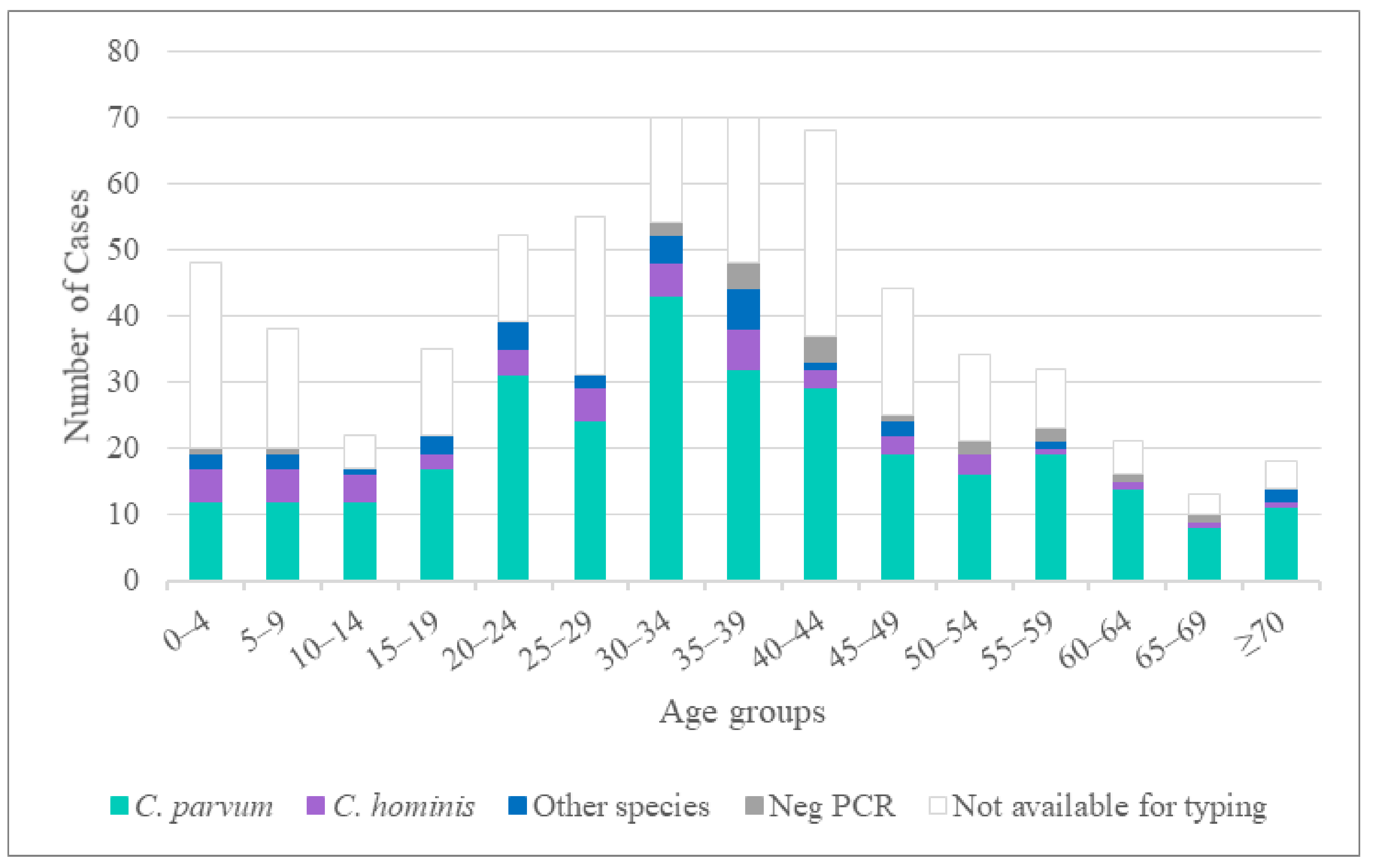
Figure 1. Number of reported cases of cryptosporidiosis detected in Sweden in 2013 and 2014 according to age group. One case with mixed C. hominis and C. parvum infection is not included in the figure.
Pathogens 10 00523 g001
Figure 2. Phylogenetic relationships between partial SSU rDNA Cryptosporidium sequences obtained in the present study and sequences retrieved from the NCBI database. Trees were constructed using the neighbor-joining method based on genetic distance calculated by the Kimura’s 2-parameter model as implemented in MEGA X. The final dataset included 749 positions. Bootstrap values ≥50% from 1000 replicates are indicated at each node. Isolates from this study are indicated in boldface.
Pathogens 10 00523 g002
Figure 3. Phylogenetic relationships between partial gp60 Cryptosporidium sequences obtained in the present study and sequences retrieved from the NCBI database. Trees were constructed using the neighbor-joining method based on genetic distance calculated by the Kimura’s 2-parameter model as implemented in MEGA X. The final dataset included 840 positions. Bootstrap values ≥50% from 1000 replicates are indicated at each node. New subtype families observed in this study are indicated by filled circles. All isolates from this study are indicated in boldface.
Pathogens 10 00523 g003
Table 1. Cryptosporidium species in samples provided by 12 participating laboratories across 11 Swedish counties and locally identified as Cryptosporidium-positive.
CountyNumber of LaboratoriesNumber of SamplesSpecies (Number of Samples)
Halland1138C. parvum (112), C. hominis (15), C. cuniculus (5), C. erinacei (2), C. meleagridis (1), non-typeable (3)
Jämtland14C. parvum (4)
Jönköping176C. parvum (58), C. hominis (5), Cryptosporidium chipmunk genotype I (3), non-typeable (10)
Kronoberg11C. felis (1)
Skåne15C. parvum (3), C. felis (1), Cryptosporidium horse genotype (1)
Stockholm292C. parvum (62), C. hominis (23), C. ubiquitum (2), C. ditrichi (1), C. felis (1), C. meleagridis (1), C. viatorum (1), non-typeable (1)
Uppsala170C. parvum (52), C. hominis (6), C. meleagridis (6), Cryptosporidium chipmunk genotype I (2), C. felis (1), C. hominis + C. parvum (1), C. suis (1), non-typeable (1)
Västerbotten14C. parvum (4)
Västernorrland12C. parvum (2)
Västra Götaland15C. parvum (2), non-typeable (3)
Örebro11non-typeable (1)
Total12398C. parvum (299), C. hominis (49), C. parvum + C. hominis (1), C. meleagridis (8), C. cuniculus (5), Cryptosporidium chipmunk genotype I (5), C. felis (4), C. erinacei (2), C. ubiquitum (2), C. ditrichi (1), C. suis (1), C. viatorum (1), Cryptosporidium horse genotype (1), non-typeable (19)
Table 2. Distribution of Cryptosporidium spp. according to area of origin of infection among 398 patients with cryptosporidiosis diagnosed in Sweden from 2013 to 2014.
Species/Genotypes Number of Samples (%)
TotalSwedenOther European CountriesAfricaAsiaNorth AmericaSouth AmericaUnknown
C. parvum299211 (71)60 (20)9 (3)5 (2)3 (1)110 (3)
C. hominis498 (16)10 (20)14 (29)13 (27)3 (6)1 (2)0
C. parvum + C. hominis10001000
C. meleagridis80008 (100)000
C. cuniculus53 (60)2 (40)00000
Cryptosporidium chipmunk genotype I55 (100)000000
C. felis43 (75)001 (25)000
C. erinacei21100000
C. ubiquitum22000000
C. suis10100000
C. viatorum10010000
Cryptosporidium horse genotype10010000
C. ditrichi11000000
None-typable 11916 (84)1 (5)2 (11)0000
All cases398250 (63)75 (19)27 (7)28 (7)6 (3)2 (1)10 (3)
1 Negative in all PCRs.
Table 3. Cryptosporidium parvum gp60 sequences generated in this study.
Species (No. of Patients)Subtype Family (No. of Patients)Subtype (No. of Patients)GenBank Acc. No. 1Origin of Infection (No. of Patients)
C. parvum (299)IIa (164)IIaA13G2R1 (2)KU852706Turkey (1)
KU852701Morocco (1)
IIaA13R1 (1)KU852702Sweden (1)
IIaA14G1R1 (13)JQ030882Cyprus (1) Georgia (2) Sweden (3)
JX183798 (IIaA14G1R1b)Sweden (7)
IIaA14G1R1r1 (6)KU852703Sweden (4) Spain (1) Uzbekistan (1)
IIaA14G2R1 (1)KF128738Greece (1)
IIaA14R1 (2)JX183797Estonia (1) Sweden (1)
IIaA15G1R1 (6)AM937012Sweden (4) unknown (1)
KU852704 (IIaA15G1R1_variant)Sweden (1)
IIaA15G2R1 (31)AF164490Dominican Republic (1) France (1) Mexico (1) Portugal (9) Portugal/Spain (1) Sweden (16) Venezuela (1) unknown (1)
IIaA16G1R1 (42)EU647727 (IIaA16G1R1b)Georgia (1) Italy (1) Serbia (1) Spain (6) Sweden (30 2)
KU852707unknown (1)
KT895368 (IIaA16G1R1b_variant)Austria (1) Sweden (1)
IIaA16G2R1 (3)DQ192505Sweden (3)
IIaA16G3R1 (2)DQ192506Portugal (1) Sweden (1)
IIaA16R1 (1)AM937010Malta (1)
IIaA17G1R1 (20)GQ983359Poland (1) Sweden (1)
JX183801 (IIaA17G1R1c)Italy (1) Sweden (11) South Africa (1)
AF403168 (IIaA17G1R1c_variant)Sweden (5)
IIaA17R1 (1)JX183800Sweden (1)
IIaA18G1R1 (12)HQ005742Finland (1)
KF289038 (IIaA18G1R1b)Sweden (2)
KT895369 (IIaA18G1R1b_variant)Portugal (1) Sweden (3)
JX183803 (IIaA18G1R1d)United Kingdom (1) Sweden (4)
IIaA18R1 (1)KU852705Sweden (1)
IIaA19G1R1 (3)KC679056Spain (1) Sweden (2)
IIaA19G2R1 (1)DQ630514Mexico (1)
IIaA20G1R1 (4)KC995127Croatia (1) Italy (1) Sweden (1) unknown (1)
IIaA21G1R1 (5)FJ917373Sweden (5)
IIaA22G1R1 (5)JX183806Sweden (4) unknown (1)
IIaA23G1R1 (2)KC995126Sweden (2)
IId (118)IIdA16G1 (5)FJ917372Sweden (1)
JX183808 (IIdA16G1b)Spain (1) Sweden (3)
IIdA17G1 (4)KU852708Italy (1) Norway (1) Spain (1) Sweden (1)
IIdA18G1 (3)AY738194France (1)
KU852709Africa (1) Sweden (1)
IIdA19G1 (16)DQ280496China (1) Morocco (2) Oman (1) Portugal (5) Sweden (2) Sweden/Portugal (1)
KU852711Sweden (3)
KU852713Sweden (1)
IIdA20G1 (24)AY738185 (IIdA20G1b)Israel (1)
JQ028866 (IIdA20G1e)Sweden (20)
KU852711Croatia (1) Sweden (1)
KU852713Croatia (1)
IIdA21G1 (3)DQ280497Portugal (1) Sweden (2)
IIdA22G1 (37)AY166806Spain (1) Sweden (15)
FJ917374 (IIdA22G1c)Estonia (1) Greece (1) Sweden (15) Sweden/US (1) unknown (1)
KR349103France (1) Sweden (1)
IIdA23G1 (5)FJ917376Ivory Coast (1) Sweden (4)
IIdA24G1 (14)JQ028865Denmark (1) Germany (1) Sweden (11 3)
JX183810 (IIdA24G1c)Sweden (1)
IIdA25G1 (6)JX043492Sweden (6)
IIdA29G1 (1)GU458803Sweden (1)
IIc (2)IIcA5G3a (1)AF164491Germany (1)
IIcA5G3j (1)HQ005749Sweden (1)
IIe (2)IIeA10G1 (1)KM539058Guinea (1)
IIeA13G1 (1)KU852716Sweden (1)
IIl (1)IIlA16R2 (1)AM937007Europe/Asia (1)
IIn (2)IInA10 (2)KU852717Tanzania/Sweden (1) Tanzania (1)
IIr (1)IIrA5G1 (1)KU852719Sweden (1)
IIs (1)IIsA10G1 (1)KU852720Sweden (1)
IIt (1)IItA13R1 (1)KU852718Tanzania (1)
Mixed subtypesIIa + IIa (1)IIaA14G2R1 + IIaA15G2R1 (1)KF128738, KF128738Italy (1)
Mixed subtypesIIa + IId (2)IIaA15G2R1 + IIdA19G1 (2)AF164490, JF691561Portugal (2)
C. parvum + C. hominis (1)Ia + IIa (1)IaA18R3 + IIaA16R1 (1)KM538987, AM937010Syria (1)
neg gp60 PCR (4) Sweden (4)
1 Sequences submitted to GenBank during this study are indicated in boldface type. Accession numbers in non-boldface type refer to reference sequences from GenBank. 2 Four samples from an outbreak [27]; 3 two samples from an outbreak [27].
Table 4. Cryptosporidium hominis gp60 sequences generated in this study.
Species
(No. of Patients)
Subtype Family
(No. of Patients)
Subtype
(No. of Patients)
GenBank Acc. No. 1Origin of Infection (No. of Patients)
C. hominis (49)Ia (12)IaA17R3 (1)KU852723India (1)
IaA18R3 (4)JF927190Sweden (2) Thailand (2)
IaA18R4 (1)FJ153246Thailand (1)
IaA20R3 (2)KU727289Tanzania (2)
IaA23R3 (1)JQ798143India (1)
IaA26R3 (1)KU852724Somalia (1)
IaA28R4 (2)KF682373US (2)
Ib (26)IbA6G3 (1)KU852722Egypt (1)
IbA9G3 (9)DQ665688Afghanistan (1) Ethiopia (1) Malawi (1) Mozambique (1) Somalia (1) Uzbekistan (1) Zambia (1)
KF974523Congo Republic (1) Uganda (1)
IbA10G2 (15)AY262031Estonia (1) Greece (1) United Arab Emirates (2) Guatemala (1) Peru (1) Spain (7) Sweden (2)
IbA13G3 (1)KM539004Burkina Faso (1)
Id (3)IdA16 (2)HQ149034China (1) Sri Lanka (1)
IdA17 (1)KU852721Tanzania (1)
Ie (1)IeA11G3T3 (1)GU214354South Africa (1)
If (2)IfA12G1R5 (2)HQ149036Germany (1) Sweden (1)
Ik (2)IkA18G1 (2 2)KU727290Sweden (2)
Ii (2)IiA17 (2 2)KF679724Thailand (2)
neg gp60 PCR (1) Sweden (1)
1 Sequences submitted to GenBank during this study are indicated in boldface type. Accession numbers in non-boldface type refer to reference sequences from GenBank. 2 Cases described in Lebbad et al., 2018 [29].
Table 5. Outbreaks and family clusters involving C. parvum and C. hominis identified in the study period.
Outbreak/ClusterMonth/YearNumber of Suspected CasesNumber of Confirmed CasesNumber of Cases TypedInformationSpeciesSubtypeGenBank Acc. No.
Outbreak 1 1Feb.
2013
1366Sweden: veterinary studentsC. parvumIIaA16G1R1 (4 cases) 1
IIdA24G1 (2 cases) 1
EU647727
(IIaA16G1R1b) JQ028865
Outbreak 2Jan.
2013
1022Sweden: foodborne, private dinner, salad was the suspected source of infectionC. parvumIIaA16G2R1DQ192505
Outbreak 3May
2014
821Sweden: foodborne, private dinner, no identified source of infectionC. parvumIIaA17R1JX183800
Outbreak 4March
2014
-2313Sweden: foodborne, restaurant, parsley was the suspected sourceC. parvumIIdA22G1AY166806
Cluster 1July
2014
533Sweden: a family, suspected contaminated water wellC. parvumIIaA15G2R1AF164490
Cluster 2Nov.
2014
222Portugal: a couple traveling togetherC. parvumIIaA15G2R1 + IIdA19G1AF164490 + JF691561
Cluster 3 2Feb.
2013
322Thailand: a father and his son traveling together 2C. hominisIiA17 2KF679724
1 Outbreak described in Kinross et al., 2015 [27]; 2 described in Lebbad et al., 2018 [29].
Table 6. Cryptosporidium gp60 sequences from non-hominis and non-parvum Cryptosporidium species generated in this study.
Species
(No. of Patients)
Subtype Family
(No. of Patients)
Subtype
(No. of Patients)
GenBank Acc. No. 1Origin of Infection (No of Patients)
C. meleagridis (8)IIIb (3)IIIbA23G1R1 (2)KJ210606 (IIIbA23G1R1a) 2Indonesia (1)
KU852727 (IIIbA23G1R1c)Malaysia (1)
IIIbA24G1R1 (1)KU852729China (1)
IIIe (4)IIIeA17G2R1 (2)KU852726China (2)
IIIeA19G2R1 (1)KJ210620 2Uzbekistan (1)
IIIeA21G2R1 (1)KU852728Indonesia (1)
IIIg (1)IIIgA22G3R1 (1)KU852730Nepal (1)
C. cuniculus (5)Va (1)VaA19 (1)KU852733Sweden (1)
Vb (4)VbA20R2 (1)KU852735 (VbA20R2b)Sweden (1)
VbA25R3 (1)KU852731Spain (1)
VbA29R4 (1)KU852734Sweden (1)
VbA31R4 (1)KU852732Greece (1)
C. erinacei (2)XIIIa (2)XIIIaA23R12 (1)KU852736Sweden (1)
XIIIaA24R9 (1)KU852737Greece (1)
C. ubiquitum (2)XIIa (2)XIIa-1 (2)KU852740Sweden (2)
C. viatorum (1)XVa (1)XVaA3b (1)KP115937 3Kenya (1)
Cryptosporidium chipmunk genotype I (5)XIVa (5)XIVaA20G2T1 (5)KP099089Sweden (5)
Cryptosporidium horse genotype (1)VIc (1)VIcA16 (1)KU852738Kenya (1)
C. felis (4) 4XIXa (3)XIXa-39 (1)MH240852 4Indonesia (1)
XIXa-43 (1)MH240856 4Sweden (1)
XIXa-68 (1)MH240883 4,5Sweden (1)
XIXb (1)XIXb-1 (1)MH240901 4Sweden (1)
C. suis (1)XXVa (1)XXVaR37 (1)MH187875Lithuania (1)
C. ditrichi (1) 6gp60 PCR neg Sweden (1)
1 Sequences submitted to GenBank during this study are indicated in boldface type. Accession numbers in non-boldface type refer to reference sequences from GenBank. 2 isolates included in Stensvold et al., 2014 [23]; 3 isolate included in Stensvold et al., 2015 [22]; 4 isolates included in Rojas et al., 2020 [21]; 5 case described in Beser et al., 2015 [30]; 6 case described in Beser et al., 2020 [31].
Publisher’s Note: MDPI stays neutral with regard to jurisdictional claims in published maps and institutional affiliations.