
Virucidal Activity of Plant Extracts against African Swine Fever Virus

 ,
,  ,
,  and
and
Abstract
:1. Introduction
2. Materials and Methods
2.1. Cells and Viruses
2.2. Virus Stock Preparation
2.3. Plant Extracts
2.4. Diluents and Interfering Substances
2.5. Test Conditions
2.6. Cytotoxicity Reduction
2.7. Medium Antiviral Activity Assay
2.8. Test Controls
2.9. Statistical Analysis
3. Results
4. Discussion
Author Contributions
Funding
Informed Consent Statement
Acknowledgments
Conflicts of Interest
References
- Arias, M.; De Torre, A.; Dixon, L.; Gallardo, C.; Jori, F.; Laddomada, A.; Martins, C.; Parkhouse, R.M.; Revilla, Y.; Rodriguez, F. African Swine Fever Virus Vaccines. Vaccines 2017, 5, 35. [Google Scholar] [CrossRef] [PubMed]
- Penrith, M.-L. African swine fever. Onderstepoort J. Vet. Res. 2009, 76, 91–95. [Google Scholar] [CrossRef]
- Malogolovkin, A.; Burmakina, G.; Titov, I.; Sereda, A.; Gogin, A.; Baryshnikova, E.; Kolbasov, D. Comparative analysis of african swine fever virus genotypes and serogroups. Emerg. Infect. Dis. 2015, 21, 312–315. [Google Scholar] [CrossRef] [PubMed]
- Quembo, C.J.; Jori, F.; Vosloo, W.; Heath, L. Genetic characterization of African swine fever virus isolates from soft ticks at the wildlife/domestic interface in Mozambique and identification of a novel genotype. Transbound. Emerg. Dis. 2018, 65, 420–431. [Google Scholar] [CrossRef] [Green Version]
- Wang, T.; Sun, Y.; Qiu, H.J. African swine fever: An unprecedented disaster and challenge to China. Infect. Dis. Poverty 2018, 7, 3–7. [Google Scholar] [CrossRef] [PubMed]
- Woźniakowski, G.; Mazur-Panasiuk, N.; Walczak, M.; Juszkiewicz, M.; Frant, M.; Niemczuk, K. Attempts at the development of a recombinant African swine fever virus strain with abrogated EP402R, 9GL, and A238L gene structure using the CRISPR/Cas9 system. J. Vet. Res. 2020, 64, 197. [Google Scholar] [CrossRef] [PubMed]
- GIW General Veterinary Inspectorate/Glówny Inspektorat Weterynarii. Afrykanski Pomór Świń (ASF). Available online: https://bip.wetgiw.gov.pl/asf/mapa/ (accessed on 18 September 2021).
- USDA United States Department of Agriculture. Livestock and Poultry: World Markets and Trade China’s Meat and Poultry Import Forecast 2018: Decline and Constrained Growth; United States Department of Agriculture: Washington, DC, USA, 2017; p. 27. [Google Scholar]
- United States Department of Agriculture. Livestock and Poultry: World Markets and Trade; USDA: Washington, DC, USA, 2016; p. 31. [Google Scholar]
- Li, X.; Tian, K. African swine fever in China. Vet. Rec. 2018, 183, 300–301. [Google Scholar] [CrossRef]
- World Organisation for Animal Health (OIE). Situational updates of ASF in Asia and the Pacific. Available online: https://rr-asia.oie.int/en/projects/asf/situational-updates-of-asf/ (accessed on 29 July 2021).
- World Organisation for Animal Health. African Swine Fever (ASF) Report N°47: 2016–2020; World Organisation for Animal Health: Paris, France, 2020; pp. 1–2. [Google Scholar]
- Gallardo, C.; Sánchez, E.G.; Pérez-Núñez, D.; Nogal, M.; de León, P.; Carrascosa, Á.L.; Nieto, R.; Soler, A.; Arias, M.L.; Revilla, Y. African swine fever virus (ASFV) protection mediated by NH/P68 and NH/P68 recombinant live-attenuated viruses. Vaccine 2018, 36, 2694–2704. [Google Scholar] [CrossRef]
- Walczak, M.; Frant, M.; Juszkiewicz, M.; Mazur-Panasiuk, N.; Szymankiewicz, K.; Bruczyńska, M.; Woźniakowski, G. Vertical transmission of anti-ASFV antibodies as one of potential causes of seropositive results among young wild boar population in Poland. Pol. J. Vet. Sci. 2020, 23, 21–25. [Google Scholar] [CrossRef]
- Pikalo, J.; Zani, L.; Hühr, J.; Beer, M.; Blome, S. Pathogenesis of African swine fever in domestic pigs and European wild boar—Lessons learned from recent animal trials. Virus Res. 2019, 271, 197614. [Google Scholar] [CrossRef]
- Mellor, P.S.; Kitching, R.P.; Wilkinson, P.J. Mechanical transmission of capripox virus and African swine fever virus by Stomoxys calcitrans. Res. Vet. Sci. 1987, 43, 109–112. [Google Scholar] [CrossRef]
- Olesen, A.S.; Lohse, L.; Frimodt, M.; Anette, H.; Halasa, T.; Belsham, G.J.; Bruun, T.; Anette, R. Infection of pigs with African swine fever virus via ingestion of stable flies (Stomoxys calcitrans). 2018, 65, 1152–1157. Transbound. Emerg. Dis. 2018, 65, 1152–1157. [Google Scholar] [CrossRef] [Green Version]
- Rosenblatt, R.A. Ecological change and the future of the human species: Can physicians make a difference? Ann. Fam. Med. 2005, 3, 173–176. [Google Scholar] [CrossRef] [PubMed]
- Juszkiewicz, M.; Walczak, M.; Wozniakowski, G. Characteristics of selected active substances used in disinfectants and their virucidal activity against ASFV. J. Vet. Res. 2019, 63, 17. [Google Scholar] [CrossRef] [PubMed] [Green Version]
- Formaldehyde Administration Occupational Safety and Health. Available online: www.osha.gov/OshDoc/data_General_Facts/formaldehyde-factsheet.pdf (accessed on 6 August 2021).
- AL-Ballawi, Z.F.S.; Redhwan, N.A.; Ali, M. In Vitro Studies of Some Medicinal Plants Extracts for Antiviral Activity against Rotavirus. IOSR J. Pharm. Biol. Sci. 2017, 12, 53–58. [Google Scholar] [CrossRef]
- Li, Y.X.; Liu, Y.B.; Ma, A.Q.; Bao, Y.; Wang, M.; Sun, Z.L. In vitro antiviral, anti-inflammatory, and antioxidant activities of the ethanol extract of Mentha piperita L. Food Sci. Biotechnol. 2017, 26, 1675–1683. [Google Scholar] [CrossRef] [PubMed]
- Sidor, A.; Gramza-Michałowska, A. Black Chokeberry Aronia melanocarpa L.—A Qualitative Composition, Phenolic Profile and Antioxidant Potential. Molecules 2019, 24, 3710. [Google Scholar] [CrossRef] [PubMed] [Green Version]
- Nikolaeva-Glomb, L.; Mukova, L.; Nikolova, N.; Badjakov, I.; Dincheva, I.; Kondakova, V.; Doumanova, L.; Galabov, A.S. In vitro antiviral activity of a series of wild berry fruit extracts against representatives of Picorna-, Orthomyxo- and Paramyxoviridae. Nat. Prod. Commun. 2014, 9, 51–54. [Google Scholar] [CrossRef] [Green Version]
- Lelešius, R.; Karpovaite, A.; Mickiene, R.; Drevinskas, T.; Tiso, N.; Ragažinskiene, O.; Kubiliene, L.; Maruška, A.; Šalomskas, A. In vitro antiviral activity of fifteen plant extracts against avian infectious bronchitis virus. BMC Vet. Res. 2019, 15, 178. [Google Scholar] [CrossRef] [Green Version]
- Gandhi, G.R.; Barreto, P.G.; dos Santos Lima, B.; de Souza Siqueira Quintans, J.; de Souza Araújo, A.A.; Narain, N.; Quintans-Júnior, L.J.; Gurgel, R.Q. Medicinal plants and natural molecules with in vitro and in vivo activity against rotavirus: A systematic review. Phytomedicine 2016, 23, 1830–1842. [Google Scholar] [CrossRef]
- Schuhmacher, A.; Reichling, J.; Schnitzler, P. Virucidal effect of peppermint oil on the enveloped viruses herpes simplex virus type 1 and type 2 in vitro. Phytomedicine 2003, 10, 504–510. [Google Scholar] [CrossRef] [PubMed] [Green Version]
- Visintini Jaime, M.F.; Redko, F.; Muschietti, L.V.; Campos, R.H.; Martino, V.S.; Cavallaro, L.V. In vitro antiviral activity of plant extracts from Asteraceae medicinal plants. Virol. J. 2013, 10, 245. [Google Scholar] [CrossRef] [Green Version]
- Malinowska, P.; Kiewlicz, J. Ekstrakty roślinne—Wielofunkcyjne składniki kosmetyków Wstęp. Zesz. Nauk. Uniw. Ekon. Pozn. 2012, 244, 9–12. [Google Scholar]
- Kohlmünzer, S. Farmakognozja; Wydawnictwo Lekarskie PZWL: Warszaw, Poland, 2003. [Google Scholar]
- Swamy, M.K. Plant-derived bioactives: Production, properties and therapeutic applications. In Plant-Derived Bioactives: Production, Properties and Therapeutic Applications; Springer Nature: Cham, Switzerland, 2020; pp. 1–619. [Google Scholar] [CrossRef]
- Silva, O.; Barbosa, S.; Diniz, A.; Valdeira, M.L.; Gomes, E. Plant extracts antiviral activity against Herpes simplex virus type 1 and African swine fever virus. Pharm. Biol. 1997, 35, 12–16. [Google Scholar] [CrossRef]
- Fasina, F.O.; Olaokun, O.O.; Oladipo, O.O.; Fasina, M.M.; Makinde, A.A.; Heath, L.; Bastos, A.D.S. Phytochemical analysis and in-vitro anti-African swine fever virus activity of extracts and fractions of Ancistrocladus uncinatus, Hutch and Dalziel (Ancistrocladaceae). BMC Vet. Res. 2013, 9, 120. [Google Scholar] [CrossRef] [Green Version]
- Gabbert, L.R.; Neilan, J.G.; Rasmussen, M. Recovery and chemical disinfection of foot-and-mouth disease and African swine fever viruses from porous concrete surfaces. J. Appl. Microbiol. 2020, 129, 1092–1101. [Google Scholar] [CrossRef]
- Hierholzer, J.C.; Killington, R.A. Virus isolation and quantitation. In Virology Methods Manual; Academic Press: San Diego, CA, USA, 1996; pp. 25–46. [Google Scholar] [CrossRef]
- Paul, D.; Kolar, P.; Hall, S.G. A review of the impact of environmental factors on the fate and transport of coronaviruses in aqueous environments. NPJ Clean Water 2021, 4, 7. [Google Scholar] [CrossRef]
- Conzelmann, C.; Weil, T.; Groß, R.; Jungke, P.; Frank, B.; Eggers, M.; Müller, J.A.; Münch, J. Antiviral activity of plant juices and green tea against SARS-CoV-2 and influenza virus in vitro. bioRxiv 2020, 1–13. [Google Scholar] [CrossRef]
- Ikuta, K.; Hashimoto, K.; Kaneko, H.; Mori, S.; Ohashi, K.; Suzutani, T. Anti-viral and anti-bacterial activities of an extract of blackcurrants (Ribes nigrum L.). Microbiol. Immunol. 2012, 56, 805–809. [Google Scholar] [CrossRef]
- Lee, J.H.; Bae, S.Y.; Oh, M.; Seok, J.H.; Kim, S.; Chung, Y.B.; Gowda K, G.; Mun, J.Y.; Chung, M.S.; Kim, K.H. Antiviral effects of black raspberry (Rubus coreanus) seed extract and its polyphenolic compounds on norovirus surrogates. Biosci. Biotechnol. Biochem. 2016, 80, 1196–1204. [Google Scholar] [CrossRef] [PubMed] [Green Version]
- Kramer, A.; Galabov, A.S.; Sattar, S.A.; Döhner, L.; Pivert, A.; Payan, C.; Wolff, M.H.; Yilmaz, A.; Steinmann, J. Virucidal activity of a new hand disinfectant with reduced ethanol content: Comparison with other alcohol-based formulations. J. Hosp. Infect. 2006, 62, 98–106. [Google Scholar] [CrossRef]
- Juszkiewicz, M.; Walczak, M.; Mazur-Panasiuk, N.; Woźniakowski, G. Effectiveness of chemical compounds used against african swine fever virus in commercial available disinfectants. Pathogens 2020, 9, 878. [Google Scholar] [CrossRef]
- Yucharoen, R.; Meepowpan, P.; Tragoolpua, Y. Inhibitory Effect of Peppermint Extracts and Menthol against Herpes Simplex Virus Infection. Chiang Mai J. Sci. 2012, 39, 97–110. [Google Scholar]
- Nolkemper, S.; Reichling, J.; Stintzing, F.C.; Carle, R.; Schnitzler, P. Antiviral Effect of Aqueous Extracts from Species of the Lamiaceae Family against Herpes simplex Virus Type 1 and Type 2 in vitro. Planta Med. 2006, 72, 1378–1382. [Google Scholar] [CrossRef] [PubMed] [Green Version]
- Schnitzler, J.R.N.S.S. Impact of ethanolic lamiaceae extracts on herpesvirus infectivity in cell culture. Complement. Med. Res. 2008, 15, 313–320. [Google Scholar] [CrossRef]


| Species (Family) | Common Name | Part Extracted | Extracts Ingredients |
|---|---|---|---|
| Ribes nigrum (Grossulariaceae) | Black currant | Seeds | Ribes nigrum oil extract-100% |
| Aronia melanocarpa (Rosaceae) | Black chokeberry | Seeds | Aronia melanocarpa oil extract-100% |
| Fragaria ananasa (Rosaceae) | Strawberry | Seeds | Fragaria ananasa oil extract-100% |
| Rubus idaeus (Rosaceae) | Raspberry | Seeds | Rubus idaeus oil extract-100% |
| Thymus vulgaris (Lamiaceae) | Thyme | Flower/Leaf | Glycerine-52.60% Water-45.0% Thymus vulgaris extract-2.00% Sodium benzoate-0.20% Potassium sorbate-0.20% |
| Equisetum arvense (Equisetaceae) | Field horsetail | Above ground parts | Glycerine-48.50% Water-48.50% Equisetum arvense extract-2.50% Sodium benzoate-0.25% Potassium sorbate-0.25% |
| Mentha piperita (Lamiaceae) | Peppermint | Leaf | Propylene glycol-76.1% Water-20.00% Mentha piperita extract-3.50% Sodium benzoate-0.20% Potassium sorbate-0.20% |
| Aloe barbadensis (Asphodelaceae) | Aloe Vera | Leaf | Aloe barbadensis-99.8% Sodium benzoate-0.1% Potassium sorbate-0.1% |
| Centella asiatica (Apiaceae) | Asiatic pennywort | Leaf | Glycerine-49.0% Water-48.50% Centella asiatica extract-2.00% Sodium benzoate-0.25% Potassium sorbate-0.25% |
| Citrus aurantifolia (Rutaceae) | Lime | Fruit | Glycerine-50% Water-47.50% Citrus aurantifolia extract-2.00% Sodium benzoate-0.25% Potassium sorbate-0.25% |
| Melissa officinalis (Lamiaceae) | Lemon balm | Leaf | Glycerine-50% Water-47.40% Melissa officinalis extract-2.00% Sodium benzoate-0.30% Potassium sorbate-0.30% |
| Cucumis sativus (Cucurbitaceae) | Cucumber | Fruit | Glycerine-50% Water-47.85% Cucumis sativus extract-1.75% Sodium benzoate-0.20% Potassium sorbate-0.20% |
| Urtica dioica (Urticaceae) | Common nettle | Leaf | Propylene glycol-79.0% Water-17.648% Urtica dioica extract-3.0% Phenoxyethanol-0.29% Methylparaben-0.062% |
| Trigonella foenum-graecum (Fabaceae) | Fenugreek | Seed | Propylene glycol-76.50% Water-20.00% Trigonella foenum-graecum extract-3.125% Phenoxyethanol-0.375% |
| Plant Extracts | Tested Concentration of the Extract (Real Concentration of Active Compound) | Log10 Difference ** (±SD) (TCID50/mL) | Virucidal Effect (Reduction ≥ 4 Log10) | ||
|---|---|---|---|---|---|
| BSA | BSA + YE | BSA | BSA + YE | ||
| Black currant | 80% (80%) | 0.3 (±0.11) | 0.1 (±0.1) | No | No |
| 60% (60%) | 0.4 (±0.11) | 0.0 (±0.0) | No | No | |
| 30% (30%) | 1.4 (±0.23) | 0.8 (±0.31) | No | No | |
| Black chokeberry | 80% (80%) | 0.33 (±0.11) | 0.25 (±0.2) | No | No |
| 60% (60%) | 0.33 (±0.11) | 0.5 (±0.00) | No | No | |
| 30% (30%) | 0.08 (±0.11) | 0.0 (±0.00) | No | No | |
| Strawberry | 80% (80%) | 0.08 (±0.11) | 0.25 (±0.00) | No | No |
| 60% (60%) | 0.83 (±0.23) | 1.33 (±0.11) | No | No | |
| 30% (30%) | 0.75 (±0.00) | 1.58 (±0.11) | No | No | |
| Raspberry | 80% (80%) | 0.75 (±0.20) | 0.58 (±0.11) | No | No |
| 60% (60%) | 0.58 (±0.20) | 0.58 (±0.11) | No | No | |
| 30% (30%) | 0.0 (±0.00) | 0.0 (±0.00) | No | No | |
| Thyme | 80% (2%) | 1.25 (±0.40) | 0.83 (±011) | No | No |
| 60% (1.2%) | 1.41 (±0.51) | 0.66 (±0.11) | No | No | |
| 30% (0.6%) | 0.25 (±0.20) | 0.0 (±0.00) | No | No | |
| Field Horsetail | 80% (2.5%) | 0.0 (±0.00) | 0.0 (±0.00) | No | No |
| 60% (1.5%) | 0.16 (±0.11) | 0.16 (±0.23) | No | No | |
| 30% (0.75%) | 0.66 (±0.23) | 0.25 (±0.35) | No | No | |
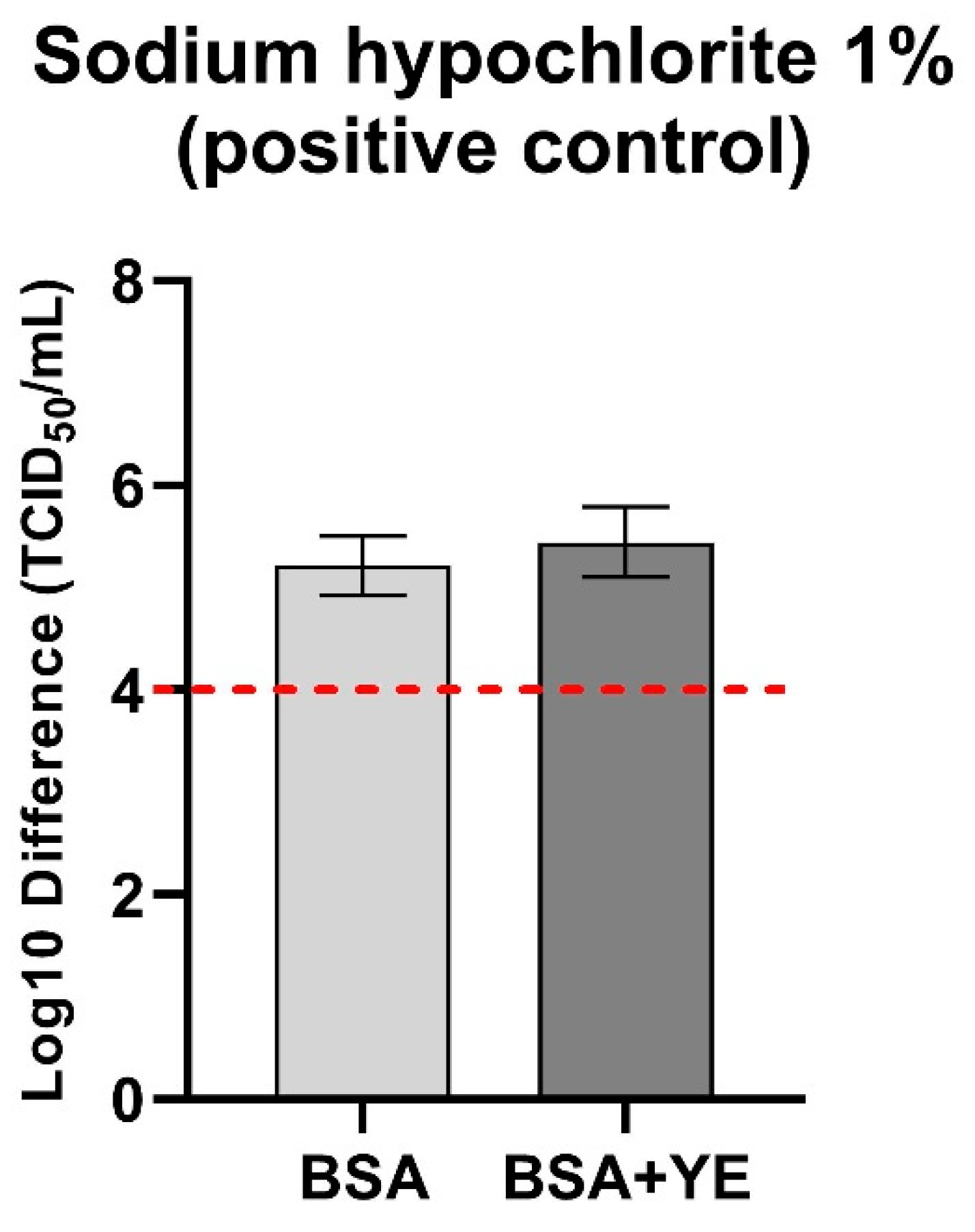
| Peppermint * | 80% (3.5%) | 0.0 cc (±0.00) | 0.0 cc (±0.00) | No | No |
| 60% (2.1%) | 1.92 cc (±0.23) | 3.16 cc (±0.35) | No | No | |
| 30% (1.05%) | 4.41 d (±0.23) | 4.17 d (±0.11) | Yes | Yes | |
| Aloe vera | 80% (80%) | 0.75 (±0.20) | 1.16 (±0.11) | No | No |
| 60% (60%) | 1.08 (±0.62) | 0.83 (±0.31) | No | No | |
| 30% (30%) | 0.83 (±0.11) | 1.16 (±0.11) | No | No | |
| Asiatic pennywort | 80% (2%) | 1.0 (±0.00) | 0.0 (±0.00) | No | No |
| 60% (1.2%) | 1.25 (±0.35) | 0.0 (±0.00) | No | No | |
| 30% (0.6%) | 0.66 (±0.31) | 0.0 (±0.00) | No | No | |
| Lime | 80% (2%) | 0.50 (±0.35) | 0.0 (±0.00) | No | No |
| 60% (1.2%) | 0.08 (±0.11) | 0.0 (±0.00) | No | No | |
| 30% (0.6%) | 0.66 (±0.11) | 0.0 (±0.00) | No | No | |
| Lemon balm | 80% (2%) | 1.91 (±0.23) | 2.25 (±0.35) | No | No |
| 60% (1.2%) | 1.5 (±0.35) | 1.83 (±0.23) | No | No | |
| 30% (0.6%) | 1.33 (±0.23) | 1.0 (±0.54) | No | No | |
| Cucumber | 80% (2%) | 0.0 (±0.00) | 0.0 (±0.00) | No | No |
| 60% (1.2%) | 0.25 (±0.35) | 0.25 (±0.35) | No | No | |
| 30% (0.6%) | 0.08 (±0.11) | 0.08 (±0.11) | No | No | |
| Common nettle | 80% (3%) | 0.0 d (±0.0) | 0.0 d (±0.0) | No | No |
| 60% (1.8%) | 0.25 (±0.20) | 1.50 (±0.20) | No | No | |
| 30% (0.9%) | 1.83 (±0.42) | 1.16 (±0.11) | No | No | |
| Fenugreek * | 80% (3%) | 0.0 cc (±0.00) | 0.0 cc (±0.00) | No | No |
| 60% (1.8%) | 2.58 cc (±0.23) | 2.4 cc (±0.11) | No | No | |
| 30% (0.9%) | 2.16 (±0.31) | 1.08 (±0.11) | No | No | |
Publisher’s Note: MDPI stays neutral with regard to jurisdictional claims in published maps and institutional affiliations. |
© 2021 by the authors. Licensee MDPI, Basel, Switzerland. This article is an open access article distributed under the terms and conditions of the Creative Commons Attribution (CC BY) license (https://creativecommons.org/licenses/by/4.0/).
Share and Cite
Juszkiewicz, M.; Walczak, M.; Woźniakowski, G.; Szczotka-Bochniarz, A. Virucidal Activity of Plant Extracts against African Swine Fever Virus. Pathogens 2021, 10, 1357. https://doi.org/10.3390/pathogens10111357
Juszkiewicz M, Walczak M, Woźniakowski G, Szczotka-Bochniarz A. Virucidal Activity of Plant Extracts against African Swine Fever Virus. Pathogens. 2021; 10(11):1357. https://doi.org/10.3390/pathogens10111357
Chicago/Turabian StyleJuszkiewicz, Małgorzata, Marek Walczak, Grzegorz Woźniakowski, and Anna Szczotka-Bochniarz. 2021. "Virucidal Activity of Plant Extracts against African Swine Fever Virus" Pathogens 10, no. 11: 1357. https://doi.org/10.3390/pathogens10111357
APA StyleJuszkiewicz, M., Walczak, M., Woźniakowski, G., & Szczotka-Bochniarz, A. (2021). Virucidal Activity of Plant Extracts against African Swine Fever Virus. Pathogens, 10(11), 1357. https://doi.org/10.3390/pathogens10111357