Article

Intracellular Behaviour of Legionella Non-pneumophila Strains within Three Amoeba Strains, Including Willaertia magna C2c Maky

R&D Department, Amoéba, 38 Avenue des Frères Montgolfier, 69680 Chassieu, France
*
Author to whom correspondence should be addressed.
Pathogens 2021, 10(10), 1350; https://doi.org/10.3390/pathogens10101350
Submission received: 10 September 2021 / Revised: 7 October 2021 / Accepted: 15 October 2021 / Published: 19 October 2021

Abstract

:
Legionellosis, an often-lethal pneumonia, is generally associated with contamination by Legionella pneumophila. This bacterium can persist in the environment and resist chemical treatment when it is internalized by amoebae. In addition, there is increasing medical proof that other Legionella species can be causative agents of Legionellosis. The objective of this study was to evaluate whether Legionella non-pneumophila (Lnp) strains were able to use the machinery of amoeba to multiply, or whether amoebae were able to limit their proliferation. Seven strains belonging to the species L. longbeachae, L. anisa, L. bozemanae, L. taurinensis, and L. dumoffii were cocultured with three amoebae, Acanthamoeba castellanii, Willaertia magna T5(S)44, and Willaertia magna C2c Maky, at two temperatures, 22 and 37 °C. We found that at 22 °C, all amoebae were able to phagocytose the seven Lnp strains and to avoid intracellular development, except for L. longbeachae, which was able to multiply inside W. magna T5(S)44. At 37 °C, four Lnp strains were able to hijack the machinery of one or two amoebae and to use it to proliferate, but none were able to multiply inside W. magna C2c Maky.

1. Introduction

The genus Legionella currently comprises more than 60 species (https://lpsn.dsmz.de/genus/legionella) (accessed on 18 October 2021) [1], of which more than half have been shown to be pathogenic to humans [2,3,4]. These bacteria occur naturally in freshwater (lakes and rivers) and wet soils. From the natural environment, the bacteria colonise anthropogenic infrastructures, especially those containing stagnant water at a temperature between 25 and 45 °C. Free-living Legionella have difficulty surviving in the environment because of their nutritional requirements. They develop in biofilms or within protists such as ciliates and amoebae [5,6]. The presence of biofilm and certain materials such as iron, zinc, and aluminium favour their survival [7].
Free-living amoebae (FLA) are ubiquitous protozoa, which have been detected in a variety of environmental and manmade habitats, including freshwater, seawater, soil, air-conditioning units and drinking water treatment plants [8,9,10,11,12,13,14,15]. In their natural environment, FLA are an essential component of microbial communities, regulating bacterial populations by feeding on them, but they can also sometimes serve as hosts, vehicles, and reservoirs for pathogenic amoeba-resistant bacteria (ARB), including Legionella species [16,17,18].
Legionella have developed mechanisms to invade FLA by inducing "coiled phagocytosis" to escape digestion by inhibiting the fusion of the phagosome with the lysosome [6,19]. After intracellular replication, Legionella progressively invade the cytoplasm, leading to lysis of the amoeba and the release of mobile Legionella. In some cases, the amoeba also releases vesicles of 1–5 µm in diameter containing the Legionella [16,20,21], allowing their survival for several months [22]. When aerosols filled with bacteria are inhaled, Legionella can infect alveolar macrophages and epithelial cells [23]. It is suspected that, when an amoebic membrane surrounds them, it promotes their fusion with the membrane of pulmonary epithelial cells, increasing the risk of infection [20]. It has been shown that growth within FLA, especially in Acanthamoeba, protects Legionella from adverse environmental conditions, because their cysts are extremely resistant to environmental stresses such as desiccation and changes in pH, osmolarity, or temperature, and can even survive chlorination or other disinfection methods [24,25,26]. Amoebic passage also enhances, in some cases, the virulence of L. pneumophila [27,28], but it has been demonstrated that Willaertia magna C2c Maky did not [29]. Moreover, this amoeba proved to efficiently resist L. pneumophila [30,31,32].
In 2019 in Europe, 11,298 cases of legionellosis were detected [33]. A recent study estimated that the actual number of cases of Legionnaires’ disease may be 1.8 to 2.7 times higher than that reported [34]. Part of the explanation for this lies in the detection method used and the designation of confirmed cases. A urinary antigen test is often used to diagnose legionellosis, but it is not sensitive enough [35]. Although not time-consuming and easily implemented, this test only detects Legionella pneumophila serotype 1 (Lp1). Moreover, in the vast majority of cases, the diagnosis is based solely on antigenicity. The result of a positive PCR test will be designated as a probable case, but not as a confirmed case [36]. Many studies demonstrate that legionellosis cases were related to Legionella non-pneumophila (Lnp) strains and that these strains are frequently isolated in the environment [2,37,38,39,40,41,42,43]. For example, Steege and Moore detected by means of qPCR on the mip gene the presence of Legionella in 95% of samples, but L. pneumophila was only detected in 1.8% of samples [44]. In Japan from 2008 to 2016, Lp1 was responsible for 81% of cases, but Lnp species such as L. bozemanae, L. dumoffii, and L. longbeachae were also isolated [37]. A recent study, using qPCR on the mip gene in hospital and community samples, demonstrated an increased prevalence of the Lnp species (84.1%) compared to L. pneumophila (15.9%) in the region of Bologna (Italy) [45].
Considering these data, the aim of this study was to evaluate the intracellular behaviour of seven Lnp strains belonging to the species L. longbeachae, L. anisa, L. bozemanae, L. taurinensis, and L. dumoffii in coculture with three FLA, Acanthamoeba castellanii, Willaertia magna T5(S)44, and Willaertia magna C2c Maky, to discover whether Lnp strains have the same intra-amoebal multiplicative properties as L. pneumophila strains. To compare our results with a previous study on three L. pneumophila strains [32], two temperatures were considered, 22 and 37 °C.

2. Results

2.1. L. Non-pneumophila Survival in an SCYEM Medium

The survival of seven L. non-pneumophila (Lnp) strains in the coculture medium was evaluated at 22 and 37 °C.
The seven bacterial strains had a better survival capacity at 22 °C than at 37 °C in the SCYEM medium (Figure 1a,b, respectively).
At 22 °C, L. longbeachae, L. anisa ATCC, and L. anisa DSM decreased 100-fold after two days, whereas L. bozemanae, L. taurinensis, L. dumoffii, and L. anisa 1025 were only reduced 2-fold. After four days, L. longbeachae and L. anisa DSM decreased to 13 and 18 CFU/mL, respectively, whereas the five other strains were able to maintain levels between 2.4 × 10³ and 4.2 × 10⁴ CFU/mL (Figure 1a).
At 37 °C, L. longbeachae, L. anisa DSM, and L. taurinensis were not detectable on the plate after three days, nor L. anisa ATCC at day 4. The three other strains, L. bozemanae, L. dumoffii, and L. anisa 1025, decreased to 25 ± 12, 24 ± 57, and 9 ± 13 CFU/mL, respectively (Figure 1b).

2.2. Amoeba Survival

The SCYEM medium allows the survival of the three amoebae during the four days of experimentation at 22 and 37 °C (Figure S1). Survival of the three amoebae in the presence of bacteria was evaluated for four days at 22 and 37 °C in an SCYEM medium.
At 22 °C, the number of W. magna C2c Maky cells remained stable with intracellular Lnp strains until the end of the experiment on day four (Figure 2a), whereas at 37 °C, the number of cells increased in L. anisa ATCC, L. anisa DSM, and L. longbeachae and decreased by a factor of two in the four other strains (Figure 2d).
At 22 °C, A. castellanii was able to grow with the seven intracellular bacterial strains until the end of the experiment at day four (Figure 2b), whereas at 37 °C, the number of cells remained stable or decreased by less than ten-fold for all strains except L. bozemanae, where the number of cells decreased approximately 100-fold from 3.31 × 10⁵ to 5.01 × 10² cells/mL (Figure 2e).
At 22 °C, the number of W. magna T5(S)44 cells remained stable with intracellular bacteria, except in L. anisa 1025, where the number of cells decreased from 2.80 × 10⁵ to 7.25 × 10⁴ cells/mL (Figure 2c). At 37 °C, the number of cells increased, except in L. bozemanae, where the number of cells remained stable (Figure 2f).

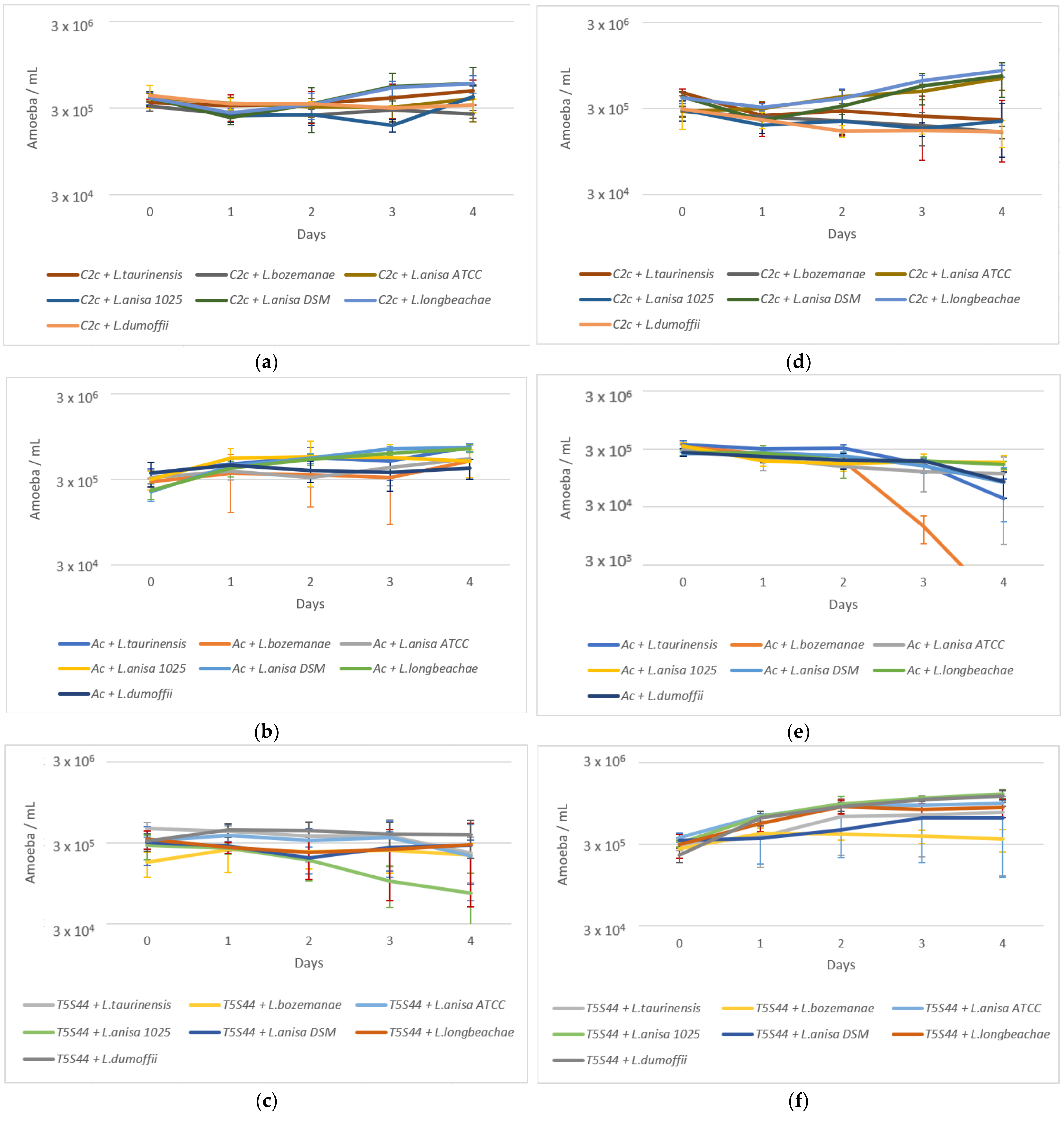
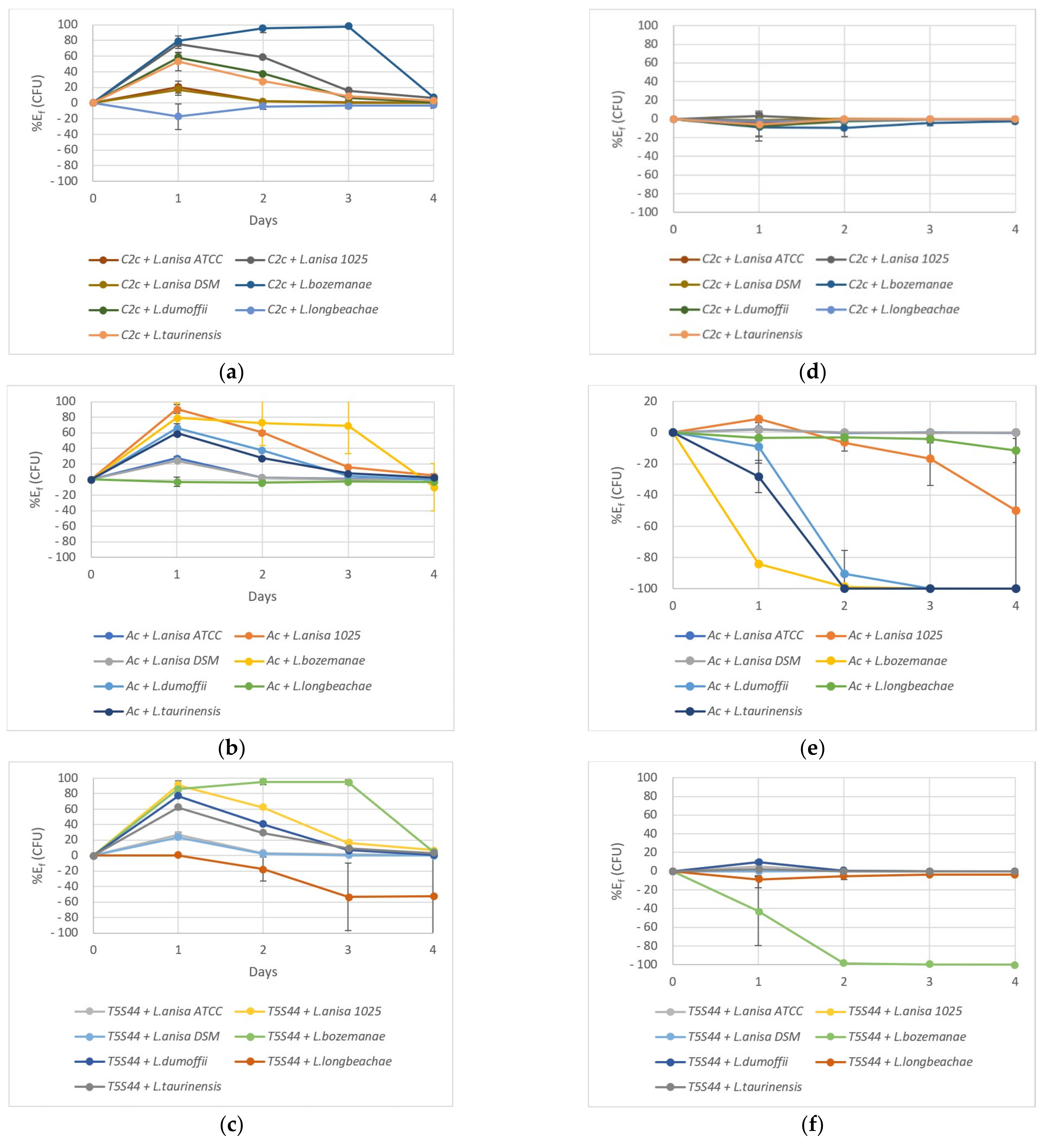
2.3. Coculture Experiments

Considering the death of the Lnp strains in SCYEM medium at 37 °C, the phagocytosis effect by the amoebae could not be easily evaluated. However, the potential of the bacterial strains to hijack the amoeba machinery could be determined because if bacterial multiplication occurs, it can only be attributed to its ability to use amoebae as bioreactors, as no bacterial multiplication in the culture medium occurred.
In order to compare the behaviour of the three amoebae in the presence of the seven Lnp strains, an efficacy percentage was calculated according to the following formula (Equation (1)):
E assay = CFU   t 0 CFU   t CFU   t 0   × 100
where CFU   t 0 is the number of intracellular bacteria at day zero and CFU   t is the number of intracellular bacteria at day one, two, three, or four.
To consider the bacterial decrease due to the culture medium effect, the same calculation was applied to the control conditions (bacteria in SCYEM without amoeba) (Equation (2)):
E control = CFU c   t 0 CFU c   t CFU c   t 0   × 100
where CFU c   t 0 is the number of bacteria at day zero in the control flask and CFU c   t is the number of bacteria in control flasks at day one, two, three, or four.
The net efficacy percentage was calculated by subtracting the medium effect from the efficacy values of cocultures according to the following formula (Equation (3)):
E f = E assay E control
where E assay is the efficacy percentage of both the amoeba and the culture medium to decrease the number of intracellular bacteria; E control is the percentage of bacteria killed by the culture medium in control conditions. E f is the net efficacy of amoeba on Lnp strains.
If E f is positive, it means that the amoeba was efficient, and consequently, bacterial death was increased by the presence of the amoeba, whereas a negative E f means that the number of living Legionella cells increased, indicating that it was able to multiply within the amoeba (as Lnp strains are not able to multiply in the SCYEM medium).
At 22 °C, W. magna C2c Maky was efficient in phagocytosing and killing L. taurinensis, L. dumoffii, L. anisa 1025, and, in particular, L. bozemanae, with 98% efficacy after 72 h (Figure 3a). At 37 °C, the amoeba efficacy could not be proven, as the bacteria in the control condition died as fast as in the presence of W. magna C2c Maky (Figure 3d). However, at both temperatures, none of the seven Legionella were able to multiply in the presence of W. magna C2c Maky (Figure S2).
A. castellanii was efficient in phagocytosing and killing L. taurinensis, L. dumoffii, L. anisa 1025, and, in particular, L. bozemanae, with 69% efficacy after 72 h (Figure 3b). On the contrary, at 37 °C, no efficacy could be proven (Figure 3e). Moreover, L. anisa 1025 (Figure S2c), L. bozemanae (Figure S2d), L. dumoffii, (Figure S2e), and L. taurinensis (Figure S2f) were able to multiply inside A. castellanii. A. castellanii was completely invaded by bacteria from day two (Figure 3e).
At 22 °C, W. magna T5(S)44 was efficient in phagocyting L. taurinensis, L. dumoffii, L. anisa 1025, and, in particular, L. bozemanae, with 95% efficacy after 48 h (Figure 3c), but it could not reduce the level of L. longbeachae, which was able to multiply inside the amoeba (Figure S2a). At 37 °C, no efficacy could be established as the bacteria in the control condition died as fast as in the presence of W. magna T5(S)44 (Figure 3f). Moreover, L. bozemanae was able to multiply within W. magna T5(S)44 cells that lost their efficacy from day two (Figure 3f) and allowed the bacterium to multiply (Figure S2d).

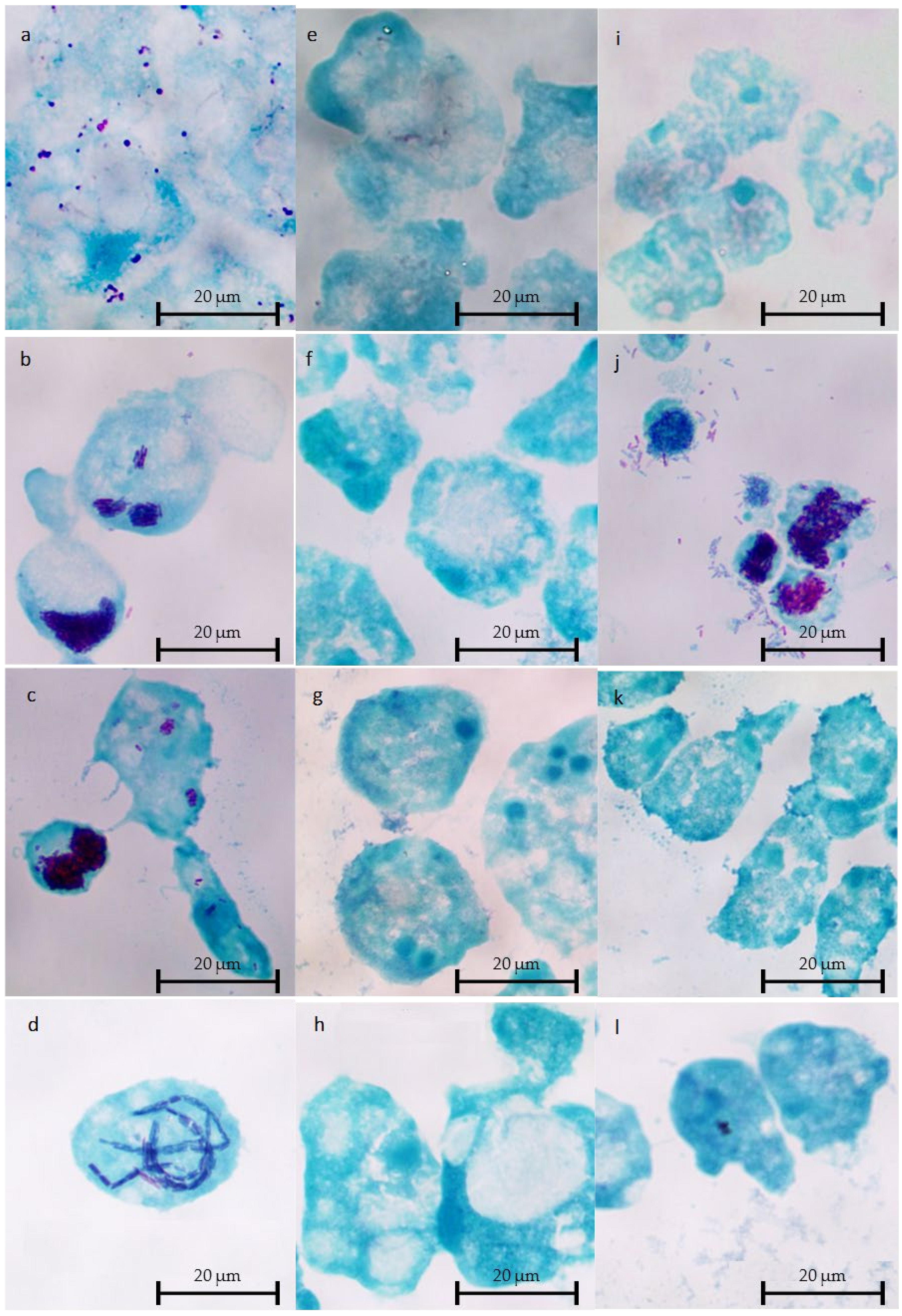
2.4. Microscopic Observations of Intracellular L. Non-pneumophila at 37 °C

Microscopic observations were performed at T0, T0 + 48 h, and T0 + 96 h.
Intracellular multiplication at 37 °C detected by the plating approach were congruent with microscopic observations. Bacterial clusters were found in the cocultures of A. castellanii with L. anisa 1025 (Figure 4a), L. bozemanae (Figure 4b), L. dumoffii (Figure 4c), and L. taurinensis (Figure 4d), and of W. magna T5(S)44 with L. bozemanae (Figure 4j). No intracellular bacterial strain multiplication was observed within W. magna C2c Maky (Figure 4e–h).

3. Discussion

Even if L. pneumophila is responsible for most legionellosis outbreaks, L. non-pneumophila (Lnp) strains are also involved in legionellosis cases, and often their responsibility is under-recognized due to diagnostic bias [2,39,46]. Even though it is now well-known that among the approximately 60 species of Legionella, 50% are able to infect humans [47], the responsibility for 4% of legionellosis cases can still not be attributed to a known species [39]. An extensive detection of Lnp strains in water distribution systems demonstrated that 16% of the sampled water was contaminated with Legionella, and that Lnp strains were prevalent [48]. In the same way, an international survey demonstrated that 43 legionellosis cases among a total of 508 were due to Lnp strains, with the most prevalent being L. longbeachae [43]. Tools developed to study the behaviour of L. pneumoniae can be used to study Lnp. A clinical strain of L. bozemanae transformed with a GFP-expressing plasmid was able to infect and replicate within A. castellanii [49]. To increase knowledge of the replication of Lnp strains inside amoeba, we studied seven non pneumophila strains, three anisa isolates, and one isolate of strains bozemanae, dumoffii, longbeachae, and taurinensis (Table 1).
L. anisa ATCC 35291, isolated from a sink faucet in Illinois, USA, and strain DSM 17627 (ATCC 35292) from tap water in California, USA, were not associated with disease [50]. These two strains were not able to multiply within the three amoebae tested in this study. In contrast, L. anisa 1025 was isolated from a human lung and was responsible for the death of the patient [51]. This strain was able to hijack A. castellanii machinery at 37 °C and to multiply within the amoeba. Its closest relative is L. pneumophila strain Lens [52]. L. anisa is rarely encountered in legionellosis cases, being responsible for only 0.2% of cases [39,43]. However, it was responsible for an outbreak in California in 1988 [53] and was also isolated from a patient in Spain [42]. It is one of the most frequently isolated Lnp species from water systems [48].
L. bozemanae DSM 16523 (ATCC 33217) originated from a human lung tissue of a patient who died in 1959 (USA); its current name was proposed in 1980 [38,54]. L. dumoffii DSM 17625 (ATCC 33279) was isolated from a cooling tower in New York. For the last ten years, L. bozemanae and L. dumoffii were responsible for, respectively, 1 and 3% of legionellosis cases in New Zealand, and remained below 1% in other countries [39]. These two strains were able to multiply within A. castellanii at 37 °C, and L. bozemanae was also able to hijack the machinery of W. magna T5(S)44 at 37 °C.
L. longbeachae DSM 10572, originally designated as Long Beach 4, was isolated from transtracheal aspirates from a fatal case of pneumonia in 1980 [55]. In the case of L. longbeachae confirmed infections, its source was exclusively associated with soils, potting mixes, and composts [56]. It is responsible for 5% of legionellosis in the US and is endemic in New Zealand and Australia [2,57]. It was unable to multiply in A. castellanii and Mono Mac 6 cells [58]. This strain was not able to develop inside amoebae at 37 °C, but it could multiply within W. magna T5(S)44 at 22 °C.
L. taurinensis DSM 21897 (ATCC 700508) was isolated from a water sample of an oxygen bubble humidifier in a hospital in Turin (Italy), where nosocomial Legionella infection occurred [59,60]. It was able to multiply within A. castellanii at 37 °C, but not in the two W. magna strains.
These data show that some Lnp strains can use amoebae for their own benefit. There is no general rule for this; this property is strain-dependent. The optimal temperature for growing A. castellanii is 28 °C. At 37 °C, A. castelanii showed limited growth, which may determine the interaction with Legionella species. However, none of the seven Lnp strains were able to multiply within W. magna C2c Maky, which is considered to be a non-permissive amoeba. Other amoebae have this property such as Micriamoeba tesseris [61] or environmental FLA isolates [62].
These results on seven Lnp strains reinforced the previous results on three L. pneumophila strains [32] to demonstrate that W. magna C2c Maky must have a very efficient process to fight amoeba-resistant bacteria and is a good candidate to be used as a biological biocide to treat industrial waters instead of, or along with, chemical treatments.

4. Materials and Methods

4.1. Microbial Cultures

W. magna C2c Maky (ATCC PTA-7824), W. magna T5(S)44 (ATCC 50036), and A. castellanii (ATCC 30010) were grown in an SCYEM medium as described in Hasni et al. [32]. Amoeba working suspensions (AWS) containing 3 × 10⁵ cells/mL were prepared.
Seven Legionella strains (Table 1) were grown and prepared as described for L. pneumophila strains in Hasni et al. [32]. Bacteria working suspensions (BWS) containing 3 × 10⁷ bacteria/mL were prepared.

4.2. Bacteria and Amoeba Survival in SCYEM Medium (Controls)

Survival of the 3 amoeba strains and 7 bacterial strains was monitored in SCYEM medium at both 22 and 37 °C for four days as described by Hasni et al. [32], except for the plating method. Samples were plated in duplicate with the easySpiral Pro apparatus (Interscience, Saint-Nom-la-Bretèche, France). Plates were incubated at 36 ± 2 °C, and CFUs were counted after 7 days with a Scan 500 reader (Interscience, Saint-Nom-la-Bretèche, France). Each condition was performed in triplicate and independently repeated (n = 6).

4.3. Coculture Assays

Amoeba and bacterial working solutions were mixed in equal quantity as described in Hasni et al. [32]. All flasks were left to stand for 2 h at 22 ± 2 °C or at 37 ± 2 °C to allow for amoeba–bacteria contact and the internalization of Legionella into amoebae. After the 2-hour contact step, each flask was gently shaken 10 times to detach amoeba cells, and the suspension was transferred into a 15 mL Flacon® tube and centrifuged twice at 500× g for 10 min. The supernatant was removed and the cells were resuspended in 10 mL of fresh SCYEM medium. This step allowed for the removal of non-internalized (i.e., extracellular) Legionella. The suspensions were transferred into a new 25 cm3 flask and incubated at 22 ± 2 °C or at 37 ± 2 °C for four days. This time point corresponded to the T0 time point of the assay. Each condition consisted of three independent flasks and was repeated independently (n = 6).

4.4. Bacteria and Amoeba Quantifications in Coculture Assays from T0 to T96h

At T0, T24h, T48h, T72h, and T96h, the supernatant was removed from each flask to detect only intracellular bacteria and replaced by 10 mL of sterile SCYEM according to Hasni et al. [32]. One millilitre was then sampled from each flask. Amoeba numbers were determined using a haemocytometer cell counting chamber method with Trypan blue. Intracellular Legionella CFUs were obtained after lysing amoeba with Triton™ X-100 at 0.02% v/v (final concentration) for 2 min according to Hasni et al. [32]. The samples were then serially 10-fold diluted in SCYEM and plated in duplicate with the easySpiral Pro apparatus. Plates were incubated at 36 ± 2 °C, and CFUs were counted after 7 days with a Scan 500 reader. Each condition was performed in triplicate and independently repeated (n = 6).

4.5. Microscopic Observations of Cocultures

Cocultures were stained by means of the Gimenez technique [63,64] at T0, T48h, and T96h. Cocultures (0.5 mL) were centrifuged at 1000× g for 5 min, 0.45 mL of the supernatant was removed, pellets were resuspended in the remaining 50 µL, and 25 µL was deposited onto glass slides. After 5 min at room temperature, the cells were thermally fixed on a flame and then stained using the Gimenez technique. Briefly, each of the glass slides was stained with fuchsin solution for 3 min and washed with water. Then, the glass slides were stained with malachite green for 10–15 s and washed, and this step was repeated twice. Finally, the glass slides were allowed to dry at room temperature. Bacteria were stained in purple, and amoebae in blue.
The observations were performed using a LEICA DM 2500 LED microscope (Leica Microsystemes SAS, Nanterre, France) under a 100× oil immersion objective.

Supplementary Materials

The following are available online at https://www.mdpi.com/article/10.3390/pathogens10101350/s1, Figure S1: Amoeba survival in SCYEM medium at 22 and 37 °C, Figure S2: Intracellular bacterial fate of bacteria.

Author Contributions

Conceptualization, S.D. and J.-B.E.; methodology, A.C. (Aurélien Croze), A.S., A.C. (Antoine Carlino) and B.Q.; formal analysis, T.C. and S.D.; writing—original draft preparation, A.C. (Aurélien Croze), S.D. and J.-B.E.; writing—review and editing, S.D., J.-B.E. and A.C. (Aurélien Croze); supervision, S.D. All authors have read and agreed to the published version of the manuscript.

Funding

This research was funded by Amoéba SA.

Data Availability Statement

Available on demand.

Acknowledgments

We are very grateful to B. La Scola for his advice throughout the completion of this study and for reviewing this article prior to publication.

Conflicts of Interest

The authors declare no conflict of interest.

References

  1. Li, P.; Vassiliadis, D.; Ong, S.Y.; Bennett-Wood, V.; Sugimoto, C.; Yamagishi, J.; Hartland, E.L.; Pasricha, S. Legionella Pneumophila Infection Rewires the Acanthamoeba Castellanii Transcriptome, Highlighting a Class of Sirtuin Genes. Front. Cell. Infect. Microbiol. 2020, 10, 428. [Google Scholar] [CrossRef]
  2. Bell, H.; Chintalapati, S.; Patel, P.; Halim, A.; Kithas, A.; Schmalzle, S.A. Legionella Longbeachae Pneumonia: Case Report and Review of Reported Cases in Non-Endemic Countries. IDCases 2021, 23, e01050. [Google Scholar] [CrossRef]
  3. Burillo, A.; Pedro-Botet, M.L.; Bouza, E. Microbiology and Epidemiology of Legionnaire’s Disease. Infect. Dis. Clin. N. Am. 2017, 31, 7–27. [Google Scholar] [CrossRef]
  4. Newton, H.J.; Ang, D.K.Y.; van Driel, I.R.; Hartland, E.L. Molecular Pathogenesis of Infections Caused by Legionella Pneumophila. Clin. Microbiol. Rev. 2010, 23, 274–298. [Google Scholar] [CrossRef] [Green Version]
  5. Borella, P.; Guerrieri, E.; Marchesi, I.; Bondi, M.; Messi, P. Water Ecology of Legionella and Protozoan: Environmental and Public Health Perspectives. Biotechnol. Annu. Rev. 2005, 11, 355–380. [Google Scholar] [CrossRef]
  6. Albert-Weissenberger, C.; Cazalet, C.; Buchrieser, C. Legionella Pneumophila—A Human Pathogen That Co-Evolved with Fresh Water Protozoa. Cell. Mol. Life Sci. CMLS 2007, 64, 432–448. [Google Scholar] [CrossRef]
  7. Abdel-Nour, M.; Duncan, C.; Low, D.E.; Guyard, C. Biofilms: The Stronghold of Legionella Pneumophila. Int. J. Mol. Sci. 2013, 14, 21660–21675. [Google Scholar] [CrossRef] [Green Version]
  8. Michel, R.; Just, H.M. Acanthamoebae, Naegleria and other free-living Amoebae in cooling and rinsing water of dental treatment units. Zentralbl. Bakteriol. Mikrobiol. Hyg. 1984, 179, 56–72. [Google Scholar]
  9. Barbaree, J.M.; Fields, B.S.; Feeley, J.C.; Gorman, G.W.; Martin, W.T. Isolation of Protozoa from Water Associated with a Legionellosis Outbreak and Demonstration of Intracellular Multiplication of Legionella Pneumophila. Appl. Environ. Microbiol. 1986, 51, 422–424. [Google Scholar] [CrossRef] [Green Version]
  10. De Jonckheere, J.F. Ecology of Acanthamoeba. Rev. Infect. Dis. 1991, 13 (Suppl. 5), S385–S387. [Google Scholar] [CrossRef]
  11. Thomas, V.; Bouchez, T.; Nicolas, V.; Robert, S.; Loret, J.F.; Lévi, Y. Amoebae in Domestic Water Systems: Resistance to Disinfection Treatments and Implication in Legionella Persistence. J. Appl. Microbiol. 2004, 97, 950–963. [Google Scholar] [CrossRef] [PubMed]
  12. Thomas, V.; Herrera-Rimann, K.; Blanc, D.S.; Greub, G. Biodiversity of Amoebae and Amoeba-Resisting Bacteria in a Hospital Water Network. Appl. Environ. Microbiol. 2006, 72, 2428–2438. [Google Scholar] [CrossRef] [PubMed] [Green Version]
  13. De Jonckheere, J.F. Isolation and Molecular Identification of Free-Living Amoebae of the Genus Naegleria from Arctic and Sub-Antarctic Regions. Eur. J. Protistol. 2006, 42, 115–123. [Google Scholar] [CrossRef] [PubMed]
  14. Thomas, V.; Loret, J.-F.; Jousset, M.; Greub, G. Biodiversity of Amoebae and Amoebae-Resisting Bacteria in a Drinking Water Treatment Plant. Environ. Microbiol. 2008, 10, 2728–2745. [Google Scholar] [CrossRef] [PubMed]
  15. Stockman, L.J.; Wright, C.J.; Visvesvara, G.S.; Fields, B.S.; Beach, M.J. Prevalence of Acanthamoeba Spp. and Other Free-Living Amoebae in Household Water, Ohio, USA—1990–1992. Parasitol. Res. 2011, 108, 621–627. [Google Scholar] [CrossRef] [PubMed]
  16. Rowbotham, T.J. Preliminary Report on the Pathogenicity of Legionella Pneumophila for Freshwater and Soil Amoebae. J. Clin. Pathol. 1980, 33, 1179–1183. [Google Scholar] [CrossRef] [PubMed] [Green Version]
  17. Michel, R.; Müller, K.-D.; Amann, R.; Schmid, E.N. Legionella-like Slender Rods Multiplying within a Strain of Acanthamoeba Sp. Isolated from Drinking Water. Parasitol. Res. 1997, 84, 84–88. [Google Scholar] [CrossRef]
  18. Greub, G.; Raoult, D. Microorganisms Resistant to Free-Living Amoebae. Clin. Microbiol. Rev. 2004, 17, 413–433. [Google Scholar] [CrossRef] [Green Version]
  19. Abu Kwaik, Y.; Gao, L.-Y.; Stone, B.J.; Venkataraman, C.; Harb, O.S. Invasion of Protozoa by Legionella Pneumophila and Its Role in Bacterial Ecology and Pathogenesis. Appl. Environ. Microbiol. 1998, 64, 3127–3133. [Google Scholar] [CrossRef] [Green Version]
  20. Berk, S.G.; Ting, R.S.; Turner, G.W.; Ashburn, R.J. Production of Respirable Vesicles Containing Live Legionella Pneumophila Cells by Two Acanthamoeba Spp. Appl. Environ. Microbiol. 1998, 64, 279–286. [Google Scholar] [CrossRef] [Green Version]
  21. Rowbotham, T.J. Current Views on the Relationships between Amoebae, Legionellae and Man. Isr. J. Med. Sci. 1986, 22, 678–689. [Google Scholar] [PubMed]
  22. Bouyer, S.; Imbert, C.; Rodier, M.-H.; Héchard, Y. Long-Term Survival of Legionella Pneumophila Associated with Acanthamoeba Castellanii Vesicles. Environ. Microbiol. 2007, 9, 1341–1344. [Google Scholar] [CrossRef]
  23. Fields, B.S. The Molecular Ecology of Legionellae. Trends Microbiol. 1996, 4, 286–290. [Google Scholar] [CrossRef]
  24. Kilvington, S.; Price, J. Survival of Legionella Pneumophila within Cysts of Acanthamoeba Polyphaga Following Chlorine Exposure. J. Appl. Bacteriol. 1990, 68, 519–525. [Google Scholar] [CrossRef] [PubMed]
  25. Dupuy, M.; Binet, M.; Bouteleux, C.; Herbelin, P.; Soreau, S.; Héchard, Y. Permissiveness of Freshly Isolated Environmental Strains of Amoebae for Growth of Legionella Pneumophila. FEMS Microbiol. Lett. 2016, 363, fnw022. [Google Scholar] [CrossRef]
  26. Hwang, M.G.; Katayama, H.; Ohgaki, S. Effect of Intracellular Resuscitation of Legionella Pneumophila in Acanthamoeba Polyphage Cells on the Antimicrobial Properties of Silver and Copper. Environ. Sci. Technol. 2006, 40, 7434–7439. [Google Scholar] [CrossRef]
  27. Cirillo, J.D.; Falkow, S.; Tompkins, L.S. Growth of Legionella Pneumophila in Acanthamoeba Castellanii Enhances Invasion. Infect. Immun. 1994, 62, 3254–3261. [Google Scholar] [CrossRef] [Green Version]
  28. Nora, T.; Lomma, M.; Gomez-Valero, L.; Buchrieser, C. Molecular Mimicry: An Important Virulence Strategy Employed by Legionella Pneumophila to Subvert Host Functions. Future Microbiol. 2009, 4, 691–701. [Google Scholar] [CrossRef]
  29. Mameri, R.M.; Bodennec, J.; Bezin, L.; Demanèche, S. Mitigation of Expression of Virulence Genes in Legionella Pneumophila Internalized in the Free-Living Amoeba Willaertia Magna C2c Maky. Pathogens 2020, 9, 447. [Google Scholar] [CrossRef]
  30. Dey, R.; Bodennec, J.; Mameri, M.O.; Pernin, P. Free-Living Freshwater Amoebae Differ in Their Susceptibility to the Pathogenic Bacterium Legionella Pneumophila. FEMS Microbiol. Lett. 2009, 290, 10–17. [Google Scholar] [CrossRef] [Green Version]
  31. Dey, R.; Mameri, M.R.; Trajkovic-Bodennec, S.; Bodennec, J.; Pernin, P. Impact of Inter-Amoebic Phagocytosis on the L. Pneumophila Growth. FEMS Microbiol. Lett. 2020, 367, fnaa147. [Google Scholar] [CrossRef]
  32. Hasni, I.; Jarry, A.; Quelard, B.; Carlino, A.; Eberst, J.-B.; Abbe, O.; Demanèche, S. Intracellular Behaviour of Three Legionella Pneumophila Strains within Three Amoeba Strains, Including Willaertia Magna C2c Maky. Pathogens 2020, 9, 105. [Google Scholar] [CrossRef] [Green Version]
  33. European Centre for Disease Prevention and Control. Legionnaires’ Disease—Annual Epidemiological Report for 2019. Available online: https://www.ecdc.europa.eu/en/publications-data/legionnaires-disease-annual-epidemiological-report-2019 (accessed on 8 September 2021).
  34. Collier, S.A.; Deng, L.; Adam, E.A.; Benedict, K.M.; Beshearse, E.M.; Blackstock, A.J.; Bruce, B.B.; Derado, G.; Edens, C.; Fullerton, K.E.; et al. Estimate of Burden and Direct Healthcare Cost of Infectious Waterborne Disease in the United States. Emerg. Infect. Dis. J. CDC 2021, 27, 140–149. [Google Scholar] [CrossRef] [PubMed]
  35. Miyata, J.; Huh, J.Y.; Ito, Y.; Kobuchi, T.; Kusukawa, K.; Hayashi, H. Can We Truly Rely on the Urinary Antigen Test for the Diagnosis? Legionella Case Report. J. Gen. Fam. Med. 2017, 18, 139–143. [Google Scholar] [CrossRef] [PubMed] [Green Version]
  36. Jarraud, S.; Lina, G. Legionella_Rapport Activité 2019_FINAL.Pdf—CNR Sites Web (10-01-2020 08:26). Available online: https://clarolineconnect.univ-lyon1.fr/resource/open/file/5322646 (accessed on 11 August 2021).
  37. Amemura-Maekawa, J.; Kura, F.; Chida, K.; Ohya, H.; Kanatani, J.; Isobe, J.; Tanaka, S.; Nakajima, H.; Hiratsuka, T.; Yoshino, S.; et al. Legionella Pneumophila and Other Legionella Species Isolated from Legionellosis Patients in Japan between 2008 and 2016. Appl. Environ. Microbiol. 2018, 84, e00721-18. [Google Scholar] [CrossRef] [PubMed] [Green Version]
  38. Brenner, D.J.; Steigerwalt, A.G.; Gorman, G.W.; Weaver, R.E.; Feeley, J.C.; Cordes, L.G.; Wilkinson, H.W.; Patton, C.; Thomason, B.M.; Sasseville, K.R.L. Legionella Bozemanii Sp. Nov. AndLegionella Dumoffii Sp. Nov.: Classification of Two Additional Species OfLegionella Associated with Human Pneumonia. Curr. Microbiol. 1980, 4, 111–116. [Google Scholar] [CrossRef]
  39. Chambers, S.T.; Slow, S.; Scott-Thomas, A.; Murdoch, D.R. Legionellosis Caused by Non-Legionella Pneumophila Species, with a Focus on Legionella Longbeachae. Microorganisms 2021, 9, 291. [Google Scholar] [CrossRef] [PubMed]
  40. Cordes, L.G.; Wilkinson, H.W.; Gorman, G.W.; Fikes, B.J.; Fraser, D.W. Atypical Legionella-like Organisms: Fastidious Water-Associated Bacteria Pathogenic for Man. Lancet Lond. Engl. 1979, 2, 927–930. [Google Scholar] [CrossRef]
  41. Lam, S.; Smith, J.; Burton, J.; Evelyn, R.; Harper, B.; Huckell, V.; Jones, E. Legionella Longbeachae Pneumonia Diagnosed by Bronchial Brushing. Can. Med. Assoc. J. 1982, 127, 223–224. [Google Scholar]
  42. Vaccaro, L.; Izquierdo, F.; Magnet, A.; Hurtado, C.; Salinas, M.A.; Gomes, T.S.; Angulo, S.; Salso, S.; Pelaez, J.; Tejeda, M.I.; et al. First Case of Legionnaire’s Disease Caused by Legionella Anisa in Spain and the Limitations on the Diagnosis of Legionella Non-Pneumophila Infections. PLoS ONE 2016, 11, e0159726. [Google Scholar] [CrossRef]
  43. Yu, V.L.; Plouffe, J.F.; Pastoris, M.C.; Stout, J.E.; Schousboe, M.; Widmer, A.; Summersgill, J.; File, T.; Heath, C.M.; Paterson, D.L.; et al. Distribution of Legionella Species and Serogroups Isolated by Culture in Patients with Sporadic Community-Acquired Legionellosis: An International Collaborative Survey. J. Infect. Dis. 2002, 186, 127–128. [Google Scholar] [CrossRef] [Green Version]
  44. Steege, L.; Moore, G. The Presence and Prevalence of Legionella Spp in Collected Rainwater and Its Aerosolisation during Common Gardening Activities. Perspect. Public Health 2018, 138, 254–260. [Google Scholar] [CrossRef]
  45. Mazzotta, M.; Salaris, S.; Pascale, M.R.; Girolamini, L.; Cristino, S. Occurrence of Legionella Spp. in Man-Made Water Sources: Isolates Distribution and Phylogenetic Characterization in the Emilia-Romagna Region. Pathogens 2021, 10, 552. [Google Scholar] [CrossRef]
  46. Priest, P.C.; Slow, S.; Chambers, S.T.; Cameron, C.M.; Balm, M.N.; Beale, M.W.; Blackmore, T.K.; Burns, A.D.; Drinković, D.; Elvy, J.A.; et al. The Burden of Legionnaires’ Disease in New Zealand (LegiNZ): A National Surveillance Study. Lancet Infect. Dis. 2019, 19, 770–777. [Google Scholar] [CrossRef]
  47. Gomes, T.S.; Gjiknuri, J.; Magnet, A.; Vaccaro, L.; Ollero, D.; Izquierdo, F.; Fenoy, S.; Hurtado, C.; Del Águila, C. The Influence of Acanthamoeba-Legionella Interaction in the Virulence of Two Different Legionella Species. Front. Microbiol. 2018, 9, 2962. [Google Scholar] [CrossRef] [PubMed] [Green Version]
  48. Salinas, M.B.; Fenoy, S.; Magnet, A.; Vaccaro, L.; Gomes, T.D.; Hurtado, C.; Ollero, D.; Valdivieso, E.; Del Águila, C.; Pozuelo, M.J.; et al. Are Pathogenic Legionella Non-Pneumophila a Common Bacteria in Water Distribution Networks? Water Res. 2021, 196, 117013. [Google Scholar] [CrossRef] [PubMed]
  49. Head, B.M.; Graham, C.I.; MacMartin, T.; Keynan, Y.; Brassinga, A.K.C. Development of a Fluorescent Tool for Studying Legionella Bozemanae Intracellular Infection. Microorganisms 2021, 9, 379. [Google Scholar] [CrossRef] [PubMed]
  50. Gorman, G.W.; Feeley, J.C.; Steigerwalt, A.; Edelstein, P.H.; Moss, C.W.; Brenner, D.J. Legionella Anisa: A New Species of Legionella Isolated from Potable Waters and a Cooling Tower. Appl. Environ. Microbiol. 1985, 49, 305–309. [Google Scholar] [CrossRef] [PubMed] [Green Version]
  51. La Scola, B.; Mezi, L.; Weiller, P.J.; Raoult, D. Isolation of Legionella Anisa Using an Amoebic Coculture Procedure. J. Clin. Microbiol. 2001, 39, 365–366. [Google Scholar] [CrossRef] [Green Version]
  52. Pagnier, I.; Croce, O.; Robert, C.; Raoult, D.; La Scola, B. Genome Sequence of Legionella Anisa, Isolated from a Respiratory Sample, Using an Amoebal Coculture Procedure. Genome Announc. 2014, 2, 2. [Google Scholar] [CrossRef] [Green Version]
  53. Fenstersheib, M.D.; Miller, M.; Diggins, C.; Liska, S.; Detwiler, L.; Werner, S.B.; Lindquist, D.; Thacker, W.L.; Benson, R.F. Outbreak of Pontiac Fever Due to Legionella Anisa. Lancet 1990, 336, 35–37. [Google Scholar] [CrossRef]
  54. Tindall, B.J. Taking a Closer Look at the Valid Publication and Authorship of Legionella Bozemanae Brenner et al. 1980, Fluoribacter Bozemanae Garrity et al. 1980, Legionella Pittsburghensis Pasculle et al. 1980, Legionella Micdadei Hébert et al. 1980 and Tatlockia Micdadei (Hébert et al. 1980) Garrity et al. 1980. Curr. Microbiol. 2020, 77, 146–153. [Google Scholar] [CrossRef] [PubMed]
  55. McKinney, R.M.; Porschen, R.K.; Edelstein, P.H.; Bissett, M.L.; Harris, P.P.; Bondell, S.P.; Steigerwalt, A.G.; Weaver, R.E.; Ein, M.E.; Lindquist, D.S.; et al. Legionella Longbeachae Species Nova, Another Etiologic Agent of Human Pneumonia. Ann. Intern. Med. 1981, 94, 739–743. [Google Scholar] [CrossRef]
  56. Whiley, H.; Bentham, R. Legionella Longbeachae and Legionellosis. Emerg. Infect. Dis. 2011, 17, 579–583. [Google Scholar] [CrossRef] [PubMed]
  57. Thornley, C.N.; Harte, D.J.; Weir, R.P.; Allen, L.J.; Knightbridge, K.J.; Wood, P.R.T. Legionella Longbeachae Detected in an Industrial Cooling Tower Linked to a Legionellosis Outbreak, New Zealand, 2015; Possible Waterborne Transmission? Epidemiol. Infect. 2017, 145, 2382–2389. [Google Scholar] [CrossRef] [Green Version]
  58. Neumeister, B.; Schöniger, S.; Faigle, M.; Eichner, M.; Dietz, K. Multiplication of Different Legionella Species in Mono Mac 6 Cells and in Acanthamoeba Castellanii. Appl. Environ. Microbiol. 1997, 63, 1219–1224. [Google Scholar] [CrossRef] [Green Version]
  59. Lo Presti, F.; Riffard, S.; Meugnier, H.; Reyrolle, M.; Lasne, Y.; Grimont, P.A.; Grimont, F.; Vandenesch, F.; Etienne, J.; Fleurette, J.; et al. Legionella Taurinensis Sp. Nov., a New Species Antigenically Similar to Legionella Spiritensis. Int. J. Syst. Bacteriol. 1999, 49 Pt 2, 397–403. [Google Scholar] [CrossRef]
  60. Franzin, L.; Gioannini, P. Legionella Taurinensis, a New Species of Legionella Isolated in Turin, Italy. Int. J. Syst. Evol. Microbiol. 2000, 50 Pt 2, 937. [Google Scholar] [CrossRef] [PubMed]
  61. Atlan, D.; Coupat-Goutaland, B.; Risler, A.; Reyrolle, M.; Souchon, M.; Briolay, J.; Jarraud, S.; Doublet, P.; Pélandakis, M. Micriamoeba Tesseris Nov. Gen. Nov. Sp.: A New Taxon of Free-Living Small-Sized Amoebae Non-Permissive to Virulent Legionellae. Protist 2012, 163, 888–902. [Google Scholar] [CrossRef] [PubMed]
  62. Bornier, F.; Zas, E.; Potheret, D.; Laaberki, M.-H.; Coupat-Goutaland, B.; Charpentier, X. Environmental Free-Living Amoebae Can Predate on Diverse Antibiotic-Resistant Human Pathogens. Appl. Environ. Microbiol. 2021, 87, AEM0074721. [Google Scholar] [CrossRef]
  63. Gimenez, D.F. Staining Rickettsiae in Yolk-Sac Cultures. Stain Technol. 1964, 39, 135–140. [Google Scholar] [CrossRef] [PubMed]
  64. Greer, P.W.; Chandler, F.W.; Hicklin, M.D. Rapid Demonstration of Legionella Pneumophila in Unembedded Tissue. An Adaptation of the Giménez Stain. Am. J. Clin. Pathol. 1980, 73, 788–790. [Google Scholar] [CrossRef] [PubMed]
Figure 1. L. non-pneumophila survival in an SCYEM medium at 22 (a) and 37 °C (b). Results are expressed as the mean ± standard deviation (SD).
Pathogens 10 01350 g001
Figure 2. Amoeba survival at 22 (ac) and 37 °C (df) in an SCYEM medium with the seven L. non-pneumophila strains. C2c: W. magna C2c Maky (a,d); Ac: A. castellanii (b,e); T5S44: W. magna T5(S)44 (c,f). Results are expressed as the mean ± SD.
Pathogens 10 01350 g002
Figure 3. Efficacy of amoeba phagocytosis at 22 (ac) and 37 °C (df) in an SCYEM medium on seven L. non-pneumophila strains. C2c: W. magna C2c Maky (a,d); Ac: A. castellanii (b,e); T5S44: W. magna T5(S)44 (c,f). Results are expressed as the mean ± SD.
Pathogens 10 01350 g003
Figure 4. Optical microscopy observation using Gimenez staining of A. castellanii (ad), W. magna C2c Maky (eh), and W. magna T5(S)44 (il) infected with L. anisa 1025 (a,e,i), L. bozemanae (b,f,j), L. dumoffii (c,g,k), and L. taurinensis (d,h,l) after 96 h of coculture at 37 °C.
Pathogens 10 01350 g004
Table 1. List of Legionella strain studied.
Bacterial NameOrigin
L. anisa ATCCATCC 35291
L. anisa 1025Clinical isolate
L. anisa DSMDSM 17627/ATCC 35292
L. bozemanaeDSM 16523/ATCC 33217
L. dumoffiiDSM 17625/ATCC 33279
L.longbeachaeDSM 10572/ATCC 33462
L. taurinensisDSM 21897/ATCC 700508
Publisher’s Note: MDPI stays neutral with regard to jurisdictional claims in published maps and institutional affiliations.

