Article

Identification of Immunogenic Linear B-Cell Epitopes in C. burnetii Outer Membrane Proteins Using Immunoinformatics Approaches Reveals Potential Targets of Persistent Infections

by
Sílvia da Silva Fontes
1,
Fernanda de Moraes Maia
1,
Laura Santa’Anna Ataides
1,
Fernando Paiva Conte
2,
Josué da Costa Lima-Junior
3,
Tatiana Rozental
4,
Matheus Ribeiro da Silva Assis
4,
Adonai Alvino Pessoa Júnior
4,
Jorlan Fernandes
4,
Elba Regina Sampaio de Lemos
4 and
Rodrigo Nunes Rodrigues-da-Silva
1,*
1
Laboratory of Monoclonal Antibodies Technology, Immunobiological Technology Institute, FIOCRUZ, Rio de Janeiro 21040-900, Brazil
2
Pilot Plant Implantation Project, Immunobiological Technology Institute, FIOCRUZ, Rio de Janeiro 21040-900, Brazil
3
Laboratory of Immunoparasitology, Oswaldo Cruz Institute, FIOCRUZ, Rio de Janeiro 21040-900, Brazil
4
Laboratory of Hantaviroses and Rickettsioses, Oswaldo Cruz Institute, FIOCRUZ, Rio de Janeiro 21040-900, Brazil
*
Author to whom correspondence should be addressed.
Pathogens 2021, 10(10), 1250; https://doi.org/10.3390/pathogens10101250
Submission received: 30 June 2021 / Revised: 11 August 2021 / Accepted: 16 August 2021 / Published: 28 September 2021
(This article belongs to the Collection Updates on Rickettsia and Coxiella)

Abstract

:
Coxiella burnetii is a global, highly infectious intracellular bacterium, able to infect a wide range of hosts and to persist for months in the environment. It is the etiological agent of Q fever—a zoonosis of global priority. Currently, there are no national surveillance data on C. burnetii’s seroprevalence for any South American country, reinforcing the necessity of developing novel and inexpensive serological tools to monitor the prevalence of infections among humans and animals—especially cattle, goats, and sheep. In this study, we used immunoinformatics and computational biology tools to predict specific linear B-cell epitopes in three C. burnetii outer membrane proteins: OMP-H (CBU_0612), Com-1 (CBU_1910), and OMP-P1 (CBU_0311). Furthermore, predicted epitopes were tested by ELISA, as synthetic peptides, against samples of patients reactive to C. burnetii in indirect immunofluorescence assay, in order to evaluate their natural immunogenicity. In this way, two linear B-cell epitopes were identified in each studied protein (OMP-H(51–59), OMP-H(91–106), Com-1(57–76), Com-1(191–206), OMP-P1(197–209), and OMP-P1(215–227)); all of them were confirmed as naturally immunogenic by the presence of specific antibodies in 77% of studied patients against at least one of the identified epitopes. Remarkably, a higher frequency of endocarditis cases was observed among patients who presented an intense humoral response to OMP-H and Com-1 epitopes. These data confirm that immunoinformatics applied to the identification of specific B-cell epitopes can be an effective strategy to improve and accelerate the development of surveillance tools against neglected diseases.

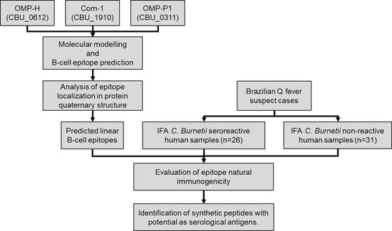
Graphical Abstract

1. Introduction

Coxiella burnetii is a polymorphic and obligate intracellular Gram-negative bacterium that is highly infectious and can persist in the environment for months. It is the causative agent of Q fever in humans and coxiellosis in animals—a worldwide disease that is 1 of 13 global priority zoonoses [1,2,3]. In addition, C. burnetii is classified as a potential agent of bioterrorism, due to its remarkable resistance to environmental stress, extremely low infectious dose, and ease of dissemination [4,5,6]. A wide range of animals is known to harbor C. burnetii, including many species of ticks, birds, and wild, domestic, and companion mammals [7,8]. Infected animals excrete bacteria in large quantities in their birth products, milk, urine, and feces, contaminating the environment. The main transmission route of C. burnetii to humans is through inhalation of aerosols contaminated by amniotic fluid and animal placenta, but other possible routes are under investigation, including consumption of raw milk and unpasteurized dairy products, blood transfusions, nosocomial transmission, during autopsies, and delivery of infected pregnant women [9,10,11,12].
Q fever is an ubiquitous zoonosis, with outbreaks reported all over the globe, except for New Zealand and Antarctica [1]. In animals, coxiellosis is often asymptomatic in livestock, but is associated with reproductive disorders causing miscarriages, premature births, infertility, mastitis, and decreased milk production, especially in ungulates and ruminants [13]. In humans, clinical outcomes vary in severity, ranging from asymptomatic infection with seroconversion, to acute Q fever—a mild, influenza-like, self-limited, febrile illness that can progress to more severe cases, atypical pneumonia, or hepatitis [14]. In both asymptomatic and acute cases, Q fever can progress as a persistent and localized infection of a specific organ—mainly infectious endocarditis—and vascular infection, although other impairments have been described that may result in death, depending on host characteristics [1,15]. To date, since the 2007 epidemic in the Netherlands [16], the number of global publications on Q fever has increased. However, despite having been listed as a notifiable disease by the World Organization for Animal Health [17], in both animals and humans, C. burnetii infections remain poorly understood, and their prevalence still is underestimated in several regions of the world [18]. Moreover, most of the countries where Q fever exists are yet to formulate sanitary control measures to control the disease in livestock and humans.
Although most human cases are asymptomatic, Q fever may manifest nonspecific symptoms that could lead to misdiagnosis with other prevalent tropical diseases, such as dengue, malaria, or leptospirosis [19]. In Brazil, Q fever is a mandatory notification disease, in the context of differential diagnosis with Brazilian spotted fever; however, there is still little information about C. burnetii’s circulation in Brazil. In recent years, case reports and seroprevalence studies reported the detection of C. burnetii in animals [20,21,22,23], humans [23,24,25,26,27] and, most recently, in artisanal cheese [28] and unpasteurized milk [29]. These data, allied to the low number of Q fever cases reported annually, suggest that Q fever might be underreported, and reinforce the urgency of the development of novel diagnostic tools that allow for large-scale epidemiological surveillance, as well as improving the inspection of products of animal origin.
Regarding the diagnosis of Q fever, the isolation of C. burnetii is rarely attempted due to its prolonged incubation period and the biosafety level required [30,31]. Currently, the Q fever/coxiellosis diagnosis is mainly based on serological (immunofluorescence assay, ELISA, and latex agglutination assay) and molecular methods (PCR). However, the poor field applicability, along with the inability to differentiate active infection from recovered individuals, or even acute and chronic Q fever, pose serious bottlenecks to the large-scale surveillance of Q fever/coxiellosis [32,33,34,35]. In this context, numerous novel antigen candidates have been proposed to improve the serodiagnosis of Q fever [36,37,38,39,40]. Among these antigens, OMP-H (CBU_0612) was described as an immunodominant marker for acute and persistent forms of Q fever [35,41], Com-1 (CBU_1910) is considered a reliable Q fever serodiagnosis marker [42,43], and OMP-P1 (CBU_0311) is a porin—highly expressed only in the replicative form of the bacterium in the cell hosts [44,45,46]. The identification of B-cell epitopes arises as a promising alternative to improve the specificity of serological tests to C. burnetii, since the use in serodiagnosis of the above-mentioned whole antigens may result in cross-reactivity with other phylogenetically related proteobacteria.
Recently, synthetic peptides have emerged as novel targets for the efficient serological diagnosis of infectious diseases of viral, bacterial, and parasitic origin [47,48,49,50,51,52,53,54]. This kind of molecule is applied via latex agglutination assay to diagnose Q fever, presenting good sensitivity and specificity for diagnosis in cattle [49,55]. This approach, compared to diagnosis based on whole antigens, presents low-cost production, higher specificity and reproducibility with no batch-to-batch variation, and large-scale production [56,57,58]; however, it still depends on the accurate identification of more immunodominant epitopes. Therefore, this study aimed to identify B-cell epitopes from C. burnetii outer membrane proteins (OMP-P1, OMP-H, and Com-1) in silico, and to validate them against samples from Brazilian exposed individuals.

2. Results

2.1. Prediction of Linear B-Cell Epitopes in C. burnetii OMPs

In order to improve the accuracy of in silico identification of epitopes, we used a combination of three prediction algorithms: BepiPred 1.0, ABCpred, and ESA. Firstly, 10 sequences were predicted as linear B-cell epitopes in studied proteins—2 sequences in OMP-H, and 4 sequences each in Com-1 and OMP-P1. Among all 10 sequences, 4 were predicted by all algorithms, while 3 were predicted by Bepipred and ABCpred, 2 by ABCpred and ESA, and 1 potential epitope by Bepipred and ESA (Table 1).

2.2. Prediction of Epitopes’ Antigenicity and Specificity

The antigenicity of predicted linear epitopes was evaluated using the VaxiJen algorithm. Based on this evaluation, eight predicted epitopes were considered antigenic, among which were the four sequences predicted as linear B-cell epitopes by the three used algorithms (OMP-H(51–59), OMP-H(98–106), Com-1(57–76), and Com-1(191–206)). The sequences Com-1(83–96) and OMP-P1(43–51) presented VaxiJen scores lower than the threshold (0.139 and 0.383, respectively), and were considered non-antigenic and excluded from the study (Table 2).
Additionally, in order to exclude epitopes conserved among proteobacteria, predicted epitopes were evaluated for their degree of conservation using BLASTp, considering a cutoff E-value of 1.0. All sequences predicted as antigenic linear B-cell epitopes were specific to C. burnetii, since no significant similarities were found when compared to phylogenetically similar bacteria and microbiota bacteria.

2.3. Epitope Exposition in Protein Quaternary Structure

Considering that OMPs naturally oligomerize in order to exclude sequences that interact with other chains of each oligomer from the study, we evaluated the localization of the predicted epitopes in protein quaternary structures. Firstly, oligomerization analysis indicates that each of the studied proteins—OMP-H, OMP-P1, and Com-1—forms homotrimers. Here, aiming to selected immunodominant epitopes, and considering contacts between chains within 3.0 A, sequences that presented more than 30% of their amino acids interacting with other chains were excluded from the study.
Regarding the exposition of epitopes on protein quaternary structures, predicted epitopes OMP-H(51–59) and OMP-H(91–106) (Figure 1a,b), Com-1(191–206) (Figure 1c,d), and OMP-P1(197–209) and OMP-P1(215–227) (Figure 1e,f) were exposed on the trimer surface, and did not interact with other chains in the oligomeric structure; meanwhile, the epitopes Com-1(57–76), Com-1(26–34) (Figure 1d), and OMP-P1(98–106) (Figure 1e) presented, 25%, 78%, and 56%, respectively, of their sequences interacting with other chains when the predicted OMP trimeric structure was analyzed. Therefore, predicted epitopes Com-1(26–34) (Figure 1d) and OMP-P1(98–106) (Figure 1f) were considered buried within the quaternary structures, and were excluded from the study.

2.4. Studied Population Description

The studied population was composed of 57 suspected Q fever cases; among them, 26 individuals (45.6%) were C. burnetii seroreactive in IFA, with antibody titers ranging from 1:64 to 1:32,768 (median: 1:128), while 31 patients (54.4%) were non-reactive to C. burnetii, and were used as the negative control group. As shown in Table 3, both groups—reactive and non-reactive to C. burnetii—presented a similar median age (33 years old).
Regarding clinical manifestations, fever and prostration were reported by 10 (38%) and 6 (23%) Q fever cases, respectively, and by 14 (45%) and 12 (39%) non-reactive individuals, respectively (Table 3). Moreover, endocarditis was diagnosed in 4 (15%) C. burnetii-reactive patients and in 2 (6%) non-reactive individuals.

2.5. Preliminary Assessment of the Potential of Epitopes as Serological Antigens

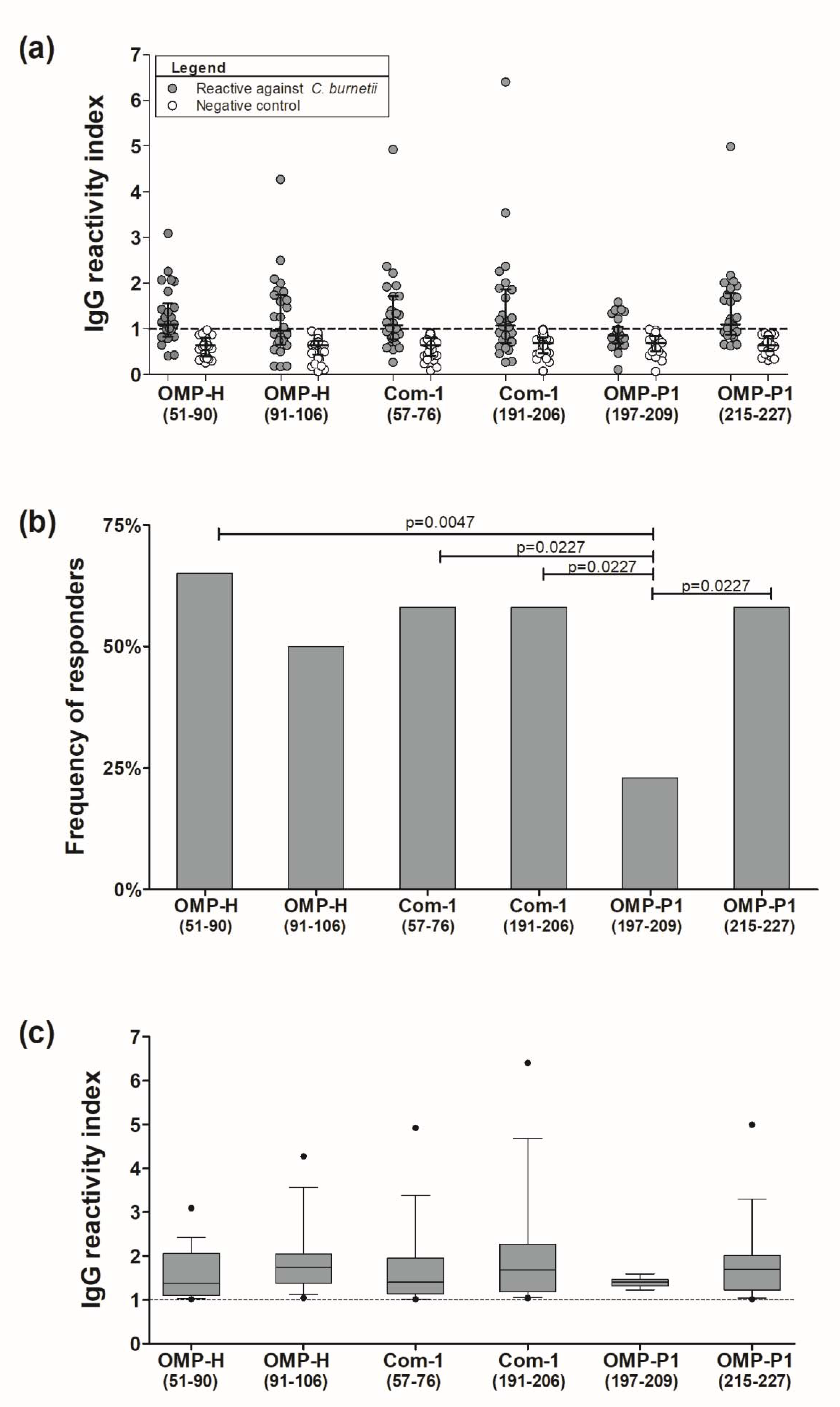
Aiming to verify the natural immunogenicity of the predicted epitopes, samples of patients both reactive and non-reactive to C. burnetii were tested by ELISA against synthetic peptides containing predicted linear B-cell epitopes. Remarkably, based on our threshold, we observed no responsivity against peptides in the negative control group, composed of C. burnetii-non-reactive individuals (Figure 2a). Moreover, in the C. burnetii-reactive group, the frequencies of response against the epitopes OMP-H(51–59) (65%), OMP-H(91–106) (50%), Com-1(57–76) (58%), Com-1(191–206) (58%), and OMP-P1(215–227) (15 responders; 58%) were higher than 50%. Moreover, the frequency of response to OMP-P1(197–209) (23%) was statistically lower than the frequencies of response to OMP-H(51–59) (p = 0.0047), Com-1(57–76), Com-1(191–206), and OMP-P1(215–227) (p = 0.0227) (Figure 2b). Despite these differences, we observed a similar magnitude of IgG-RI against synthetic peptides, ranging from 1.01 to 6.4, in which the median reactivity indices were 1.37 (IQ: 1.095–2.055) against OMP-H(51–59), 1.74 (IQ: 1.37–2.045) against OMP-H(91–106), 1.4 (IQ: 1.14–1.95) against Com-1(57–76), 1.68 (IQ: 1.18–2.26) against Com-1(191–206), 1.39 (IQ:1.31–1.46) against OMP-P1(197–209), and 1.69 (IQ: 1.22–2.01) against OMP-P1(215–227) (Figure 2c).
As shown in Table 4, 77% of studied individuals (n = 20) presented antibodies against at least one of the identified epitopes. Regarding the reactivity against each studied protein epitope, 18 patients (69%) responded to at least one of the OMP-H epitopes, 17 patients were reactive to at least one of the Com-1 epitopes, while 15 individuals presented antibodies against at least one of the OMP-P1 epitopes.

2.6. Associations between Humoral Response to Synthetic Peptides and Clinical Features

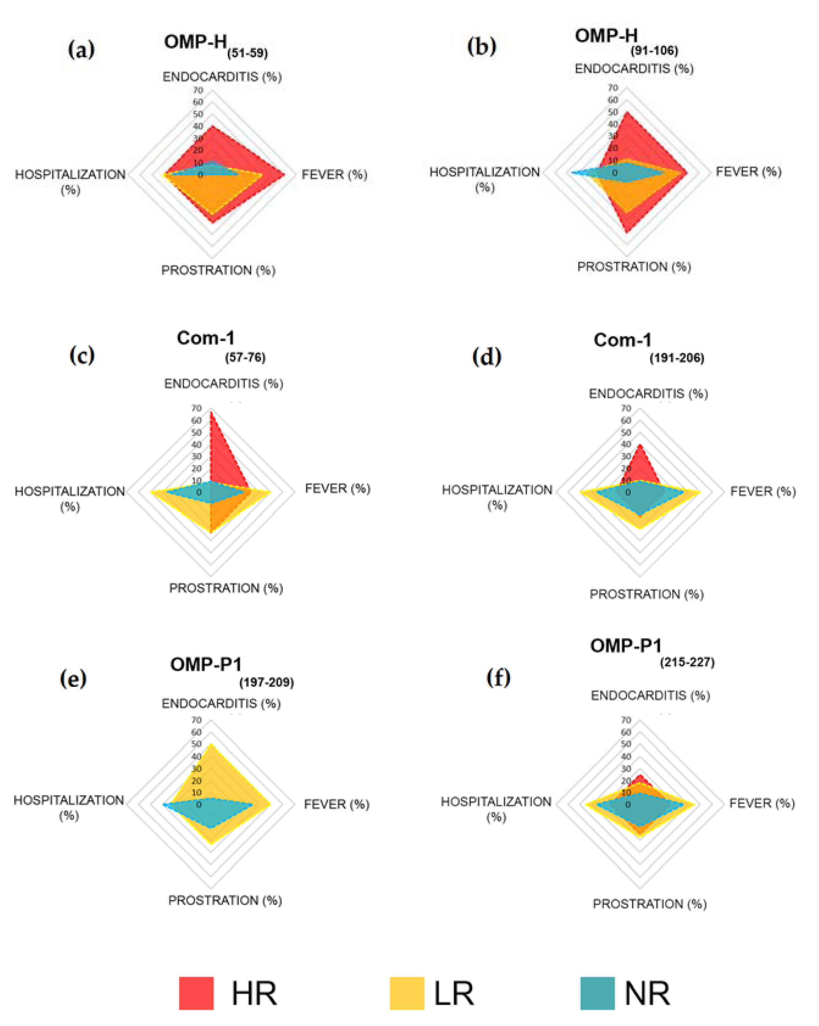
As previously shown in Table 4, between 20% and 33% of responders to the epitopes OMP-H(51–59), OMP-H(91–106), Com-1(57–76), Com-1(191–206), and OMP-P1(215–227) presented high levels of specific antibodies. Aiming to explore these data, we compared the clinical features of high responders (HR: RI > 2), low responders (LR: 1 > RI ≤ 2), and non-responders (NR: RI ≤ 1) to each studied peptide. Remarkably, 75% of patients reactive to C. burnetii who presented endocarditis (n = 6) also demonstrated a high level of antibodies to at least one of the identified epitopes. The frequency of endocarditis seems to be higher in HR than in LR and NR to OMP-H(51–59) (HR: 40%; LR: 8% and NR: 11%; Figure 3a), OMP-H(91–106) (HR: 50%, LR: 11% and NR: 8%; Figure 3b), Com-1(57–76) (HR: 67%, LR: 8% and NR: 9%; Figure 3c), and Com-1(191–206) (HR: 40%, LR: 10% and NR: 9%; Figure 3d). Moreover, despite the six responders to OMP-P1(197–209) having presented a low specific antibody level, 50% presented endocarditis, while only 5% of NR to this epitope presented this complication (Figure 3e).
Regarding another clinical feature, prostration seems to be more frequent in patients who present antibodies against OMP-H(51–59) (HR: 40%, LR: 33%; NR: 0; Figure 3a), OMP-H(91–106) (HR: 50%, LR: 33% and NR: 8; Figure 3b), and Com-1(57–76) (HR:33%, LR:33% and NR: 9%; Figure 3c). Remarkably, an apparently minor hospitalization rate was observed in high responders to Com-1(57–76) (HR:0, LR: 50%, NR: 36%; Figure 3c) and Com-1(191–206) (HR: 20%, LR: 50%, NR: 36%; Figure 3d). Finally, the antibody response against OMP-P1(215–227) seems not to be related to clinical features, with no differences between HR, LR, and NR to this epitope (Figure 3f).

3. Discussion

Most C. burnetii infections are asymptomatic or associated with nonspecific symptoms. Based on this, seroprevalence surveys arise as a potential way to evaluate the real prevalence of C. burnetii [19]. However, despite studies in the Netherlands [59,60], Australia [61], the USA [62], Bhutan [63], Northern Ireland [64], Cyprus [65], and South America [66,67], the true global prevalence of C. burnetii infections is still unknown due to the dearth of representative national seroprevalence surveys in the world. In this scenario, the identification of linear B-cell epitopes arises as a potential approach to improve and accelerate the development of novel diagnostic tests that could allow effective C. burnetii seroprevalence surveys.
In this study, we identified B-cell epitopes in antigen candidates for the development of serological tests for C. burnetii, and showed their immunogenicity in individuals naturally exposed to infection. In this way, we explored three C. burnetii outer membrane proteins (OMPs): OMP-H (CBU_0612), an outer membrane chaperon protein highlighted as a very strong immunodominant marker for both acute and persistent forms of Q fever [35,41]; Com-1 (CBU_1910), a strongly immunogenic protein [38,40] that is considered a reliable Q fever serodiagnosis marker [42,43]; and OMP-P1, a porin, highly expressed in large-cell variants (LCVs)—the replicative form of the bacterium in the cell hosts—and downregulated in small-cell variants (SCVs), the metabolically inactive and resistant form found in the environment [44,45,46]. However, the use of whole antigens in serodiagnosis may result in cross-reactivity with other related proteobacteria. Based on this, the B-cell epitopes’ identification arises as a promising alternative to improve the specificity of serological tests for C. burnetii.
In recent years, combinations of prediction algorithms have been used to improve the accuracy of linear B-cell epitope prediction against viruses [68,69,70,71], fungi [72], protozoa [73,74], and bacteria [75,76,77]. However, there was only one in silico study of C. burnetii, in which six different algorithms were individually used to explore OMP-H and Com-1, predicting between 1–7 linear B-cell epitopes in each studied protein [78]. In this study, using a combination of prediction algorithms—Bepipred, ABCpred, and Emini Surface Accessibility prediction—four sequences (OMP-H(51–59), OMP-H(91–106), Com-1(57–76), and Com-1(191–206)) were predicted as epitopes by the three used algorithms, while six sequences were predicted by only two of them. Moreover, combining this initial epitope identification with the antigenic analysis, we predicted only two epitopes in OMPH, three in Com-1, and three in OMP-P1. This reduction in the number of predicted epitopes in comparison to those reported by Jaydari et al. [78] may be related to the improvement in the accuracy of in silico analyses by the combination of prediction algorithms. Moreover, the specificities of the predicted epitopes were verified by comparison with proteobacteria proteins. Remarkably, despite all of the predicted epitopes being conserved among C. burnetii strains described in the UniProt database (data not shown), the scarcity of studies concerning OMPs’ polymorphism hampers conclusions about the real conservation of the identified epitopes in C. burnetii strains.
Furthermore, in a previous study, our group hypothesized that predicted epitopes that fail in experimental validation could be buried in protein quaternary structures [71], upon which prediction algorithms would be unable to evaluate these structures. Based on this hypothesis, and considering the oligomerization of similar proteins [45,79,80], we evaluated the localization of predicted epitopes in oligomeric structures of studied proteins. To do this, we modeled the quaternary structures of the studied proteins, and observed that epitopes Com-1(26–34) and OMP-P1(98–106) were buried in Com-1 and OMP-P1 homoligomers (Figure 1). These two epitopes—Com-1(26–34) and OMP-P1(98–106)—were excluded from the study, while the other six predicted epitopes (OMP-H(51–59), OMP-H(91–106), Com-1(57–76), Com-1(191–206), OMP-P1(197–209), and OMP-P1(215–227)) were selected and experimentally validated. From our point of view, the evaluation of the predicted epitopes’ exposition in the protein oligomeric structure can be a critical step to improve the accuracy of epitope prediction, and the lack of those analyses could explain—at least in part—the low validation rates (15%) of predicted epitopes for some infectious agents seen in Brucella sp. proteins [81,82,83]. On the other hand, despite the improvements in methodologies and the results of in silico studies, the evaluation of reactivity to predicted linear B-cell epitopes remains rare, and this was the first study using samples from Brazilian C. burnetii-exposed individuals to confirm the natural immunogenicity of B-cell epitopes predicted in this bacterium’s proteins.
In Brazil, although the presence of C. burnetii has been demonstrated in animals [20,21,22,23], humans [21,23,24,25,26,27] and, most recently, in artisanal cheese [28] and unpasteurized milk [29], Q fever remains poorly understood and reported. Moreover, despite the possibility of mistaking Q fever for influenza, dengue, malaria, leptospirosis, and other infectious diseases [30], in Brazil, only suspected rickettsiosis cases are investigated as Q fever, corroborating the underreporting of this zoonosis, and explaining the limited number of C. burnetii-reactive samples in this study (n = 26). Remarkably, similar-sized samples were previously used to successfully define C. burnetii proteins as antigens to serological tests [37,84,85,86], indicating that the number of patients used in our study was sufficient to prove the natural immunogenicity of the identified epitopes.
In this way, samples of the studied population were tested by ELISA against synthetic peptides, representing the sequences of six predicted linear B-cell epitopes (OMP-H(51–59), OMP-H(91–106), Com-1(57–76), Com-1(191–206), OMP-P1(197–209), and OMP-P1(215–227)). Our data showed high specificity of the selected B-cell epitopes, such that all unreactive patients to C. burnetii according to IFA were also non-responsive against the tested synthetic peptides (Figure 2a). Moreover, all of the synthetic peptides were recognized by C. burnetii-reactive patients, confirming their natural immunogenicity. In this context, a previous study using a synthetic peptide containing the sequence of epitope Com-1(57–76) in a latex agglutination test (LAT) reported a performance (sensitivity and sensibility) of ~75% in cattle samples, when compared to a commercial ELISA test [49]. Although more studies are necessary to evaluate the true specificity and sensitivity of these epitopes, immunodominant epitope combinations can be used to improve the accuracy of serological diagnosis of C. burnetii. This strategy can be applied especially to commercial ELISA tests for C. burnetii, which have presented low sensitivity and specificity when compared to IFA [87,88]. Here, while the reactivity to a single peptide ranged from 23% to 65% of studied patients, the combination of epitopes increased sensitivity, allowing the detection of specific antibodies against at least one epitope of the same protein in ~60–70% of C. burnetii-seroreactive individuals. Moreover, 77% of the C. burnetii-reactive group was also reactive to at least one of the identified epitopes (Table 4). These data reinforce the need to identify linear B-cell epitopes in other promising serological markers—such as OmpA [89], C. burnetii macrophage infectivity potentiator protein (Cb-Mip) [90], and YbgF [85]—in order to improve the development of novel tools for the diagnosis of C. burnetii.
Despite the similarities in the magnitude of response against the identified epitopes, where the epitopes OMP-H(51–59), OMP-H(91–106), Com-1(57–76), Com-1(191–206), and OMP-P1(215–227) were recognized in more than 50% of the studied samples, we observed that ~20–30% of responders presented a high level of antibodies (R.I. > 2.0), while all responders to OMP-P1(197–209)—the peptide with lower recognition rates (Figure 2b)—presented a low level (1.0 > RI ≤ 2.0) of specific antibodies against this peptide. The frequency of high responders to specific antigens is not well explored for C. burnetii; however, grouping individuals according to RI was a strategy previously employed by our group in a study that reported high responders against Plasmodium vivax recombinant protein, suggesting an association between frequency of recently exposed individuals and high humoral response to this protein [91]. Based on this previous experience, in order to investigate associations between clinical features and the level of antibodies against the identified epitopes, we evaluated the frequency of cases of endocarditis, fever, prostration, and hospitalization among patients reactive to C. burnetii, comparing these features between individuals who presented high levels of antibodies (R.I. > 2), low levels of antibodies (1.0 > R.I. ≤ 2.0), and who were non-responsive (R.I. ≤ 1) to an identified epitope. Remarkably, we observed that 75% of the studied patients with endocarditis presented a high level of antibodies against at least one of the identified epitopes, corroborating that the intense humoral response may be a marker in chronic/persistent cases of Q fever, which was already suggested by the antibody response against Com-1 recombinant protein [43].
Regarding the response against each epitope, we observed that endocarditis seems to be more frequent in patients who are HR to the OMP-H and Com-1 epitopes (Figure 3), suggesting that an intense humoral response to the Com-1 and OMP-H epitopes may be associated with chronic Q fever complications. This hypothesis corroborates the potential of recombinant OMP-H as an antigen for the serological diagnosis of C. burnetii endocarditis cases [37]; however, more studies involving a higher number of patients and complications are still necessary to prove this hypothesis. Moreover, we observed low hospitalization rates in high responders to Com-1 epitopes, while we observed a low frequency of prostration reports in non-responders to OMP-H epitopes when compared to LR and HR to OMP-H epitopes. Finally, the humoral response against OMP-P1 seems not to be associated with clinical features, given that OMP-P1(197–209) was the epitope with the lowest recognition rate, and did not present high responders, while we observed no differences in clinical features among HR, LR, and NR to OMP-P1(215–227). These data corroborate the study of Qingfeng Li et al., which showed that recombinant OMP-P1 induced a low antibody level, but a high cellular immune response, in immunized mice [92]. Here, we observed that OMP-P1 linear B-cell epitopes are poorly immunogenic, but more studies aiming to identify its TCD4 and TCD8 epitopes are still necessary, and may be used to improve vaccine development [93].
In conclusion, despite the limitations in sample size and study design, six linear B-cell epitopes were identified by a combination of in silico tools and, subsequently, confirmed as naturally immunogenic in individuals exposed to C. burnetii. This was the first study evaluating the natural immunogenicity of the predicted linear B-cell epitopes in the C. burnetii outer membrane proteins OMP-H, Com-1, and OMP-P1 using samples of human patients. Therefore, more serosurveys regarding these epitopes are encouraged, and are essential in order to estimate their real specificity and sensitivity. Currently, synthetic peptides representing B-cell epitope sequences are considered a valuable tool as novel molecules for the reliable and rapid diagnosis of infectious diseases [58,94], with promising results for Q fever/coxiellosis [49,55], as corroborated by this study. When compared to whole proteins, the use of synthetic peptides as diagnostic antigens allows a higher specificity and reproducibility with no batch-to-batch variation, along with easy and low-cost production. In this context, we believe that the identification of B-cell epitopes may be an effective strategy to improve and accelerate the development of surveillance tools for Q fever and other neglected diseases.

4. Materials and Methods

4.1. Sequence Data

For in silico analyzes and 3-dimensional (3D) structure modeling, the full sequences of OMP-H (ID: CBU_0612), Com-1 (CBU_1910), and OMP-P1 (ID: CBU_0311) were obtained from the UniProt database (https://www.uniprot.org/ accessed on 1 June 2021).

4.2. B-Cell Epitope Prediction

Firstly, based on information from the UniProt database, transmembrane regions, signal peptides, and cytoplasmic regions of the studied proteins were excluded from B-cell epitope predictions. The identification of linear B-cell epitopes in the extracellular regions of the studied proteins was performed based on the combination of three different algorithms: Bepipred 1.0 (threshold: 0.35), Emini Surface Accessibility prediction (ESA) (threshold 1.0), and ABCpred (threshold 0.75). Sequences predicted by at least two of the used algorithms were considered predicted B-cell epitopes, and were evaluated for antigenicity using VaxiJen (http://www.ddg-pharmfac.net/vaxijen/VaxiJen/VaxiJen.html accessed on 1 June 2021) (threshold 0.4).
Briefly, Bepipred takes a single sequence in FASTA format input, and each amino acid receives a prediction score based on hidden Markov model profiles of known antigens and incorporates propensity scale methods based on hydrophilicity and secondary structure prediction. For each input sequence, the server outputs a prediction score [95]. Meanwhile, ESA calculates the surface accessibility of hexapeptides, with values greater than 1.0 indicating an increased probability of being found on the surface [96]. Moreover, ABCpred is the first server developed based on a recurrent neural network (machine-based technique), using fixed-length patterns to predict B-cell epitopes in an antigen sequence, with 65.93% accuracy [97]. VaxiJen is the first server for alignment-independent prediction of protective antigens; it was developed to allow classification of antigens based solely on the physicochemical properties of proteins, without recourse to sequence alignment. A bacterial protein dataset, threshold 0.4, was used to derive models for the prediction of whole-protein antigenicity, showing prediction accuracy from 70% to 89% [98,99]. All prediction algorithms were accessed in March 2019.

4.3. Evaluation of B-Cell Epitopes’ Degree of Conservation

To evaluate the degree of conservation of the predicted epitopes, the identified sequences were compared for similarity with bacteria from the phylum Proteobacteria (Francisella tularensis, Legionella Pneumophila, Escherichia coli, Pseudomonas Aeruginosa, Rickettsia rickettsii, Ehrlichia chaffeensis, Bartonella henselae, Brucella melitensis, Afipia felis, and Campylobacter jejuni) and microbiota bacteria (Enterobacter spp., Klebsiella spp., Citrobacter spp., and Enterococcus coli) using BLASTp (https://blast.ncbi.nlm.nih.gov/Blast.cgi?PAGE=Proteins accessed on 21 June 2021). When compared to this group of organisms, the sequences that presented E-values higher than 1 were considered to be non-conserved B-cell epitopes.

4.4. Three-Dimensional (3D) Structure Modeling

The 3D structures of the studied OMPs were modeled using the Robetta server (http://new.robetta.org/ accessed on 21 June 2021); this server is continually evaluated through continuous automated model evaluation (CAMEO), and its primary service is to predict the 3D structure of a protein given its amino acid sequence [100,101,102,103]. The PDB formats reported from this server were visualized using PyMOL V1 viewer software [104,105].

4.5. Evaluation of Epitopes’ Exposure in the Protein Quaternary Structures

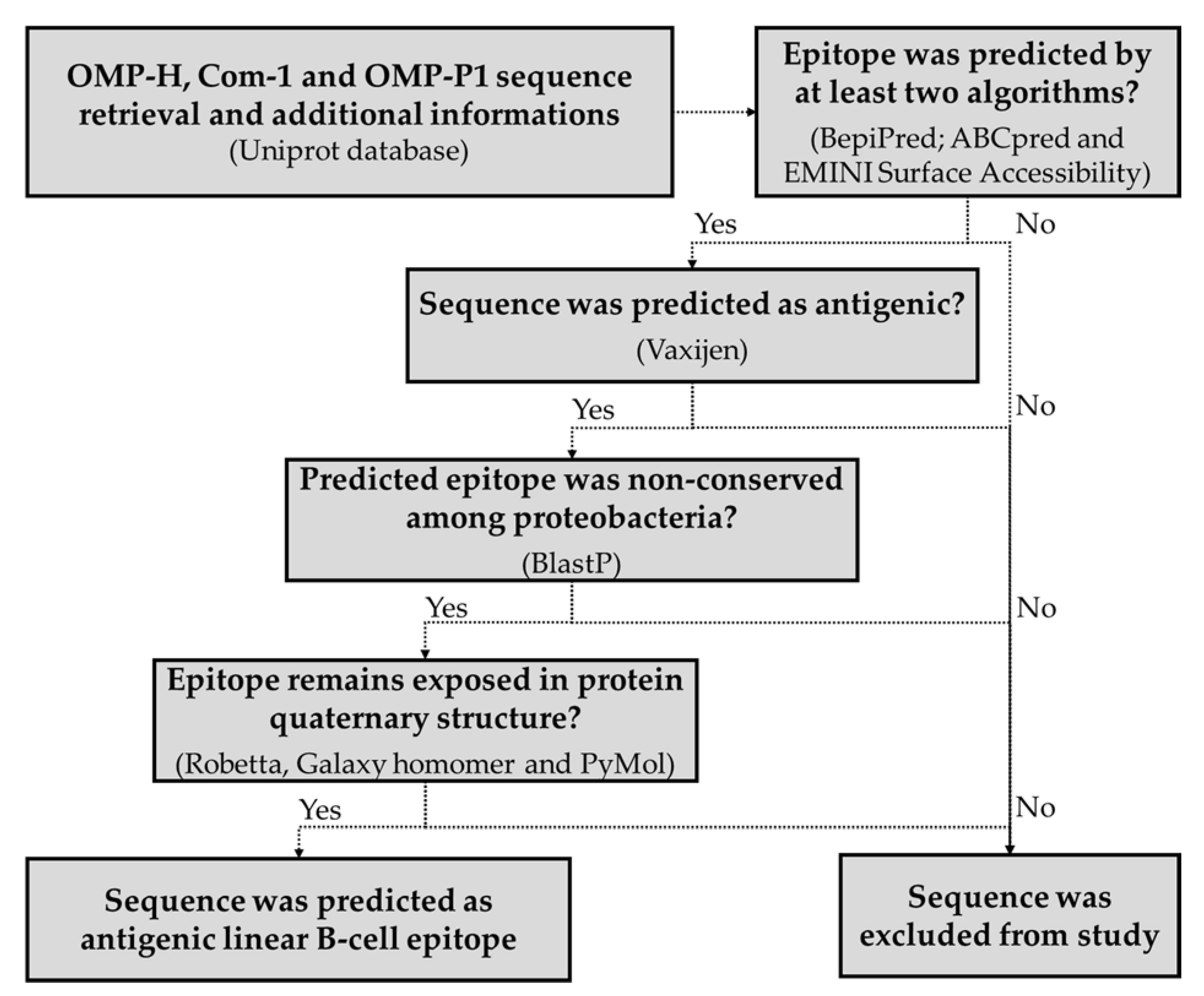
Considering that the OMPs generally form oligomers, the modeled 3D structures of the studied proteins were submitted to the GalaxyHomomer server (http://galaxy.seoklab.org/cgi-bin/submit.cgi?type=HOMOMER accessed on 21 June 2021), which performs comparative modeling of quaternary structures [106,107]. In this study, it was defined that the sequences that had at least 30% of their amino acids interacting with other chains were considered hidden in the quaternary structure of the protein, and were excluded from further analyses. Finally, sequences predicted as antigenic and linear B-cell epitopes—non-conserved among proteobacteria, and exposed in quaternary structures—were selected for experimental validation. The schematic abstract of the prediction of B-cell epitopes is represented in Figure 4.

4.6. Peptide Synthesis

Sequences predicted as antigenic linear B-cell epitopes were chemically synthesized by the company GenOne Biotechnologies, Brazil. Analytical chromatography of the peptides demonstrated a purity of approximately > 95%, and mass spectrometry analysis of the peptides indicated their expected mass.

4.7. Studied Population

A total of 57 samples of suspected Q fever cases were provided by the Brazilian National Reference Center for Rickettsioses—Laboratory of Hantaviruses and Rickettsiosis—Oswaldo Cruz Foundation. Serum samples were previously tested by IFA for the presence of immunoglobulin G antibodies (IgG) against Coxiella burnetii, using a commercial kit from Scimedx® (Dover, NJ, USA) [108]. Firstly, suspected samples were screened at a 1:64 dilution, and those that were considered positive were further diluted to determine the IFA´s end titers. Negative and positive controls were included for each test run. The study was reviewed and approved by the Oswaldo Cruz Foundation Ethical Committee and the National Ethical Committee of Brazil (number CAAE: 39056120.6.0000.5248).

4.8. Experimental Confirmation of the Antigenicity of the Predicted Epitopes

Samples of suspected and confirmed Q fever cases were screened for the presence of naturally acquired antibodies against the synthetic peptides via ELISA. Briefly, MaxiSorp 96-well plates (Nunc, Rochester, NY, USA) were coated with 100 μg/mL of a peptide. After overnight incubation at 4 °C, plates were washed with phosphate-buffered saline (PBS) and blocked with PBS-containing 5% non-fat dry milk (PBS-M) for 1 h at 37 °C. Individual plasma samples diluted 1:100 on PBS-M were added in duplicate wells, and the plates were incubated at 37 °C for 2 h. After three washes with PBS-Tween20 (0.05%), bound antibodies were detected with peroxidase-conjugated goat anti-human IgG (Sigma St. Louis, MO, USA), followed by TMB (3,3’,5,5’-tetramethylbenzidine). The absorbance was read at 450 nm using an xMark™ microplate absorbance spectrophotometer (Bio-Rad, Hercules, CA, USA). The results for total IgG were expressed as the reactivity index (RI)—the ratio between the mean optical density (OD) of tested samples and the mean OD of 31 negative controls plus 2 standard deviations (SD). Subjects were considered IgG responders to a particular antigen if the RI was higher than 1.

4.9. Statistical Analysis

All statistical analyses were carried out using Prism 5.0 for Windows (GraphPad Software, Inc., San Diego, CA, USA). The one-sample Kolmogorov–Smirnoff test was used to determine whether a variable was normally distributed. Differences in frequencies of IgG responders to synthetic peptides were evaluated using Fisher’s exact test. The Mann–Whitney test was used to compare reactivity indices against synthetic peptides between responders to each epitope. A two-sided p-value < 0.05 was considered significant.

5. Conclusions

Our data corroborate the use of immunoinformatics approaches to identify epitopes as targets of humoral response that can be further explored in the development of novel surveillance tools for Q fever and other neglected diseases. Here, exploring linear B-cell epitopes in C. burnetii—OMP-H, Com-1, and OMP-P1—we experimentally confirmed the natural immunogenicity of two epitopes in each studied protein. Moreover, our data suggest that an intense humoral response to OMP-H and Com-1 epitopes may be related to Q fever complications, such as endocarditis.

Author Contributions

Conceptualization, R.N.R.-d.-S., F.d.P.C., T.R., and E.R.S.d.L.; methodology, S.d.S.F., F.d.M.M., L.S.A., M.R.d.S.A., and A.A.P.J.; writing—original draft preparation, R.N.R.-d.-S. and J.d.C.L.-J.; writing—review and editing, E.R.S.d.L., J.F., and F.d.P.C.; funding acquisition: R.N.R.-d.-S. and E.R.S.d.L. All authors have read and agreed to the published version of the manuscript.

Funding

This research was funded by Fiocruz—InovaFiocruz-Geração de conhecimento: novos talentos, grant number VPPCB-008-FIO-18; Coordenação de Aperfeiçoamento de Pessoal de Nível Superior—Brazil (CAPES), grant number 001, FAPERJ (project number E-26/202.575/2019), and the Brazilian National Council for Scientific and Technological Development—CNPq (project number 303024/2019-4).

Institutional Review Board Statement

This study was conducted in accordance with the guidelines of the Declaration of Helsinki, and approved by the Oswaldo Cruz Foundation Ethical Committee and the National Ethical Committee of Brazil (number CAAE: 39056120.6.0000.5248).

Informed Consent Statement

Informed consent was obtained from all subjects involved in the study.

Data Availability Statement

Publicly available datasets were analyzed in this study. This data can be found here: https://www.uniprot.org/ accessed on 16 August 2021.

Conflicts of Interest

The authors declare no conflict of interest, and the funders had no role in the design of the study, in the collection, analyses, or interpretation of data, in the writing of the manuscript, or in the decision to publish the results.

References

  1. Eldin, C.; Melenotte, C.; Mediannikov, O.; Ghigo, E.; Million, M.; Edouard, S.; Mege, J.L.; Maurin, M.; Raoult, D. From Q fever to Coxiella burnetii infection: A paradigm change. Clin. Microbiol. Rev. 2017, 30, 115–190. [Google Scholar] [CrossRef] [Green Version]
  2. ILRI. Mapping of Poverty and Likely Zoonoses Hotspots; Department for International Development, UK, 2012. Available online: https://cgspace.cgiar.org/bitstream/handle/10568/21161/ZooMap_July2012_final.pdf (accessed on 19 August 2021).
  3. Sahu, R.; Rawool, D.B.; Dhaka, P.; Yadav, J.P.; Mishra, S.P.; Kumar, M.; Vergis, J.; Malik, S.S.; Barbuddhe, S.B. Current perspectives on the occurrence of Q fever: Highlighting the need for systematic surveillance for a neglected zoonotic disease in Indian subcontinent. Environ. Microbiol. Rep. 2021, 13, 138–158. [Google Scholar] [CrossRef] [PubMed]
  4. Maurin, M.; Raoult, D. Q fever. Clin. Microbiol. Rev. 1999, 12, 518–553. [Google Scholar] [CrossRef] [PubMed] [Green Version]
  5. Asamoah, J.K.K.; Jin, Z.; Sun, G.Q.; Li, M.Y. A deterministic model for Q fever transmission dynamics within dairy cattle herds: Using sensitivity analysis and optimal controls. Comput. Math. Methods Med. 2020, 2020, 6820608. [Google Scholar] [CrossRef] [PubMed]
  6. Rotz, L.D.; Khan, A.S.; Lillibridge, S.R.; Ostroff, S.M.; Hughes, J.M. Public health assessment of potential biological terrorism agents. Emerg. Infect. Dis. 2002, 8, 225–230. [Google Scholar] [CrossRef]
  7. Woldehiwet, Z. Q fever (coxiellosis): Epidemiology and pathogenesis. Res. Vet. Sci. 2004, 77, 93–100. [Google Scholar] [CrossRef] [PubMed]
  8. Bauer, A.E.; Hubbard, K.R.; Johnson, A.J.; Messick, J.B.; Weng, H.Y.; Pogranichniy, R.M. A cross sectional study evaluating the prevalence of Coxiella burnetii, potential risk factors for infection, and agreement between diagnostic methods in goats in Indiana. Prev. Vet. Med. 2016, 126, 131–137. [Google Scholar] [CrossRef]
  9. Patil, S.M.; Regunath, H. Q Fever; StatPearls Publishing: Treasure Island, FL, USA, 2020. Available online: https://www.ncbi.nlm.nih.gov/books/NBK556095/ (accessed on 19 August 2021).
  10. Fishbein, D.B.; Raoult, D. A cluster of Coxiella burnetii infections associated with exposure to vaccinated goats and their unpasteurized dairy products. Am. J. Trop. Med. Hyg. 1992, 47, 35–40. [Google Scholar] [CrossRef] [PubMed]
  11. Esmaeili, S.; Mobarez, A.M.; Khalili, M.; Mostafavi, E. High prevalence and risk factors of Coxiella burnetii in milk of dairy animals with a history of abortion in Iran. Comp. Immunol. Microbiol. Infect. Dis. 2019, 63, 127–130. [Google Scholar] [CrossRef] [PubMed]
  12. Angelakis, E.; Raoult, D. Emergence of Q fever. Iran. J. Public Health 2011, 40, 1–18. [Google Scholar]
  13. Van den Brom, R.; van Engelen, E.; Roest, H.I.; van der Hoek, W.; Vellema, P. Coxiella burnetii infections in sheep or goats: An opinionated review. Vet. Microbiol. 2015, 181, 119–129. [Google Scholar] [CrossRef] [PubMed]
  14. van Schaik, E.J.; Chen, C.; Mertens, K.; Weber, M.M.; Samuel, J.E. Molecular pathogenesis of the obligate intracellular bacterium Coxiella burnetii. Nat. Rev. Microbiol. 2013, 11, 561–573. [Google Scholar] [CrossRef] [PubMed] [Green Version]
  15. Million, M.; Thuny, F.; Richet, H.; Raoult, D. Long-term outcome of Q fever endocarditis: A 26-year personal survey. Lancet Infect. Dis. 2010, 10, 527–535. [Google Scholar] [CrossRef]
  16. Delsing, C.E.; Kullberg, B.J.; Bleeker-Rovers, C.P. Q fever in the Netherlands from 2007 to 2010. Neth. J. Med. 2010, 68, 382–387. [Google Scholar]
  17. Porter, S.R.; Czaplicki, G.; Mainil, J.; Guatteo, R.; Saegerman, C. Q fever: Current state of knowledge and perspectives of research of a neglected zoonosis. Int. J. Microbiol. 2011, 2011, 248418. [Google Scholar] [CrossRef] [PubMed]
  18. Marrie, T.J.; Raoult, D. Q fever—A review and issues for the next century. Int. J. Antimicrob. Agents 1997, 8, 145–161. [Google Scholar] [CrossRef]
  19. Anderson, A.; Bijlmer, H.; Fournier, P.E.; Graves, S.; Hartzell, J.; Kersh, G.J.; Limonard, G.; Marrie, T.J.; Massung, R.F.; McQuiston, J.H.; et al. Diagnosis and management of Q fever—United States, 2013: Recommendations from CDC and the Q fever working group. Morb. Mortal. Wkly. Rep. Recomm. Rep. 2013, 62, 1–30. [Google Scholar]
  20. Mares-Guia, M.A.; Rozental, T.; Guterres, A.; Gomes, R.; Almeida, D.N.; Moreira, N.S.; Barreira, J.D.; Favacho, A.R.; Santana, A.L.; Lemos, E.R. Molecular identification of the agent of Q fever—Coxiella burnetii—in domestic animals in State of Rio de Janeiro, Brazil. Rev. Soc. Bras. Med. Trop. 2014, 47, 231–234. [Google Scholar] [CrossRef] [PubMed] [Green Version]
  21. Mioni, M.S.R.; Costa, F.B.; Ribeiro, B.L.D.; Teixeira, W.S.R.; Pelicia, V.C.; Labruna, M.B.; Rousset, E.; Sidi-Boumedine, K.; Thiery, R.; Megid, J. Coxiella burnetii in slaughterhouses in Brazil: A public health concern. PLoS ONE 2020, 15, e0241246. [Google Scholar] [CrossRef]
  22. Zanatto, D.C.S.; Gatto, I.R.H.; Labruna, M.B.; Jusi, M.M.G.; Samara, S.I.; Machado, R.Z.; Andre, M.R. Coxiella burnetii associated with BVDV (Bovine Viral Diarrhea Virus), BoHV (Bovine Herpesvirus), Leptospira spp., Neospora caninum, Toxoplasma gondii and Trypanosoma vivax in reproductive disorders in cattle. Rev. Bras. Parasitol. Vet. 2019, 28, 245–257. [Google Scholar] [CrossRef]
  23. Souza, E.A.R.; Castro, E.M.S.; Oliveira, G.M.B.; Azevedo, S.S.; Peixoto, R.M.; Labruna, M.B.; Horta, M.C. Serological diagnosis and risk factors for Coxiella burnetii in goats and sheep in a semi-arid region of Northeastern Brazil. Rev. Bras. Parasitol. Vet. 2018, 27, 514–520. [Google Scholar] [CrossRef] [PubMed]
  24. Mares-Guia, M.A.; Rozental, T.; Guterres, A.; Ferreira Mdos, S.; Botticini Rde, G.; Terra, A.K.; Marraschi, S.; Bochner, R.; Lemos, E.R. Molecular identification of Q fever in patients with a suspected diagnosis of dengue in Brazil in 2013–2014. Am. J. Trop. Med. Hyg. 2016, 94, 1090–1094. [Google Scholar] [CrossRef] [PubMed] [Green Version]
  25. Ribeiro-Netto, A.; Nikitin, T.; Valentini, H.; Ribeiro, I.F. Study of Q fever in S Ao Paulo. I. Incidence in dairy cattle in the Para’ibo Valley. Rev. Inst. Med. Trop. Sao Paulo 1964, 6, 137–141. [Google Scholar]
  26. Mioni, M.S.R.; Sidi-Boumedine, K.; Dalanezi, F.M.; Joaquim, S.F.; Denadai, R.; Teixeira, W.S.R.; Labruna, M.B.; Megid, J. New genotypes of Coxiella burnetii circulating in Brazil and Argentina. Pathogens 2019, 9, 30. [Google Scholar] [CrossRef] [Green Version]
  27. Rozental, T.; Silva, A.; Oliveira, R.C.; Favacho, A.R.M.; Oliveira, M.L.A.; Bastos, F.I.; Lemos, E.R.S. Seroprevalence of bartonella spp., Coxiella burnetii, and Hantavirus among people who inject drugs in Rio de Janeiro, Brazil: A retrospective assessment of a biobank. Rev. Inst. Med. Trop. Sao Paulo 2018, 60, e31. [Google Scholar] [CrossRef]
  28. Rozental, T.; Faria, L.S.; Forneas, D.; Guterres, A.; Ribeiro, J.B.; Araujo, F.R.; Lemos, E.R.S.; Silva, M.R. First molecular detection of Coxiella burnetii in Brazilian artisanal cheese: A neglected food safety hazard in ready-to-eat raw-milk product. Braz. J. Infect. Dis. 2020, 24, 208–212. [Google Scholar] [CrossRef]
  29. de Souza Ribeiro Mioni, M.; Ribeiro, B.L.D.; Peres, M.G.; Teixeira, W.S.R.; Pelicia, V.C.; Motta, R.G.; Labruna, M.B.; Ribeiro, M.G.; Sidi-Boumedine, K.; Megid, J. Real-time quantitative PCR-based detection of Coxiella burnetii in unpasteurized cow’s milk sold for human consumption. Zoonoses Public Health 2019, 66, 695–700. [Google Scholar] [CrossRef]
  30. Vaidya, V.M.; Malik, S.V.; Bhilegaonkar, K.N.; Rathore, R.S.; Kaur, S.; Barbuddhe, S.B. Prevalence of Q fever in domestic animals with reproductive disorders. Comp. Immunol. Microbiol. Infect. Dis. 2010, 33, 307–321. [Google Scholar] [CrossRef] [PubMed]
  31. Vincent, G.A.; Graves, S.R.; Robson, J.M.; Nguyen, C.; Hussain-Yusuf, H.; Islam, A.; Fenwick, S.G.; Stenos, J. Isolation of Coxiella burnetii from serum of patients with acute Q fever. J. Microbiol. Methods 2015, 119, 74–78. [Google Scholar] [CrossRef] [PubMed]
  32. Madariaga, M.G.; Rezai, K.; Trenholme, G.M.; Weinstein, R.A. Q fever: A biological weapon in your backyard. Lancet Infect. Dis. 2003, 3, 709–721. [Google Scholar] [CrossRef]
  33. Fournier, P.E.; Marrie, T.J.; Raoult, D. Diagnosis of Q fever. J. Clin. Microbiol. 1998, 36, 1823–1834. [Google Scholar] [CrossRef] [Green Version]
  34. Stephen, S.; Ambroise, S.; Pradeep, J.; Gunasekaran, D.; Sangeetha, B.; Sarangapani, K. Unreliability of three commercial Coxiella burnetii phase II IgM ELISA kits for the seroscreening of acute Q fever in human cases. Indian J. Med Res. 2017, 146, 386–391. [Google Scholar] [CrossRef] [PubMed]
  35. Kowalczewska, M.; Nappez, C.; Vincentelli, R.; La Scola, B.; Raoult, D. Protein candidates for Q fever serodiagnosis. FEMS Immunol. Med. Microbiol. 2012, 64, 140–142. [Google Scholar] [CrossRef] [PubMed] [Green Version]
  36. Beare, P.A.; Chen, C.; Bouman, T.; Pablo, J.; Unal, B.; Cockrell, D.C.; Brown, W.C.; Barbian, K.D.; Porcella, S.F.; Samuel, J.E.; et al. Candidate antigens for Q fever serodiagnosis revealed by immunoscreening of a Coxiella burnetii protein microarray. Clin. Vaccine Immunol. 2008, 15, 1771–1779. [Google Scholar] [CrossRef] [PubMed] [Green Version]
  37. Sekeyova, Z.; Kowalczewska, M.; Decloquement, P.; Pelletier, N.; Spitalska, E.; Raoult, D. Identification of protein candidates for the serodiagnosis of Q fever endocarditis by an immunoproteomic approach. Eur. J. Clin. Microbiol. Infect. Dis. 2009, 28, 287–295. [Google Scholar] [CrossRef]
  38. Xiong, X.; Wang, X.; Wen, B.; Graves, S.; Stenos, J. Potential serodiagnostic markers for Q fever identified in Coxiella burnetii by immunoproteomic and protein microarray approaches. BMC Microbiol. 2012, 12, 35. [Google Scholar] [CrossRef] [PubMed] [Green Version]
  39. Vigil, A.; Ortega, R.; Nakajima-Sasaki, R.; Pablo, J.; Molina, D.M.; Chao, C.C.; Chen, H.W.; Ching, W.M.; Felgner, P.L. Genome-wide profiling of humoral immune response to Coxiella burnetii infection by protein microarray. Proteomics 2010, 10, 2259–2269. [Google Scholar] [CrossRef] [PubMed] [Green Version]
  40. Jiao, J.; Xiong, X.; Qi, Y.; Gong, W.; Duan, C.; Yang, X.; Wen, B. Serological characterization of surface-exposed proteins of Coxiella burnetii. Microbiology 2014, 160, 2718–2731. [Google Scholar] [CrossRef]
  41. Skultety, L. Workshop on Q fever. Acta Virol. 2017, 61, 347–348. [Google Scholar] [CrossRef] [Green Version]
  42. Flores-Ramirez, G.; Danchenko, M.; Quevedo-Diaz, M.; Skultety, L. Reliable tool for detection of novel Coxiella burnetii antigens, using immobilized human polyclonal antibodies. J. Chromatogr. B Anal. Technol. Biomed. Life Sci. 2017, 1047, 84–91. [Google Scholar] [CrossRef]
  43. Vranakis, I.; Mathioudaki, E.; Kokkini, S.; Psaroulaki, A. Com1 as a promising protein for the differential diagnosis of the two forms of Q fever. Pathogens 2019, 8, 242. [Google Scholar] [CrossRef] [Green Version]
  44. Sandoz, K.M.; Popham, D.L.; Beare, P.A.; Sturdevant, D.E.; Hansen, B.; Nair, V.; Heinzen, R.A. Transcriptional profiling of Coxiella burnetii reveals extensive cell wall remodeling in the small cell variant developmental form. PLoS ONE 2016, 11, e0149957. [Google Scholar] [CrossRef] [Green Version]
  45. Varghees, S.; Kiss, K.; Frans, G.; Braha, O.; Samuel, J.E. Cloning and porin activity of the major outer membrane protein P1 from Coxiella burnetii. Infect. Immun. 2002, 70, 6741–6750. [Google Scholar] [CrossRef] [Green Version]
  46. McCaul, T.F.; Banerjee-Bhatnagar, N.; Williams, J.C. Antigenic differences between Coxiella burnetii cells revealed by postembedding immunoelectron microscopy and immunoblotting. Infect. Immun. 1991, 59, 3243–3253. [Google Scholar] [CrossRef] [Green Version]
  47. Guevarra, L.A., Jr.; Boado, K.J.O.; Cenidoza, F.B.B.; Imbao, M.; Sia, M.J.G.; Dalmacio, L.M.M. A synthetic peptide analog of in silico-predicted immunogenic epitope unique to dengue virus serotype 2 NS1 antigen specifically binds immunoglobulin G antibodies raised in rabbits. Microbiol. Immunol. 2020, 64, 153–161. [Google Scholar] [CrossRef]
  48. Kohler, H.; Liebler-Tenorio, E.; Hughes, V.; Stevenson, K.; Bakker, D.; Willemsen, P.; Bay, S.; Ganneau, C.; Biet, F.; Vordermeier, H.M. Interferon-gamma response of mycobacterium avium subsp. Paratuberculosis infected goats to recombinant and synthetic mycobacterial antigens. Front. Vet. Sci. 2021, 8, 645251. [Google Scholar] [CrossRef] [PubMed]
  49. Kumar, M.; Malik, S.S.; Vergis, J.; Ramanjeneya, S.; Sahu, R.; Pathak, R.; Yadav, J.P.; Dhaka, P.; Barbuddhe, S.B.; Rawool, D.B. Development of the Com1 synthetic peptide-based Latex Agglutination Test (LAT) and its comparative evaluation with commercial indirect-ELISA for sero-screening of coxiellosis in cattle. J. Microbiol. Methods 2019, 162, 83–85. [Google Scholar] [CrossRef] [PubMed]
  50. Middleton, S.; Steinbach, S.; Coad, M.; McGill, K.; Brady, C.; Duignan, A.; Wiseman, J.; Gormley, E.; Jones, G.J.; Vordermeier, H.M. A molecularly defined skin test reagent for the diagnosis of bovine tuberculosis compatible with vaccination against Johne’s Disease. Sci. Rep. 2021, 11, 2929. [Google Scholar] [CrossRef] [PubMed]
  51. Polvere, I.; Parrella, A.; Casamassa, G.; D’Andrea, S.; Tizzano, A.; Cardinale, G.; Voccola, S.; Porcaro, P.; Stilo, R.; Vito, P.; et al. Seroprevalence of anti-SARS-CoV-2 IgG and IgM among adults over 65 years old in the south of Italy. Diagnostics 2021, 11, 483. [Google Scholar] [CrossRef] [PubMed]
  52. Arumugam, S.; Nayak, S.; Williams, T.; di Santa Maria, F.S.; Guedes, M.S.; Chaves, R.C.; Linder, V.; Marques, A.R.; Horn, E.J.; Wong, S.J.; et al. A multiplexed serologic test for diagnosis of lyme disease for point-of-care use. J. Clin. Microbiol. 2019, 57. [Google Scholar] [CrossRef]
  53. Murphy, N.; Macchiaverna, N.P.; Cardinal, M.V.; Bhattacharyya, T.; Mertens, P.; Zeippen, N.; Gustin, Y.; Gilleman, Q.; Gurtler, R.E.; Miles, M.A. Lineage-specific rapid diagnostic tests can resolve trypanosoma cruzi TcII/V/VI ecological and epidemiological associations in the argentine chaco. Parasit Vectors 2019, 12, 424. [Google Scholar] [CrossRef] [Green Version]
  54. Prado, I.C.; Mendes, V.G.; Souza, A.L.A.; Dutra, R.F.; De-Simone, S.G. Electrochemical immunosensor for differential diagnostic of Wuchereria bancrofti using a synthetic peptide. Biosens. Bioelectron. 2018, 113, 9–15. [Google Scholar] [CrossRef] [PubMed]
  55. Sahu, R.; Rawool, D.B.; Vinod, V.K.; Malik, S.V.S.; Barbuddhe, S.B. Current approaches for the detection of Coxiella burnetii infection in humans and animals. J. Microbiol. Methods 2020, 179, 106087. [Google Scholar] [CrossRef] [PubMed]
  56. Noya, O.; Patarroyo, M.E.; Guzman, F.; Alarcon de Noya, B. Immunodiagnosis of parasitic diseases with synthetic peptides. Curr. Protein Pept. Sci. 2003, 4, 299–308. [Google Scholar] [CrossRef] [PubMed]
  57. Gomara, M.J.; Haro, I. Synthetic peptides for the immunodiagnosis of human diseases. Curr. Med. Chem. 2007, 14, 531–546. [Google Scholar] [CrossRef]
  58. Kashyap, R.S.; Shekhawat, S.D.; Nayak, A.R.; Purohit, H.J.; Taori, G.M.; Daginawala, H.F. Diagnosis of tuberculosis infection based on synthetic peptides from Mycobacterium tuberculosis antigen 85 complex. Clin. Neurol. Neurosurg. 2013, 115, 678–683. [Google Scholar] [CrossRef]
  59. Dijkstra, F.; van der Hoek, W.; Wijers, N.; Schimmer, B.; Rietveld, A.; Wijkmans, C.J.; Vellema, P.; Schneeberger, P.M. The 2007–2010 Q fever epidemic in The Netherlands: Characteristics of notified acute Q fever patients and the association with dairy goat farming. FEMS Immunol. Med. Microbiol. 2012, 64, 3–12. [Google Scholar] [CrossRef]
  60. Schimmer, B.; Notermans, D.W.; Harms, M.G.; Reimerink, J.H.; Bakker, J.; Schneeberger, P.; Mollema, L.; Teunis, P.; van Pelt, W.; van Duynhoven, Y. Low seroprevalence of Q fever in The Netherlands prior to a series of large outbreaks. Epidemiol. Infect. 2012, 140, 27–35. [Google Scholar] [CrossRef]
  61. Gidding, H.F.; Peng, C.Q.; Graves, S.; Massey, P.D.; Nguyen, C.; Stenos, J.; Quinn, H.E.; McIntyre, P.B.; Durrheim, D.N.; Wood, N. Q fever seroprevalence in Australia suggests one in twenty people have been exposed. Epidemiol. Infect. 2020, 148, e18. [Google Scholar] [CrossRef] [Green Version]
  62. Anderson, A.D.; Kruszon-Moran, D.; Loftis, A.D.; McQuillan, G.; Nicholson, W.L.; Priestley, R.A.; Candee, A.J.; Patterson, N.E.; Massung, R.F. Seroprevalence of Q fever in the United States, 2003–2004. Am. J. Trop. Med. Hyg. 2009, 81, 691–694. [Google Scholar] [CrossRef]
  63. Tshokey, T.; Stenos, J.; Durrheim, D.N.; Eastwood, K.; Nguyen, C.; Graves, S.R. Seroprevalence of rickettsial infections and Q fever in Bhutan. PLoS Negl. Trop. Dis. 2017, 11, e0006107. [Google Scholar] [CrossRef] [PubMed] [Green Version]
  64. McCaughey, C.; McKenna, J.; McKenna, C.; Coyle, P.V.; O’Neill, H.J.; Wyatt, D.E.; Smyth, B.; Murray, L.J. Human seroprevalence to Coxiella burnetii (Q fever) in Northern Ireland. Zoonoses Public Health 2008, 55, 189–194. [Google Scholar] [CrossRef] [PubMed]
  65. Psaroulaki, A.; Hadjichristodoulou, C.; Loukaides, F.; Soteriades, E.; Konstantinidis, A.; Papastergiou, P.; Ioannidou, M.C.; Tselentis, Y. Epidemiological study of Q fever in humans, ruminant animals, and ticks in Cyprus using a geographical information system. Eur. J. Clin. Microbiol. Infect. Dis. 2006, 25, 576–586. [Google Scholar] [CrossRef] [PubMed]
  66. Tapia, T.; Olivares, M.F.; Stenos, J.; Iglesias, R.; Diaz, N.; Vergara, N.; Sotomayor, V.; Gallegos, D.; Soares Magalhaes, R.J.; Acevedo, J.; et al. National seroprevalence of Coxiella burnetii in Chile, 2016–2017. Pathogens 2021, 10, 531. [Google Scholar] [CrossRef]
  67. Epelboin, L.; Nacher, M.; Mahamat, A.; de Santi, V.P.; Berlioz-Arthaud, A.; Eldin, C.; Abboud, P.; Briolant, S.; Mosnier, E.; Mdo, S.M.G.; et al. Q fever in French guiana: Tip of the iceberg or epidemiological exception? PLoS Negl. Trop. Dis. 2016, 10, e0004598. [Google Scholar] [CrossRef] [Green Version]
  68. Salarpour, A.; Toroghi, R.; Brujeni, G.N.; Momayez, R. In silico prediction of linear B-cell epitopes for S1 protein of two Iranian 793/B isolates and their changes after 90 serial passaging. Vet. Res. Forum Int. Q. J. 2020, 11, 365–370. [Google Scholar] [CrossRef]
  69. Moeini, H.; Afridi, S.Q.; Donakonda, S.; Knolle, P.A.; Protzer, U.; Hoffmann, D. Linear B-Cell epitopes in human norovirus GII.4 capsid protein elicit blockade antibodies. Vaccines 2021, 9, 52. [Google Scholar] [CrossRef]
  70. Sianez-Estrada, L.I.; Rivera-Benitez, J.F.; Rosas-Murrieta, N.H.; Reyes-Leyva, J.; Santos-Lopez, G.; Herrera-Camacho, I. Immunoinformatics approach for predicting epitopes in HN and F proteins of porcine rubulavirus. PLoS ONE 2020, 15, e0239785. [Google Scholar] [CrossRef]
  71. Conte, F.P.; Tinoco, B.C.; Santos Chaves, T.; Oliveira, R.C.; Figueira Mansur, J.; Mohana-Borges, R.; Lemos, E.R.S.; Neves, P.; Rodrigues-da-Silva, R.N. Identification and validation of specific B-cell epitopes of hantaviruses associated to hemorrhagic fever and renal syndrome. PLoS Negl. Trop. Dis. 2019, 13, e0007915. [Google Scholar] [CrossRef] [Green Version]
  72. Almeida, M.A.; Almeida-Paes, R.; Guimaraes, A.J.; Valente, R.H.; Soares, C.M.A.; Zancope-Oliveira, R.M. Immunoproteomics reveals pathogen’s antigens involved in homo sapiens-histoplasma capsulatum interaction and specific linear B-cell epitopes in histoplasmosis. Front. Cell. Infect. Microbiol. 2020, 10, 591121. [Google Scholar] [CrossRef]
  73. Matos, A.D.S.; Rodrigues-da-Silva, R.N.; Soares, I.F.; Baptista, B.O.; de Souza, R.M.; Bitencourt-Chaves, L.; Totino, P.R.R.; Sanchez-Arcila, J.C.; Daniel-Ribeiro, C.T.; Lopez-Camacho, C.; et al. Antibody responses against plasmodium vivax TRAP recombinant and synthetic antigens in naturally exposed individuals from the brazilian amazon. Front. Immunol. 2019, 10, 2230. [Google Scholar] [CrossRef]
  74. Campos, M.P.; Figueiredo, F.B.; Morgado, F.N.; Renzetti, A.; de Souza, S.M.M.; Pereira, S.A.; Rodrigues-Da-Silva, R.N.; Lima-Junior, J.D.C.; De Luca, P.M. Leishmania infantum virulence factor A2 protein: Linear B-cell epitope mapping and identification of three main linear B-cell epitopes in vaccinated and naturally infected dogs. Front. Immunol. 2018, 9, 1690. [Google Scholar] [CrossRef]
  75. Zargaran, F.N.; Akya, A.; Rezaeian, S.; Ghadiri, K.; Lorestani, R.C.; Madanchi, H.; Safaei, S.; Rostamian, M. B cell epitopes of four fimbriae antigens of klebsiella pneumoniae: A comprehensive in silico study for vaccine development. Int. J. Pept. Res. Ther. 2021, 27, 875–886. [Google Scholar] [CrossRef]
  76. Feodorova, V.A.; Lyapina, A.M.; Khizhnyakova, M.A.; Zaitsev, S.S.; Saltykov, Y.V.; Motin, V.L. Yersinia pestis antigen F1 but not LcrV induced humoral and cellular immune responses in humans immunized with live plague vaccine-comparison of immunoinformatic and immunological approaches. Vaccines 2020, 8, 698. [Google Scholar] [CrossRef]
  77. Tas, S.K.; Kirkik, D.; Ozturk, K.; Tanoglu, A. Determination of B- and T-cell epitopes for Helicobacter pylori cagPAI: An in silico approach. Turk. J. Gastroenterol. 2020, 31, 713–720. [Google Scholar] [CrossRef]
  78. Jaydari, A.; Forouharmehr, A.; Nazifi, N. Determination of immunodominant scaffolds of Com1 and OmpH antigens of Coxiella burnetii. Microb. Pathog. 2019, 126, 298–309. [Google Scholar] [CrossRef] [PubMed]
  79. Chevalier, G.; Duclohier, H.; Thomas, D.; Shechter, E.; Wroblewski, H. Purification and characterization of protein H, the major porin of Pasteurella multocida. J. Bacteriol. 1993, 175, 266–276. [Google Scholar] [CrossRef] [PubMed] [Green Version]
  80. Jameson-Lee, M.; Garduno, R.A.; Hoffman, P.S. DsbA2 (27 kDa Com1-like protein) of legionella pneumophila catalyses extracytoplasmic disulphide-bond formation in proteins including the Dot/Icm type IV secretion system. Mol. Microbiol. 2011, 80, 835–852. [Google Scholar] [CrossRef] [Green Version]
  81. Yamaguchi, H.; Miura, H.; Ohsumi, K.; Osaki, T.; Taguchi, H.; Yamamoto, T.; Hanawa, T.; Ogata, S.; Kamiya, S. Analysis of the epitopes recognized by mouse monoclonal antibodies directed to Yersinia enterocolitica heat-shock protein 60. Microbiol. Immunol. 1996, 40, 77–80. [Google Scholar] [CrossRef] [Green Version]
  82. Vizcaino, N.; Zygmunt, M.S.; Verger, J.M.; Grayon, M.; Cloeckaert, A. Localization and characterization of a specific linear epitope of the Brucella DnaK protein. FEMS Microbiol. Lett. 1997, 154, 117–122. [Google Scholar] [CrossRef]
  83. Wang, W.; Wu, J.; Qiao, J.; Weng, Y.; Zhang, H.; Liao, Q.; Qiu, J.; Chen, C.; Allain, J.P.; Li, C. Evaluation of humoral and cellular immune responses to BP26 and OMP31 epitopes in the attenuated Brucella melitensis vaccinated sheep. Vaccine 2014, 32, 825–833. [Google Scholar] [CrossRef]
  84. Chen, C.; Bouman, T.J.; Beare, P.A.; Mertens, K.; Zhang, G.Q.; Russell-Lodrigue, K.E.; Hogaboam, J.P.; Peters, B.; Felgner, P.L.; Brown, W.C.; et al. A systematic approach to evaluate humoral and cellular immune responses to Coxiella burnetii immunoreactive antigens. Clin. Microbiol. Infect. 2009, 15 (Suppl. 2), 156–157. [Google Scholar] [CrossRef] [PubMed] [Green Version]
  85. Psaroulaki, A.; Mathioudaki, E.; Vranakis, I.; Chochlakis, D.; Yachnakis, E.; Kokkini, S.; Xie, H.; Tsiotis, G. In the search of potential serodiagnostic proteins to discriminate between acute and chronic Q fever in humans. Some promising outcomes. Front. Cell. Infect. Microbiol. 2020, 10, 557027. [Google Scholar] [CrossRef] [PubMed]
  86. Sekeyova, Z.; Kowalczewska, M.; Vincentelli, R.; Decloquement, P.; Flores-Ramirez, G.; Skultety, L.; Raoult, D. Characterization of antigens for Q fever serodiagnostics. Acta Virol. 2010, 54, 173–180. [Google Scholar] [CrossRef]
  87. Dangel, L.; Koempf, D.; Fischer, S.F. Comparison of different commercially available enzyme-linked immunosorbent assays with immunofluorescence test for detection of phase II IgG and IgM antibodies to Coxiella burnetii. J. Clin. Microbiol. 2020, 58. [Google Scholar] [CrossRef]
  88. Herremans, T.; Hogema, B.M.; Nabuurs, M.; Peeters, M.; Wegdam-Blans, M.; Schneeberger, P.; Nijhuis, C.; Notermans, D.W.; Galama, J.; Horrevorts, A.; et al. Comparison of the performance of IFA, CFA, and ELISA assays for the serodiagnosis of acute Q fever by quality assessment. Diagn. Microbiol. Infect. Dis. 2013, 75, 16–21. [Google Scholar] [CrossRef]
  89. Martinez, E.; Cantet, F.; Fava, L.; Norville, I.; Bonazzi, M. Identification of OmpA, a Coxiella burnetii protein involved in host cell invasion, by multi-phenotypic high-content screening. PLoS Pathog. 2014, 10, e1004013. [Google Scholar] [CrossRef] [Green Version]
  90. Seshu, J.; McIvor, K.L.; Mallavia, L.P. Antibodies are generated during infection to Coxiella burnetii macrophage infectivity potentiator protein (Cb-Mip). Microbiol. Immunol. 1997, 41, 371–376. [Google Scholar] [CrossRef] [PubMed] [Green Version]
  91. Rodrigues-da-Silva, R.N.; Soares, I.F.; Lopez-Camacho, C.; da Silva, J.H.M.; Perce-da-Silva, D.S.; Teva, A.; Franco, A.M.R.; Pinheiro, F.G.; Chaves, L.B.; Pratt-Riccio, L.R.; et al. Plasmodium vivax Cell-traversal protein for ookinetes and sporozoites: Naturally acquired humoral immune response and b-cell epitope mapping in brazilian amazon inhabitants. Front. Immunol. 2017, 8, 77. [Google Scholar] [CrossRef] [Green Version]
  92. Li, Q.; Niu, D.; Wen, B.; Chen, M.; Qiu, L.; Zhang, J. Protective immunity against Q fever induced with a recombinant P1 antigen fused with HspB of Coxiella burnetii. Ann. N. Y. Acad. Sci. 2005, 1063, 130–142. [Google Scholar] [CrossRef] [PubMed]
  93. Scholzen, A.; Richard, G.; Moise, L.; Baeten, L.A.; Reeves, P.M.; Martin, W.D.; Brauns, T.A.; Boyle, C.M.; Paul, S.R.; Bucala, R.; et al. Promiscuous Coxiella burnetii CD4 epitope clusters associated with human recall responses are candidates for a novel T-cell targeted multi-epitope Q fever vaccine. Front. Immunol. 2019, 10, 207. [Google Scholar] [CrossRef]
  94. Prado, I.C.; Chino, M.; Santos, A.L.D.; Souza, A.L.A.; Pinho, L.G.; Lemos, E.R.S.; De-Simone, S.G. Development of an electrochemical immunosensor for the diagnostic testing of spotted fever using synthetic peptides. Biosens. Bioelectron. 2018, 100, 115–121. [Google Scholar] [CrossRef]
  95. Larsen, J.E.; Lund, O.; Nielsen, M. Improved method for predicting linear B-cell epitopes. Immunome Res. 2006, 2, 2. [Google Scholar] [CrossRef] [PubMed] [Green Version]
  96. Emini, E.A.; Hughes, J.V.; Perlow, D.S.; Boger, J. Induction of hepatitis a virus-neutralizing antibody by a virus-specific synthetic peptide. J. Virol. 1985, 55, 836–839. [Google Scholar] [CrossRef] [Green Version]
  97. Saha, S.; Raghava, G.P. Prediction of continuous B-cell epitopes in an antigen using recurrent neural network. Proteins 2006, 65, 40–48. [Google Scholar] [CrossRef]
  98. Zaharieva, N.; Dimitrov, I.; Flower, D.R.; Doytchinova, I. VaxiJen dataset of bacterial immunogens: An update. Curr. Comput. Aided Drug Des. 2019, 15, 398–400. [Google Scholar] [CrossRef] [PubMed]
  99. Doytchinova, I.A.; Flower, D.R. VaxiJen: A server for prediction of protective antigens, tumour antigens and subunit vaccines. BMC Bioinform. 2007, 8, 4. [Google Scholar] [CrossRef] [PubMed] [Green Version]
  100. Park, H.; Kim, D.E.; Ovchinnikov, S.; Baker, D.; DiMaio, F. Automatic structure prediction of oligomeric assemblies using Robetta in CASP12. Proteins 2018, 86 (Suppl. 1), 283–291. [Google Scholar] [CrossRef] [PubMed]
  101. Chivian, D.; Kim, D.E.; Malmstrom, L.; Schonbrun, J.; Rohl, C.A.; Baker, D. Prediction of CASP6 structures using automated Robetta protocols. Proteins 2005, 61 (Suppl. 7), 157–166. [Google Scholar] [CrossRef]
  102. Kim, D.E.; Chivian, D.; Baker, D. Protein structure prediction and analysis using the Robetta server. Nucleic Acids Res. 2004, 32, W526–W531. [Google Scholar] [CrossRef] [PubMed] [Green Version]
  103. Chivian, D.; Kim, D.E.; Malmstrom, L.; Bradley, P.; Robertson, T.; Murphy, P.; Strauss, C.E.; Bonneau, R.; Rohl, C.A.; Baker, D. Automated prediction of CASP-5 structures using the Robetta server. Proteins 2003, 53 (Suppl. 6), 524–533. [Google Scholar] [CrossRef]
  104. Gierut, A.M.; Dabrowski-Tumanski, P.; Niemyska, W.; Millett, K.C.; Sulkowska, J.I. PyLink: A PyMOL plugin to identify links. Bioinformatics 2019, 35, 3166–3168. [Google Scholar] [CrossRef] [PubMed]
  105. Grell, L.; Parkin, C.; Slatest, L.; Craig, P.A. EZ-Viz, a tool for simplifying molecular viewing in PyMOL. BioChem. Mol. Biol. Educ. 2006, 34, 402–407. [Google Scholar] [CrossRef]
  106. Baek, M.; Park, T.; Heo, L.; Seok, C. Modeling protein homo-oligomer structures with GalaxyHomomer web server. Methods Mol. Biol 2020, 2165, 127–137. [Google Scholar] [CrossRef] [PubMed]
  107. Baek, M.; Park, T.; Heo, L.; Park, C.; Seok, C. GalaxyHomomer: A web server for protein homo-oligomer structure prediction from a monomer sequence or structure. Nucleic Acids Res. 2017, 45, W320–W324. [Google Scholar] [CrossRef] [Green Version]
  108. Dupont, H.T.; Thirion, X.; Raoult, D. Q fever serology: Cutoff determination for microimmunofluorescence. Clin. Diagn. Lab. Immunol. 1994, 1, 189–196. [Google Scholar] [CrossRef] [PubMed]
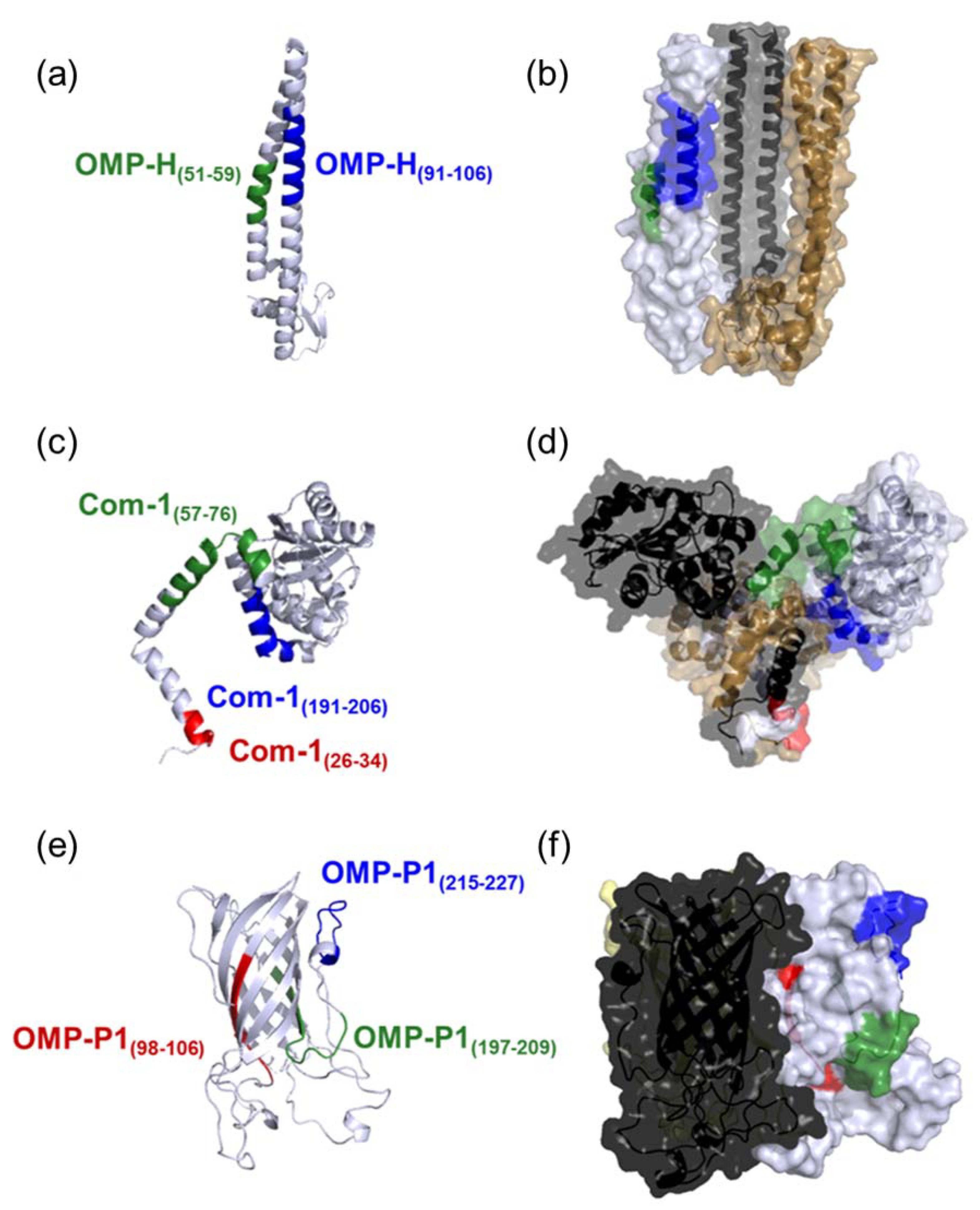
Figure 1. Conformational structure of the studied proteins: (a) OMP-H monomeric structure (gray). (b) Surface of OMP-H trimeric structure. Epitopes OMP-H(51–59) and OMP-H(91–106) are highlighted in green and blue, respectively. (c) Com-1 monomeric structure (gray). (d) Surface of Com-1 trimeric structure. Epitopes Com-1(26–34), Com-1(57–76), and Com-1(191–206) are highlighted in red, green, and blue, respectively. (e) OMP-P1 monomeric structure (gray). (f) Surface of OMP-P1 trimeric structure. Epitopes OMP-P1(98–106), OMP-P1 (197–209), and OMP-P1 (215–227) are highlighted in red, green, and blue, respectively. The chains of trimeric structures (b,d,f) are indicated in gray, gold, and black.
Pathogens 10 01250 g001
Figure 2. Evaluation of the natural immunogenicity of predicted C. burnetii epitopes: (a) Reactivity indices against predicted epitopes of patients reactive (gray points) and non-reactive (white points) to C. burnetii in IFA. (b) Frequencies of response against predicted epitopes. (c) The magnitude of IgG response against synthetic peptides. The reactivity indices (RI) represent the ratios between the optical density of each reaction to C. burnetii and the cutoff value, defined as the mean of the control group’s optical densities plus twice their standard deviation. Each point represents an individual R.I. against one of the synthetic peptides (OMP-H(51–59), OMP-H(91–106), Com-1(57–76), Com-1(191–206), OMP-P1(197–209),, or OMP-P1(215–227)), the traced line indicates the cutoff, while lines represent the median and interquartile range (SEM) of reactive (gray points) and non-reactive (white points) patients to the studied peptides. The comparison of frequencies was done using Fisher´s exact test, and statistical differences were represented by p-values among bars. The magnitudes of response against predicted epitopes are indicated by gray boxes and whiskers (10th–90th percentile), with outliers –indicated by black points in the last graph.
Pathogens 10 01250 g002
Figure 3. Comparison of age and clinical features between high responders (red), low responders (yellow), and non-responders (blue) to the studied epitopes. Indicated numbers represent the frequencies (%) of reported hospitalizations, fever, prostration, and endocarditis cases in high responders (RI > 2), low responders (1.0 > R.I. ≤ 2.0), and non-responders (R.I. ≤ 1) to the epitopes (a) OMP-H(51–59), (b) OMP-H(91–106), (c) Com-1(57–76), (d) Com-1(191–206), (e) OMP-P1(197–209), and (f) OMP-P1(215–227).
Pathogens 10 01250 g003
Figure 4. Schematic summary of B-cell epitope prediction.
Pathogens 10 01250 g004
Table 1. Linear B-cell epitopes identified in silico in the C. burnetii OMPs.
ProteinSequenceBepipredABCpredESA
OMP-H51-QFSPQREKM-59×××
91-EIQNDESTLRQQQQQF-106×××
Com-126-FSFSPQQVK-34××-
57-ALQKKTEAQQEEHAQQAIKE-76×××
83-FNDPASPVAGNPHGN-96××-
191-KKDMDNPAIQKQLRDN-206×××
OMP-P143-GYKSYTYDQ-51×-×
98-KAQYQYDNV-106-××
197-LSYDYALYRSKSN-209 ××
215-SATASAEGTAIG-226××
Table 2. Antigenicity evaluation of the predicted epitopes.
ProteinEpitopeLength (mers)Vaxijen Score
OMP-HOMP-H(51–59)90.524
OMP-H(91–106)160.671
Com-1Com-1(26–34)91.435
Com-1(57–76)200.923
Com-1(83–96)140.139
Com-1(191–206)160.418
OMP-P1OMP-P1(43–51)90.383
OMP-P1(98–106)90.598
OMP-P1(197–209)130.598
OMP-P1(215–227)131.359
Table 3. Summary of the studied population’s clinical and epidemiological data.
Overall
(n = 57)
C. burnetii Seroreactive
(n = 26)
C. burnetii Non-Reactive
(n = 31)
Age-Median (IR)33 (22–49)33 (21–53)33 (23–48)
Gender, n (%)
Male32 (66%)17 (65%)15 (48%)
Female25 (42%)9 (35%)16 (52%)
Symptomatology, n (%)
Fever24 (42%)10 (38%)14 (45%)
Nausea7 (12%)4 (15%)3 (10%)
Endocarditis6 (11%)4 (15%)2 (6%)
Hemorrhagic manifestations1 (2%)01 (3%)
Myalgia13 (23%)4 (15%)9 (29%)
Respiratory manifestations11 (19%)3 (12%)8 (26%)
Prostration18 (32%)6 (23%)12 (39%)
Table 4. Heatmap analysis of C. burnetii-seroreactive patients based on reactivity indices against predicted linear B-cell epitopes. Each cell represents the R.I. of one sample against an epitope.
GroupIDOMP-H(51–59)OMP-H(91–106)Com-1(57–76)Com-1(191–206)OMP-P1(197–209)OMP-P1(215–227)
C. burnetiiseroreactive patients318/120.8270.6710.8130.9200.7650.912
487/121.0060.7361.0200.8560.6100.869
69/130.8220.7360.5930.5830.8690.856
207/170.6510.8910.8900.9100.6481.060
464/160.7910.7440.8240.7740.6630.668
466/161.0381.2790.2711.0890.4710.931
471/161.0941.0420.9461.0451.4181.259
104/121.4301.6341.9161.8871.4201.695
551/121.8161.6011.3180.7160.8491.625
118/132.2651.2601.4081.2421.5851.224
463/161.1000.8261.0013.5400.8921.014
126/131.2501.4750.6110.2710.6890.941
134/130.9970.8820.8370.2960.7401.757
130/162.0752.0061.7121.1800.8242..014
140/131.1370.1931.1321.0600.9150.901
151/131.0550.1801.1371.2980.9210.877
204/131.2500.1921.3191.1970.7961.141
253/130.9811.7441.3401.8640.9932.038
264/133.0864.2704.9166.4040.9964.994
304/131.3691.8081.7162.3710.9901.885
81/140.4320.4980.5890.4620.8140.663
20/160.8350.5500.6640.6060.6580.818
480/160.4120.6400.5525400.1100.628
490/172.0722.0882.21520061.3381.940
239/181.4691.8351.94816761.2161.604
311/182.0422.4972.37222561.3792.175
Each cell represents the R.I. of one sample against an epitope. Each line represents R.I. of one C. burnetii seroreactive patient against each studied peptide (colums). Non-responders (R.I. ≤ 1) are indicated by light blue cells; low responders (1.0 > R.I. ≤ 2.0) are indicated by orange cells, and high responders (R.I. > 2.0) are indicated by dark red cells.
Publisher’s Note: MDPI stays neutral with regard to jurisdictional claims in published maps and institutional affiliations.