
Article

Passive Film Degradation and Microbiologically Influenced Corrosion Mechanism of β Titanium Alloy Induced by Pseudomonas aeruginosa Biofilms

1
School of Materials & Southern Marine Science and Engineering Guangdong Laboratory (Zhuhai), Sun Yat-sen University, Shenzhen 518107, China
2
School of Materials Science and Engineering, Sun Yat-sen University, Guangzhou 510006, China
3
School of Design, The Hong Kong Polytechnic University, Hong Kong 999077, China
4
Baoti Group Ltd., Baoji 721014, China
*
Authors to whom correspondence should be addressed.
Metals 2026, 16(8), 840; https://doi.org/10.3390/met16080840
Submission received: 27 June 2026 / Revised: 17 July 2026 / Accepted: 20 July 2026 / Published: 2 August 2026
(This article belongs to the Section Corrosion and Protection)

Abstract

This study investigated microbiologically influenced corrosion (MIC) and passive-film degradation of a Ti-15Mo-3Al-2.7Nb-0.25Si β titanium alloy exposed to Pseudomonas aeruginosa. The alloy comprised a continuous β-Ti matrix with dispersed α-Ti precipitates, and this α/β dual-phase microstructure provided a potential microstructural basis for spatial variations in passivation behavior among different microregions. During immersion, P. aeruginosa formed a heterogeneous biofilm of bacterial cells and extracellular polymeric substances, altering interfacial mass transfer, oxygen distribution, and local chemistry. Relative to the sterile control, the inoculated group showed increases in maximum pit depth from 2.4 to 4.1 μm and corrosion current density from 8.72 to 17.2 nA cm−2, while the charge-transfer resistance decreased to 4.32 MΩ cm2 after 14 d, confirming enhanced localized corrosion. Mott-Schottky and XPS analyses showed that the donor density increased from 1.07 × 10¹⁹ to 1.29 × 1019 cm−3 and the Ti4+ fraction decreased from 72.29% to 66.74% and the relative Ti0 fraction increased from 7.01% to 17.59%, reflecting increased defect accumulation within the film, impaired passive-film integrity, and reduced local protective capability. P. aeruginosa therefore increases the MIC susceptibility of this β titanium alloy by biofilm-induced interfacial microenvironmental heterogeneity. These findings support MIC assessment and integrated antifouling-anticorrosion surface design for marine β titanium alloys.

1. Introduction

Marine engineering equipment is exposed for prolonged periods to complex environments characterized by high salinity, fluctuations in dissolved oxygen, sediment deposition, and biofouling. The heterogeneous coverage of biofilms and sediments continuously alters oxygen and ion transport at material surfaces, resulting in pronounced spatiotemporal heterogeneity in the local electrochemical state [1,2,3]. Titanium and its alloys are important candidates for lightweight and highly reliable marine components because of their low density, high specific strength, favorable fatigue performance, and excellent corrosion resistance in seawater. Their corrosion resistance primarily derives from a nanoscale TiO2-based passive film that rapidly forms on the surface, effectively suppresses anodic dissolution, and enables repassivation after local damage [4,5,6]. However, under conditions of local oxygen depletion, acidification, Cl enrichment, and restricted mass transport induced by biofilm coverage, the chemical composition, defect state, and semiconducting properties of the passive film may be altered. These changes can weaken the barrier performance and repassivation capability of the film, thereby increasing susceptibility to localized corrosion and microbiologically influenced corrosion (MIC) [7,8,9]. Therefore, evaluation of the marine MIC behavior of titanium alloys should consider not only the regulation of the initial passive film by alloy composition and microstructure, but also the effects of the biofilm-modified local environment on passive-film stability and repair processes.
β titanium alloys combine high specific strength, good formability, and a tunable response to heat treatment, and their microstructure and properties are governed primarily by the stability of the β matrix and the precipitation behavior of the α phase [10,11]. Ti-15Mo-3Al-2.7Nb-0.25Si (TB8) is a high-strength titanium alloy dominated by the β-Ti phase, in which Mo and Nb serve as the principal β-stabilizing elements, whereas Al and Si contribute to solid-solution strengthening and the regulation of precipitation behavior [12,13]. In addition to controlling the phase constitution, Mo and Nb may participate in passive-film formation and modify film stability. In a study of the corrosion behavior of Ti-Mo alloys in hydrochloric acid, Zheng et al. found that heat-treatment-induced changes in the α/β microstructure affected passive-film dissolution and local microgalvanic interactions, indicating that the corrosion-resistance contribution of Mo alloying is closely related to both the matrix microstructure and the surface-film state [14]. Çaha et al. confirmed through atomic-scale characterization that Nb participates in the formation of a composite passive film on Ti-Nb alloys [15]. Ji et al. further demonstrated that Nb addition improved the stability of passive films on Ti-Zr alloys and reduced their susceptibility to pitting corrosion [16]. Studies of Ti-Nb alloys with different Nb contents also showed that increasing the Nb content resulted in a positive shift in the open-circuit potential, a decrease in passive current density, and an increase in interfacial charge-transfer resistance [17]. These findings suggest that the influence of Mo and Nb on the corrosion behavior of titanium alloys is associated not only with β-phase stabilization, but also with the combined regulation of passive-film composition, defect state, and repassivation kinetics. In contrast, the small amount of Si in TB8 primarily contributes to the regulation of precipitation behavior and microstructural stability, and its influence on corrosion behavior may be exerted indirectly through changes in phase distribution and the local microstructural state [18,19]. Therefore, compared with commercially pure Ti and conventional α + β Ti-6Al-4V alloys, the β-matrix-dominated microstructure and relatively high Mo and Nb contents of TB8 may result in distinct passive-film formation mechanisms and local electrochemical responses. However, current understanding is derived mainly from studies conducted in sterile acidic or chloride-containing solutions and therefore does not clarify the stability of Mo- and Nb-containing composite passive films within the localized environment created by microbial biofilms.
Pseudomonas aeruginosa (P. aeruginosa) is a common biofilm-forming bacterium in marine environments and exhibits strong surface adhesion, colonization capability, and environmental adaptability [20,21]. After a material is immersed in a bacteria-containing medium, cells can attach to the surface, proliferate, and secrete extracellular polymeric substances (EPS), gradually forming a biofilm with heterogeneous spatial distribution and surface coverage [22,23]. Such nonuniform coverage can restrict local oxygen and ion transport and alter the interfacial pH, Cl concentration, and redox state, thereby interfering with passive-film growth, stabilization, and repair processes [24,25,26]. Previous studies have shown that P. aeruginosa biofilms can increase the corrosion current density of commercially pure Ti, reduce interfacial charge-transfer resistance, and promote the formation of lower-valence Ti oxides, such as Ti2O3, together with localized corrosion [27]. Phenazine-1-carboxylate secreted by P. aeruginosa can also act as a redox-active mediator, accelerating passive-film degradation and pit propagation on Ti-6Al-4V alloy [28]. These findings demonstrate that Ti and its alloys are not completely immune to MIC induced by P. aeruginosa. However, TB8 differs from commercially pure Ti and Ti-6Al-4V in both matrix structure and alloying-element composition, and its passive film may therefore respond differently to biofilm-induced local mass-transfer limitations and changes in interfacial redox conditions. In particular, how Mo- and Nb-containing composite passive films undergo defect accumulation, chemical-state evolution, and localized destabilization during bacterial attachment and biofilm development remains insufficiently understood.
Current studies on MIC of titanium alloys have focused mainly on commercially pure Ti, Ti-6Al-4V, and antibacterial-modified systems, whereas the corrosion behavior of Mo- and Nb-rich β titanium alloys in microbial environments has received limited attention. In particular, whether P. aeruginosa attachment and biofilm evolution can weaken the intrinsic passivation stability of TB8, and how semiconductor defects in the passive film, Ti chemical states, and localized corrosion evolve in a coupled manner, remain to be clarified. Accordingly, the Ti-15Mo-3Al-2.7Nb-0.25Si alloy was selected as the research material in this study. Its α/β phase constitution, precipitate distribution, and local orientation characteristics were first characterized. Changes in surface morphology and electrochemical behavior during P. aeruginosa attachment and biofilm evolution were then investigated, while Mott-Schottky analysis and XPS were used to characterize semiconductor defects and surface chemical states of the passive film. By establishing the relationships among matrix microstructure, biofilm coverage, passive-film degradation, and localized corrosion, this study clarifies the passive-film evolution characteristics and MIC response of the TB8 alloy in a model P. aeruginosa environment.

2. Experimental Procedures and Characterization

2.1. Materials, Bacterial Strain, and Culture Medium

TB8 titanium alloy was used in this study. Specimens for microbiologically influenced corrosion experiments were sequentially ground using abrasive papers from #400 to #4000, followed by ultrasonic cleaning in acetone, anhydrous ethanol, and deionized water to remove surface contaminants. The specimens were sterilized by ultraviolet irradiation for 30 min and stored under vacuum until use.
A marine Gram-negative bacterium, Pseudomonas aeruginosa (MCCC 1A00099), was selected as the test strain. Bacterial cultivation and immersion experiments were conducted in 2216E medium with a pH between 7.0 and 7.2. Erlenmeyer flasks containing 50 mL of 2216E liquid medium were sterilized at 121 °C for 20 min. Subsequently, 1 mL of P. aeruginosa glycerol stock was added, and the cultures were incubated in a thermostatic shaker at 37 °C and 120 r min−1 for 24 h. Subsequently, the bacterial suspension was microscopically counted using a hemocytometer to determine the total cell concentration, and the initial inoculum concentration was adjusted to 1 × 10⁷ cells·mL−1.

2.2. Microstructural Characterization

The initial microstructure of the TB8 titanium alloy was examined using a field emission scanning electron microscope (FE SEM, Hitachi SU5000, Tokyo, Japan). High energy synchrotron X ray diffraction (HEXRD) experiments were conducted at the BL12SW high energy X ray beamline of the Shanghai Synchrotron Radiation Facility (SSRF). A monochromatic X ray beam with an energy of 100 keV and a beam size of 100 μm × 100 μm was used to identify the phase constitution and acquire two dimensional diffraction patterns. Electron backscatter diffraction (EBSD) analysis was performed using a system equipped with a Bruker eFlash FS detector to obtain crystallographic orientation, phase distribution, and local misorientation information.

2.3. Biofilm Morphology Characterization

Specimens were immersed in 2216E liquid medium inoculated with P. aeruginosa for 14 d. After immersion, the specimens were removed and gently rinsed several times with phosphate-buffered saline (PBS) to remove nonadherent cells. The biofilms were then fixed in PBS containing 2.5% glutaraldehyde at 4 °C for 12 h. After fixation, the specimens were dehydrated sequentially in ethanol solutions with volume fractions of 25%, 50%, 75%, 95%, and 100%, with each step lasting 10 min. The dehydrated specimens were dried under high purity nitrogen before subsequent characterization.
The surface morphology of the biofilms was examined using a field emission scanning electron microscope (FE SEM, Hitachi SU5000, Japan). Before SEM observation, a thin gold layer was sputtered onto the specimen surface to improve electrical conductivity. In addition, the specimens were stained with SYTO 9 and propidium iodide (PI) for 30 min in the dark. A confocal laser scanning microscope (CLSM, TCS SP8 X, Leica, Wetzlar, Germany) was then used for three dimensional reconstruction and biofilm thickness measurement.

2.4. Pitting Morphology Characterization

To remove surface corrosion products, the specimens were ultrasonically cleaned in a corrosion product removal solution and anhydrous ethanol for 3 min each. The corrosion product removal solution was prepared from 500 mL of hydrochloric acid with a mass fraction of 37%, 500 mL of deionized water, and 3.5 g of hexamethylenetetramine. After cleaning, the specimens were dried under high purity nitrogen (N2) and examined using a three dimensional optical profilometer to determine pit depth.

2.5. Electrochemical Measurements

Electrochemical measurements were performed using an electrochemical workstation (Gamry Interface 1010E, Warminster, PA, USA) and a conventional three-electrode cell to evaluate the corrosion behavior of TB8 titanium alloy specimens in sterile and P. aeruginosa-inoculated 2216E media. The specimen served as the working electrode with an exposed area of 1.0 cm2, while a platinum sheet and an Ag/AgCl electrode served as the counter and reference electrodes, respectively. All electrochemical measurements were conducted after open circuit potential (OCP) stabilization for 3600 s.
Electrochemical impedance spectroscopy (EIS) measurements were performed over a frequency range from 100 kHz to 0.01 Hz using a sinusoidal perturbation amplitude of 5 mV. Potentiodynamic polarization measurements were performed from −0.3 to 2.0 V relative to OCP at a scan rate of 0.5 mV s−1. Linear polarization resistance measurements were conducted within ±5 mV of OCP at a scan rate of 0.125 mV s−1. Each electrochemical test was performed using three parallel specimens.
In addition, Mott–Schottky measurements were performed to assess the semiconducting properties of the passive films formed on specimens immersed in different media. The measurements were conducted at a fixed frequency of 1000 Hz with an ac perturbation amplitude of 10 mV and an anodic scan rate of 10 mV s−1 over a potential range from −1.0 to 1.0 V relative to the saturated calomel electrode (SCE).

2.6. XPS Analysis

X-ray photoelectron spectroscopy (XPS, Thermo VG ESCALAB250, Waltham, MA, USA) was used to analyze the chemical composition and chemical states of the passive films formed on the alloy surface. The binding energies of all characteristic peaks were calibrated against the C 1s peak at 284.6 eV. Peak fitting of the XPS spectra was performed using Avantage software (Version 5.9).

3. Results

3.1. Microstructure and Phase Analysis

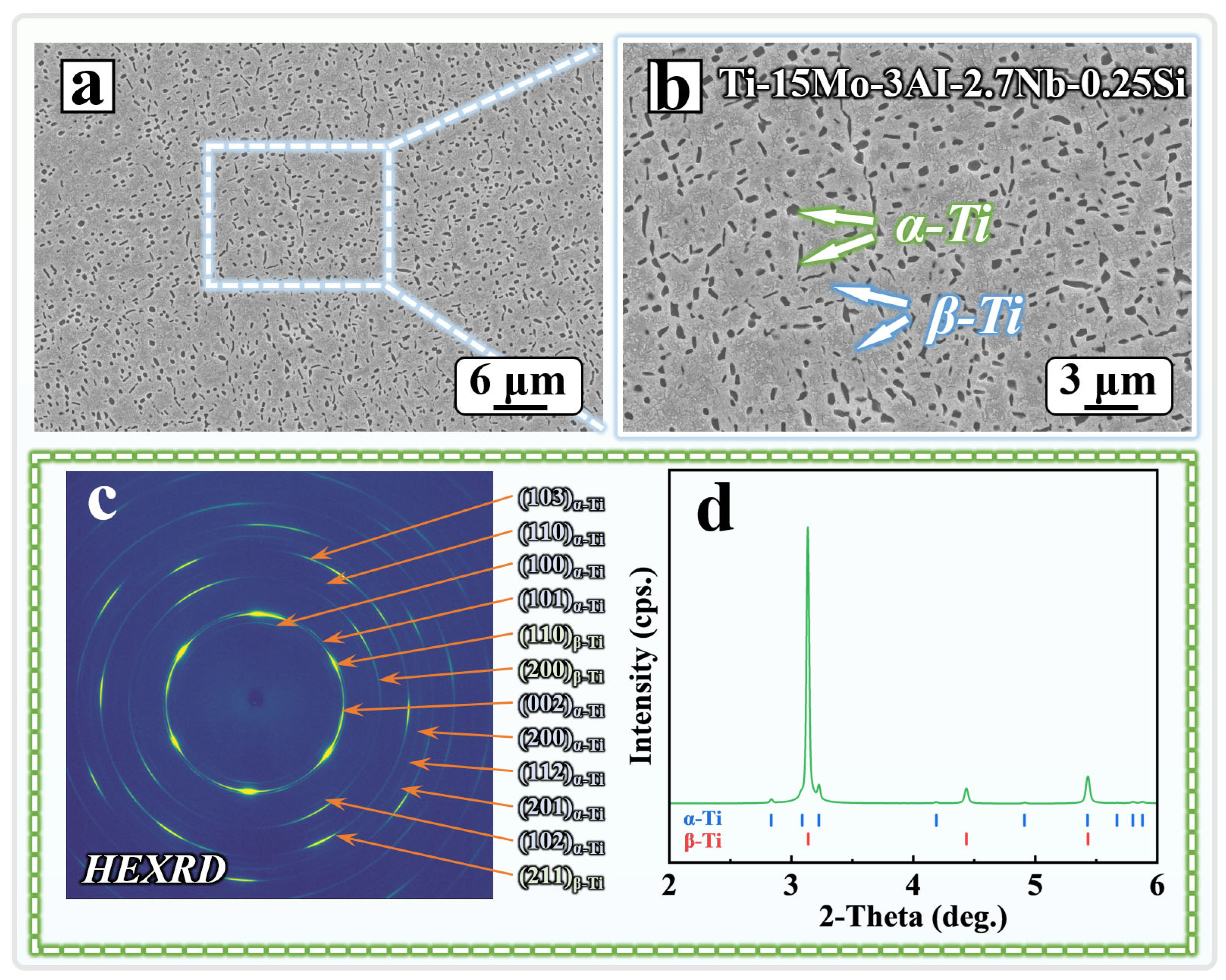
Figure 1 presents the initial microstructure and high-energy synchrotron X-ray diffraction results of the Ti–15Mo–3Al–2.7Nb–0.25Si β titanium alloy. As shown in Figure 1a, the alloy exhibited a relatively uniform microstructure, without obvious agglomeration of coarse second-phase particles or continuous precipitation features. The higher magnification SEM image in Figure 1b shows that the alloy consisted of a continuous β-Ti matrix and dispersed α-Ti precipitates. The α-Ti precipitates were mainly present as fine particles or short rods within the β-Ti matrix, resulting in an α + β dual-phase microstructure composed of a continuous β-Ti matrix and dispersed α-Ti precipitates. These observations indicate that the TB8 alloy possessed an initial microstructure dominated by the β-Ti phase with a small fraction of dispersed α-Ti precipitates.
Figure 1c shows the two-dimensional high-energy synchrotron X-ray diffraction (HE-XRD) pattern of the alloy. The diffraction rings can be indexed to the characteristic reflections of the hexagonal close-packed α-Ti phase and the body-centered cubic β-Ti phase. The major reflections of the α-Ti phase correspond to the (100)α, (002)α, (101)α, (102)α, (103)α, (112)α, and (201)α planes, whereas the β-Ti phase exhibits the characteristic (110)β, (200)β, and (211)β diffraction rings. The coexistence of the α-Ti and β-Ti diffraction rings further confirms the α + β dual-phase constitution of the alloy in its initial state.
In addition, several diffraction rings show nonuniform intensity enhancement along the azimuthal direction and appear as arcs rather than fully uniform continuous rings. This feature indicates orientation-dependent diffraction contributions from grains with different crystallographic orientations and suggests a degree of preferred orientation or retained texture within the alloy [29,30]. Figure 1d presents the one-dimensional diffraction profile obtained by azimuthal integration of the two-dimensional HE-XRD pattern. The positions of the major diffraction peaks agree with the standard diffraction positions of α-Ti and β-Ti, further confirming the coexistence of the two phases.
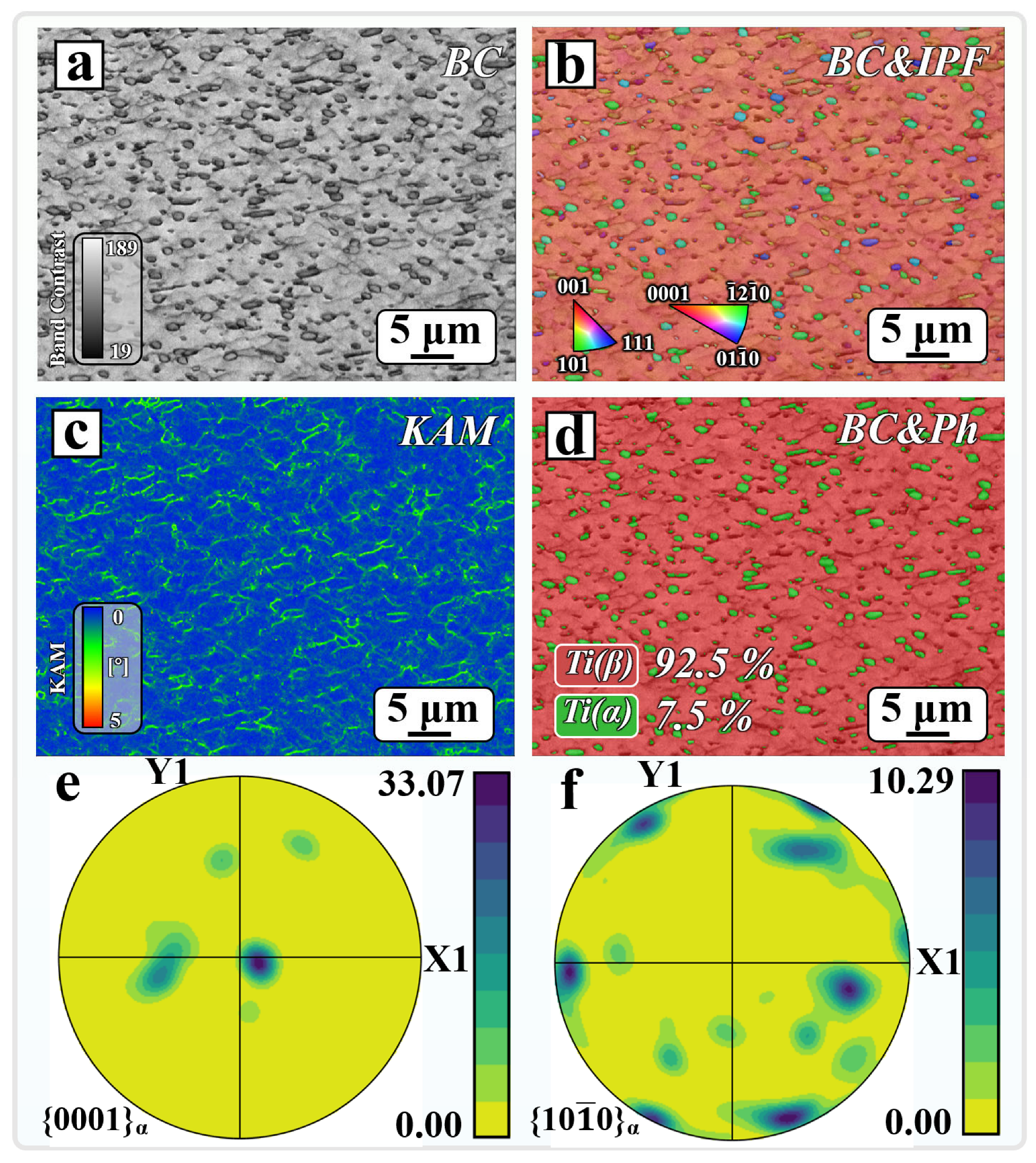
To further analyze the initial microstructural features and crystallographic state of the Ti–15Mo–3Al–2.7Nb–0.25Si β titanium alloy, EBSD characterization was performed, as shown in Figure 2. Figure 2a presents the band contrast map. The β-Ti matrix generally exhibits relatively high and uniform diffraction pattern quality, whereas the dispersed particle-shaped regions show lower band contrast. Combined with the phase distribution map in Figure 2d, the discrete second-phase regions are identified as α-Ti, while the continuous regions correspond to the β-Ti matrix.
The overlay of the band contrast and inverse pole figure maps in Figure 2b further indicates that the β-Ti phase is continuously distributed throughout the scanned region and serves as the matrix phase. In contrast, the α-Ti phase is present as fine, discontinuous particles or short rods within the β-Ti matrix. The β-Ti matrix is dominated by similar orientation colors, whereas the α-Ti phase exhibits distinct orientation colors, indicating that the two phases possess different local crystallographic orientation characteristics.
Figure 2d presents the EBSD phase distribution map. The area fractions of β-Ti and α-Ti are approximately 92.5% and 7.5%, respectively, within the mapped region. This result is consistent with the coexistence of α-Ti and β-Ti diffraction peaks in Figure 1c,d, confirming that the alloy is dominated by the β-Ti phase with a minor fraction of α-Ti precipitates in its initial state.
Figure 2c presents the kernel average misorientation map of the alloy. Overall, the β-Ti matrix is dominated by blue regions with low kernel average misorientation (KAM) values, indicating relatively low local orientation gradients within the matrix. In contrast, green to yellow regions become more pronounced near α/β interfaces and around some α-Ti precipitates, indicating higher local misorientation and accumulated lattice distortion in these regions. This feature may be associated with lattice mismatch, transformation strain generated during the transformation from β-Ti to α-Ti, and strain accommodation between the two phases. Because α-Ti and β-Ti have different crystal structures and elastic responses, local strain concentration is more likely to develop near α/β interfaces, thereby increasing local lattice distortion.
Figure 2e,f show the {0001} basal and {10-10} prismatic pole figures of the α-Ti phase, respectively. The maximum pole density of the α-Ti {0001} pole figure reaches 33.07 m.r.d., which is substantially higher than that of a random orientation distribution, indicating a pronounced basal preferred orientation. In comparison, the maximum pole density of the {10-10} pole figure is 10.29 m.r.d., revealing a weaker but still distinguishable prismatic texture component. These results indicate that the α-Ti precipitates are not randomly oriented but exhibit a degree of orientation selection. Their texture characteristics are consistent with the classical Burgers orientation relationship that may operate during the transformation from β-Ti to α-Ti, namely {0001}α // {110}β and <11-20>α // <111>β [31,32].
The EBSD results show that the Ti-15Mo-3Al-2.7Nb-0.25Si alloy consists of a continuous β-Ti matrix containing a small fraction of finely dispersed α-Ti precipitates, with relatively high local orientation gradients and lattice distortion near the α/β interfaces. Given the differences in crystal structure, elemental partitioning, and local strain state between α-Ti and β-Ti, this microstructural heterogeneity may affect the surface reactivity, passive film formation, and local electrochemical response in different microstructural regions.
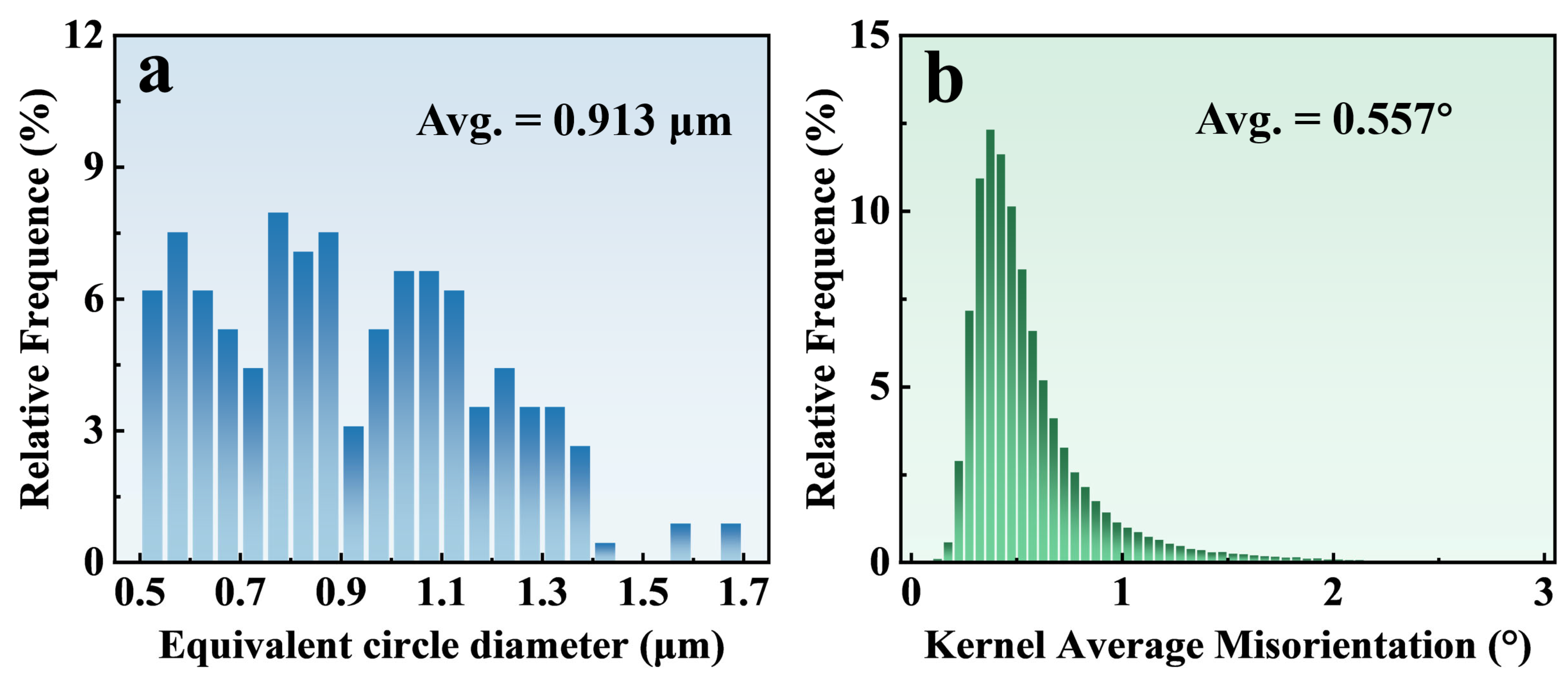
To quantitatively characterize the size of the α-Ti precipitates and the local misorientation state in the Ti-15Mo-3Al-2.7Nb-0.25Si β titanium alloy, statistical analyses of equivalent circle diameter and kernel average misorientation were performed based on the EBSD results, as shown in Figure 3. Figure 3a presents the equivalent circle diameter distribution of the α-Ti precipitates. The equivalent circle diameters of the α-Ti precipitates are mainly distributed from 0.5 to 1.3 μm, while a small number of precipitates reach approximately 1.7 μm. The average equivalent circle diameter is 0.913 μm. Overall, the α-Ti phase is dominated by fine precipitates with a certain degree of size dispersion.
For β titanium alloys, the size, morphology, and spatial distribution of α-Ti precipitates are important microstructural parameters influencing microstructural stability and service performance. Fine and dispersed α-Ti precipitates can generate numerous α/β interphase boundary regions within the β-Ti matrix. Because the two phases differ in crystal structure, elemental partitioning, and local electronic structure, these boundary regions and their adjacent areas may exhibit passive film formation and local electrochemical responses distinct from those within the β-Ti matrix [33].
Figure 3b shows the statistical distribution of local kernel average misorientation values. The average KAM value of the alloy is 0.557°, and most measured points are concentrated in the low KAM range, indicating relatively limited local orientation gradients within the overall β-Ti matrix. Meanwhile, the KAM distribution is right skewed, with a small fraction of regions exhibiting KAM values above 1°. Combined with the spatial distribution shown in Figure 2c, the higher KAM regions are mainly located near α/β interphase boundaries and around some α-Ti precipitates, indicating higher local misorientation and accumulated lattice distortion in these regions.
This feature may be associated with lattice mismatch, elemental redistribution, and precipitation strain generated during α-Ti precipitation from the β-Ti matrix. The α-Ti phase has a hexagonal close packed structure, whereas the β-Ti phase has a body centered cubic structure, resulting in a substantial crystallographic difference between the two phases. During α-Ti nucleation, growth, and subsequent cooling, local lattice distortion and strain concentration may develop near α/β interphase boundaries to satisfy interfacial compatibility and strain accommodation requirements [34].
In summary, the Ti-15Mo-3Al-2.7Nb-0.25Si alloy exhibits an α + β dual-phase microstructure composed of a continuous β-Ti matrix and fine dispersed α-Ti precipitates, with an average equivalent circle diameter of approximately 0.913 μm for the α-Ti precipitates. Although the alloy has a relatively low average KAM value, regions with elevated local orientation gradients and lattice distortion accumulation are present near α/β interphase boundaries. This microstructural heterogeneity may result in variations in surface reactivity, passive film evolution, and local electrochemical response across different microstructural regions, thereby providing a microstructural basis for subsequent analysis of localized corrosion susceptibility in the presence of P. aeruginosa biofilms.

3.2. Biofilm Characterization

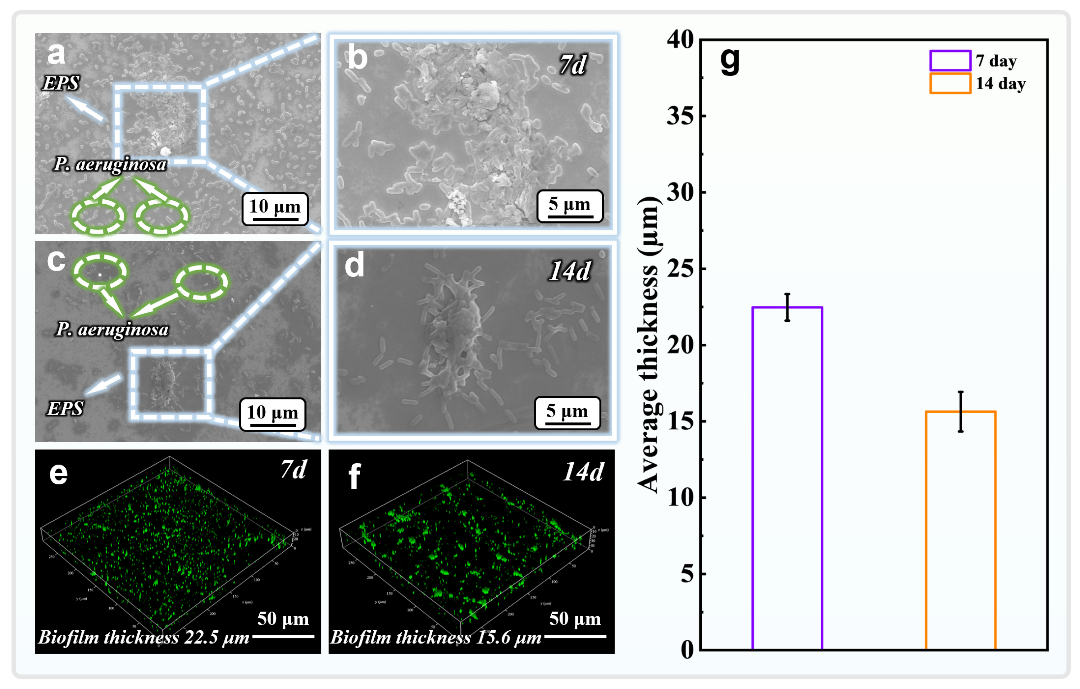
Figure 4 shows the surface morphologies of biofilms formed on the Ti-15Mo-3Al-2.7Nb-0.25Si alloy after immersion in culture medium inoculated with P. aeruginosa for 7 and 14 d. SEM observations show that, after 7 d of immersion, numerous rod-shaped P. aeruginosa cells adhered to the alloy surface. The cells were interconnected by visible extracellular polymeric substances (EPS) and locally aggregated, forming a biofilm with relatively continuous surface coverage (Figure 4a,b).
After 14 d of immersion, rod-shaped bacterial cells and EPS remained visible on the surface, but the cells were more dispersed, local aggregates were less frequent, and the extent of biofilm coverage decreased (Figure 4c,d). Three-dimensional CLSM reconstructions further supported this trend. At 7 d, a relatively thick and dense biofilm formed on the alloy surface, with an average thickness of approximately 22.5 μm. After 14 d, the average biofilm thickness decreased to approximately 15.6 μm (Figure 4e–g). This decrease in biofilm thickness may be associated with nutrient depletion, metabolite accumulation, and partial bacterial detachment during the later stage of cultivation, indicating a higher level of biofilm attachment at 7 d than at 14 d. Although the biofilm was thinner after 14 d, residual bacterial cells and EPS may still create locally occluded regions at the alloy/solution interface. These regions can alter oxygen transport, restrict ion transport, and modify local interfacial chemistry, thereby affecting passive film stability and subsequent microbiologically influenced corrosion processes [35].

3.3. Pitting Morphology

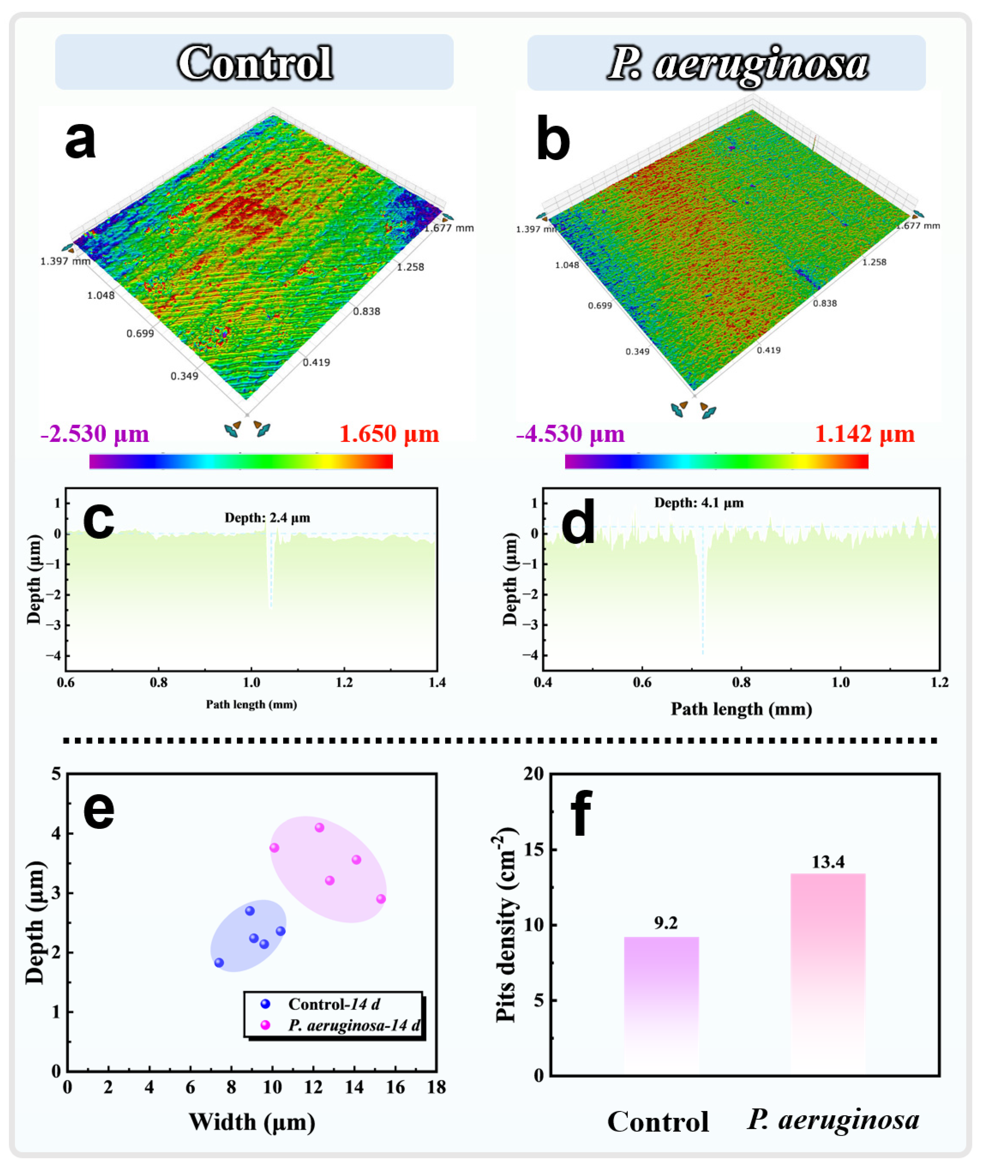
Figure 5 presents the three-dimensional corrosion morphologies, cross-sectional profiles, and pitting statistics of the Ti-15Mo-3Al-2.7Nb-0.25Si alloy after 14 d of immersion in sterile and P. aeruginosa inoculated media. Only a small number of shallow and isolated corrosion pits were observed on the sterile control surface, with relatively limited height variation. The cross-sectional profile showed a maximum pit depth of approximately 2.4 μm (Figure 5a,c). In contrast, more pronounced localized corrosion was observed after exposure to P. aeruginosa, and the maximum pit depth increased to approximately 4.1 μm (Figure 5b,d).
The pit size distribution further shows that immersion in the P. aeruginosa inoculated medium shifted the pit population toward greater depths and widths, indicating enhanced pit development under biofilm exposure (Figure 5e). In addition, the pit density increased from 9.2 cm−2 in the sterile control to 13.4 cm−2 in the P. aeruginosa group (Figure 5f), suggesting that the presence of P. aeruginosa increased the localized corrosion susceptibility of the alloy during immersion.
These results indicate that the effect of P. aeruginosa on the corrosion behavior of the Ti-15Mo-3Al-2.7Nb-0.25Si alloy was primarily manifested as an intensification of localized corrosion. Under sterile conditions, the Ti-based passive film formed on the alloy surface could suppress Cl-induced localized dissolution to some extent, resulting in relatively low pit numbers and depths. The α-Ti precipitates and β-Ti matrix differ in crystal structure, elemental partitioning, and local strain state. This microstructural heterogeneity may result in differences in passive film formation and local electrochemical response among different microstructural regions.
After inoculation with P. aeruginosa, bacteria and extracellular polymeric substances formed nonuniform coverage on the alloy surface. This coverage may have further altered oxygen transport, ion migration, and interfacial redox conditions between biofilm-covered and relatively uncovered regions, thereby intensifying differences in the local interfacial environment. This spatial heterogeneity may promote local differences in oxygen concentration and ion enrichment, leading to gradual defect accumulation and reduced passive film stability in localized regions [16]. Although the average biofilm thickness decreased after 14 d, the residual bacteria and EPS may still have maintained nonuniform interfacial coverage. In addition, the local environmental changes and passive film damage caused by the biofilm during the early stage of immersion may have exerted cumulative effects. Once the passive film became locally destabilized, the freshly exposed metallic surface came into contact with the Cl-containing medium, thereby further promoting pit nucleation and propagation. Therefore, the corrosion acceleration associated with P. aeruginosa was not primarily characterized by uniform dissolution but was more likely related to the combined effects of the intrinsic microstructural heterogeneity of the alloy and nonuniform biofilm coverage on the local interfacial environment and passive film stability.

3.4. Electrochemical Corrosion Behavior of TB8 Induced by P. aeruginosa

3.4.1. OCP, Linear Polarization Resistance (LPR)

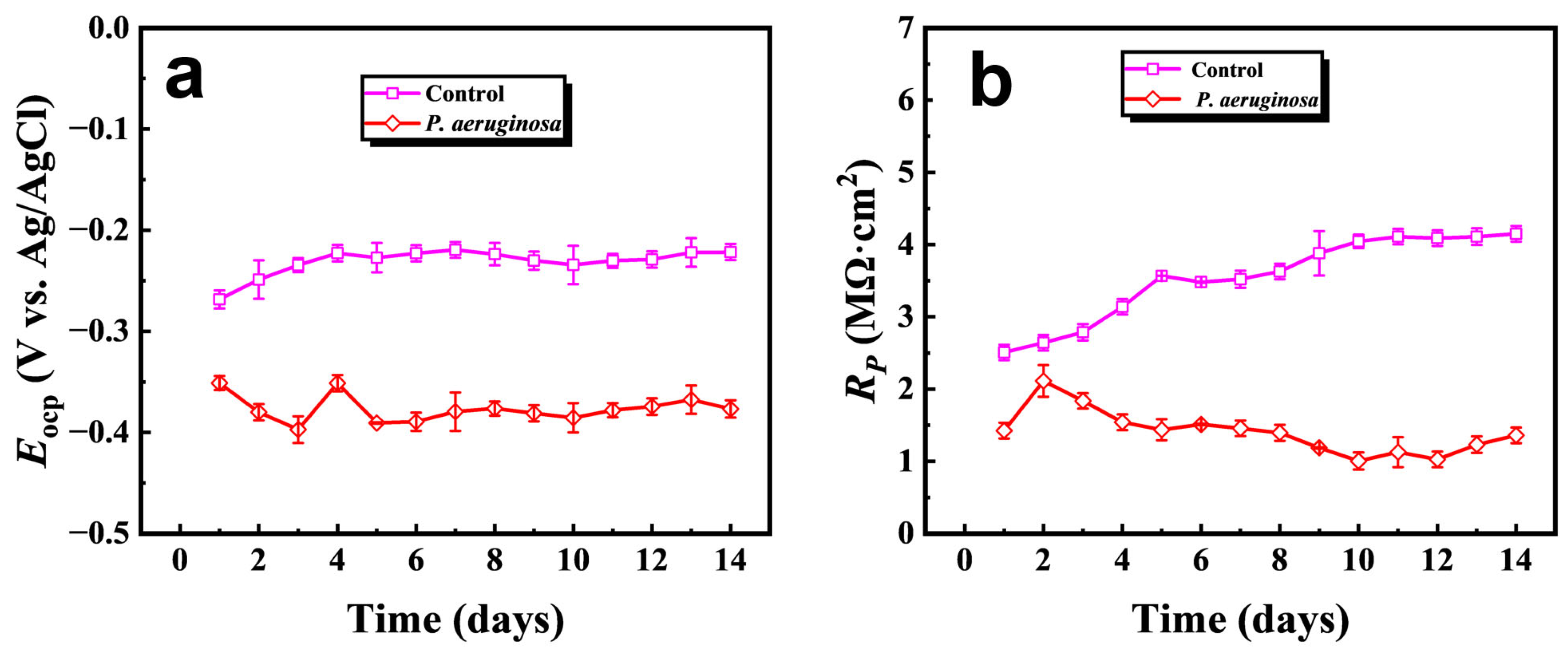
Figure 6 shows the evolution of open circuit potential (Eocp) and polarization resistance (Rp) of the Ti-15Mo-3Al-2.7Nb-0.25Si alloy during 14 d of immersion in sterile and P. aeruginosa-inoculated media. As shown in Figure 6a, the Eocp of the sterile control was initially approximately −0.27 V, then gradually shifted in the positive direction and stabilized at approximately −0.23 V. This behavior indicates that the electrochemical state of the alloy/solution interface became progressively more stable, which is consistent with the gradual establishment and relative stabilization of the passive film.
In contrast, after inoculation with P. aeruginosa, the Eocp remained between −0.35 and −0.40 V, approximately 0.13 to 0.17 V lower than that of the sterile control. This negative shift indicates that bacterial attachment and biofilm formation altered the electrochemical state of the alloy/solution interface, resulting in a more negative corrosion potential.
Figure 6b further shows a marked difference in the evolution of Rp between the two groups. The Rp of the sterile control increased continuously from approximately 2.5 MΩ cm2 to approximately 4.1 MΩ cm2, indicating a gradual increase in the overall polarization resistance of the alloy surface during immersion. In contrast, the Rp of the P. aeruginosa group remained lower than that of the sterile control throughout the immersion period. Although it transiently increased to approximately 2.0 MΩ cm2 after 2 d of immersion, it subsequently decreased rapidly and remained between approximately 1.0 and 1.5 MΩ cm2 from 7 to 14 d. This behavior indicates that sustained biofilm exposure prevented the interface from maintaining the increase in polarization resistance observed under sterile conditions, thereby reducing the overall corrosion resistance of the alloy surface.
In conjunction with the preceding biofilm and pitting morphology results, the promotion of localized corrosion by P. aeruginosa may be associated with the heterogeneous biofilm formed on the alloy surface. Bacterial cells and EPS can modify local oxygen and ion transport, generating different interfacial microenvironments between biofilm covered and relatively uncovered regions. These conditions may lead to local oxygen concentration differences, heterogeneous electrochemical responses, and passive film defect accumulation.
The negative shift in Eocp and the decrease in Rp indicate that the P. aeruginosa biofilm reduced the stability of the alloy/solution interface and increased the localized corrosion susceptibility of the alloy. This electrochemical response is consistent with the increase in pit depth and pit density observed after 14 d, indicating that MIC in this system may involve the combined effects of biofilm induced interfacial microenvironmental changes and local passive film destabilization.

3.4.2. Potentiodynamic Polarization

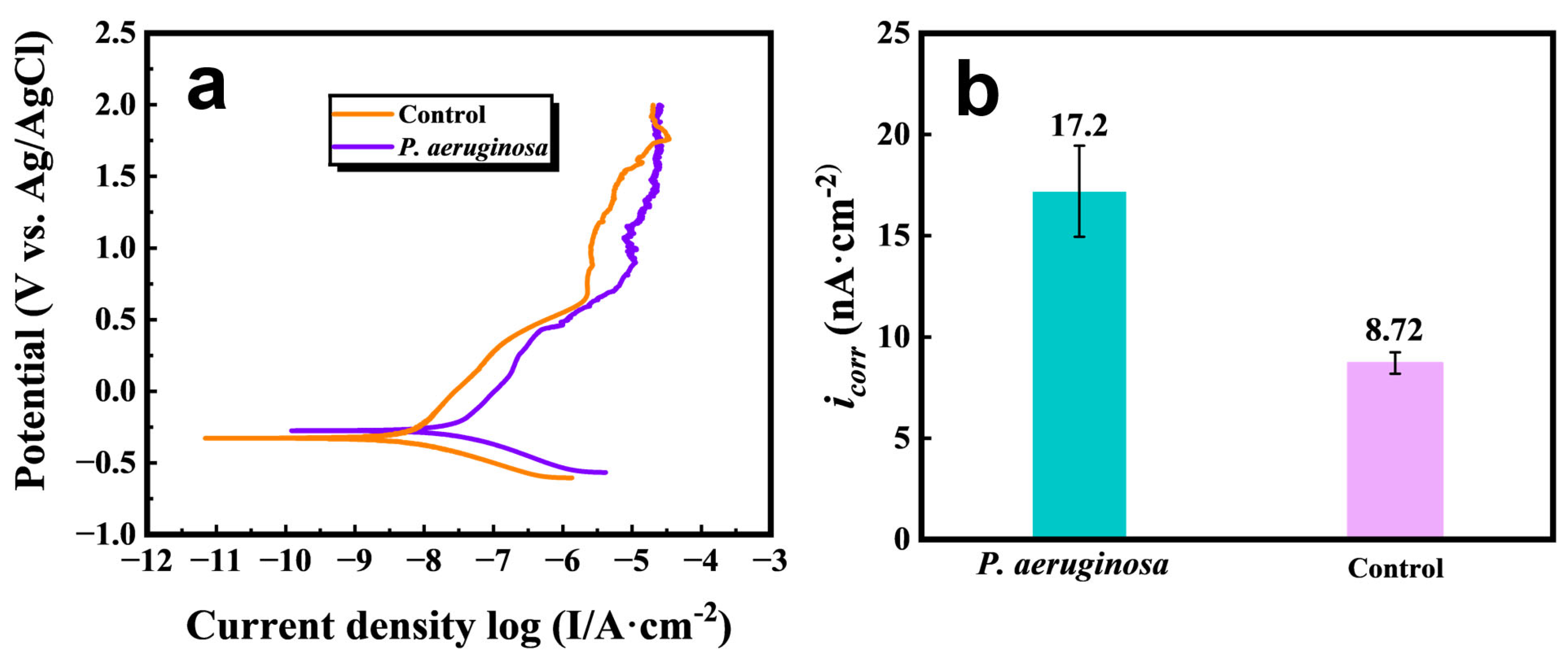
Figure 7 presents the potentiodynamic polarization curves and corrosion current density values of the Ti-15Mo-3Al-2.7Nb-0.25Si alloy after 14 d of immersion in sterile and P. aeruginosa-inoculated media. As shown in Figure 7a, both groups exhibit distinct passive regions, indicating that the alloy can still form a Ti-based passive film with a certain protective effect in the chloride-containing medium.
However, compared with the sterile control, the alloy exposed to P. aeruginosa shows a higher anodic current response over a comparable potential range, indicating that biofilm exposure weakened the ability of the passive film to suppress anodic dissolution. The corrosion current densities obtained from polarization curve fitting further support this trend. The icorr value increased from 8.72 nA cm−2 for the sterile control to 17.2 nA cm−2 for the P. aeruginosa group, corresponding to an approximately twofold increase (Figure 7b).
These results indicate that the P. aeruginosa biofilm increased the corrosion susceptibility of the Ti-15Mo-3Al-2.7Nb-0.25Si alloy. Bacterial attachment and EPS coverage can generate a heterogeneous interfacial microenvironment on the alloy surface, causing spatial differences in local oxygen transport, chloride distribution, and metabolite accumulation. These changes may weaken passive film stability and promote localized anodic dissolution.
It is noteworthy that the polarization curve of the inoculated group still retains a passive region, indicating that P. aeruginosa exposure did not cause complete active dissolution of the alloy. Instead, it is more likely to promote localized corrosion development by reducing passive film protectiveness and increasing localized corrosion susceptibility. This interpretation is consistent with the increased pit depth and pit density observed after immersion.

3.4.3. EIS

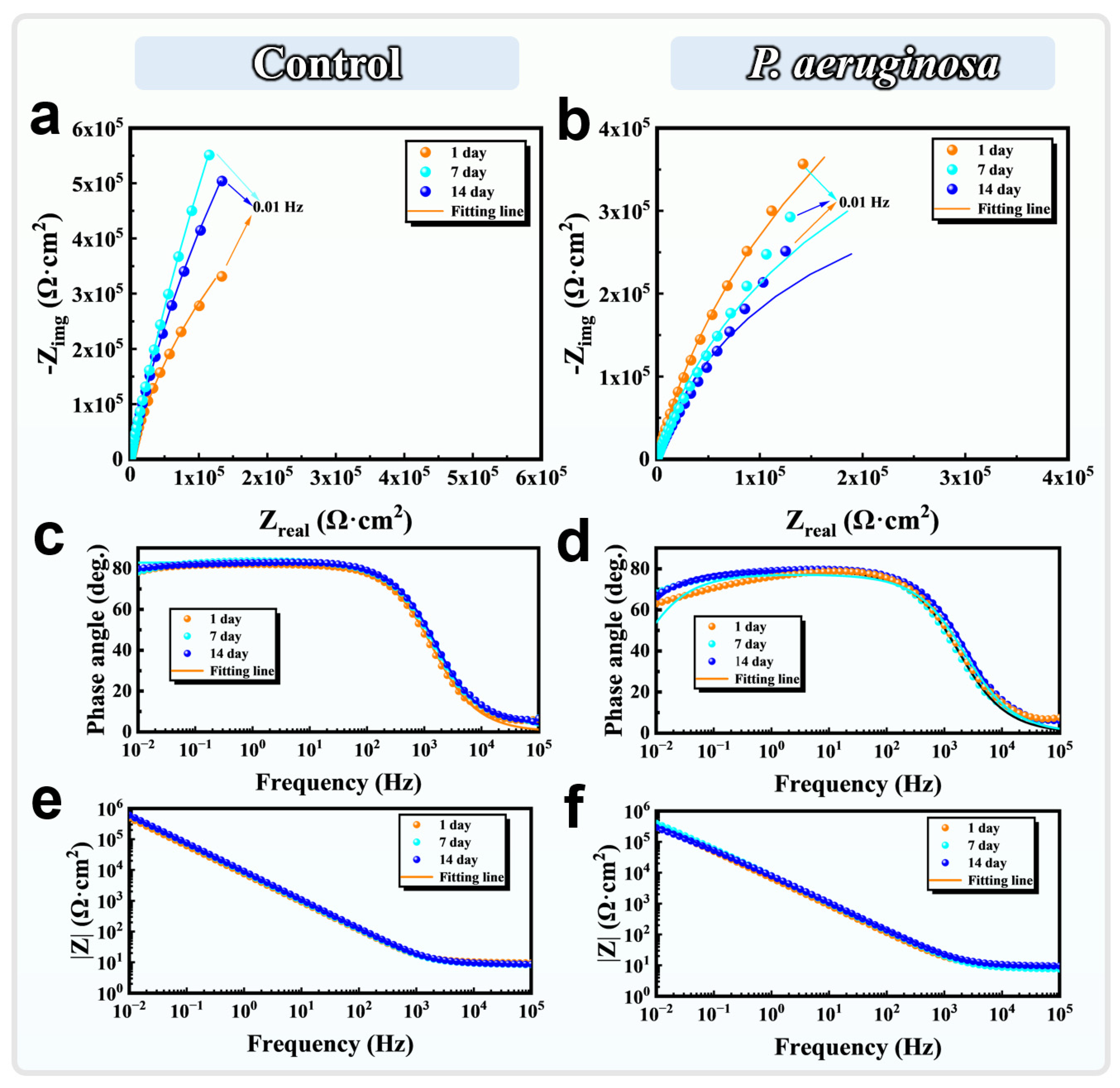
Figure 8 presents the EIS results of the Ti-15Mo-3Al-2.7Nb-0.25Si alloy after immersion in sterile control and P. aeruginosa inoculated systems for 1, 7, and 14 d. The Nyquist plots of the sterile control exhibit relatively large capacitive responses in the low frequency region, and the low frequency impedance reaches its highest value after 7 d, indicating that the passive film on the alloy surface gradually formed during immersion and exhibited a strong shielding effect (Figure 8a). Correspondingly, the Bode phase angle plot of the sterile group maintains a high phase angle plateau close to 80° over a broad frequency range, indicating that the interfacial process was mainly controlled by the capacitive behavior of a stable passive film (Figure 8c) [36]. The impedance modulus at low frequency, f = 0.01 Hz, also remains at a relatively high level, further indicating that the passive film on the alloy surface in the sterile environment possessed good integrity and barrier performance (Figure 8e) [37].
Compared with the sterile control, the Nyquist curves of the P. aeruginosa-inoculated system exhibit obviously smaller overall dimensions and lower low-frequency impedance responses, indicating that bacterial activity weakened the corrosion impedance of the alloy/solution interface (Figure 8b). In the Bode plots, the phase angle plateau of the inoculated group is slightly lower than that of the sterile group and changes with increasing immersion time, indicating that biofilm coverage and passive film response jointly participated in the interfacial electrochemical process (Figure 8d). Furthermore, the low frequency |Z| values of the inoculated group are lower than those of the sterile group and decrease with increasing immersion time (Figure 8f), indicating that sustained exposure to P. aeruginosa reduced the ability of the passive film to impede ion transport and charge transfer processes.
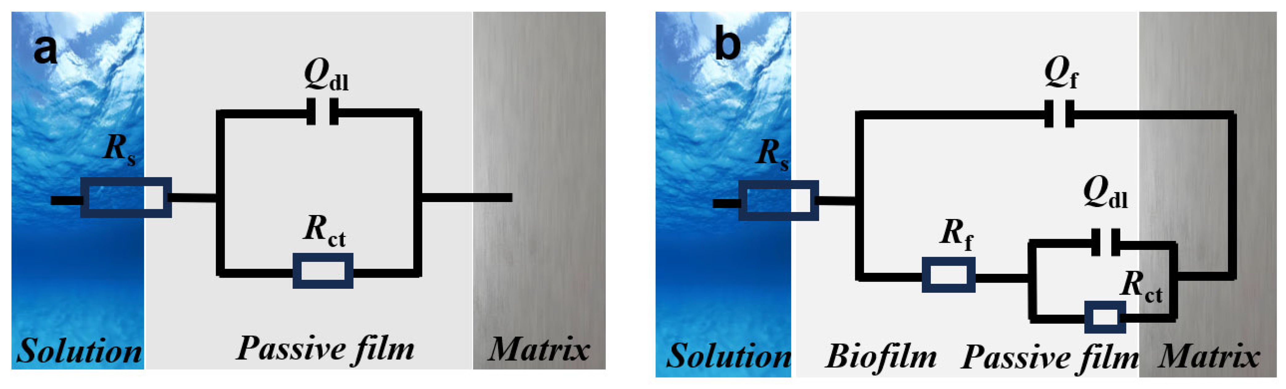
Based on the differences in interfacial structure, a single time constant equivalent circuit was used to fit the sterile system (Figure 9a), whereas a double time constant equivalent circuit was used for the inoculated system because of the presence of a biofilm or corrosion product layer (Figure 9b). The fitting results are presented in Table 1 and Table 2. For the sterile group, Rct increased from 10.94 MΩ cm2 after 1 d to 16.13 MΩ cm2 after 7 d, and then decreased to 12.19 MΩ cm2 after 14 d. This evolution reflects the gradual stabilization of the passive film during the initial immersion stage. After prolonged immersion, Cl action may have increased local film defects, thereby causing a slight decrease in charge transfer resistance. For the inoculated group, Rct gradually decreased from 8.41 MΩ cm2 after 1 d to 6.29 MΩ cm2 after 7 d and 4.32 MΩ cm2 after 14 d, corresponding to a progressive acceleration of the charge transfer process at the alloy surface. These EIS results are consistent with the negative shift in open circuit potential, the decrease in polarization resistance, and the increases in pit depth and pit density. Collectively, the results suggest that P. aeruginosa mainly weakened the stability of the Ti-based passive film and enhanced the localized corrosion susceptibility of the alloy through biofilm-induced interfacial microenvironmental heterogeneity.

3.4.4. Analysis of Donor Defect Density in the Passive Film

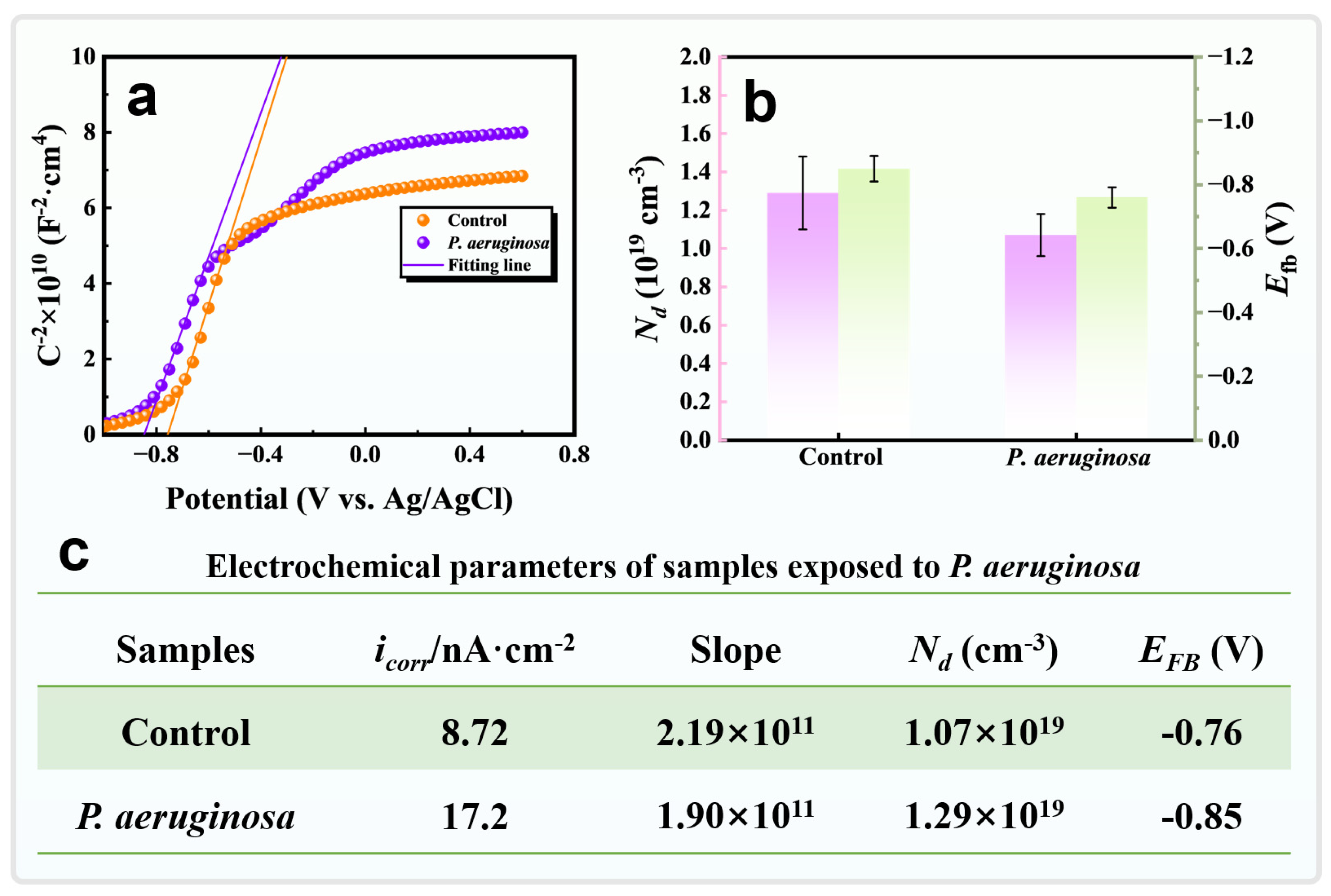
Figure 10 presents the Mott-Schottky curves and related electrochemical parameters of the Ti-15Mo-3Al-2.7Nb-0.25Si alloy after 14 d of immersion in sterile and P. aeruginosa-inoculated media. The linear regions of the Mott–Schottky curves for both groups exhibit positive slopes, demonstrating that the surface passive films possess typical n-type semiconductor characteristics (Figure 10a) [38,39]. For Ti-based passive films, n-type conductivity is commonly associated with donor defects, such as oxygen vacancies and titanium interstitials [40]. For an n-type semiconductor, the Mott-Schottky relationship can be expressed as follows:
1 C sc 2 = 1 ε ε 0 e N d A 2 ( E E f b K T e )
where C is the capacitance of the space charge layer, ε0 is the vacuum permittivity, ε is the dielectric constant of the passive film, e is the elementary charge, Efb is the flat-band potential, Nd is the donor density, k is the Boltzmann constant, and T is the absolute temperature. The values of ε0, e, and k were taken as 8.854 × 10⁻¹⁴ F cm−1, 1.602 × 10⁻¹⁹ C, and 1.38 × 10⁻²³ J K−1, respectively. For TiO2, ε was taken as 45 [41]. The donor density, Nd, was calculated from the slope of the linear region of the Mott-Schottky plot.
According to the slope of the linear region, the donor density of the sterile control was 1.07 × 1019 cm−3, whereas that of the P. aeruginosa group increased to 1.29 × 1019 cm−3 (Figure 10b,c). The higher Nd value suggests an increased concentration of donor-type defects in the passive film after bacterial exposure, which enhances charge transport through the film and consequently weakens the barrier effect of the passive film against ion migration and interfacial charge transfer processes.
Meanwhile, the flat band potential, Efb, of the P. aeruginosa group shifted negatively from −0.76 V for the sterile control to −0.85 V (Figure 10b,c), showing that biofilm exposure modified the electronic structure and charge distribution at the passive film/electrolyte interface. This negative shift is consistent with the previously observed negative shift in open circuit potential and the decrease in Rp, supporting the conclusion that the P. aeruginosa biofilm reduced passive film stability.
In conjunction with the potentiodynamic polarization results, icorr increased from 8.72 nA cm−2 for the sterile control to 17.2 nA cm−2 for the inoculated group, demonstrating a good correspondence between the increase in donor type defects and accelerated corrosion kinetics. Therefore, the interfacial microenvironmental changes induced by P. aeruginosa not only promoted localized pitting development but also weakened the protective ability of the Ti-based passive film by increasing passive film defect density and modifying its semiconductor properties.

3.5. Passive Film Composition Analysis

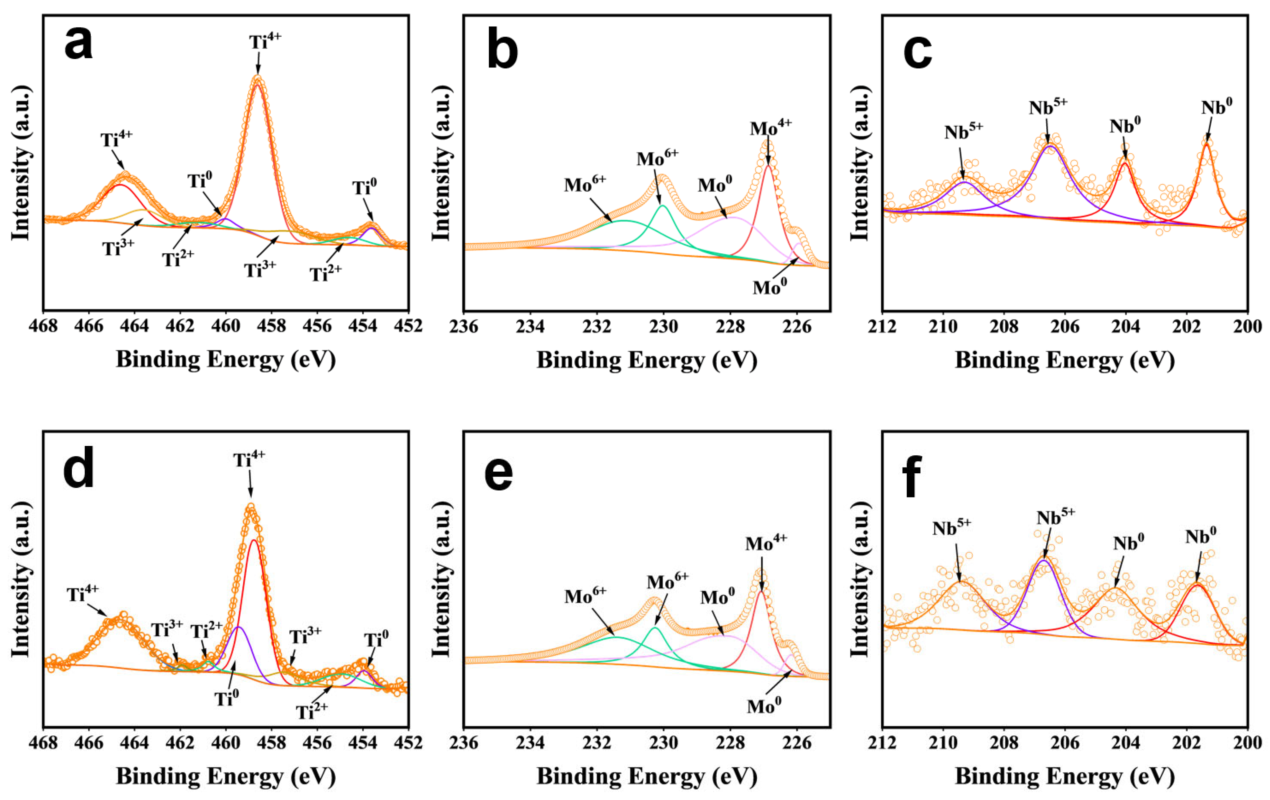
Figure 11 presents the high-resolution Ti 2p, Mo 3d, and Nb 3d XPS spectra of the Ti-15Mo-3Al-2.7Nb-0.25Si alloy after 14 d of immersion in sterile control and P. aeruginosa-inoculated systems. Figure 11a–c correspond to the sterile control group, whereas Figure 11d–f correspond to the P. aeruginosa-inoculated group. The Ti 2p spectra of both groups can be deconvoluted into Ti4+, Ti3+, Ti2+, and Ti0 components, which are assigned to TiO2, Ti2O3, TiO, and metallic Ti, respectively.
TiO2, Ti2O3, and TiO are commonly regarded as the principal oxide species in passive films on titanium alloys. TiO2 is generally associated with the outer region of the passive film, whereas lower valence titanium oxides, including Ti2O3 and TiO, are typically located closer to the metal/film interface. This distribution of chemical states is consistent with a compositional gradient extending from a TiO2-enriched outer region through an inner suboxide region toward the metallic substrate.
The growth of passive films on titanium alloys is commonly associated with the progressive oxidation of lower valence titanium oxides to TiO2. Representative reactions can be written as follows [42]:
2 T i + 3 H 2 O T i 2 O 3 + 6 H + + 6 e
T i 2 O 3 + 4 O H 2 T i O ( O H ) 2 + H 2 O + 4 e
T i O ( O H ) 2 T i O 2 + H 2 O
Equation (2) describes the oxidation of Ti2+ in TiO to Ti2O3 during the initial stage of passive film formation. Ti2O3 then undergoes further hydroxylation to form the Ti(IV) intermediate TiO(OH)2 (Equation (3)). Finally, TiO(OH)2 is converted into the thermodynamically more stable TiO2 through dehydration (Equation (4)). These reactions represent a typical pathway for the formation of Ti based passive films on titanium alloys in aqueous environments.
The XPS fitting results are summarized in Table 3. The surface of the sterile control group was dominated by TiO2, with a relative fraction of 72.29%, whereas the relative fractions of Ti2O3, TiO, and Ti0 were 14.68%, 6.02%, and 7.01%, respectively (Figure 11a). The high TiO2 fraction and weak Ti0 signal indicate that a highly oxidized, TiO2-dominated passive film formed on the alloy surface under sterile conditions, with a relatively limited contribution of the metallic substrate to the XPS signal.
After inoculation with P. aeruginosa, the relative fractions of TiO2 and Ti2O3 decreased to 66.74% and 7.16%, respectively, whereas those of TiO and Ti0 increased to 8.51% and 17.59%, respectively (Figure 11d). In particular, the pronounced enhancement of the Ti0 signal indicates an increased relative contribution from the metallic substrate within the XPS information depth. This change is consistent with a reduction in the effective thickness of the passive film and/or compromised local film continuity. Meanwhile, the relative fraction of Ti3+ decreased, whereas that of Ti2+ increased only slightly, indicating that the different lower-valence Ti components did not exhibit a consistent trend. Therefore, the decrease in the relative fraction of TiO2 may be associated not only with changes in the oxidation-state composition of the residual film but also with the peak-area normalization effect arising from the enhanced Ti0 signal.
The high-resolution Mo 3d spectra can be deconvoluted into Mo6+, Mo4+, and Mo0 components, which are assigned to high-valence Mo oxides, MoO2, and metallic Mo, respectively (Figure 11b,e) [43,44]. In the sterile control, the relative contents of Mo6+, Mo4+, and Mo0 were 42.59%, 23.09%, and 34.32%, respectively. After inoculation with P. aeruginosa, the Mo6+ and Mo4+ fractions decreased to 40.38% and 21.79%, respectively, whereas the Mo0 fraction increased to 37.83%. The overall slight decrease in oxidized Mo species, together with the increase in the metallic Mo signal, indicates that the contribution of Mo-related oxides within the surface film was reduced after biofilm exposure, while the contribution from metallic Mo in the substrate detected by XPS increased accordingly. Because the Mo6+ and Mo4+ fractions decreased simultaneously, this change is more likely associated with a reduction in the effective passive-film thickness or a loss of local film continuity rather than a simple transformation of high-valence Mo oxides into lower-valence species.
The Nb 3d spectra mainly consisted of Nb5+ and Nb0 components, corresponding to Nb2O5 and metallic Nb, respectively (Figure 11c,f) [45]. In the sterile control, the relative contents of Nb5+ and Nb0 were 56.76% and 43.24%, respectively, indicating that Nb in the surface film was present predominantly as Nb2O5. Previous studies have shown that Nb2O5-containing composite passive films exhibit favorable chemical stability and compactness, thereby enhancing resistance to aggressive-ion migration and interfacial charge transfer [46,47]. After inoculation with P. aeruginosa, the Nb5+ fraction decreased to 50.34%, whereas the Nb0 fraction increased to 49.66%. The decrease in the relative Nb2O5 content, accompanied by the increase in the metallic Nb signal, indicates a greater contribution from the Nb-containing substrate within the XPS sampling depth, further supporting a reduction in the effective passive-film thickness and/or loss of local film continuity. The reduced contribution of Nb oxides to film protection may further weaken the stability and barrier performance of the passive film.
Combined with the Mott-Schottky results, the increased donor density and negative shift in the flat-band potential of the inoculated group indicate that biofilm exposure altered the point-defect equilibrium within the passive film and the charge distribution at the film/solution interface. Local oxygen depletion, restricted mass transport, ion retention, and changes in interfacial redox conditions caused by the heterogeneous coverage of bacterial cells and EPS may inhibit complete oxidation of the passive film and local repassivation, thereby promoting the progressive accumulation of donor-type defects such as oxygen vacancies and Ti interstitials. The increased defect concentration further facilitates electronic and ionic transport through the film and reduces its resistance to interfacial charge transfer and Cl migration.
Therefore, the XPS, Mott-Schottky, and electrochemical results collectively demonstrate that, after exposure to P. aeruginosa, the passive film on the TB8 alloy underwent an evolution from defect accumulation to degradation of its effective barrier structure. This process was characterized by an increase in donor-type defects, reduced relative contributions of TiO2 and Mo/Nb oxides, enhanced signals from the metallic substrate, and weakened local film continuity. These changes corresponded closely to the negative shift in open-circuit potential, decreases in polarization resistance and charge-transfer resistance, and the increase in pit depth. The initial passivation stability provided by Mo and Nb was weakened under the heterogeneous interfacial environment induced by the biofilm, ultimately promoting localized passive-film destabilization and pitting development.

4. Discussion

The Ti-15Mo-3Al-2.7Nb-0.25Si alloy was dominated by a continuous β-Ti matrix containing a small fraction of dispersed α-Ti precipitates. Compared with commercially pure Ti, which exhibits a near-single-phase α structure, and the conventional α + β Ti-6Al-4V alloy, the higher Mo and Nb contents in TB8 enhanced the stability of the β matrix and may have contributed to the maintenance of a relatively stable initial passive state by regulating the microstructure and surface-film composition [10,11,48]. Meanwhile, differences in crystal structure, elemental partitioning, and local orientation gradients between the α-Ti precipitates and the β-Ti matrix introduced a certain degree of microstructural heterogeneity near the α/β interfaces. Such heterogeneity may result in spatial variations in passive-film formation and repair kinetics among different microstructural regions, thereby providing a potential microstructural basis for localized film destabilization [49]. The KAM results reflect variations in local misorientation and lattice curvature near the α/β interfaces and can therefore be used to characterize the degree of microstructural heterogeneity; however, regions with elevated KAM values should not be directly regarded as preferential sites for pit initiation.
After inoculation with P. aeruginosa, bacterial cells and EPS formed a spatially heterogeneous biofilm on the alloy surface, transforming the initially relatively uniform alloy/solution interface into a complex interface comprising cell-rich regions, EPS-enriched regions, and locally exposed areas. Biofilm coverage restricted oxygen and ion transport and generated spatial differences in oxygen concentration, Cl distribution, and interfacial redox conditions between covered and uncovered regions [50]. The average biofilm thickness decreased from 22.5 μm after 7 d to 15.6 μm after 14 d, which may be associated with limitations in nutrient and oxygen transport during the later stage of cultivation, biofilm structural reorganization, and partial detachment of bacterial cells and EPS. Although the biofilm became thinner at the later stage, the residual bacterial cells and EPS were still capable of maintaining a heterogeneous interfacial environment. Therefore, the localized corrosion observed after 14 d was more likely the cumulative consequence of biofilm activity throughout the immersion period rather than a direct result of biofilm detachment itself.
The electrochemical, Mott–Schottky, and XPS results collectively demonstrate that P. aeruginosa primarily promoted localized passive-film degradation rather than generalized active dissolution of the alloy. After inoculation, the open-circuit potential shifted negatively, the polarization resistance and charge-transfer resistance decreased, and the corrosion current density increased from 8.72 to 17.2 nA cm−2, indicating enhanced interfacial charge transfer and anodic dissolution. Meanwhile, the donor density increased from 1.07 × 10¹⁹ to 1.29 × 1019 cm−3, suggesting an increased concentration of donor-type defects, such as oxygen vacancies and Ti interstitials, within the passive film. An increased defect concentration can facilitate electronic and ionic transport through the film and provide transport pathways for Cl migration and localized dissolution, causing the rate of film degradation to progressively exceed the rate of repassivation [42].
After inoculation, the relative Ti0 content increased markedly from 7.01% to 17.59%, indicating a greater contribution from the metallic substrate within the XPS sampling depth and reflecting a decrease in the effective passive-film thickness and/or a loss of local film continuity. The decrease in Ti3+ content, together with only a slight increase in Ti2+ content, indicates that passive-film degradation did not involve a uniform and complete transformation of TiO2 into lower-valence titanium oxides. Instead, the degradation process more likely involved defect accumulation within the film, localized thinning, and a reduction in barrier performance in specific regions [51]. When the locally damaged passive film cannot be repaired in a timely manner, the freshly exposed metal surface comes into contact with the Cl-containing medium. Metal-ion hydrolysis within the pits, inward migration of Cl, and restricted mass transport then further sustain the locally aggressive environment, promoting the transition from metastable to stable pitting. Accordingly, the increase in pit density from 9.2 to 13.4 cm−2 and in maximum pit depth from 2.4 to 4.1 μm after inoculation reflects increases in both the number of localized corrosion initiation events and the capacity for sustained pit propagation.
Previous studies have shown that P. aeruginosa can decrease the charge-transfer resistance, degrade the passive film, and enhance pitting corrosion of commercially pure Ti and TC4 alloys [27,28]. Compared with these conventional titanium materials, TB8 is distinguished by the coexistence of a Mo/Nb-stabilized continuous β matrix and α/β microstructural heterogeneity introduced by dispersed α-Ti precipitates. Its MIC behavior is therefore governed not only by interactions between the TiO2-based passive film and the biofilm, but also by the combined effects of β-stabilizing elements, α-phase precipitation, and interfacial strain on the local passive state [52]. Recent studies on β titanium alloys have also shown that a relatively stable passive state under sterile conditions does not necessarily correspond to higher MIC resistance, because biofilms can still weaken passive-film protection by amplifying phase-dependent and interfacial environmental heterogeneity [33,34,44]. The present study therefore reveals a characteristic corrosion response of TB8 in which a relatively stable initial passive state coexists with biofilm-induced localized passive-film destabilization. This finding indicates that the MIC susceptibility of high-Mo/Nb β titanium alloys is governed by the dynamic balance among alloy microstructure, passive-film defect repair capability, and the biofilm-modified interfacial environment.
Overall, the MIC process of the TB8 alloy can be summarized as follows. The α/β microstructural heterogeneity establishes potential spatial differences in local passivation, while the heterogeneous biofilm further alters interfacial mass transport and redox conditions. These changes promote the accumulation of donor-type defects and reduce the effective thickness of the passive film, ultimately shifting the local breakdown–repassivation balance toward sustained dissolution and accelerating pit initiation and propagation. Although the corrosion current density increased to 17.2 nA cm−2, the alloy still exhibited pronounced passive behavior. Therefore, these results primarily indicate the promotion of early-stage localized corrosion processes rather than the occurrence of engineering-relevant structural failure within 14 d.

5. Conclusions

This study elucidated the localized corrosion behavior and passive film degradation characteristics of the Ti-15Mo-3Al-2.7Nb-0.25Si β titanium alloy in the presence of P. aeruginosa biofilms. The main conclusions are as follows:
(1) The Ti-15Mo-3Al-2.7Nb-0.25Si alloy consisted of a continuous β-Ti matrix and dispersed α-Ti precipitates. EBSD revealed area fractions of approximately 92.5% for β-Ti and 7.5% for α-Ti, while the α-Ti precipitates exhibited an average equivalent circle diameter of 0.913 μm. Higher local orientation gradients and lattice distortion were observed near the α/β interfaces, providing a potential microstructural basis for spatial variations in passivation behavior among different microregions.
(2) After attachment to the alloy surface, P. aeruginosa formed a heterogeneous biofilm composed of bacterial cells and EPS, thereby increasing the localized corrosion susceptibility of the alloy. The biofilm coverage altered local oxygen and ion transport and promoted spatial heterogeneity in the interfacial microenvironment. Compared with the sterile system, exposure to P. aeruginosa increased the maximum pit depth from 2.4 to 4.1 μm and the pit density from 9.2 to 13.4 cm−2. These changes show that the corrosion damage shifted from shallow and isolated pitting toward deeper and more densely distributed localized attack.
(3) Electrochemical and surface chemical results collectively revealed passive film degradation following exposure to P. aeruginosa. The icorr value increased from 8.72 to 17.2 nA cm−2, while Rct decreased to 4.32 MΩ cm2 after 14 d. Meanwhile, the donor density increased from 1.07 × 10¹⁹ to 1.29 × 1019 cm−3, the relative Ti4+ content decreased from 72.29% to 66.74%, and the relative Ti0 content increased to 17.59%. Biofilm-induced local oxygen depletion, ion enrichment, and changes in interfacial redox conditions promoted defect accumulation and loss of passive-film integrity, thereby weakening its local barrier effect and accelerating pit initiation and propagation.
Therefore, optimizing the solution treatment and aging conditions to regulate the size and spatial distribution of α-Ti precipitates and reduce microstructural and local strain heterogeneity near the α/β interfaces may provide an effective strategy for further improving passive film stability and resistance to microbiologically influenced corrosion of the TB8 alloy.

Author Contributions

Q.Z.: Conceptualization, Methodology, Validation, Formal analysis, Investigation, Data curation, Writing—original draft preparation, Visualization. Y.T.: Methodology, Validation, Formal analysis, Investigation, Data curation, Writing—original draft preparation, Visualization. D.L.: Conceptualization, Methodology, Supervision, Writing—review and editing. H.Z.: Methodology, Validation, Formal analysis, Investigation, Data curation. J.C.: Software, Validation, Formal analysis, Data curation, Visualization. Z.Z.: Methodology, Validation, Investigation, Resources, Data curation. Q.F.: Resources, Validation, Investigation, Data curation. W.G.: Conceptualization, Methodology, Resources, Supervision, Writing—review and editing. Q.W.: Conceptualization, Methodology, Resources, Supervision, Writing—review and editing, Funding acquisition. H.Y.: Resources, Supervision, Writing—review and editing, Project administration. D.S.: Conceptualization, Resources, Supervision, Writing—review and editing, Funding acquisition. All authors have read and agreed to the published version of the manuscript.

Funding

This work is supported by Guangdong Basic and Applied Basic ResearchFoundation (No.2023B1515250006), Opening Project Fund of Materials Service Safety Assessment Facilities, Southern Marine Science and Engineering Guangdong Laboratory (Zhuhai) (Nos. SML2023SP242 and SML2024SP005).

Data Availability Statement

The original contributions presented in this study are included in the article. Further inquiries can be directed to the corresponding author.

Conflicts of Interest

Qiuyuan Feng was employed by Baoti Group Ltd., Baoji 721014, China. The remaining authors declare that the research was conducted in the absence of any commercial or financial relationships that could be construed as a potential conflict of interest.

References

  1. Yan, S.K.; Song, G.L.; Li, Z.X.; Wang, H.N.; Zheng, D.J.; Cao, F.Y.; Horynova, M.; Dargusch, M.S.; Zhou, L. A state-of-the-art review on passivation and biofouling of Ti and its alloys in marine environments. J. Mater. Sci. Technol. 2018, 34, 421–435. [Google Scholar] [CrossRef] [Scilit]
  2. Tuck, B.; Watkin, E.; Somers, A.; Machuca, L.L. A critical review of marine biofilms on metallic materials. npj Mater. Degrad. 2022, 6, 25. [Google Scholar] [CrossRef] [Scilit]
  3. Xu, D.K.; Gu, T.; Lovley, D.R. Microbially mediated metal corrosion. Nat. Rev. Microbiol. 2023, 21, 705–718. [Google Scholar] [CrossRef] [Scilit] [PubMed]
  4. Tian, Y.X.; Zhang, Q.N.; Liu, D.; Li, Y.Z.; Gao, W.; Hu, K.K.; Liao, Y.; Liu, J.X.; Yu, H.Y.; Sun, D.B.; et al. Composition-driven phase structures in laser-deposited titanium-steel composites: Microstructural evolution and interfacial strengthening mechanisms. Adv. Compos. Hybrid Mater. 2026, 9, 194. [Google Scholar] [CrossRef] [Scilit]
  5. Li, J.Q.; Zhang, D.Y.; Chen, X.B.; Xu, D.K.; Qiu, D.; Wang, F.H.; Easton, M. Laser directed energy deposited, ultrafine-grained functional titanium–copper alloys tailored for marine environments: Antibacterial and anti-microbial corrosion studies. J. Mater. Sci. Technol. 2023, 166, 21–33. [Google Scholar] [CrossRef] [Scilit]
  6. Seo, D.I.; Lee, J.B. Localized corrosion and repassivation behaviors of additively manufactured titanium alloys in simulated biomedical solutions. npj Mater. Degrad. 2023, 7, 44. [Google Scholar] [CrossRef] [Scilit]
  7. Meng, X.Z.; Li, X.R.; Li, F.; Yan, H.J.; Zhang, Q.H.; Wu, L.K.; Di Tommaso, D.; Cao, F.H. Molecular insights into the stability of titanium in electrolytes containing chlorine and fluorine ions. Langmuir 2023, 39, 17853–17861. [Google Scholar] [CrossRef] [Scilit] [PubMed]
  8. Huang, Q.Y.; Xiao, Y.H.; Li, X.R.; Zhu, C.B.; Sun, Q.Q.; Wu, L.K.; Cao, F.H. Influence of near-yield stress on corrosion and passivation behavior of titanium in chloride solutions. Corros. Sci. 2025, 255, 113092. [Google Scholar] [CrossRef] [Scilit]
  9. Li, N.B.; Wang, L.T.; Zhou, Z.H.; Jiang, X.Z.; Yu, H.Y.; Sun, D.B. The interaction mechanism of titanium alloy TC4 between passive film and sulfate reducing bacteria biofilm in marine environment. Appl. Surf. Sci. 2025, 690, 162620. [Google Scholar] [CrossRef] [Scilit]
  10. Calazans Neto, J.V.; Celles, C.A.S.; de Andrade, C.S.A.F.; Afonso, C.R.M.; Nagay, B.E.; Barão, V.A.R. Recent advances and prospects in β-type titanium alloys for dental implant applications. ACS Biomater. Sci. Eng. 2024, 10, 6029–6060. [Google Scholar] [CrossRef] [Scilit] [PubMed]
  11. Ballor, J.A.; Li, T.; Prima, F.; Boehlert, C.J.; Devaraj, A. A review of the metastable omega phase in beta titanium alloys: The phase transformation mechanisms and its effect on mechanical properties. Int. Mater. Rev. 2023, 68, 26–45. [Google Scholar] [CrossRef] [Scilit]
  12. Kang, X.D.; Jiang, H.Y.; Du, Z.X.; Gong, T.H.; Liu, J.W.; Guo, W.X.; Cheng, J.; Liu, J.S.; Li, G.W. Regulation of microstructure to optimize mechanical properties of Ti-15Mo-3Al-2.7Nb-0.2Si via solution-duplex ageing. Metals 2023, 13, 869. [Google Scholar] [CrossRef] [Scilit]
  13. Jiang, H.Y.; Du, Z.X.; Wang, D.; Gong, T.H.; Cui, X.M.; Liu, F.; Cheng, J.; Chen, W.Z. Preparation of multiscale α phase by heat treatments and its effect on tensile properties in metastable β titanium alloy sheet. Metals 2021, 11, 1708. [Google Scholar] [CrossRef] [Scilit]
  14. Zheng, C.W.; Chen, R.R.; Wang, Q.; Wei, W.; Su, B.X.; Ding, X.; Fu, H.Z. Great enhancement in corrosion resistance of Ti alloy by Mo element regulating β grain boundary and microstructure. Corros. Sci. 2025, 256, 113237. [Google Scholar] [CrossRef] [Scilit]
  15. Çaha, I.; Alves, A.C.; Chirico, C.; Pinto, A.M.; Tsipas, S.; Gordo, E.; Bondarchuk, O.; Deepak, F.L.; Toptan, F. Atomic-scale investigations of passive film formation on Ti–Nb alloys. Appl. Surf. Sci. 2023, 615, 156282. [Google Scholar] [CrossRef] [Scilit]
  16. Ji, P.F.; Li, B.; Chen, B.H.; Wang, F.; Ma, W.; Zhang, X.Y.; Ma, M.Z.; Liu, R.P. Effect of Nb addition on the stability and biological corrosion resistance of Ti–Zr alloy passivation films. Corros. Sci. 2020, 170, 108696. [Google Scholar] [CrossRef] [Scilit]
  17. Liu, H.; Wang, Z.X.; Cheng, J.; Li, N.; Liang, S.X.; Zhang, L.; Shang, F.M.; Dobuvyy, O.; Chen, L.Y. Nb-content-dependent passivation behavior of Ti–Nb alloys for biomedical applications. J. Mater. Res. Technol. 2023, 27, 7882–7894. [Google Scholar] [CrossRef] [Scilit]
  18. Xu, T.W.; Zhang, S.S.; Liang, S.; Cui, N.; Cao, L.; Wan, Y. Precipitation behaviour during the β → α/ω phase transformation and its effect on the mechanical performance of a Ti-15Mo-2.7Nb-3Al-0.2Si alloy. Sci. Rep. 2019, 9, 17628. [Google Scholar] [CrossRef] [Scilit] [PubMed]
  19. Xu, T.W.; Li, J.S.; Zhang, S.S.; Zhang, F.S.; Liu, X.H. Cold deformation behavior of the Ti-15Mo-3Al-2.7Nb-0.2Si alloy and its effect on α precipitation and tensile properties in aging treatment. J. Alloys Compd. 2016, 682, 404–411. [Google Scholar] [CrossRef] [Scilit]
  20. Gao, W.; Zhang, Q.N.; Tian, Y.X.; Liu, D.; Wang, Q.; Yu, H.Y.; Sun, D.B. Unveiling microstructural heterogeneity, mechanical properties, and microbiologically induced selective corrosion in Q355 welded joints. J. Mater. Res. Technol. 2026, 40, 618–637. [Google Scholar] [CrossRef] [Scilit]
  21. Feng, Z.H.; Wei, K.M.; Dong, H.C.; Ma, J.; Wang, J.G.; Liu, D.; Li, J.H.; Zhang, X.Y. Antagonistic effect of β phase and passive film on corrosion resistance of TiZrAlV series alloys in marine Pseudomonas aeruginosa environment. Appl. Surf. Sci. 2025, 706, 163550. [Google Scholar] [CrossRef] [Scilit]
  22. Flemming, H.C.; van Hullebusch, E.D.; Little, B.J.; Neu, T.R.; Nielsen, P.H.; Seviour, T.; Stoodley, P.; Wingender, J.; Wuertz, S. Microbial extracellular polymeric substances in the environment, technology and medicine. Nat. Rev. Microbiol. 2025, 23, 87–105. [Google Scholar] [CrossRef] [Scilit] [PubMed]
  23. Wang, Y.N.; Zhang, R.Y.; Duan, J.Z.; Hou, B.R. Extracellular polymeric substances and biocorrosion/biofouling: Recent advances and future perspectives. Int. J. Mol. Sci. 2022, 23, 5566. [Google Scholar] [CrossRef] [Scilit] [PubMed]
  24. Qi, P.; Zeng, Y.; Zhang, D.; Sun, Y.; Wang, P. The biofilm–metal interface: A hotspot for microbiologically influenced corrosion. Cell Rep. Phys. Sci. 2025, 6, 102500. [Google Scholar] [CrossRef] [Scilit]
  25. Jones, L.M.; Salta, M.; Skovhus, T.L.; Thomas, K.G.; Illson, T.; Wharton, J.; Webb, J.S. Effects of sulphate-reducing bacteria mixed-species biofilms on microbiologically influenced corrosion. Environ. Microbiol. 2025, 27, e70116. [Google Scholar] [CrossRef] [Scilit] [PubMed]
  26. Huang, L.Y.; Chang, W.W.; Zhang, D.W.; Huang, Y.; Li, Z.Y.; Lou, Y.T.; Qian, H.C.; Jiang, C.Y.; Li, X.G.; Mol, A.M. Acceleration of corrosion of 304 stainless steel by outward extracellular electron transfer of Pseudomonas aeruginosa biofilm. Corros. Sci. 2022, 199, 110159. [Google Scholar] [CrossRef] [Scilit]
  27. Khan, M.S.; Li, Z.; Yang, K.; Xu, D.K.; Yang, C.; Liu, D.; Lekbach, Y.; Zhou, E.; Kalnaowakul, P. Microbiologically influenced corrosion of titanium caused by aerobic marine bacterium Pseudomonas aeruginosa. J. Mater. Sci. Technol. 2019, 35, 216–222. [Google Scholar] [CrossRef] [Scilit]
  28. Liu, D.; Yang, H.; Li, J.; Li, J.; Dong, Y.; Yang, C.; Jin, Y.; Lekbach, Y.; Li, Z.; Hernandez, D.; et al. Electron transfer mediator PCN secreted by aerobic marine Pseudomonas aeruginosa accelerates microbiologically influenced corrosion of TC4 titanium alloy. J. Mater. Sci. Technol. 2021, 79, 101–108. [Google Scholar] [CrossRef] [Scilit]
  29. Zhao, P.Y.; Shen, C.; Savage, M.F.; Li, J.; Niezgoda, S.R.; Mills, M.J.; Wang, Y.Z. Slip transmission assisted by Shockley partials across α/β interfaces in Ti-alloys. Acta Mater. 2019, 171, 291–305. [Google Scholar] [CrossRef] [Scilit]
  30. Chong, Y.; Deng, G.Y.; Yi, J.; Shibata, A.; Tsuji, N. On the strain hardening abilities of α+β titanium alloys: The roles of strain partitioning and interface length density. J. Alloys Compd. 2019, 811, 152040. [Google Scholar] [CrossRef] [Scilit]
  31. Qiu, D.; Shi, R.; Zhang, D.; Lu, W.; Wang, Y. Variant selection by dislocations during α precipitation in α/β titanium alloys. Acta Mater. 2015, 88, 218–231. [Google Scholar] [CrossRef] [Scilit]
  32. Shi, R.; Wang, Y. Variant selection of grain boundary α by special prior β grain boundaries in titanium alloys. Acta Mater. 2014, 75, 156–166. [Google Scholar] [CrossRef] [Scilit]
  33. Lu, W.Q.; Liu, Y.J.; Wu, X.; Liu, X.C.; Wang, J.C. Corrosion behavior and microstructural effects on passivation film mechanisms in forged Ti-5Al-5Mo-5V-1Cr-1Fe titanium alloy under laser surface remelting. Corros. Sci. 2024, 241, 112542. [Google Scholar] [CrossRef] [Scilit]
  34. Zheng, Y.F.; Williams, R.E.A.; Viswanathan, G.B.; Clark, W.A.T.; Fraser, H.L. Determination of the structure of α–β interfaces in metastable β-Ti alloys. Acta Mater. 2018, 150, 25–39. [Google Scholar] [CrossRef] [Scilit]
  35. Ramírez, C.G.; Monsalve, A.; Montero, C.; Espinoza, J.; Araujo, J.; Vejar, N.; Azócar, M.; Gulppi, M.; Jiménez-Arevalo, V.M.; Zhou, X.; et al. Microbiologically influenced corrosion of Al–Cu–Li alloy by Pseudomonas aeruginosa. J. Mater. Res. Technol. 2025, 36, 5286–5297. [Google Scholar] [CrossRef] [Scilit]
  36. Pan, J.; Thierry, D.; Leygraf, C. Electrochemical impedance spectroscopy study of the passive oxide film on titanium for implant application. Electrochim. Acta 1996, 41, 1143–1153. [Google Scholar] [CrossRef] [Scilit]
  37. Claros, C.A.E.; Campanelli, L.C.; Jorge Junior, A.M.; Leprêtre, J.C.; Bolfarini, C.; Kiminami, C.S.; Botta, W.J.; Roche, V. Corrosion behaviour of biomedical β-titanium alloys with the surface modified by chemical etching and electrochemical methods. Corros. Sci. 2021, 188, 109544. [Google Scholar] [CrossRef] [Scilit]
  38. Li, X.; Wang, L.W.; Fan, L.; Cui, Z.; Sun, M. Effect of temperature and dissolved oxygen on the passivation behavior of Ti–6Al–3Nb–2Zr–1Mo alloy in artificial seawater. J. Mater. Res. Technol. 2022, 17, 374–391. [Google Scholar] [CrossRef] [Scilit]
  39. Zhong, M.H.; Su, B.X.; Jin, Y.L.; Cui, G.G.; Li, Z.W.; Zhou, J.C.; Yang, Y.; Zhang, Q.D.; Wang, B.B.; Yang, Q.; et al. Effect of electron beam surface melting on the microstructure and corrosion behavior of Ti-4Al-4Zr-2Sn-1.5Mo-Nb-V alloy. npj Mater. Degrad. 2026, 10, 72. [Google Scholar] [CrossRef] [Scilit]
  40. Kadiri, M.; Tanji, A.; Fan, X.S.; Liaw, P.K.; Mahlia, T.M.I.; Hermawan, H. Corrosion of TiHfZrNbx high-entropy alloys in a simulated condition of proton exchange membrane water electrolyser. Electrochim. Acta 2025, 521, 145925. [Google Scholar] [CrossRef] [Scilit]
  41. Roh, B.; Macdonald, D.D. Passivity of titanium: Part II, the defect structure of the anodic oxide film. J. Solid State Electrochem. 2019, 23, 1967–1979. [Google Scholar] [CrossRef] [Scilit]
  42. Qin, P.; Chen, L.Y.; Liu, Y.J.; Jia, Z.; Liang, S.X.; Zhao, C.H.; Sun, H.; Zhang, L.C. Corrosion and passivation behavior of laser powder bed fusion produced Ti-6Al-4V in static/dynamic NaCl solutions with different concentrations. Corros. Sci. 2021, 191, 109728. [Google Scholar] [CrossRef] [Scilit]
  43. Wei, Y.; Pan, Z.M.; Fu, Y.; Yu, W.; He, S.L.; Yuan, Q.Y.; Luo, H.; Li, X.G. Effect of annealing temperatures on microstructural evolution and corrosion behavior of Ti–Mo titanium alloy in hydrochloric acid. Corros. Sci. 2022, 197, 110079. [Google Scholar] [CrossRef] [Scilit]
  44. Kaseem, M.; Choe, H.C. The effect of in-situ reactive incorporation of MoOx on the corrosion behavior of Ti–6Al–4V alloy coated via micro-arc oxidation coating. Corros. Sci. 2021, 192, 109764. [Google Scholar] [CrossRef] [Scilit]
  45. Santos, R.R.; Elias, C.N.; Ferreira, E.A.; Huguenin, J.A.O.; Farias, E.E.; de Souza, M.L.; da Silva, L. Characterization of thin oxide layers formed by Ti–Nb alloy anodization. J. Mater. Res. Technol. 2024, 33, 2519–2529. [Google Scholar] [CrossRef] [Scilit]
  46. do Nascimento, J.P.L.; Ferreira, M.O.A.; Gelamo, R.V.; Scarmínio, J.; Steffen, T.T.; da Silva, B.P.; Aoki, I.V.; dos Santos, A.G., Jr.; de Castro, V.V.; Malfatti, C.F.; et al. Enhancing the corrosion protection of Ti–6Al–4V alloy through reactive sputtering niobium oxide thin films. Surf. Coat. Technol. 2021, 428, 127854. [Google Scholar] [CrossRef] [Scilit]
  47. Liu, C.; Yan, Z.L.; Yang, J.; Wei, P.G.; Zhang, D.; Wang, Q.; Zhang, X.; Hao, Y.L.; Yang, D.H. Corrosion and biological behaviors of biomedical Ti–24Nb–4Zr–8Sn alloy under an oxidative stress microenvironment. ACS Appl. Mater. Interfaces 2024, 16, 18503–18521. [Google Scholar] [CrossRef] [Scilit] [PubMed]
  48. Wang, L.; Wang, S.Y. Quantitative analysis of self-healing properties and microstructure of Ti–5Al–5Mo–5V–1Cr–1Fe alloy by quasi-in-situ XPS. J. Alloys Compd. 2025, 1012, 178509. [Google Scholar] [CrossRef] [Scilit]
  49. Sun, C.H.; Xiao, R.L.; Li, H.R.; Ruan, Y. Effects of phase selection and microsegregation on corrosion behaviors of Ti–Al–Mo alloys. Corros. Sci. 2022, 200, 110232. [Google Scholar] [CrossRef] [Scilit]
  50. Wu, J.J.; Chen, Z.Q.; Li, G.; Teng, K.; Ge, L.; Chen, Y.X.; Li, L.; Qu, Q. Visualisation and quantification of biofilm-substrate interface microenvironments based on a fungal-bacterial interaction model: An in-depth investigation into microbially mediated corrosion processes. Corros. Sci. 2025, 246, 112725. [Google Scholar] [CrossRef] [Scilit]
  51. Arroussi, M.; Yang, K.; Jia, Q.; Bai, C.G.; Wang, W.K.; Zhang, R.Y. Mechanistic insights into Pseudomonas aeruginosa biofilm-driven passive film degradation of Ti–9Mn alloy in marine environment. Corros. Commun. 2026; in press. [CrossRef] [Scilit]
  52. Arroussi, M.; Jia, Q.; Zhao, J.L.; Xia, Z.Z.; Bai, C.G.; Yang, K. Modification of passive film on Ti–6Al–4V alloy induced by Bacillus vietnamensis. Mater. Chem. Phys. 2024, 313, 128679. [Google Scholar] [CrossRef] [Scilit]
Figure 1. Initial microstructure and high-energy synchrotron X-ray diffraction results of the Ti-15Mo-3Al-2.7Nb-0.25Si β titanium alloy: (a) low-magnification SEM image; (b) high-magnification SEM image; (c) two-dimensional high-energy synchrotron X-ray diffraction (HE-XRD) pattern; (d) one-dimensional diffraction profile obtained by full azimuthal integration of the two-dimensional diffraction pattern.
Metals 16 00840 g001
Figure 2. EBSD microstructure, phase distribution, local misorientation, and texture characteristics of the Ti-15Mo-3Al-2.7Nb-0.25Si β titanium alloy: (a) band contrast (BC) map; (b) overlay of the BC and inverse pole figure (IPF) maps; (c) kernel average misorientation (KAM) map; (d) overlay of the BC and phase distribution maps; (e) {0001} basal pole figure of the α-Ti phase; (f) {10-10} prismatic pole figure of the α-Ti phase.
Metals 16 00840 g002
Figure 3. Statistical results of α-Ti precipitate size and local misorientation in the Ti-15Mo-3Al-2.7Nb-0.25Si β titanium alloy: (a) equivalent circle diameter distribution of α-Ti precipitates; (b) KAM distribution.
Metals 16 00840 g003
Figure 4. Surface biofilm morphologies and thickness statistics of the Ti-15Mo-3Al-2.7Nb-0.25Si alloy after 7 and 14 d of immersion in P. aeruginosa-inoculated medium: (a,c) low-magnification SEM images; (b,d) high-magnification SEM images of the corresponding regions; (e,f) three-dimensional CLSM reconstructions; (g) average biofilm thickness.
Metals 16 00840 g004
Figure 5. Three-dimensional surface morphologies and localized corrosion statistics of the Ti-15Mo-3Al-2.7Nb-0.25Si alloy after 14 d of immersion in sterile and P. aeruginosa-inoculated media: (a,b) three-dimensional corrosion morphologies; (c,d) cross-sectional depth profiles along selected paths; (e) pit depth–width distribution; (f) pit density.
Metals 16 00840 g005
Figure 6. Evolution of (a) open circuit potential, Eocp, and (b) polarization resistance, Rp, of the Ti-15Mo-3Al-2.7Nb-0.25Si alloy during 14 d of immersion in sterile and P. aeruginosa-inoculated media.
Metals 16 00840 g006
Figure 7. Potentiodynamic polarization behavior of the Ti-15Mo-3Al-2.7Nb-0.25Si alloy after 14 d of immersion in sterile and P. aeruginosa-inoculated media: (a) polarization curves; (b) fitted corrosion current density.
Metals 16 00840 g007
Figure 8. EIS responses of the Ti-15Mo-3Al-2.7Nb-0.25Si alloy after 1, 7, and 14 d of immersion in sterile and P. aeruginosa-inoculated media: (a,b) Nyquist plots; (c,d) Bode phase angle plots; (e,f) Bode impedance modulus plots.
Metals 16 00840 g008
Figure 9. Equivalent fitting circuits for EIS results in different culture media: (a) Control; (b) P. aeruginosa.
Metals 16 00840 g009
Figure 10. Mott-Schottky analysis and related electrochemical parameters of the Ti-15Mo-3Al-2.7Nb-0.25Si alloy after 14 d of immersion in sterile and P. aeruginosa-inoculated media: (a) Mott-Schottky plots; (b) donor density, Nd, and flat-band potential, Efb; (c) fitted electrochemical parameters.
Metals 16 00840 g010
Figure 11. High-resolution Ti 2p (a,d), Mo 3d (b,e) and Nb 3d (c,f) XPS spectra of the Ti-15Mo-3Al-2.7Nb-0.25Si alloy after 14 d of immersion in (ac) sterile medium and (df) P. aeruginosa-inoculated medium.
Metals 16 00840 g011
Table 1. Fitted EIS parameters of the Ti-15Mo-3Al-2.7Nb-0.25Si alloy during 14 days in the sterile medium.
Time (d)Rs
(Ω cm2)
QfRf
(KΩ cm2)
QdlRct (MΩ cm2)∑χ2
(×10−4)
(10−5 F cm−2 sn)n(10−5 F cm−2 sn)n
18.15 ± 0.42///2.11 ± 0.120.90 ± 0.0810.94 ± 0.525.43 ± 0.02
78.16 ± 0.39///2.19 ± 0.100.90 ± 0.0616.13 ± 0.257.12 ± 0.06
148.31 ± 0.42///2.54 ± 0.170.91 ± 0.0412.19 ± 0.414.31 ± 0.03
Table 2. Fitted EIS parameters of the Ti-15Mo-3Al-2.7Nb-0.25Si alloy during 14 days in the inoculated medium.
Time (d)Rs
(Ω cm2)
QfRf
(KΩ cm2)
QdlRct (MΩ cm2)∑χ2
(×10−4)
(10−5 F cm−2 sn)n(10−5 F cm−2 sn)n
110.31 ± 0.778.56 ± 0.660.81 ± 0.018.94 ± 0.232.36 ± 0.370.89 ± 0.078.41 ± 0.193.14 ± 0.02
79.47 ± 0.328.43 ± 0.340.82 ± 0.0210.23 ± 0.612.19 ± 0.210.90 ± 0.046.29 ± 0.444.78 ± 0.08
149.31 ± 0.446.13 ± 0.210.83 ± 0.058.79 ± 0.423.46 ± 0.330.91 ± 0.054.32 ± 0.162.69 ± 0.05
Table 3. Relative contents of Ti, Mo, and Nb species in different chemical states determined by Ti 2p, Mo 3d, and Nb 3d XPS peak fitting for the Ti-15Mo-3Al-2.7Nb-0.25Si alloy after 14 d of immersion in sterile and P. aeruginosa-inoculated media.
ElementChemical StateControl (at%)P. aeruginosa (at%)
TiTi4+72.2966.74
Ti3+14.687.16
Ti2+6.028.51
Ti07.0117.59
MoMo6+42.5940.38
Mo4+23.0921.79
Mo034.3237.83
NbNb5+56.7650.34
Nb043.2449.66
Disclaimer/Publisher’s Note: The statements, opinions and data contained in all publications are solely those of the individual author(s) and contributor(s) and not of MDPI and/or the editor(s). MDPI and/or the editor(s) disclaim responsibility for any injury to people or property resulting from any ideas, methods, instructions or products referred to in the content.