
Article

The Influence of Copper on the Corrosion Resistance of CoCrMo-xCu Alloy in Several Biological Solutions for Biomedical Applications

1
School of Metallurgy, Northeastern University, Shenyang 110819, China
2
School of Materials Science and Engineering, Key Lab. for Anisotropy and Texture of Materials, Education Ministry of China, Northeastern University, Shenyang 110819, China
*
Author to whom correspondence should be addressed.
Metals 2026, 16(5), 498; https://doi.org/10.3390/met16050498
Submission received: 28 March 2026 / Revised: 26 April 2026 / Accepted: 29 April 2026 / Published: 1 May 2026
(This article belongs to the Section Corrosion and Protection)

Abstract

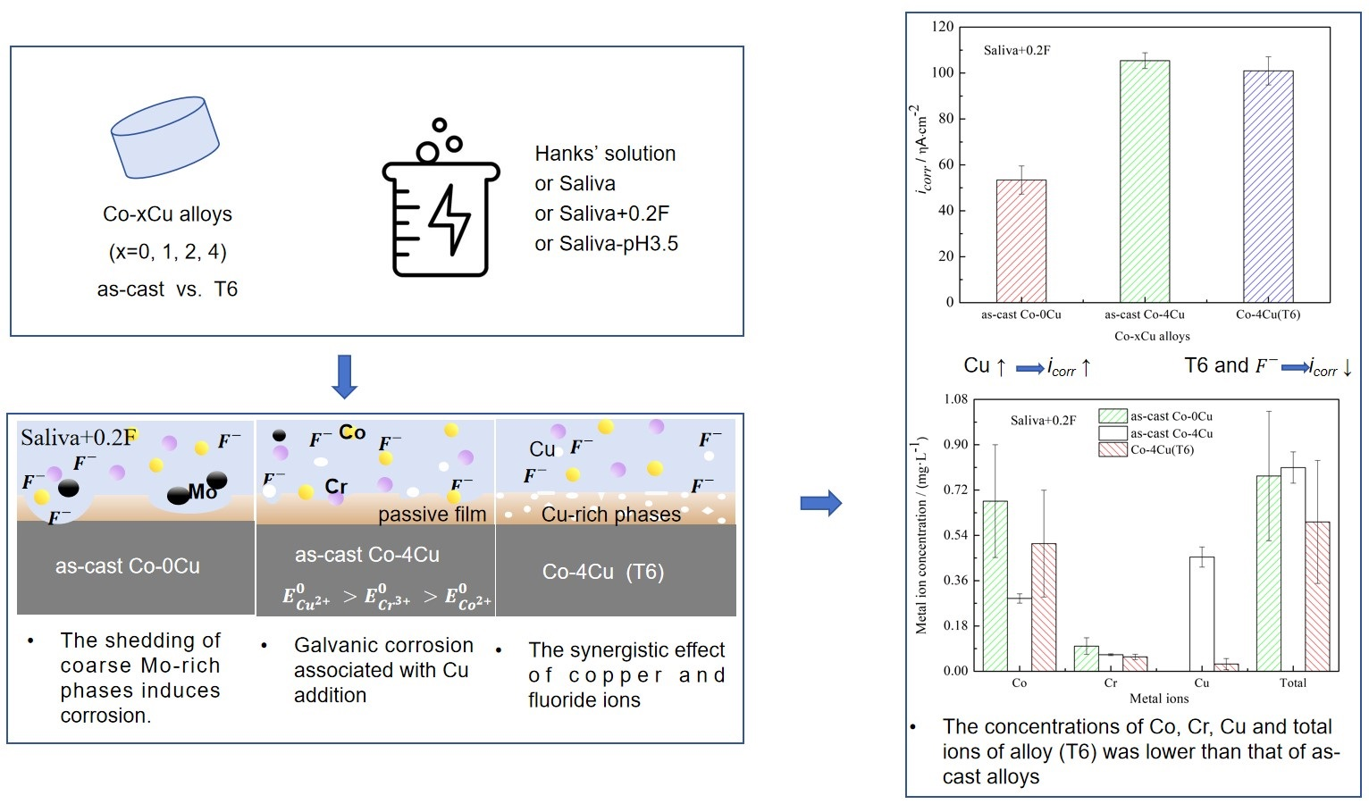
CoCrMo alloys are widely used as orthopedic and dental implants, owing to their superior mechanical properties, wear resistance, and biocompatibility. Copper (Cu) ion exhibits strong antibacterial activity, making it a promising alloying element. A systematic study was conducted on the corrosion resistance and ion release behavior of CoCrMo-xCu (Co-xCu) alloys in both as-cast and heat-treated states in different simulated solutions. The results indicated that the corrosion resistance of Co-xCu alloys decreased with the increasing Cu content, which was mainly attributed to the formation of micro-galvanic couples between the alloy matrix and Cu-rich phases. The synergistic effect of heat treatment and an appropriate Cu content can effectively improve the corrosion resistance of the alloys, and the corrosion current density (icorr) of Cu-containing cobalt alloys was comparable to that of Cu-free cobalt alloys. Maximum concentrations of Co, Cr, and Cu ions released from Co-xCu alloys were lower than the corresponding recommended safety limits. Through the combined optimization of Cu content and heat treatment, the metal ion release levels of Cu-containing cobalt alloys can be reduced to values even lower than those of Cu-free cobalt alloys.

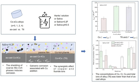
Graphical Abstract

1. Introduction

Co-based alloys are renowned for their excellent properties, including superior corrosion resistance compared to 316L stainless steel [1,2] and enhanced tribological performance over Ti alloys [3]. Consequently, they are extensively utilized in dental and orthopedic implants due to their high corrosion resistance, excellent biocompatibility, and suitable mechanical properties [1,2]. Among these alloys, CoCrMo alloys stand out for their well-rounded properties, primarily attributed to the alloying elements Cr and Mo [4,5,6,7]. In aqueous environments, CoCrMo alloys spontaneously form a nano-scale oxide layer on their surface, which significantly enhances their corrosion resistance. Additionally, Mo refines the grain structure and improves the stability and continuity of the passive film [6,8,9]. These attributes render CoCrMo alloys particularly suitable for long-term biomedical applications, such as hip implants, oral implants, and dental restorations.
Bacterial infections and inflammation present substantial challenges in the clinical use of surgical biomaterials, such as CoCrMo alloy oral implants. Research has shown that the human oral cavity contains up to 500 different bacterial species, including Staphylococcus aureus (S. aureus) and Escherichia coli (E. coli) [10,11]. Once bacteria attach and colonize, biofilm formation can lead to inflammation in surrounding tissues, negatively impacting the long-term success of implants [12,13]. Copper (Cu), a vital trace element in the human body [14,15], is known for its good antibacterial properties [16,17,18]. For example, Ren et al. [10] demonstrated that adding an appropriate amount of Cu to CoCrCu alloys enhanced the antibacterial and antibiofilm performance. Additional studies have confirmed that incorporating Cu into Co-based alloys maintained their mechanical properties [19] and corrosion resistance in 0.9% NaCl solution [20,21,22] while also providing significant antibacterial effects [10,22].
These findings suggest that adding an optimal amount of Cu to CoCrMo alloys holds significant potential for dental implant applications. Therefore, thoroughly evaluating the properties of CoCrMo-xCu alloys is crucial. Gan et al. [21] investigated the effect of Cu content on the tribological performance and corrosion resistance of CoCrMo alloys in a 0.9% NaCl solution. Their results showed that adding 2 wt% or 3 wt% Cu improved the corrosion resistance, while 4 wt% Cu did not offer the same benefit. Moradi et al. [23] found that Cu addition significantly altered the microstructure of CoCrMo alloys, enhancing their corrosion resistance in a 0.9% NaCl solution and reducing Co ion release into simulated solutions. Our previous research demonstrated that Cu suppressed the martensitic transformation of CoCrMo-xCu alloys [24]. In addition, studies on hot deformation behavior indicated that Cu addition did not affect the deformation temperature but expanded the stable process zones [25].
Although our previous work [22] studied the corrosion behavior of CoCrMo-based alloys, it was only conducted in a simple sodium chloride solution and mainly focused on the basic corrosion performance. This study compared the corrosion behavior in various biologically related solutions that were closer to real clinical environments, elucidated the dual role of the Cu element, and investigated the synergistic effect of Cu content, solution composition, and heat treatment on the corrosion resistance of cobalt alloys. It provided a more comprehensive evaluation basis for the greater expansion of medical applications of Cu-containing cobalt alloys.

2. Materials and Methods

2.1. Materials

Co-xCu alloys (Co-29Cr-6Mo-xCu, where x = 0, 1, 2, and 4 wt%) were fabricated via the casting method in accordance with the ISO 5832-4:2024 standard [26]. Based on the Cu content, the alloys were designated as Co-0Cu, Co-1Cu, Co-2Cu, and Co-4Cu, respectively. The Co-29Cr-6Mo alloy and pure copper were melted at 1520 °C in a WK-2 vacuum non-consumable arc melting furnace manufactured by Hefei Kejing Materials Technology Co., Ltd., Hefei, China. The molten material was then poured into a preheated ceramic mold to form cast ingots. The chemical compositions of the alloys were determined using an X-ray fluorescence spectrometer manufactured by Shimadzu Corporation, Kyoto, Japan. The nominal and measured chemical compositions of the Co-xCu alloys used in this study are listed in Table 1. The slight loss of Cu (measured vs. nominal contents) was attributed to Cu evaporation during arc melting. The consistently increasing trend in the measured Cu contents guaranteed the reliability of conclusions regarding the effect of Cu content.
Samples for testing were cut from the ingots via wire electrical discharge machining (WEDM). The heat treatment protocol involved heating the samples at 1060 °C for 24 h, followed by heating at 1250 °C for 4 h, subsequent water quenching, and finally aging at 650 °C for 16 h. In this paper, samples in the as-cast condition were referred to as as-cast Co-xCu, while the heat-treated samples were labeled as Co-xCu(T6). All samples were sequentially wet-ground using 500#, 1000#, 1500#, and 2000# grit silicon carbide papers and then ultrasonically decreased with distilled water and acetone.

2.2. Electrochemical Testing

Electrochemical tests were performed in accordance with ISO 10271:2001 standard [27] using a VersaSTAT3 electrochemical workstation supplied by Ametek Group, Berwyn, PA, USA. A three-electrode system was employed, consisting of a Co-xCu sample as the working electrode, a Pt electrode as the counter electrode, and a saturated calomel electrode as the reference electrode. To replicate various biological conditions, the tests were carried out in Hanks’ solution, saliva, saliva with 0.2% sodium fluoride (Saliva+0.2F), and saliva with pH 3.5 (Saliva-pH3.5) at 37 ± 1 °C. The artificial saliva solution was prepared according to ISO 10271:2020, which is the international standard for corrosion testing of dental metallic materials. The detailed chemical composition of these four simulated solutions is provided in Table 2. The artificial saliva used was a modified ISO 10271:2021 formulation, with adjustments in lactic acid concentration, fluoride content, and pH to simulate different oral conditions. The pH was adjusted to the desired value using NaOH and HCl. The fluoride concentration (2 g/L NaF) was set to simulate severe oral fluoride exposure scenarios (e.g., clinical fluoride varnish application), which can effectively highlight differences in the corrosion resistance of the alloys.
The sample was immersed in the simulated solution, and its open circuit potential (OCP) was monitored for 3600 s. To ensure quasi-steady-state conditions for reliable data collection, EIS measurements were performed only after the OCP drift rate fell below 0.1 mV/s. Potentiodynamic polarization curves were obtained over a potential range of −0.5 V to 0.75 V at a scanning rate of 0.5 mV/s. This approach is consistent with common analytical practices for the corrosion evaluation of CoCrMo-based biomedical alloys [22]. Electrochemical impedance spectroscopy (EIS) measurements were conducted over a frequency range from 105 Hz to 0.01 Hz, with an AC amplitude of 10 mV. The equivalent circuit parameters were derived from EIS data with ZSimpWin 3.20 software. For each group, three parallel samples were measured to calculate a mean value with standard deviation.

2.3. Metal Ion Release

Samples with a dimension of 13 mm × 13 mm × 2 mm were prepared. Prior to immersion tests, they were ground sequentially with 500#, 1000#, 1500#, and 2000# grit silicon carbide papers, then polished, followed by cleaning with anhydrous ethanol and deionized water for 5 min. The samples were immersed in containers filled with the test solution for seven days at 37 ± 1 °C. In accordance with ISO 10993-12:2021 [28], the ratio of the sample surface area to the volume of the simulated solution was maintained at 1.5 cm2·mL−1. The concentrations of released Co, Cr, and Cu ions were measured using inductively coupled plasma mass spectrometry (ICP-MS), using an Optima 5300DV instrument manufactured by PerkinElmer, Waltham, MA, USA.

2.4. Surface Morphology Analysis

The surface microstructural morphologies of CoCrMo-xCu alloys before and after heat treatment were characterized by field emission scanning electron microscopy (FESEM, JSM-6360LV, JEOL Ltd., Tokyo, Japan). The micro-area elemental composition was further analyzed by energy-dispersive X-ray spectroscopy (EDS) equipped on the FESEM system. After 7 days of immersion in Hanks’ solution, Saliva, Saliva+0.2F, and Saliva-pH3.5, the surface morphologies of the Co-4Cu(T6) alloy were observed by FESEM (JEOL JSM-6360LV) at an accelerating voltage of 15 kV.
All samples were cleaned with ultrasonic waves before SEM observation.

3. Results

3.1. Microstructure

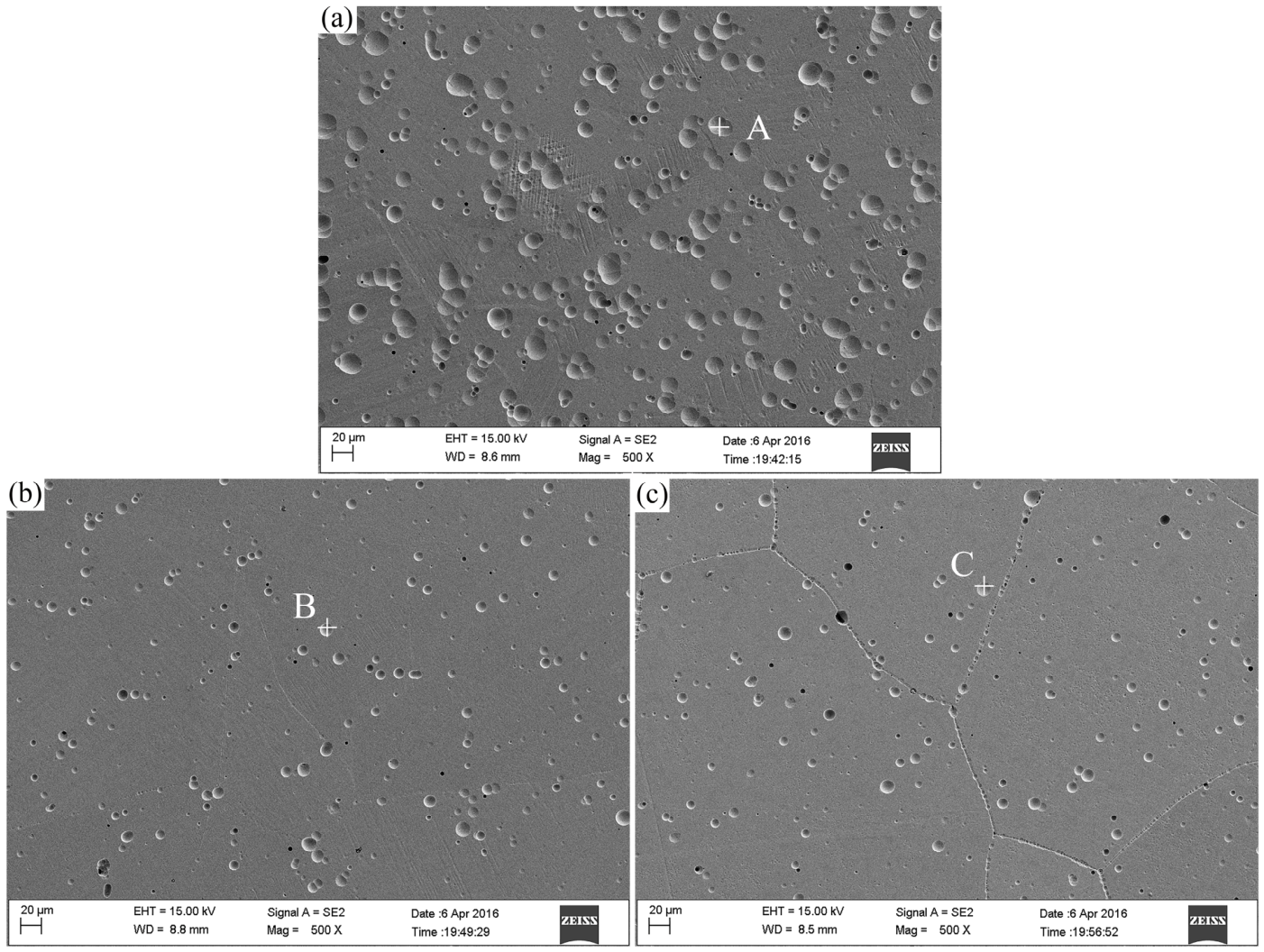
According to our previous XRD, EBSD, and SEM investigations [24,25], martensitic transformation occurs during the heat treatment process of as-cast Co-xCu alloys, and the microstructure is mainly composed of a cobalt-based matrix and martensitic phase. Heat treatment significantly reduces grain size and promotes phase transformation. Figure 1 shows SEM micrographs of as-cast Co-0Cu, Co-2Cu, and Co-4Cu alloys. The EDS quantitative results of elemental mass fractions (wt%) at distinct points (A, B, and C) are summarized in Table 3. The as-cast Co-1Cu alloy shows a microstructure similar to that of Co-2Cu, with no obvious differences in grain size or phase morphology. Mo-rich phases formed due to alloy melting defects exhibit a regular circular morphology. This morphology results in low interfacial energy, which promotes particle detachment during corrosion and appears as black pits in the microstructure. The Co-xCu alloys exhibit a similar microstructure characterized by coarse grains. However, as the Cu content increases, grain boundaries become more pronounced and well-defined. Additionally, both the number and size of black pits decrease with increasing Cu content.
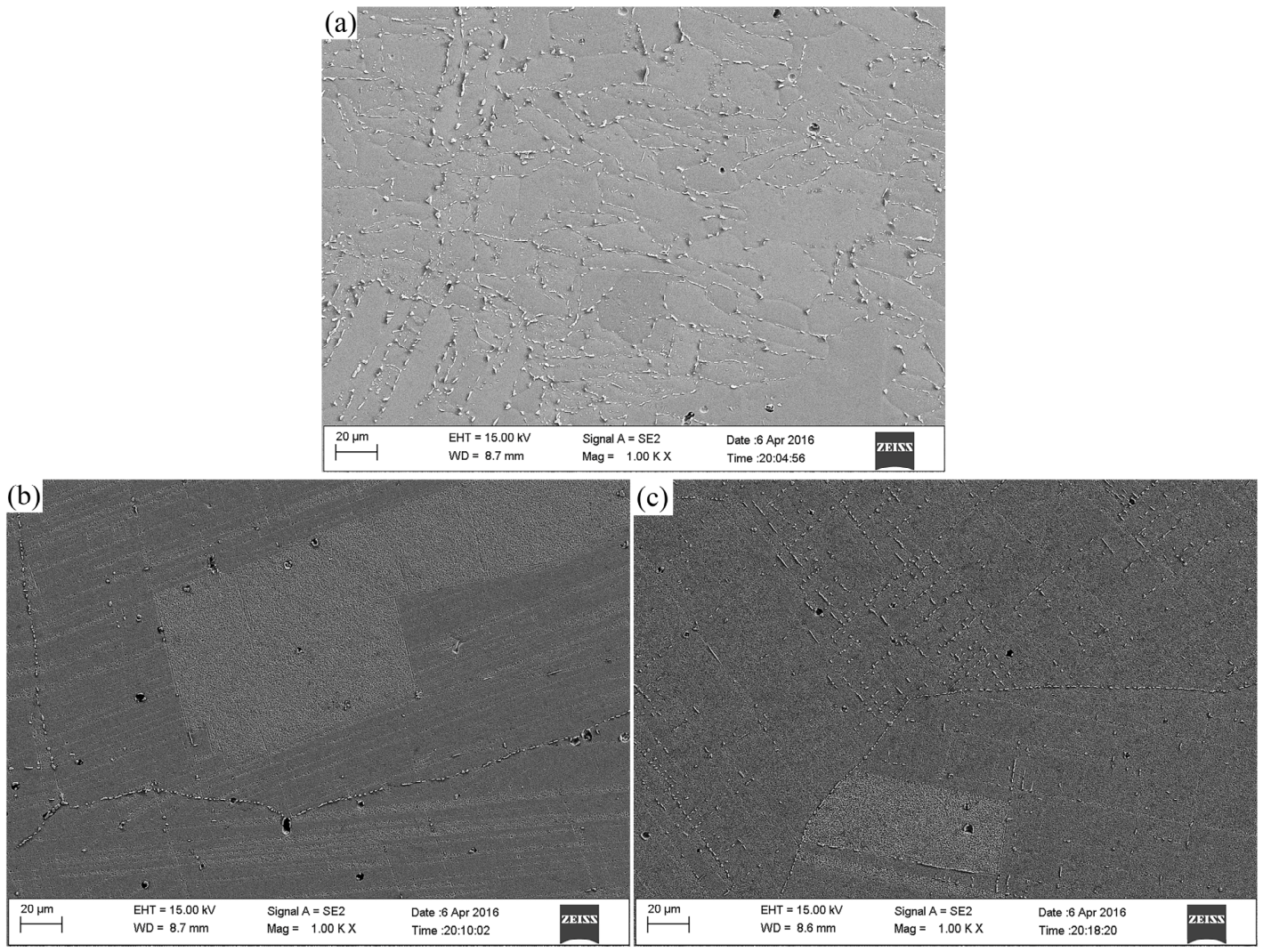
Figure 2 presents SEM images of the heat-treated Co-0Cu, Co-2Cu, and Co-4Cu alloys. The microstructure of the heat-treated Co–1Cu alloy is similar to the above alloys without obvious differences in grain size and phase morphology. After the heat treatment, austenite transforms into martensite. However, as the Cu content increases, the austenite content rises, while the martensite content declines. The number of black pits decreases significantly, and the grain boundaries appear more distinct. With the addition of Cu, elemental segregation occurs at the grain boundaries.

3.2. OCP Curves

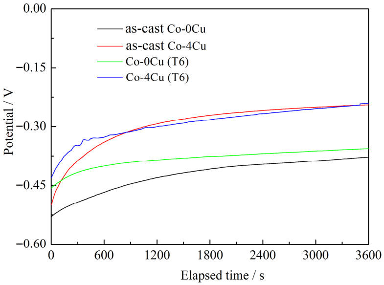
Figure 3 illustrates the OCP variation of as-cast and T6 Co-xCu alloys (x = 0 and 4) in Hanks’ solution at 37 ± 1 °C. The steady-state open circuit potential (EOCP) values for all tested samples under different conditions are summarized in Table 4. Compared with the Co-0Cu control sample, the increase in Cu content shifts the EOCP of both as-cast and T6-treated Co-xCu alloys toward more positive potentials. In Hanks’ and Saliva solutions, heat treated Co-xCu alloys show a nobler EOCP than their as-cast counterparts. By contrast, in Saliva+0.2F and Saliva-pH3.5 solutions, heat-treated Co-0Cu and Co-1Cu alloys possess more negative EOCP values than the as-cast ones. With the rise of Cu addition, the EOCP of heat-treated Co-xCu alloys gradually approaches that of the as-cast samples and eventually becomes even nobler.

3.3. Corrosion Potential and Corrosion Current Density

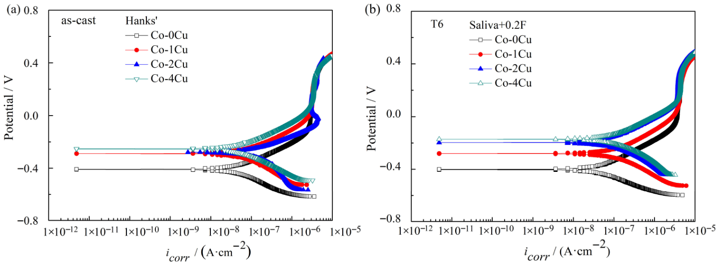
Figure 4 shows the typical potentiodynamic polarization curves of as-cast and Co-xCu(T6) alloys in the representative simulated solutions. The free corrosion potential (Ecorr) and corrosion current density (icorr) extracted from the curves are summarized in Table 4 and Table 5. In the present study, corrosion resistance is comprehensively evaluated based on a more positive Ecorr and a lower icorr. A more positive Ecorr indicates higher thermodynamic stability of the passive film, while a lower icorr indicates a slower corrosion kinetic rate. Table 5 reveals that the addition of Cu shifts Ecorr toward a more noble direction. For Co-xCu alloys immersed in Hanks’ solution and Saliva, the heat treatment also shifts Ecorr toward a more noble direction. In Saliva+0.2F, the heat treatment causes the Ecorr of Co-0Cu and Co-1Cu alloys to shift more negatively, whereas the Ecorr of Co-2Cu(T6) and Co-4Cu(T6) alloys shifts towards a more noble direction. In Saliva-pH3.5, the heat treatment results in a more negative shift in the Ecorr of Co-0Cu, Co-1Cu, and Co-2Cu alloys, while the Ecorr of the Co-4Cu(T6) alloy shifts toward a more noble position.
Table 6 indicates that the addition of Cu leads to a gradual increase in the icorr of Co-xCu alloys. In Hanks’ solution, Cu-containing cobalt alloys show a slight decrease in icorr after the heat treatment. When these alloys are immersed in Saliva, the icorr initially decreases and then increases, compared with the as-cast Co-xCu alloys. In contrast, in Saliva+0.2F, the icorr first increases and then decreases after heat treatment. In Saliva-pH3.5, the icorr slightly increases after heat treatment. The icorr values for both Cu-containing and Cu-free cobalt alloys remain within the same order of magnitude. Notably, the icorr of cobalt alloys in Saliva+0.2F is the highest, suggesting that fluoride ions attack passive film of Co-xCu alloys.

3.4. Electrochemical Impedance Spectroscopy

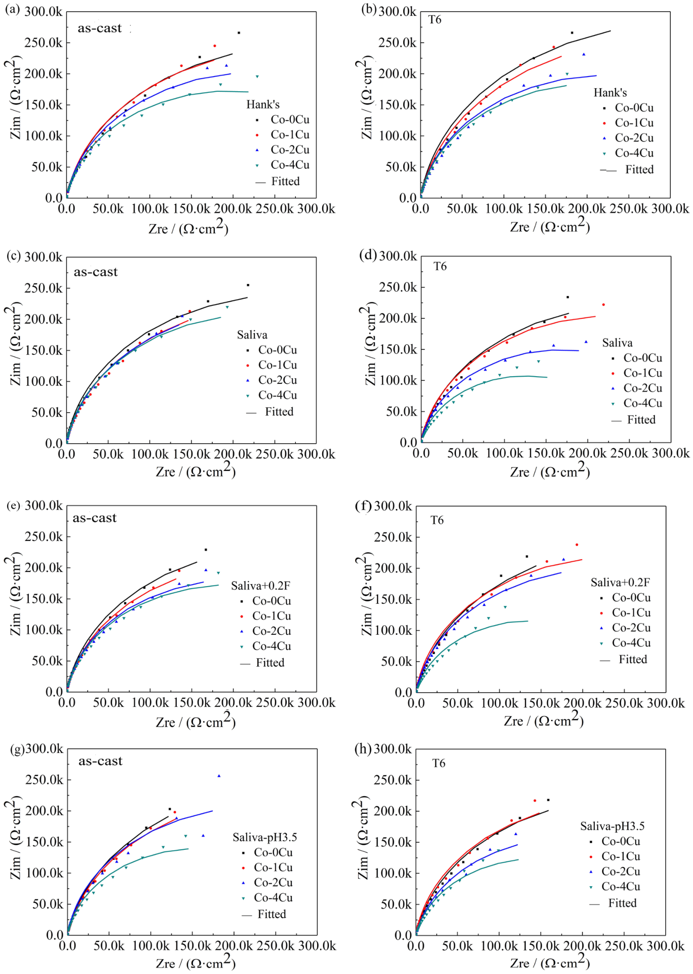
Figure 5 presents both experimental and simulated Nyquist plots for Co-xCu alloys in various solutions: Hanks’, Saliva, Saliva+0.2F, and Saliva-pH3.5. In the high-frequency region, the Nyquist plots for Co-xCu alloys show a single semicircle, typical of the electrochemical impedance spectrum of a monolayer film. At low frequencies, the impedance behavior is characterized by a straight line, with a slope close to 45°. As shown in Figure 5, the capacitance arc radius of Co-xCu alloys decreases gradually with increasing Cu content, suggesting that the addition of Cu reduces the corrosion resistance. Furthermore, the capacitance arc of cobalt alloys in the Saliva+0.2F solution is slightly smaller, indicating that fluoride ions have a minor erosive effect on the alloys. However, as shown in Table 5, the synergy of Cu and heat treatment enhances the resistance of cobalt alloys to fluoride ion erosion.
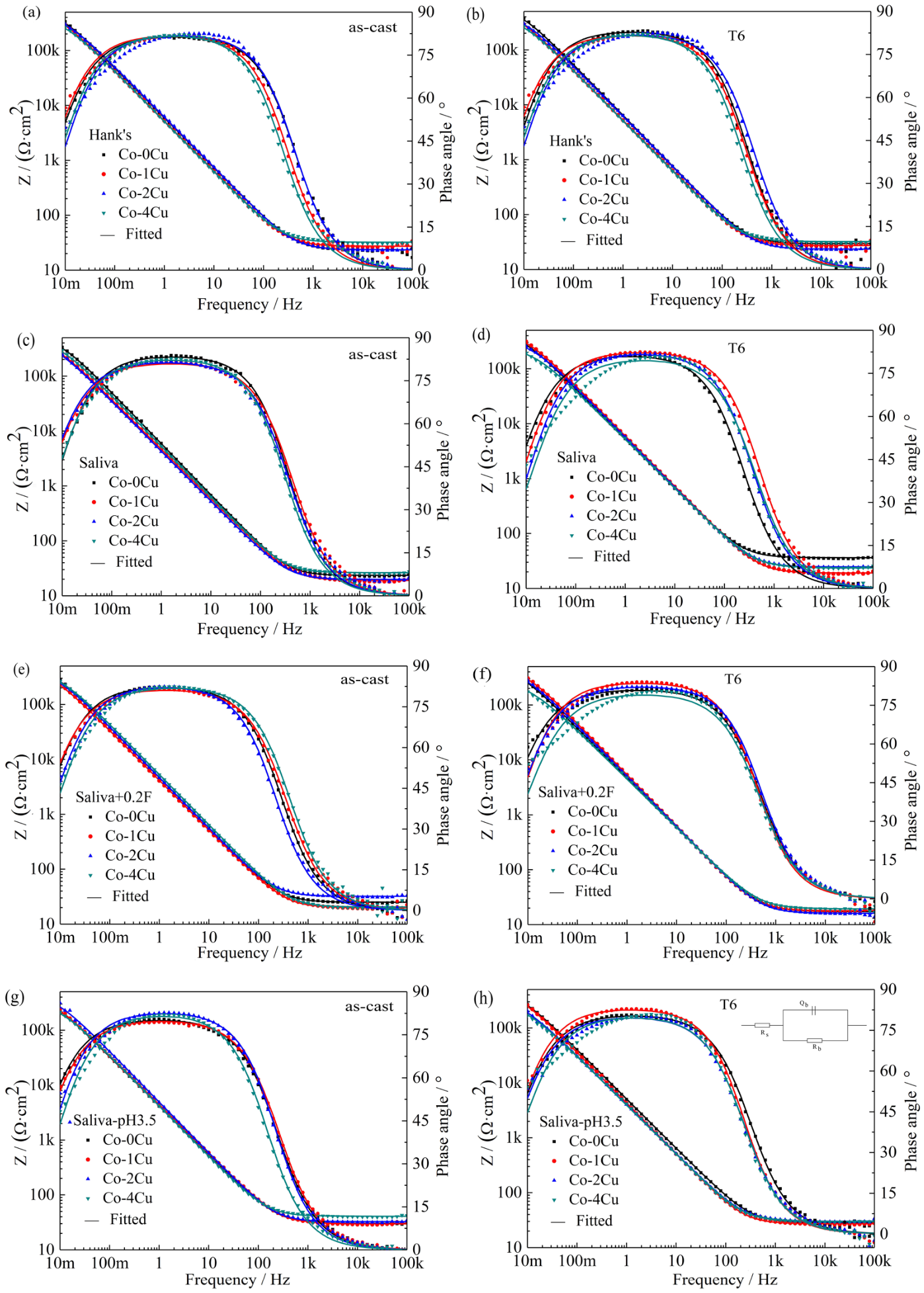
Figure 5 presents the Bode impedance and theta angle plots for Co-xCu alloys in Hanks’ solution, Saliva, Saliva+0.2F, and Saliva-pH3.5 solutions. In the high-frequency range (105–103 Hz), the impedance curves have a slope near zero, indicating typical solution resistance behavior. In the low-frequency range (103–10−2 Hz), the impedance curves form a straight line, with a slope of approximately −1, characteristic of a monolayer film’s impedance. This suggests that the passive film formed on Co-xCu alloys in these solutions exhibits typical monolayer characteristics, with only minor differences. In the high-frequency range (105–103 Hz), the phase angle nears 0°, underscoring the predominance of solution resistance. A broad peak emerges in the mid-frequency range (10−2–10−1 Hz), suggesting the existence of a monolayer passive film. The phase angle peaks at 85°, signifying that the passive film functions as a highly resistive and low-capacitance insulating layer. The phase angle curves for Co-xCu alloys in these simulated solutions largely overlap, indicating no significant difference in corrosion resistance between them.
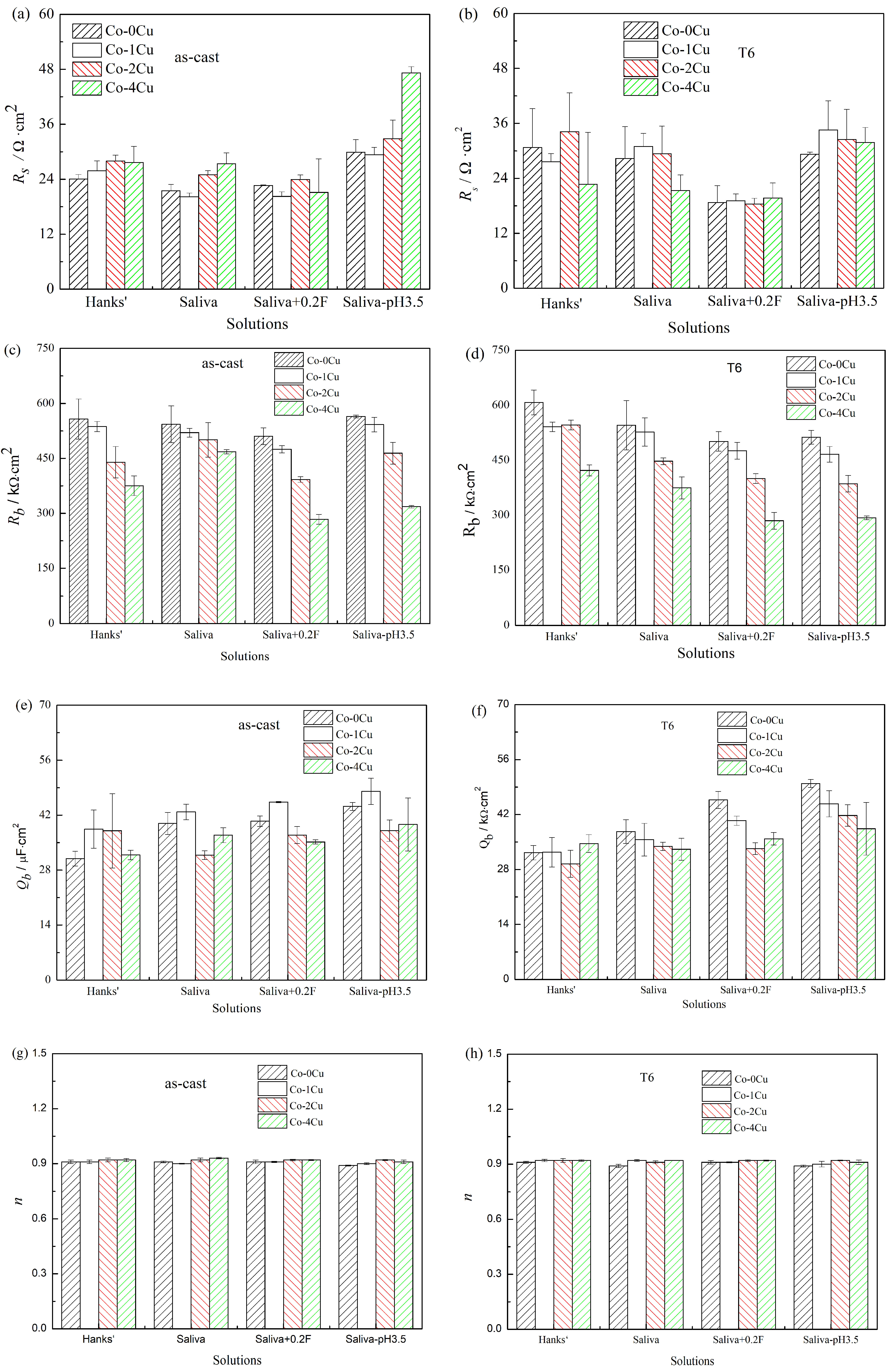
Based on the EIS curves of Co-xCu alloys in various simulated solutions and the equivalent circuit model Rs(QbRb) depicted in Figure 6, the electrochemical parameters Rs, Rb, Qb, and n obtained by fitting are detailed in Figure 7. This single-layer model has been verified to effectively characterize the overall protective behavior of the passive film [8,22], with all fittings exhibiting satisfactory goodness-of-fit (χ2 < 10−3). Here, Rs represents the solution resistance between the working and reference electrodes; Rb indicates the charge-transfer resistance; Qb denotes the double-layer capacitance; and n is a dispersion coefficient ranging from 0 to 1. The impedance (Rb) of the passive film on Co-xCu alloys decreased with increasing Cu content, and tended to stabilize at approximately 105 Ω·cm2. The relatively low Qb values suggest the formation of a stable and dense passive film on the Co-xCu alloy surfaces in these simulated solutions, which imparts excellent corrosion resistance. With n close to 1, the passive film on Co-xCu alloys closely resembles an ideal capacitor. The Co-xCu alloys in the Saliva+0.2F solution exhibited a reduced Rb value, indicating that fluoride ions exerted an erosive effect on the passive film formed on the alloy surface.
In summary, the corrosion resistance of Co-xCu alloys in Hanks’ solution, Saliva, Saliva+0.2F, and Saliva-pH3.5 solutions decreased as the Cu content increased. Additionally, the corrosion resistance of Co-xCu alloys varied slightly due to alloy composition, microstructure, heat treatments, and properties of the simulated solutions, but it was in the same order of magnitude as that of Cu-free cobalt alloys.

3.5. Metal Ion Release of Co-xCu Alloys

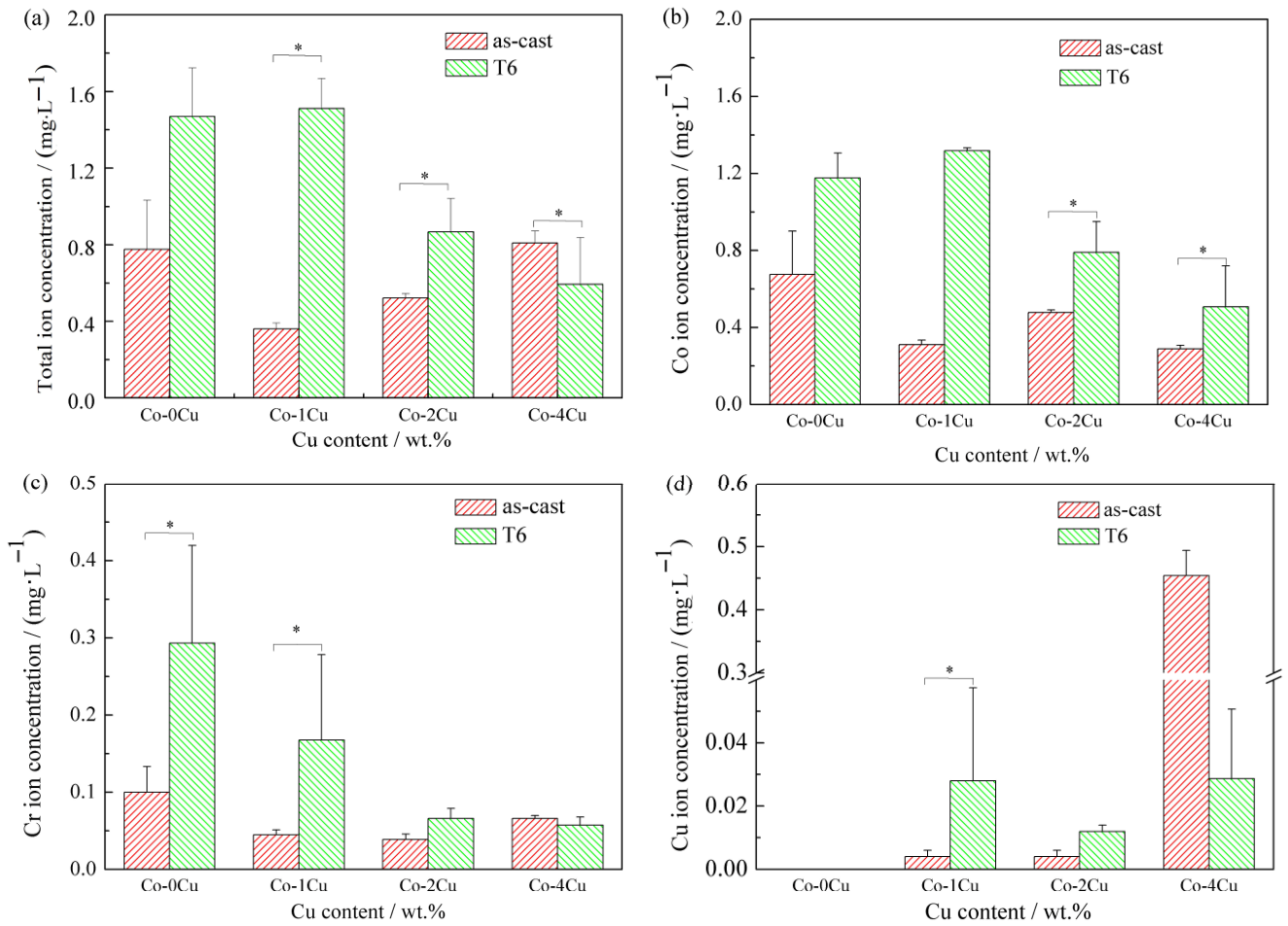
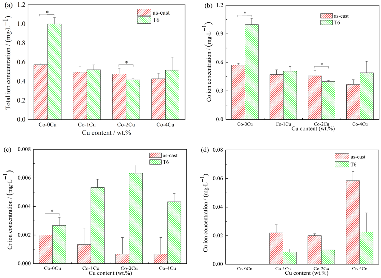
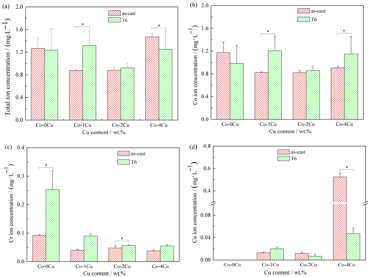
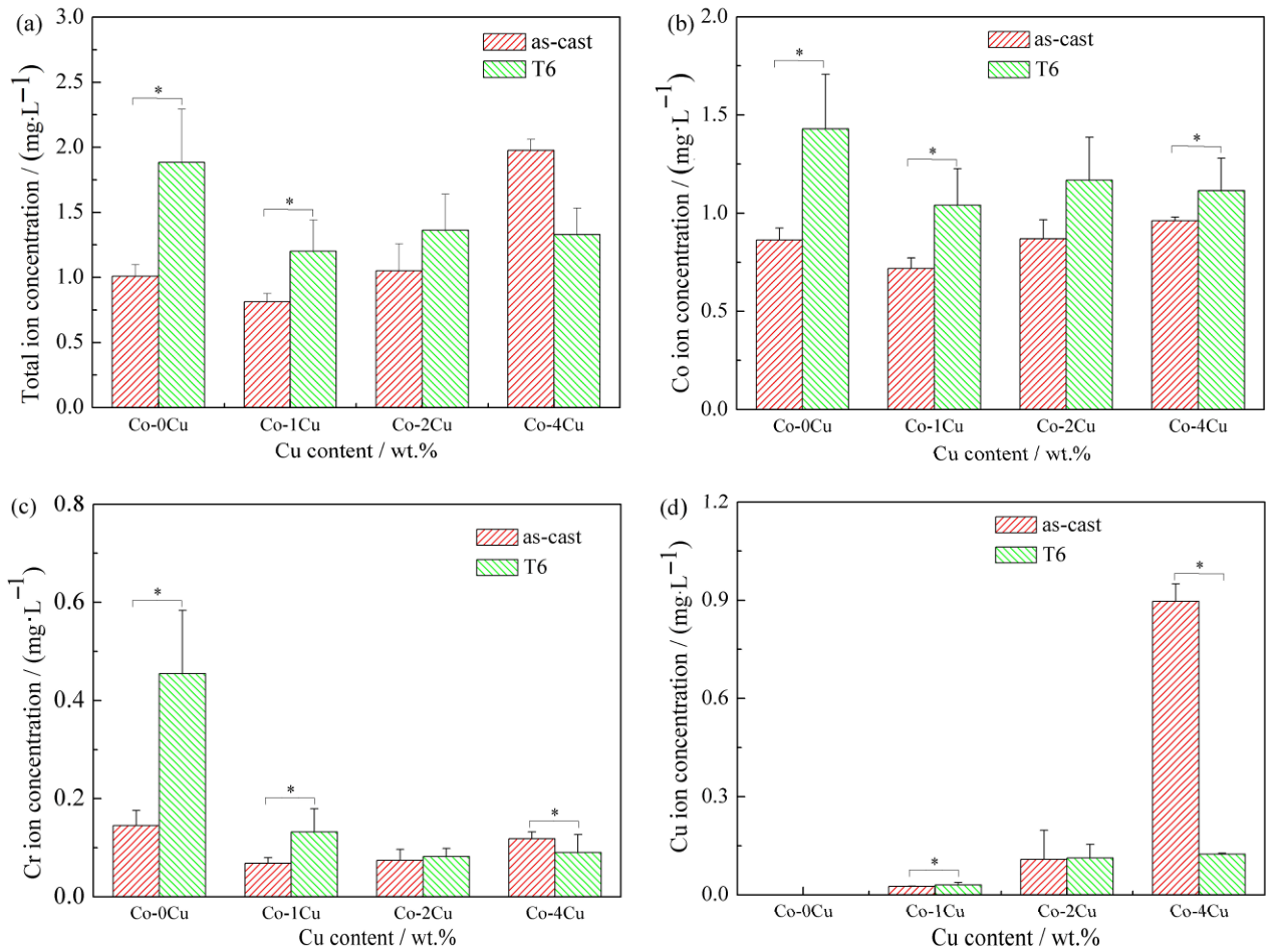
The concentrations of Co, Cr, and Cu ions released from Co-xCu alloys over seven days in Hanks’ solution, Saliva, Saliva+0.2F, and Saliva-pH3.5 solutions, along with the total amounts of metal ions, are shown in Figure 8, Figure 9, Figure 10 and Figure 11. In all four simulated solutions, the release of the Co ion predominantly influenced the dissolution behavior of Co-xCu alloys, with Co ion concentrations significantly exceeding those of Cr and Cu ions released. The addition of Cu resulted in the release of Cu ions, and the concentration of released Cu ions increased with increasing Cu content in the alloys. The highest concentration of the Cu ion released from Cu-containing cobalt alloys was 0.897 mg·L−1, observed in the as-cast Co-4Cu alloy in Saliva-pH3.5.
In Hanks’ solution, the concentration of Co ions released from Cu-free cobalt alloys exceeded that from Cu-containing cobalt alloys. The highest concentration of Co ions from Cu-containing alloys was 0.508 mg·L−1, which was observed in the Co-Cu(T6) alloy. Initially, the as-cast Cu-free cobalt alloy released more Cr ions than the Cu-containing alloys; however, after the heat treatment, the Cu-free alloy released fewer Cr ions. The maximum concentration of Cr ions released from Cu-containing cobalt alloys was 0.006 mg·L−1, which was noted in the Co-2Cu(T6) alloy. Additionally, the total concentration of Co, Cr, and Cu ions peaked at approximately 0.521 mg·L−1 in the Co-Cu(T6) alloy. This value was lower than that measured for the Cu-free cobalt alloys under the same conditions.
In Saliva, the concentration of Co ions released from as-cast Cu-free cobalt alloys was higher than that from as-cast Cu-containing cobalt alloys. Conversely, the Co ion concentration from Co-xCu(T6) alloys varied with the Cu element. The highest Co ion concentration (1.207 mg·L−1) was measured for the Co-Cu(T6) alloy. Additionally, the Cr ion concentration released from Cu-free cobalt alloys exceeded that from Cu-containing alloys. The peak Cr ion concentration from Cu-containing alloys was 0.09 mg·L−1, which occurred in the Co-Cu(T6) alloy. Furthermore, the maximum total concentration of Co, Cr, and Cu ions released from Cu-containing cobalt alloys reached 1.47 mg·L−1, as measured in the as-cast Co-Cu alloy. This value was slightly higher than that measured for Cu-free cobalt alloys under the same conditions (1.267mg·L−1).
In the Saliva+0.2F solution, Cu-free cobalt alloys released a higher concentration of Co ions, compared with as-cast Cu-containing cobalt alloys. Among Cu-containing alloys, as-cast cobalt alloys released fewer Co ions than their heat-treated counterparts. The highest Co ion concentration from Cu-containing cobalt alloys (1.317 mg·L−1) was measured in the Co-Cu(T6) alloy. Cu-free alloys also released more Cr ions than Cu-containing ones. Among Cu-containing alloys, the Co-Cu(T6) alloy had the highest Cr ion concentration at approximately 0.167 mg·L−1. The Co-Cu(T6) alloy also had the maximum total concentration of Co, Cr, and Cu ions from Cu-containing cobalt alloys, at about 1.512 mg·L−1. This was slightly higher than that measured for Cu-free cobalt alloys under the same conditions (1.469mg·L−1).
In Saliva-pH3.5, the concentration of Co ions released from as-cast cobalt alloys was lower than that from heat-treated cobalt alloys. Among Co-xCu(T6) alloys, Co ions were released in higher concentrations from Cu-free cobalt alloys, compared to Cu-containing ones. The highest concentration of Co ions from Cu-containing alloys, approximately 1.168 mg·L−1, was observed in the Co-2Cu (T6) alloy. Similarly, Cr ions were released in greater concentrations from Cu-free cobalt alloys than from those containing Cu. The peak concentration of Cr ions from Cu-containing alloys was about 0.132 mg·L−1, measured in the Co-Cu(T6) alloy. The highest total concentration of Co, Cr, and Cu ions released from a Cu-containing alloy, approximately 1.976 mg·L−1, was recorded in the as-cast Co-4Cu alloy. This was slightly higher than that measured for the heat-treated Cu-free cobalt alloys under the same conditions (1.884 mg·L−1). In addition, the total concentrations of Co, Cr, and Cu ions released from the heat-treated Co-1Cu and Co-4Cu alloys were 1.201 mg·L−1 and 1.329 mg·L−1, respectively, both of which were lower than the 1.884 mg·L−1 recorded for the Cu-free cobalt alloys.
In summary, the maximum concentrations of Co, Cr, and Cu ions released from Cu-containing cobalt alloys in simulated solutions were 1.317 mg·L−1, 0.167 mg·L−1, and 0.897 mg·L−1, respectively. The combined maximum concentration of Co, Cr, and Cu ions released from these alloys was 1.976 mg·L−1, which was slightly higher than that measured for the heat-treated Cu-free cobalt alloys (1.884 mg·L−1).

3.6. Surface Morphology

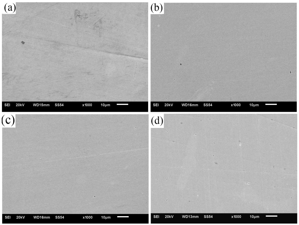
Although the corrosion resistance of Co-xCu alloys decreased with rising Cu content, it remained comparable to that of Cu-free cobalt alloys. Our previous research demonstrated that the antibacterial properties of Co-xCu alloys were improved as Cu content increased [22]. Additionally, the microstructure of as-cast cobalt alloys revealed numerous black pit-like defects caused by the detachment of Mo-rich phases, and these defects negatively affected the continuity and stability of the passive film. Using the Co-4Cu(T6) alloy as an example, we analyzed the surface morphology of the Cu-containing cobalt alloy after heat treatment and immersion in different simulated solutions, as shown in Figure 12. The surface of the Co-4Cu(T6) alloy exhibited only a few corrosion pits and no signs of general corrosion, indicating that the dense passive film on the alloy surface effectively prevented further corrosion.

4. Discussion

The corrosion resistance of Co-xCu alloys in solutions such as Hanks’ solution, Saliva, Saliva+0.2F, or Saliva-pH3.5 depends on the balance between the formation and dissolution of the passive film on the alloy surface. This balance is influenced by the chemical composition and microstructure of the Co-xCu alloys, as well as the physicochemical properties of the simulated solution. The passive film on the Co-xCu alloy surface primarily consists of Cr2O3/Cr(OH)3, with smaller amounts of cobalt oxide/hydroxide and molybdenum oxide/hydroxide [29]. Co-xCu alloys consist of a γ phase with a face-centered cubic (FCC) structure and an ε phase with a hexagonal close-packed (HCP) structure. The addition of Cu, however, can suppress martensitic transformation by significantly expanding the γ phase field [21,24].
The addition of Cu directly affects the alloy’s microstructure and corrosion resistance, exerting dual effects from the perspectives of chemical composition and microstructural segregation. On the one hand, the Cu-rich phases precipitated in the alloy can induce the enrichment of corrosion-resistant elements (such as Cr) on the alloy surface, enhance the stability of the passive film, and promote the formation of a dense and continuous passive film. For example, the formation of stable CuF2 products between Cu and fluoride ions can further “repair” the passive film on the surface of cobalt alloys. Meanwhile, Cu acts as an austenitizing element that can effectively prevent martensitic transformation, thereby avoiding structural inhomogeneities unfavorable to corrosion resistance. In addition, heat treatment optimizes the microstructure: it significantly reduces casting defects (such as circular pits caused by the detachment of Mo-rich phases) and replaces coarse grains with fine, smooth grains, forming a more uniform and complete microstructure. On the other hand, the standard electrode potentials of Co2+, Cr3+, and Cu2+ are presented in Equations (1)–(3). Notably, the standard electrode potentials of Co and Cr are significantly lower than that of Cu. This potential difference between Cu-rich phases and the alloy matrix triggers micro-galvanic couples, with the matrix acting as the low-potential anode, leading to accelerated dissolution. During the heat treatment process, accompanied by martensitic transformation, finer Cu-rich phases precipitate on the alloy matrix, which may form new micro-galvanic couples and further deteriorate the corrosion resistance. Moreover, with the increase in Cu content, Cu segregation at grain boundaries of as-cast cobalt alloys becomes significantly more pronounced, and the number of circular pits formed by the detachment of Mo-rich phases also increases. These defects provide pathways for corrosive media to penetrate, thereby weakening the alloy’s corrosion resistance. In conclusion, Cu addition exerts dual effects on the corrosion resistance of the alloy: the phase formation of Cu-rich phases and the inhibition of martensitic transformation improve corrosion resistance, whereas micro-galvanic couples and Cu segregation-induced surface defects degrade it.
Co2+ + 2e → Co E0 = −0.277 V,
Cr3+ + 3e → Cr E0 = −0.740 V,
Cu2+ + 2e → Cu E0 = 0.337 V,
The corrosion resistance of the alloy is closely related to the corrosivity of the service environment. For the copper-free cobalt alloy, the highest icorr was observed in Saliva+0.2F. This is because fluoride ions can adsorb on the alloy surface, inducing local breakdown of the passive film and accelerating localized dissolution. The neutral artificial Saliva, which contains specific ions, interacts with the passive film and reduces its protective ability, resulting in slightly higher corrosivity than that of Hanks’ solution. In Saliva-pH3.5, the high concentration of H+ promotes the rapid formation of passive films, which effectively inhibits anodic dissolution and thus leads to a lower corrosion current density than that in neutral Saliva. Hanks’ solution, as a mild neutral medium with weak corrosivity, has a slower rate of passive film formation and a relatively loose film structure, resulting in a slightly higher icorr than that in Saliva-pH3.5. The effect of heat treatment on the corrosion performance of the Cu-free cobalt alloy is environment-dependent. In Hanks’ solution and Saliva, heat treatment improves the uniformity of the alloy structure, reduces casting defects, and promotes the formation of a stable, dense, passive film, thereby reducing the icorr and improving corrosion resistance. However, in Saliva+0.2F and Saliva-pH3.5, the homogenized microstructure introduced by heat treatment provides more diffusion channels for aggressive ions (H+, F), accelerating anodic dissolution and leading to a higher icorr than that of the as-cast state.
With increasing Cu content from 0 to 4 wt%, the icorr values show a gradual increase across all media and heat treatment states. This slight elevation is attributed to the formation of Cu-rich precipitates, which may form micro-galvanic couples with the Co matrix and accelerate anodic dissolution. However, all values remain at the same order of magnitude, indicating that the overall corrosion resistance is not significantly compromised by Cu addition. With respect to Cu-containing cobalt alloys, the corrosion resistance after heat treatment is also closely related to the immersion medium. For Cu-containing cobalt alloys in Hanks’ solution, the improvement of the corrosion resistance after heat treatment is attributed to microstructural refinement and the enhanced stability of the passive film. For Cu-containing cobalt alloys in Saliva, the corrosion resistance decreased after heat treatment, mainly due to the precipitation of fine Cu-rich phases, which form micro-galvanic couples with the cobalt matrix, significantly enhancing local electrochemical activity and accelerating anodic dissolution. In addition, Cl and PO43− ions in Saliva further promote micro-galvanic couples between the precipitated phases and the cobalt matrix, weaken the protective effect of the passive film, and lead to a decrease in corrosion resistance after heat treatment. For Cu-containing cobalt alloys in Saliva-pH3.5, the corrosion resistance after heat treatment is slightly lower than that of as-cast Cu-containing cobalt alloys, which may be due to the enhanced micro-galvanic couples between the Cu-rich phases and the alloy matrix in the acidic environment. For Cu-containing cobalt alloys in Saliva+0.2F, the corrosion resistance after heat treatment was superior to that of the as-cast Cu-containing cobalt alloys. This can be explained by the combined effects of microstructure homogenization and the formation of Cu-enriched stable passive films. Cu released from the precipitates can react with fluoride ions to form stable Cu-containing fluorinated compounds on the alloy surface. These compounds effectively inhibit the further adsorption and penetration of fluoride ions, alleviating the destructive effect of fluoride on the passive film. Overall, despite the environment-dependent effect of heat treatment and the slight increase in icorr with Cu content, all Cu-containing cobalt alloys exhibit a corrosion resistance comparable to that of the Cu-free cobalt alloys in these media. This confirms that the addition of Cu does not significantly degrade the inherent corrosion resistance of the cobalt alloys.
Cu, an essential nutrient, has complex effects on the human body, offering both benefits and potential harm. Our previous research demonstrated that fine Cu phases significantly contributed to antibacterial properties. Released Cu ions can enhance the growth of human endothelial cells, inhibit excessive proliferation of arterial smooth muscle cells, and prevent platelet adhesion, thereby lowering thrombosis risk [14]. However, excessive Cu ions can be cytotoxic, causing symptoms like nausea, gastrointestinal distress, and neurodegenerative diseases [30]. Similarly, metal ions such as Co2+ and Cr3+ can be toxic if they exceed critical concentration thresholds. Among these, Co2+ is the most toxic, while Cr3+ is the least. The toxicity of metal ions is closely related to the concentration of ions released from Co-xCu alloys. Studies suggest that Co, Cr, and Cu ion concentrations should not exceed 50 µM (2.993 mg·L−1), 1000 µM (52.188 mg·L−1), and 50 µM (3.204 mg·L−1), respectively [31]. In our experiments, the maximum concentration of Cu ions released from Cu-containing cobalt alloys was approximately 0.897 mg·L−1 (14 µM), well below the 50 µM threshold. Similarly, the maximum concentration of Co ions was 1.317 mg·L−1 (22 µM), also far below 50 µM. For Cr ions, the maximum concentration released was 0.167 mg·L−1 (3.2 µM), significantly under the recommended 1000 µM limit. In summary, the maximum concentrations of Co, Cr, and Cu ions released from Co-Cu alloys were significantly lower than the recommended thresholds. In addition, the highest total ion concentration (1.976 mg·L−1) was observed for the as-cast Co-4Cu alloy in the acidic Saliva-pH3.5 solution. The stability of the passive film decreases in acidic environments, which accelerates the dissolution of Co-xCu alloys. The higher Cu content and its uneven distribution in the as-cast Co-4Cu alloy further exacerbate micro-galvanic couples, leading to the elevated release of Co, Cr, and Cu ions. The oral cavity may experience transient acidic conditions due to dietary exposure or local inflammation, thereby increasing the corrosion risk of metal biomaterials. However, this is slightly higher than measured for the heat-treated Cu-free cobalt alloys under the same conditions (1.884 mg·L−1). The total concentrations of Co, Cr, and Cu ions released from the heat-treated Co-Cu and Co-4Cu alloys in the Saliva-pH3.5 solution were 1.201 mg·L−1 and 1.329 mg·L−1, respectively, both of which were lower than the 1.884 mg·L−1 recorded for the Cu-free cobalt alloys. In summary, the concentrations of Co, Cr, and Cu metal ions released from the Cu-containing cobalt alloys were comparable to those from the Cu-free cobalt alloys. Moreover, through the combined optimization of Cu content and heat treatment, the resulting metal ion release levels can be even lower than those of the Cu-free cobalt alloy.

5. Conclusions

This study investigated the effect of Cu content on the corrosion resistance of Co-xCu alloys in different solutions, including Hanks’ solution, Saliva, Saliva+0.2F, and Saliva-pH3.5. It also evaluated the biosafety of the released metal ions. The conclusions are summarized as follows:
(1)
The corrosion resistance of Co-xCu alloys decreased with increasing Cu content, which was mainly attributed to galvanic corrosion between the alloy matrix and Cu-rich phases.
(2)
The synergistic effect of heat treatment and the addition of Cu significantly improved the resistance of cobalt alloys to fluoride ions.
(3)
The corrosion resistance of Cu-containing cobalt alloys was comparable to that of Cu-free cobalt alloys.
(4)
The maximum release concentrations of metal ions Co3+, Cr3+, and Cu2+ were measured as 1.317 mg·L−1, 0.167 mg·L−1, and 0.897 mg·L−1, respectively, and all values were lower than the corresponding recommended safety limits.

Author Contributions

Validation, W.L.; Investigation, W.L.; Data curation, X.W.; Writing—original draft, X.W.; Writing—review & editing, E.Z.; Project administration, E.Z. All authors have read and agreed to the published version of the manuscript.

Funding

The authors gratefully acknowledge the financial support from the National Foreign Expert Program of China (S20250108).

Data Availability Statement

The original contributions presented in this study are included in the article. Further inquiries can be directed to the corresponding author.

Acknowledgments

We are grateful to Her-Hsiung Huang for the fruitful discussion.

Conflicts of Interest

The authors declare that the research was conducted in the absence of any commercial or financial relationships that could be construed as a potential conflict of interest. The funders had no role in the design of the study; in the collection, analyses, or interpretation of data; in the writing of the manuscript; or in the decision to publish the results.

References

  1. Fellah, M.; Hezil, N.; Bouras, D.; Obrosov, A.; Mohammed, A.S.; Montagne, A.; Abd-Elmonem, A.; El Din, S.M.; Weiß, S. Structural, mechanical and tribological performance of a nano structured biomaterial Co–Cr–Mo alloy synthesized via mechanical alloying. J. Mater. Res. Technol. 2023, 25, 2152–2165. [Google Scholar]
  2. Niinomi, M. Recent metallic materials for biomedical applications. Metall. Mater. Trans. A 2002, 33, 477–486. [Google Scholar]
  3. Mischler, S.; Munoz, A.I. Wear of CoCrMo alloys used in metal-on-metal hip joints: A tribocorrosion appraisal. Wear 2013, 297, 1081–1094. [Google Scholar]
  4. Buciumeanu, M.; Bagheri, A.; Souza, J.C.M.; Silva, F.S.; Henriques, B. Tribocorrosion behavior of hot pressed CoCrMo alloys in artificial saliva. Tribol. Int. 2016, 97, 423–430. [Google Scholar]
  5. Saji, V.S.; Choe, H.-C. Electrochemical behavior of Co-Cr and Ni-Cr dental cast alloys. Trans. Nonferrous Met. Soc. China 2009, 19, 785–790. [Google Scholar]
  6. Hou, Y.; Li, Y.; Wang, F.; Zhang, C.; Koizumi, Y.; Chiba, A. Influence of Mo concentration on corrosion resistance to HF acid solution of Ni–Co–Cr–Mo alloys with and without Cu. Corros. Sci. 2015, 99, 185–193. [Google Scholar]
  7. Zhang, X.; Zagidulin, D.; Shoesmith, D.W. Characterization of film properties on the Ni Cr Mo Alloy C-2000. Electrochim. Acta 2013, 89, 814–822. [Google Scholar]
  8. Narayanan, D.; Liu, M.; Martinez, A.; Brooks, R.; Kuttolamadom, M.; Castaneda, H. Effect of Mo content on the passivation and localized corrosion behavior of laser engineered net shaped (LENS) Co-Cr-Mo alloys in a simulated physiological solution. Addit. Manuf. 2023, 77, 103812. [Google Scholar]
  9. Bojinov, M.; Fabricius, G.; Laitinen, T.; Makela, K.; Saario, T.; Sundholm, G. Influence of molybdenum on the conduction mechanism inpassive films on iron–chromium alloys in sulphuric acidsolution. Electrochim. Acta 2001, 46, 1339–1358. [Google Scholar]
  10. Ren, L.; Memarzadeh, K.; Zhang, S.; Sun, Z.; Yang, C.; Ren, G.; Allaker, R.P.; Yang, K. A novel coping metal material CoCrCu alloy fabricated by selective laser melting with antimicrobial and antibiofilm properties. Mater. Sci. Eng. C 2016, 67, 461–467. [Google Scholar]
  11. Zaura, E.; Keijser, B.J.F.; Huse, S.M.; Crielaard, W. Defining the healthy “core microbiome” of oral microbial communities. BMC Microbiol. 2009, 9, 259. [Google Scholar] [CrossRef] [Scilit]
  12. Fu, S.; Zhang, Y.; Yang, Y.; Liu, X.; Zhang, X.; Yang, L.; Xu, D.; Wang, F.; Qin, G.; Zhang, E. An antibacterial mechanism of titanium alloy based on micro-area potential difference induced reactive oxygen species. J. Mater. Sci. Technol. 2022, 119, 75–86. [Google Scholar] [CrossRef] [Scilit]
  13. Arciola, C.R.; Campoccia, D.; Montanaro, L. Implant infections: Adhesion, biofilm formation and immune evasion. Nat. Rev. Microbiol. 2018, 16, 397–409. [Google Scholar] [CrossRef] [Scilit]
  14. Iyer, M.; Anand, U.; Thiruvenkataswamy, S.; Babu, H.W.S.; Narayanasamy, A.; Prajapati, V.K.; Tiwari, C.K.; Gopalakrishnan, A.V.; Bontempi, E.; Sonne, C.; et al. A review of chromium (Cr) epigenetic toxicity and health hazards. Sci. Total Environ. 2023, 882, 163483. [Google Scholar] [CrossRef] [Scilit]
  15. Tapiero, H.; Townsend, D.M.; Tew, K.D. Trace elements in human physiology and pathology. Copper. Biomed. Pharmacother. 2003, 57, 386–398. [Google Scholar] [CrossRef] [Scilit]
  16. Liu, R.; Tang, Y.; Zeng, L.; Zhao, Y.; Ma, Z.; Sun, Z.; Xiang, L.; Ren, L.; Yang, K. In vitro and in vivo studies of anti-bacterial copper-bearing titanium alloy for dental application. Dent. Mater. 2018, 34, 1112–1126. [Google Scholar] [CrossRef] [Scilit]
  17. Liu, H.; Xu, D.; Yang, K.; Liu, H.; Cheng, Y.F. Corrosion of antibacterial Cu-bearing 316L stainless steels in the presence of sulfate reducing bacteria. Corros. Sci. 2018, 132, 46–55. [Google Scholar] [CrossRef] [Scilit]
  18. Mahmoudi, P.; Akbarpour, M.R.; Lakeh, H.B.; Jing, F.; Hadidi, M.R.; Akhavan, B. Antibacterial Ti–Cu implants: A critical review on mechanisms of action. Mater. Today Bio 2022, 17, 100447. [Google Scholar] [CrossRef] [Scilit]
  19. Lu, Y.; Zhao, C.; Ren, L.; Guo, S.; Gan, Y.; Yang, C.; Wu, S.; Lin, J.; Huang, T.; Yang, K.; et al. Preliminary assessment of metal-porcelain bonding strength of CoCrW alloy after 3 wt.% Cu addition. Mater. Sci. Eng. C 2016, 63, 37–45. [Google Scholar] [CrossRef] [Scilit]
  20. Wang, S.; Yang, C.; Ren, L.; Shen, M.; Yang, K. Study on antibacterial performance of Cu-bearing cobalt-based alloy. Mater. Lett. 2014, 129, 88–90. [Google Scholar] [CrossRef] [Scilit]
  21. Gan, Y.; Zhou, M.; Ji, C.; Huang, G.; Chen, Y.; Li, L.; Huang, T.; Lu, Y.; Lin, J. Tailoring the tribology property and corrosion resistance of selective laser melted CoCrMo alloys by varying copper content. Mater. Des. 2023, 228, 111869. [Google Scholar] [CrossRef] [Scilit]
  22. Zhang, E.; Liu, C. A new antibacterial Co-Cr-Mo-Cu alloy: Preparation, biocorrosion, mechanical and antibacterial property. Mater. Sci. Eng. C 2016, 69, 134–143. [Google Scholar] [CrossRef] [Scilit]
  23. Moradi, S.M.; Zangeneh, S.; Vardak, S.; Bahrami, R. New Co-Cr-Mo-Nb-Cu alloy for implant applications: Properties characterization. J. Alloys Compd. 2022, 925, 166387. [Google Scholar] [CrossRef] [Scilit]
  24. Wang, R.; Qin, G.; Zhang, E. Effect of Cu on Martensite Transformation of CoCrMo alloy for biomedical application. J. Mater. Sci. Technol. 2020, 52, 127–135. [Google Scholar] [CrossRef] [Scilit]
  25. Zhang, E.; Ge, Y.; Qin, G. Hot deformation behavior of an antibacterial Co–29Cr–6Mo–1.8Cu alloy and its effect on mechanical property and corrosion resistance. J. Mater. Sci. Technol. 2018, 34, 523–533. [Google Scholar] [CrossRef] [Scilit]
  26. ISO 5832-4:2024; Implants for Surgery—Metallic Materials Part 4: Cobalt-Chromium-Molybdenum Casting Alloy. International Organization for Standardization: Geneva, Switzerland, 2024.
  27. ISO 10271:2001; Dentistry Metallic Materials—Corrosion Test Method. International Organization for Standardization: Geneva, Switzerland, 2001.
  28. ISO 10993-12:2021; Biological Evaluation of Medical Devices—Part 12: Sample Preparation and Reference Materials. International Organization for Standardization: Geneva, Switzerland, 2021; p. 21.
  29. Bao, Y.; Muñoz, A.I.; Jolles, B.M.; Mischler, S. Assessment of in-vivo corrosion of Ti and CoCrMo joint implants by electrochemical measurements in human synovial liquids. Electrochim. Acta 2024, 476, 143708. [Google Scholar] [CrossRef] [Scilit]
  30. Wang, R.; Wang, R.; Chen, D.; Qin, G.; Zhang, E. Novel CoCrWNi alloys with Cu addition: Microstructure, mechanical properties, corrosion properties and biocompatibility. J. Alloys Compd. 2020, 824, 153924. [Google Scholar] [CrossRef] [Scilit]
  31. Issa, Y.; Brunton, P.; Waters, C.M.; Watts, D.C. Cytotoxicity of metal ions to human oligodendroglial cells and human gingival fibroblasts assessed by mitochondrial dehydrogenase activity. Dent. Mater. 2008, 24, 281–287. [Google Scholar] [CrossRef] [Scilit]
Figure 1. SEM images of the as-cast Co-xCu alloys: (a) Co-0Cu, (b) Co-2Cu, and (c) Co-4Cu.
Metals 16 00498 g001
Figure 2. SEM images of Co-xCu (T6) alloys: (a) Co-0Cu, (b) Co-2Cu, and (c) Co-4Cu.
Metals 16 00498 g002
Figure 3. OCP curves of as-cast and T6 Co-xCu alloys (x = 0 and 4) in Hanks’ solution.
Metals 16 00498 g003
Figure 4. Typical potentiodynamic polarization curves of Co-xCu alloys: (a) as-cast state in Hanks’ solution; (b) heat-treated in Saliva+0.2F.
Metals 16 00498 g004
Figure 5. Experimental and fitted Nyquist diagrams of as-cast Co-xCu alloys (a,c,e,g) and Co-xCu(T6) alloys (b,d,f,h) in Hanks’, Saliva, Saliva+0.2F, and Saliva-pH3.5.
Metals 16 00498 g005
Figure 6. Experimental and simulated Bode plots for as-cast Co-xCu alloys (a,c,e,g) and Co-xCu(T6) alloys (b,d,f,h) in various solutions: Hanks’, Saliva, Saliva+0.2F, and Saliva-pH3.5. An equivalent circuit is inserted in (h) as an example.
Metals 16 00498 g006
Figure 7. Equivalent circuit parameters Rs (a,b), Rb (c,d), Qb (e,f), and n (g,h) for EIS spectra of Co-xCu alloys in various simulated solutions.
Metals 16 00498 g007
Figure 8. Concentration of Co, Cr, and Cu metal ions released from cobalt alloys in Hanks’ solution: (a) total concentration, (b) Co, (c) Cr, and (d) Cu. * p < 0.05, indicating a statistically significant difference between the as-cast and T6 conditions for the same alloys.
Metals 16 00498 g008
Figure 9. Concentration of Co, Cr, and Cu metal ions released from cobalt alloys in Saliva. (a) total concentration, (b) Co, (c) Cr, and (d) Cu. * p < 0.05, indicating a statistically significant difference between the as-cast and T6 conditions for the same alloys.
Metals 16 00498 g009
Figure 10. Concentrations of Co, Cr, and Cu metal ions released from cobalt alloys in Saliva+0.2F: (a) total concentration, (b) Co, (c) Cr, and (d) Cu. * p < 0.05, indicating a statistically significant difference between the as-cast and T6 conditions for the same alloys.
Metals 16 00498 g010
Figure 11. Concentrations of Co, Cr, and Cu metal ions released from cobalt alloys in Saliva-pH3.5: (a) total concentration, (b) Co, (c) Cr, and (d) Cu. * p < 0.05, indicating a statistically significant difference between the as-cast and T6 conditions for the same alloys.
Metals 16 00498 g011
Figure 12. Surface morphology of the Co-4Cu (T6) alloy after seven days in different solutions: (a) Hanks’, (b) Saliva, (c) Saliva+0.2F, and (d) Saliva-pH3.5.
Metals 16 00498 g012
Table 1. Chemical composition of Co-xCu alloys (mass fraction, ±0.1 wt%).
AlloysCrMoCuCo
NominalMeasuredNominalMeasuredNominalMeasured
Co-0Cu2928.966.1--Balance
Co-1Cu10.8Balance
Co-2Cu21.8Balance
Co-4Cu43.6Balance
Table 2. Chemical composition (g/L) and pH of the test solutions.
SolutionHanks’SalivaSaliva+0.2FSaliva-pH3.5
Lactic acid-101010
NaCl85.855.855.85
NaF--2-
KCl0.4---
CaCl2·H2O0.140.140.140.14
NaHCO30.35---
MgSO4·7H2O0.06---
MgCl2·6H2O0.1---
Na2HPO40.060.060.060.06
KH2PO40.06---
Glucose1---
pH7.46.86.83.5
Table 3. EDS analysis of different points (A, B, C) in Co-xCu alloys.
ElementsMass Fraction (wt%)
Point APoint BPoint C
Co64.6663.9363.03
Cr29.7828.5927.87
Mo5.565.655.33
Cu01.833.77
Table 4. Summary of steady-state OCP values of CoCrMo-xCu alloys in different biological solutions.
SolutionConditionCo-0CuCo-1CuCo-2CuCo-4Cu
Hanks’ solutionAs-cast−380 ± 12−363 ± 29−300 ± 33−249 ± 40
T6−364 ± 18−292 ± 21−256 ± 22−244 ± 13
SalivaAs-cast−320 ± 18−253 ± 28−247 ± 24−227 ± 12
T6−313 ± 19−240 ± 13−197 ± 43−218 ± 28
Saliva+0.2FAs-cast−303 ± 32−250 ± 1−243 ± 14−204 ± 20
T6−338 ± 10−264 ± 8−217 ± 24−194 ± 20
Saliva-pH3.5As-cast−206 ± 10−119 ± 10−93 ± 16−103 ± 24
T6−211 ± 6−171 ± 36−112 ± 9−95 ± 7
Table 5. Ecorr of Co-xCu alloys in different simulated biological solutions (mV).
Simulated SolutionsHeat TreatmentCo-0CuCo-1CuCo-2CuCo-4Cu
Hanks’As-cast−409 ± 16−288 ± 25−278 ± 24−247 ± 44
T6−347 ± 16−287 ± 14−253 ± 22−246 ± 28
SalivaAs-cast−371 ± 47−265 ± 14−238 ± 38−230 ± 26
T6−331 ± 28−253 ± 12−225 ± 18−204 ± 36
Saliva+0.2FAs-cast−336 ± 34−235 ± 12−232 ± 9−208 ± 23
T6−394 ± 22−273 ± 15−198 ± 24−173 ± 16
Saliva-pH3.5As-cast−289 ± 15−154 ±9−122 ± 31−110 ± 21
T6−301 ± 32−240 ±9−130 ± 12−107 ± 47
Table 6. icorr of Co-xCu alloys in different simulated biological solutions (nA·cm−2).
Simulated SolutionsHeat TreatmentCo-0CuCo-1CuCo-2CuCo-4Cu
Hanks’As-cast38.54 ± 10.0137.32 ± 6.9377.21 ± 12.2285.33 ± 13.55
T632.73 ± 18.3233.51 ± 14.2457.15 ± 22.5674.18 ± 16.77
SalivaAs-cast51.52 ± 12.2157.72 ± 17.3368.15 ± 1.9375.41 ± 23.66
T645.54 ± 6.0561.32 ± 10.9275.25 ± 2.5786.11 ± 16.97
Saliva+0.2FAs-cast53.42 ± 6.1370.15 ± 4.9192.82 ± 5.91105.41 ± 3.45
T655.21 ± 2.4265.23 ± 13.3788.31 ± 2.96100.96 ± 6.11
Saliva-pH3.5As-cast34.93 ± 6.9345.61 ± 2.9252.05 ± 4.4463.76 ± 2.01
T646.26 ± 7.0847.3 ± 8.7457.12 ± 3.4166.81 ± 2.25
Disclaimer/Publisher’s Note: The statements, opinions and data contained in all publications are solely those of the individual author(s) and contributor(s) and not of MDPI and/or the editor(s). MDPI and/or the editor(s) disclaim responsibility for any injury to people or property resulting from any ideas, methods, instructions or products referred to in the content.