Abstract
This study reports the hydrothermal synthesis, characterization, and Fenton-like catalytic performance of CeO2–CoFe2O4 nanocomposites for degrading Congo Red (CR) dye and the oxytetracycline (OTC) antibiotic. A series of Ce-doped cobalt ferrite samples was prepared using a hydrothermal reaction. Additionally, the 50Ce-CFO sample was further activated with H2O2 treatment. XRD, FTIR, and SEM analyses confirmed the formation of a spinel phase alongside segregated CeO2, which acts as a grain-growth inhibitor. The increased Ce content promotes particle amorphization. FTIR showed changes in the intensity of the M–O stretching band, indicating Ce-induced bond polarization in the spinel lattice. In H2O2 decomposition tests, the 50Ce-CFO catalyst fully decomposes H2O2 in 160 min, while the activated sample completes it in 125 min. Fenton-like degradation of CR and OTC by untreated and activated 50Ce-CFO sample followed pseudo-first-order kinetics. Catalyst stability was confirmed using post-reaction XRD, FTIR, and SEM analyses. Incorporation of CeO2 into CoFe2O4 refines the crystallite size, increases the BET surface area, and enhances adsorption capacity, while the Ce4+/Ce3+ redox couple promotes reactive oxygen species generation. Owing to this dual structural and catalytic role, the CeO2-CoFe2O4 composites exhibit significantly improved Fenton-like catalytic activity, enabling the efficient degradation of organic pollutants.
1. Introduction
Recently, spinel ferrite catalysts have attracted considerable attention due to their advantages over homogeneous Fenton catalysts [1,2]. They are thermally stable, chemically resistant in aggressive environments (which is important when purifying water with varying pH), exhibit long-term activity and stability, and possess magnetic properties that enable their easy removal using an applied magnetic field [3,4,5,6]. A unique feature of ferrite materials is their ability to be modified, forming composite materials and a wide range of cation-substituted spinels with controlled morphology and properties [7,8]. Among various cations, cerium ions occupy a special place, since Ce-containing spinel ferrites and their composites have proven themselves as active catalysts for catalytic water purification [9]. It is worth noting that CeO2 oxide, one of the most common rare-earth oxides, due to the presence of the Ce4+/Ce3+ redox pair, is well known for its ability to effectively catalyze various processes, in particular, advanced oxidation processes used for water purification [10,11,12]. In heterostructures and composites, CeO2 can accelerate electron transfer and increase the catalytic activity of materials. Most of the researches present the results of studies indicating that this occurs due to the formation of oxygen vacancies, which contribute to better charge carrier transfer [13,14]. As an example, CuFe2O4/CoFe2O4/CeO2 nanocomposites were successfully synthesized via a self-initiated combustion method [15]. CoFe2O4/CeO2 nanocomposites were synthesized by combining precipitation and hydrothermal methods [16]. Pure CeO2 was first prepared using cerium(III) nitrate hexahydrate in ethylene glycol, with ammonium hydroxide as the precipitating agent. The resulting CeO2 powders were then introduced into an aqueous solution containing cobalt nitrate hexahydrate and iron nitrate nonahydrate to form CoFe2O4/CeO2 nanocomposites. These CoFe2O4/CeO2 nanocomposites exhibit promising properties as magnetically separable photocatalysts for the degradation of organic pollutants in aqueous systems [16]. A novel eco-friendly composite adsorbent, Ce-BTC@MCC, was synthesized by integrating a cerium-based metal-organic framework with microcrystalline cellulose (MCC), and its structure and performance were systematically characterized in the study [17].
It should be noted that composites of CeO2 with spinel compounds, including ferrites, are widely studied as membranes for solid oxide fuel cells [13], catalysts for water gas shift reactions [18], electrocatalysts for water-splitting [19,20], and, importantly, as adsorbents, photocatalysts, and Fenton-like catalysts for the purification of polluted waters [21,22,23]. CeO2 is notable for its excellent ability to activate hydrogen peroxide and peroxomonosulfate, exhibiting peroxidase-like, catalase-like, and haloperoxidase-like activities [24,25,26]. For example, in a study by [27], the Fe3O4/CeO2 nanocomposite produced through the impregnation method was evaluated as a catalyst for degrading 4-chlorophenol in aqueous solutions with hydrogen peroxide. It demonstrated a pseudo-first-order rate constant of 0.11 min−1 at 30 °C and pH 3.0, with a hydrogen peroxide utilization efficiency reaching 79.2%. This nanocomposite maintained high stability even after six reaction cycles. The authors proposed a mechanism for hydroxyl radical generation that comprises surface-bound •OHads and free •OHfree hydroxyl radicals. These radicals form due to the interaction of Fe2+ and Ce3+ ions with H2O2 on the catalyst surface, and iron ions are subsequently washed out from the catalyst [27]. Another study [28] involved CoFe2O4-CeO2 composites synthesized via an impregnation-hydrothermal method, which were used as catalysts for activating peroxymonosulfate in the removal of atrazine. A complete oxidation of atrazine was achieved using a 15%CoFe2O4-CeO2/PMS system, with a pseudo-first-order rate constant of 0.224 min−1. The findings indicated that the redox potential of the Ce(IV)/Ce(III) couple is higher than that of Fe(II)/Fe(III), making electron transfer from ≡Fe(II)–OH− to ≡Ce(IV)–OH− thermodynamically favorable, thereby promoting atrazine degradation [28].
The incorporation of CeO2 into iron-based catalysts has been shown to significantly enhance catalytic performance through synergistic redox interactions between Ce4+/Ce3+ and Fe3+/Fe2+ couples, promoting efficient H2O2 activation. Several studies have reported that oxygen vacancies (OVs) and surface-bound HO• radicals play critical roles in driving pollutant degradation in such systems. For example, in Fe2O3-CeO2, catalyst surface OVs were shown to enhance HO• formation under O2, and the surface-bounded HO• radicals played a crucial role in SMR degradation in the Fenton-like system [29]. In another study [30], the novel Fe0-Fe3O4/CeO2/C material was designed as an efficient electro-Fenton reagent that exhibits excellent electrocatalytic activity and good stability. It was investigated that the cycling of Ce4+/Ce3+ and Fe3+/Fe2+ redox couples promotes the generation of HO•. The authors in the study [30] used Mulliken charge distribution analysis to qualitatively assess the electron transport path and hypothesize that electrons are transferred from C and Fe0-Fe3O4 to Ce; therefore, CeO2 served as the electron-rich micro-region for the Fenton-like reaction. In UV–Fenton systems using Fe1Ce1Ti1 composites, ≡Fe2+ surface sites reacted with H2O2 to produce adsorbed HO•, while thermodynamically favorable electron transfer from ≡Fe2+ to ≡Ce4+ supported continuous radical generation through Ce–Fe redox cycling [31]. Furthermore, chitosan/FeOOH/CeO2 microspheres synthesized via a one-step co-precipitation method demonstrated high surface Ce(III) content (38.4%), where Ce(III) acted as the primary active center responsible for H2O2 adsorption and activation, while the presence of Fe(II) at the heterojunction facilitated the regeneration of Ce(III) [32]. Thus, these findings collectively underline the synergistic effects between cerium and iron species in Fenton-like systems and the essential contribution of CeO2 to enhancing catalytic activity and stability.
To the best of our knowledge, the catalytic system CeO2-CoFe2O4/H2O2 for the degradation of Congo Red and oxytetracycline has not been previously reported. Thus, this study aims to synthesize a cobalt ferrite-CeO2 composite catalyst using the hydrothermal method and to explore its effectiveness in activating hydrogen peroxide to degrade Congo Red dye and the oxytetracycline antibiotic. Unlike earlier studies, where CeO2 and CoFe2O4 were often prepared separately and then combined, in our approach, cerium oxide is formed in situ simultaneously with the spinel phase during hydrothermal synthesis.
2. Materials and Methods
2.1. Chemicals
Cobalt(II) nitrate hexahydrate Co(NO3)2·6H2O (CAS 10026-22-9), iron(III) nitrate nonahydrate Fe(NO3)3·9H2O (CAS 7782-61-8), and cerium(III) nitrate hexahydrate Ce(NO3)3·6H2O (CAS 10294-41-4) were purchased from Merck (Darmstadt, Germany). Sodium hydroxide, NaOH, was purchased from Chemland (Stargard, Poland). Congo Red C32H22N6Na2O6S2 (CAS 573-58-0) and oxytetracycline hydrochloride C22H24N2O9·HCl (CAS 2058-46-0) were purchased from Merck. Hydrogen peroxide (30% solution) (CAS 7722-84-1). Deionized water used in the experiments was sourced using the Hydrolab HLP system (Hydrolab, Straszyn, Poland).
2.2. Synthesis
The synthesis of Ce-containing cobalt ferrite composites was carried out using a hydrothermal method. The molar ratios of Co:Fe:Ce were 1:2:0, 1:1.75:0.25, and 1:1.5:0.5. The exact masses of the corresponding precursors are presented in Table S1. First, the weighed amounts of metal salts were added to 50 mL of distilled water and stirred for 1 h. Then, 70 mL of 5M solution of sodium hydroxide was added dropwise over 2 h to the metal salt solution while heating at 60 °C with continuous stirring at 600 rpm. The mixture was then stirred overnight at 60 °C. The final solution was transferred into a Teflon autoclave and heated at 180 °C for 12 h. The autoclave was allowed to cool naturally. Finally, a black magnetic powder was obtained, which was centrifuged and washed several times with distilled water until the pH reached 7. The collected powder was dried at 50 °C. The dried powder was then annealed at 500 °C for 2 h with a heating rate of 2.5 °C/min. The annealed samples were labeled as CFO, 25Ce-CFO, and 50Ce-CFO. The 50Ce-CFO sample was further treated with 10 wt% H2O2 for 1 h to enhance surface activation. The treatment was conducted in a beaker placed in an ice bath to control the exothermic reaction and maintain a low temperature during the oxidation process.
2.3. Characterization Techniques
X-ray diffraction (XRD) patterns were recorded using a Rigaku MiniFlex 600 diffractometer (Rigaku, Tokyo, Japan) with Cu Kα radiation (λ = 1.5406 Å) to determine the crystalline structure and phase composition of the synthesized Ce-containing cobalt ferrite composites. The scanning step size was 0.02° (2θ), and the scan rate was 1°/min, covering a 2θ range suitable for identifying ferrite phases. The average crystallite size of the cobalt ferrite and CeO2 phases was estimated using the Scherrer equation, applied to the (311) and (111) diffraction peaks, respectively. The morphology and surface features of the samples were examined using a Helios 5 Hydra DualBeam microscope (Thermo Fisher Scientific, Waltham, MA, USA). ATR-FTIR spectra were collected with a Thermo Scientific Nicolet Summit FTIR spectrometer equipped with a diamond ATR window (Thermo Fisher Scientific, Waltham, MA, USA), using the OMNIC Paradigm software (v.2.2). The spectra were recorded over the wavenumber range of 4000–400 cm−1 with a resolution of 4 cm−1 and 64 scans to identify functional groups and verify the presence of characteristic metal–oxygen bonds in the ferrite composites. The specific surface area of the catalysts was determined at −196 °C using N2 adsorption/desorption isotherms (Autosorb iQ-MP, Quantachrome instruments, Boynton Beach, FL, USA). The samples were outgassed under vacuum at 180 °C for 24 h before the analysis.
2.4. Hydrogen Peroxide Decomposition Tests
Experiments on hydrogen peroxide decomposition were performed in batch mode using a 10 mM H2O2 solution with 1 g/L of catalyst. The reaction mixtures were maintained at continuous stirring at 600 rpm to ensure homogeneous dispersion of the catalyst. Aliquots (1 mL) were withdrawn at predetermined time intervals, and the residual concentration of H2O2 was determined by measuring absorbance at 240 nm using a UV-1900 Shimadzu UV–vis spectrophotometer (Shimadzu, Kyoto, Japan), considering the molar absorption coefficient of H2O2 at 240 nm as ε = 43.6 M−1·cm−1. The experimental data were analyzed through nonlinear fitting with the pseudo-first-order (PFO) kinetic model [33].
2.5. Fenton-like Oxidation of Congo Red and Oxytetracycline
Fenton-like degradation experiments for Congo Red (CR) and oxytetracycline (OTC) were conducted using the synthesized catalysts, with hydrogen peroxide serving as the oxidant. The reactions were carried out in solutions containing 10 mg/L of CR (or OTC), 10 mM H2O2, and 1 g/L of catalyst. The reaction mixtures were maintained at a constant temperature with continuous stirring. At predetermined time intervals, aliquots were collected, and the residual concentration of CR or OTC was determined using a UV-1900 Shimadzu UV–vis spectrophotometer by measuring absorbance at 498 nm or 354 nm, respectively. The experimental data were analyzed through nonlinear fitting with the pseudo-first-order (PFO) kinetic model [33].
3. Results
3.1. Structure and Morphology Studies
XRD patterns of as-synthesized and annealed at 500 °C samples are presented in Figure 1. The as-synthesized CFO sample exhibits a well-defined cubic spinel structure with a lattice constant of 8.334 ± 0.003 Å. The obtained value is reduced comparatively to data reported for bulk cobalt ferrite (typically ranging from 8.376 to 8.390 Å) [8], but the lattice constant decrease may be attributed to the nanoscale nature of the synthesized material. In our case, the average crystallite size of CFO calculated using the Scherrer equation is about 18 nm, which can induce lattice change relative to bulk ferrite. Cobalt ferrite nanoparticles with a size below 11–16 nm exhibit lattice constants of about 8.36 Å, due to surface stress and altered cation distribution at the nanoscale [6]. It can be stated that a decrease in cobalt ferrite crystallite size is accompanied by a reduction in the lattice parameter [34]. The close lattice parameter (8.32 Å) was observed for cobalt ferrite, which was prepared by a co-precipitation method [35]. Thus, surface stress induced by Laplacian pressure in nanoscale particles can lead to a reduction in the lattice constant. The primary phases identified for 25Ce-CFO material are CoFe2O4 and CeO2, both crystallizing in a cubic structure (Fd-3m space group) (Figure 1a). Notably, the diffraction pattern also reveals sharp and intense peaks assigned to goethite (α-FeOOH), suggesting the formation of a highly crystalline goethite phase within the composite. The 50Ce-CFO sample contains two crystalline phases—CoFe2O4 and CeO2, both exhibiting a cubic structure with space group Fd-3m (Figure 1a). The increased cerium content suppresses the formation of secondary iron oxide phases during synthesis. The average crystallite size of the cobalt ferrite phase in the 50Ce-CFO sample was estimated to be approximately 17 nm, indicating the formation of well-defined nanocrystals. In contrast, the CeO2 phase exhibits significantly smaller particle size, with an average crystallite size of about 4 nm. This difference suggests that cerium oxide forms as highly dispersed nanoclusters on the surface or at the grain boundaries of the spinel matrix, which may enhance the interfacial contact and contribute to the synergistic catalytic behavior of the composite.
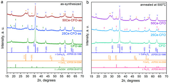
Figure 1.
XRD patterns of (a) as-synthesized and (b) annealed at 500 °C samples.
Annealing at 500 °C led to an increase in the lattice parameter of the CFO sample to 8.347 ± 0.002 Å, accompanied by a growth in crystallite size to 20 nm (Figure 1b). This behavior can be attributed to thermal relaxation of internal strain and improved crystallinity of the spinel phase. After annealing at 500 °C, the 25Ce-CFO sample consists of CoFe2O4, CeO2, and hematite (α-Fe2O3) (Figure 1b). B. Liu et al. [23] obtained similar results, where the hematite phase is formed in the system with Fe excess. Annealing does not change the phase composition of the 50Ce-CFO material, which consists of two crystalline phases: CoFe2O4 and CeO2 (Figure 1b). An increase in the lattice parameter of the cobalt ferrite phase is observed from 8.326 ± 0.009 Å to 8.340 ± 0.003 Å, accompanied by a minor reduction in crystallite size to approximately 15 nm. The increase in lattice parameter is caused by the relaxation of microstrain and surface stresses, as well as partial cation redistribution due to annealing at 500 °C. The similar Δa values for CFO and 50Ce-CFO support this strain-driven origin. The reduction in crystallite size of 50Ce-CFO can be attributed to Zener pinning [36]: CeO2 nanodomains segregated at spinel grain boundaries form a pinning pressure, which suppresses boundary migration and may cause fragmentation of ferrite crystallites, which is observed as an integral decrease in crystallite size.
Figure 2 demonstrates the surface morphology of as-synthesized (Figure 2a–c) and annealed at 500 °C (Figure 2d–f) Ce-containing composites. All hydrothermally synthesized samples display a round particle shape, exhibiting agglomeration into aggregates due to the magnetic characteristics of cobalt ferrite. The presence of Ce ions increases the amount of aggregates, likely due to the formation of the CeO2 phase. Additionally, the use of hydrothermal synthesis is advantageous for producing uniform nanoparticles, as the samples retain sizes in the 25–50 nm range even after annealing at 500 °C (Figure 2d–f).
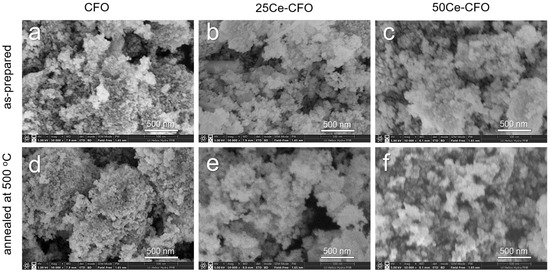
Figure 2.
The surface morphology of Ce-containing cobalt ferrites: (a–c) as-synthesized and (d–f) annealed at 500 °C.
After annealing, the sample CFO still exhibits well-defined small nanograins that tend to aggregate (Figure 2d). Its structure remains dense. Conversely, the 25Ce-CFO sample after annealing at 500 °C (Figure 2e) exhibits a more porous structure, with less densely packed particles. The individual particles appear slightly larger than those found in pure cobalt ferrite. This could be attributed to the presence of Ce ions, which may slow down crystallization and result in less compact aggregation, creating a looser morphology. The 50Ce-CFO sample (Figure 2f) is significantly more amorphous. The SEM image reveals a smoother texture rather than individual particles, likely due to considerable distortion in the cobalt ferrite crystal lattice caused by the higher Ce content. Given that CeO2 segregates as a separate phase on the surface of cobalt ferrite, it acts as a growth inhibitor and restricts grain growth.
The FTIR spectra of as-synthesized and annealed Ce-doped cobalt ferrites (Figure 3) show clear changes after adding cerium. In the as-synthesized samples (Figure 3a), broad absorption bands appear around 3330–3350 cm−1 for O–H stretching and at ~1640 cm−1 for H–O–H bending vibrations [37]. These bands are in all samples but become more intense in the Ce-doped ferrites, suggesting that cerium affects the surface’s hydroxylation. The peaks at ~798 cm−1 and ~903 cm−1 are often attributed to bending/stretching modes of surface-bound nitrate groups, which may occur if metal nitrate salts were used during synthesis. The M–O (Co–O, Fe–O, and Ce–O) stretching vibrations near 540–570 cm−1, common in spinel ferrites [23,28], are broader and less intense in the Ce-doped samples (25Ce-CFO and 50Ce-CFO) compared to undoped CoFe2O4. This indicates that CeO2 presence significantly distorts the ferrite lattice. After heating to 500 °C (Figure 3b), the O–H and H–O–H bands decrease in all samples, which is expected due to dehydration and increased crystallinity [38]. The M–O band at ~535–550 cm−1 becomes more intense in the Ce-containing samples, which suggests that the CeO2 phase causes bond polarisation in the spinel lattice after heat treatment. This distortion is likely due to the lattice mismatch: CeO2 (fluorite structure) and CoFe2O4 (spinel structure) have different lattice constants; therefore, at the interface, lattice strain develops, distorting Co–O and Fe–O bond lengths and angles. The sample 50Ce-CFO has additionally been treated with 10% H2O2 for 1 h to activate the surface. It should be noted that the intensity of the 543 cm−1 peak in the FT-IR spectra is slightly increased after H2O2 activation (Figure 3b), which may indicate better ordering of the Fe–O bonds and increased exposure or ordering of Ce–O sites, which is particularly important since the Ce3+/Ce4+ redox sites directly contribute to the catalytic activation of H2O2. In addition, the enhanced Ce–O surface sites improve the concentration of oxygen vacancies, promoting the formation of reactive oxygen species [39].
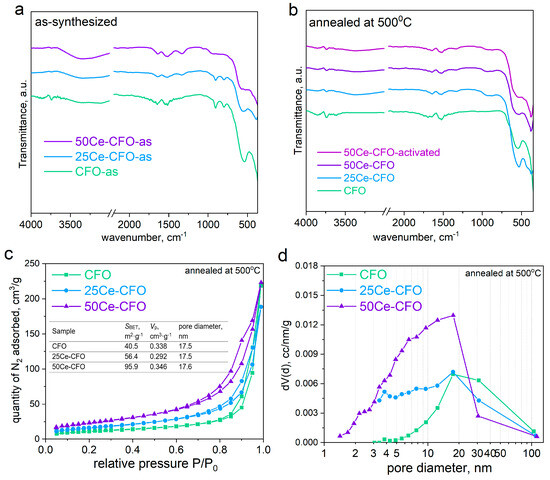
Figure 3.
(a,b) FTIR spectra of Ce-containing cobalt ferrite: (a) as-synthesized; (b) annealed at 500 °C. (c) N2 adsorption/desorption isotherms. (d) Pore size distribution.
Figure 3c depicts the N2 adsorption/desorption isotherms of the CFO, 25Ce-CFO, and 50Ce-CFO samples. The curves correspond to type IV BET isotherms with an H3-type hysteresis loop according to the IUPAC classification. The specific surface area of CoFe2O4 is 40.5 m2/g, increasing to 56.4 m2/g for the 25Ce-CFO and 95.9 m2/g for the 50Ce-CFO sample. The pore volumes are 0.338 cm3/g for CoFe2O4, 0.292 cm3/g for 25Ce-CFO, and 0.346 cm3/g for the 50Ce-CFO sample. It is obvious that Ce incorporation significantly modifies the textural properties of cobalt ferrite. While the total pore volume remains nearly constant, the specific surface area increases significantly, which can be attributed to the formation of very small CeO2 crystallites with a high external surface area that is finely dispersed among the spinel particles. The average pore diameter of 17.5 nm is observed for both CFO and 25Ce-CFO samples, and 17.6 nm for the 50Ce-CFO sample. The pore size distribution curves are presented in Figure 3d. The PSD curve for the CFO sample exhibits a main mesopore peak centered at 18 nm, indicating a predominantly mesoporous structure with large pore sizes up to 100 nm. The PSD curve for the 25Ce-CFO sample shows a similar main peak and contribution from smaller mesopores in the 5–18 nm range. The PSD curve for the 50Ce-CFO sample displays a strong shift toward smaller mesopores in the 2–18 nm range, with the highest intensity between 3 and 18 nm, and a reduced contribution from pores larger than 30 nm. Thus, the presence of abundant small mesopores for the 50Ce-CFO sample increases total surface area without significantly increasing total pore volume.
3.2. Catalytic Activity in H2O2 Decomposition
The catalytic activity of cerium-containing samples in H2O2 decomposition is illustrated in Figure 4. Among the annealed samples, the 50Ce-CFO sample exhibits the highest activity in H2O2 decomposition, fully decomposing 10 mM H2O2 in 160 min (Figure 4a). In contrast, the CFO and 25Ce-CFO samples show decomposition rates of 66% and 77%, respectively (as shown in Figure 4a). Notably, the activated sample significantly outperforms these others by completely depleting H2O2 in just 125 min. This enhanced performance can be attributed to the formation of surface oxygen vacancies and Ce3+ species, as treatment with H2O2 can reduce Ce4+ to Ce3+, thus creating Ce4+/Ce3+ redox pairs along with oxygen vacancies in CeO2 [40]. The kinetics of H2O2 depletion closely align with a pseudo-first-order kinetic model, demonstrating excellent correlations, with R2 values exceeding 0.994 (Figure 4b). The adherence to the PFO model indicates that the catalyst surfaces possess stable active sites, such as redox pairs like Co3+/Co2+, Fe3+/Fe2+, and Ce4+/Ce3+, that are uniformly distributed and present in substantial excess, ensuring that their concentration does not limit the reaction. Moreover, the decomposition of H2O2 occurs in the solution phase near the catalyst surface and does not require primary adsorption of H2O2 [27]. Importantly, there is a lack of radical chain effects; if the reaction were to proceed via radicals (such as HO• or HOO•), which could accelerate H2O2 decomposition, the kinetics would be more complex or exhibit autocatalytic behavior [41,42]. The rate coefficients were calculated (Figure 4c), revealing that the highest values were observed for both catalysts, 50Ce-CFO and 50Ce-CFO-activated, 0.0151 and 0.0236 min−1, respectively. Additionally, the post-synthesis activation of the catalyst surface with H2O2 further improves efficiency: the 50Ce-CFO-activated sample has a rate coefficient that is nearly 56% greater than that of the untreated 50Ce-CFO sample. This finding confirms that the treatment of the sample with H2O2 effectively increases the exposure of catalytic sites, probably cleaning the surface, and creates additional oxygen vacancies [40].
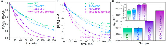
Figure 4.
Catalytic activity of Ce-containing cobalt ferrites in H2O2 decomposition: (a) kinetic curves; (b) pseudo-first-order kinetic model; (c) rate coefficient vs. Ce content (inset: percentage of H2O2 decomposition).
The phase stability and changes in surface morphology of the catalysts have been monitored using XRD, FTIR, and SEM techniques (Figure 5). XRD patterns (Figure 5a,b) showed no crucial changes in phase composition, except for the slight amorphization of the structure due to slight changes in the main (311) peak. After activation of the 50Ce-CFO material (Figure 5b), the lattice parameter of the cobalt ferrite phase was measured at 8.331 ± 0.003 Å, while the average crystallite size remained unchanged at approximately 15 nm. Similarly, the average crystallite size of the CeO2 phase was determined to be 4 nm, showing no significant variation within the experimental error range.
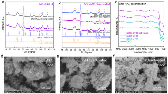
Figure 5.
Stability of the catalysts: (a) XRD of 50Ce-CFO catalyst before and after H2O2 decomposition; (b) XRD of 50Ce-CFO catalyst before activation, after activation, and after H2O2 catalytic decomposition. (c) FTIR spectra of the used catalysts. (d–f) SEM images of catalysts.
FTIR spectra (Figure 5c) demonstrate the preservation of the catalysts’ structure. A slight decrease in the intensity of the M–O peak is noted for the Ce-containing samples. This reduction may be due to the transformation of the ordered region into an amorphous phase in an oxidative environment, which weakens the M–O vibrations observed in the FTIR spectra. SEM images (Figure 5d–f) illustrate the morphological differences between the used 50Ce-CFO, the activated 50Ce-CFO, and the used activated 50Ce-CFO samples. Aggregated particles are visible in the 50Ce-CFO sample, indicating partial surface clogging caused by use in the catalytic process (Figure 5d). The 50Ce-CFO-activated sample exhibits a porous structure with visible particle separation (Figure 5e). The used 50Ce-CFO-activated sample shows signs of renewed aggregation and partial clogging due to the use (Figure 5f).
3.3. Catalytic Degradation of Congo Red Dye and Antibiotic Oxytetracycline
Two model pollutants—Congo Red dye and antibiotic oxytetracycline—have been chosen to evaluate the Fenton-like catalytic activity of Ce-CFO composites. Comparing the Fenton-like oxidation activity of the most active catalysts, 50Ce-CFO and 50Ce-CFO-activated, toward these two organic pollutants has been performed to understand the catalytic mechanisms. The corresponding UV–vis spectra of the aforementioned pollutants and their decolorisation in time are depicted in Figure 6. Control experiments were performed to better assess the catalytic effect (Figure 7a,b). The blank tests without a catalyst but with 10 mM H2O2 showed no degradation of CR and OTC over 120 min, indicating that 10 mM H2O2 alone does not decompose the pollutants under the tested conditions. Similarly, control experiments involving only adsorption of CR and OTC (without the addition of H2O2) were carried out to estimate the ability of the samples to adsorb the pollutants (Figure 7a,b). It is worth noting that the samples demonstrate good adsorption properties, and the adsorption activity of the activated 50Ce-CFO sample was slightly higher compared to the untreated sample. The adsorption of CR reached 39% for the activated 50Ce-CFO and 33% for the untreated 50Ce-CFO sample after 90 min, while OTC adsorption was 18% and 14%, respectively, after 45 min. The good adsorption properties of the samples can be attributed to the high specific surface area and the presence of mesopores in the Ce-containing samples (Figure 3c,d).
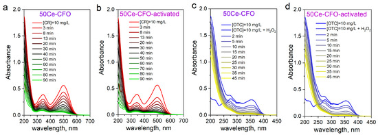
Figure 6.
UV–vis spectra of (a,b) Congo Red dye and (c,d) oxytetracycline in time degraded using untreated and activated 50Ce-CFO samples (conditions: [catalyst] = 1 g/L, [CR] = 10 mg/L, [OTC] = 10 mg/L, [H2O2] = 10 mM, T = 295 K, pH~7).
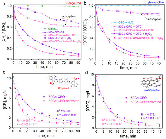
Figure 7.
Adsorption and catalytic properties of untreated and activated 50Ce-CFO sample: (a) Congo Red removal; (b) OTC removal; (c) PFO model for CR degradation; (d) PFO model for OTC degradation (k is rate coefficient; conditions: [catalyst] = 1 g/L, [CR] = 10 mg/L, [OTC] = 10 mg/L, [H2O2] = 10 mM, T = 295 K, pH~7).
Figure 7a,b demonstrates the kinetics of CR and OTC degradation in the presence of 10 mM H2O2 and synthesized samples. It can be seen that the degradation of OTC occurs two times faster compared to CR (45 and 90 min, respectively). The reason lies in their molecular structure. OTC contains several electron-rich functional groups, such as phenolic –OH, enolic C=C, and amine –NH2 groups. These groups make OTC easy to attack by hydroxyl radicals, leading to the molecule’s breakdown [43,44]. In contrast, CR has stable azo (–N=N–) bonds and sulfonated aromatic rings, which are more resistant to oxidative attacks because they have delocalized electron density [45]. In addition, the bonds in OTC, like C–O and C–N, are weaker than those in CR; therefore, they break down more easily when exposed to radicals. Moreover, it is known that hydroxyl radicals tend to react quickly with compounds that have electron-donating groups (like hydroxyl and amine groups in OTC) and react more slowly with compounds that have electron-withdrawing groups (like the sulfonate groups in CR) [46,47]. This difference helps explain why OTC breaks down faster than CR under Fenton-like conditions.
A comparison of the catalytic activity of both samples reveals that the activated sample exhibits higher performance. As shown in Figure S1, the untreated 50Ce-CFO sample achieved degradation efficiencies of 90% for Congo Red and 93% for oxytetracycline. In contrast, the activated sample reached higher degradation levels of 94% for CR and 96% for OTC, indicating enhanced catalytic activity after activation. The experimental data were analyzed through nonlinear fitting with the pseudo-first-order (PFO) kinetic model (Figure 7c,d) [33]. It can be seen that the degradation rate of Congo Red with the activated sample is higher (k = 0.1024 min−1) than with the untreated 50Ce-CFO sample (k = 0.0444 min−1), whereas for OTC degradation, the rates remain nearly the same. This can be explained by the higher affinity of Congo Red for adsorption on the catalyst surface (what was proved by adsorption experiments, Figure 7a,b), which promotes higher interaction with reactive oxygen species, while OTC molecules adsorb less efficiently and, therefore, do not benefit significantly from surface activation.
Figure 8 shows the changes in the structural stability of the used catalysts, checked with XRD and FTIR techniques. Figure 8a,b shows that after catalytic application of the 50Ce-CFO sample for the decomposition of Congo Red, the cobalt ferrite phase in the sample retains a lattice parameter of 8.332 ± 0.004 Å, with an average crystallite size of 18 nm. These values indicate that the spinel structure remains structurally stable under reaction conditions, with no significant phase degradation or lattice distortion. The apparent increase in crystallite size may be attributed to the leaching and subsequent reduction in the relative amount of smaller particles, which leads to a shift in the average crystallite size toward larger domains. This effect reflects selective removal rather than actual particle growth during the catalytic process. At the same time, the average crystallite size of the CeO2 remains equal to approximately 4 nm. After the use of the 50Ce-CFO material as a catalyst for oxytetracycline degradation, the lattice parameter of the cobalt ferrite phase remained unchanged with an average crystallite size of 15 nm. The CeO2 phase also retains particle size unchanged. These results indicate that no significant structural changes occur in the composite during the catalytic process, confirming its phase and microstructural stability under the applied reaction conditions.
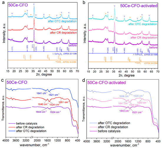
Figure 8.
Structure stability of used catalysts: (a) XRD patterns of the used 50Ce-CFO and (b) activated and used 50Ce-CFO-activated sample. (c) FTIR spectra of pristine and used 50Ce-CFO sample; (d) FTIR spectra of pristine and used 50Ce-CFO-activated sample.
After catalytic application of the activated 50Ce-CFO material in the degradation of Congo Red, the lattice parameter of the spinel phase is measured as 8.329 Å. The crystallite size of the cobalt ferrite component increased to 17 nm, reproducing the trend previously observed for the non-activated sample after use. The CeO2 phase exhibited an average crystallite size of 4.5 nm. After using the activated 50Ce-CFO material for catalytic degradation of oxytetracycline, the lattice parameter of the spinel phase of the composite was about 8.330 Å, while the average crystallite size was approximately 15 nm. The CeO2 phase exhibited a crystallite size of about 4 nm. These results indicate that the composite retains its multiphase structure and nanocrystalline character after catalytic use, with no significant structural degradation, although slight variations in crystallite size and lattice parameter may reflect surface restructuring or minor particle redistribution during the reaction.
The comparison of FTIR spectra before and after the CR and OTC degradation (Figure 8c,d) showed signs of their attachment to the catalyst’s surface. And the primary process, which accompanies the catalytic process, is the adsorption of pollutant molecules. That is why the peaks characteristic of the CR and OTC chemical structure appear in the spectrum of the used catalysts compared to pristine samples. The spectral peaks observed in the range of approximately 1524 cm−1 are associated with the C=C vibrations of the aromatic rings and the stretching of the azo group (–N=N–) within the structure of CR [48]. Additionally, the peaks located around 1050 cm−1 are attributed to the asymmetric stretching of the S=O bonds in the CR molecule, which contains sulfonate –SO3− groups [49]. For oxytetracycline, specific FTIR peaks are associated with its functional groups. The more intense peaks around 1640 cm−1 correspond to C=O stretching from amide and ketone groups within OTC’s structure [37]. The absence of intensive OTC-related peaks in the FTIR spectra of the catalysts after the reaction suggests that OTC adsorption on the catalyst surface was minimal, confirming that the degradation process was predominantly catalytic rather than adsorption-driven (which is also in agreement with Figure 7b).
The plausible reaction schemes could describe the Fenton-like degradation mechanism catalyzed by the 50Ce-CFO sample. Firstly, the activation of H2O2 occurs on the catalyst surface involving the Ce, Co, and Fe, which can change their oxidation states (Co3+/Co2+, Fe3+/Fe2+, Ce4+/Ce3+) and boost Fenton-like reactions, producing the hydroxyl radicals [14,27]:
≡Co2+ + H2O2 → ≡Co3+ + HO• + OH−
≡Fe3+ + H2O2 → ≡Fe2+ + HOO• + H+
≡Fe2+ + H2O2 → ≡Fe3+ + HO• + OH−
≡Fe3+ + HOO• → ≡Fe2+ + O2 + H+
≡Ce4+ + ≡Fe2+ → ≡Ce3+ + ≡Fe3+
≡Ce3+ + H2O2 → ≡Ce4+ + HO• + OH−
≡Ce3+ + H2O2 + H+ → ≡Ce4+ + HO• + H2O
The higher-valent metal cations might be regenerated, involving the electrons from the pollutant molecule [27]:
≡Co3+/≡Fe3+ + e− → ≡Co2+/≡Fe2+
≡Ce4+ + e− → ≡Ce3+
Finally, the CR/OTC molecule undergoes radical attack, which causes bond cleavage and leads to the formation of intermediates and general mineralization.
The activated sample demonstrates higher catalytic activity due to post-synthesis treatment with 10% H2O2. This treatment modified the surface, increasing the number of active sites, improving the redox cycling, and, probably, forming the oxygen vacancies, especially from the CeO2 phase:
Oxygen vacancies act as electron-rich sites, which facilitate the adsorption and activation of H2O2 molecules, which was observed in Figure 4a, leading to more efficient generation of hydroxyl radicals. The presence of vacancies improves the adsorption of pollutant molecules (CR or OTC) through electrostatic attraction, bringing them closer to active sites.
Table S2 presents a comparative overview of Ce-containing heterogeneous Fenton-like catalysts reported in the literature, including details on synthesis methods, target pollutants, experimental conditions, and pollutant removal efficiencies. It can be seen that the synthesized 50Ce-CFO and 50Ce-CFO-activated catalysts in this study demonstrate relatively high efficiencies in CR and OTC removal. The incorporation of CeO2 plays a key role in tailoring the properties of the composite. CeO2 nanodomains suppress the growth of CoFe2O4 crystallites through Zener pinning, leading to smaller crystallite size, increased BET surface area, and enhanced adsorption capacity. In addition, the redox-active Ce4+/Ce3+ couple promotes the generation of reactive oxygen species, thereby improving the Fenton-like catalytic activity. Thus, CeO2 provides a dual function–structural refinement and catalytic promotion, resulting in superior performance of the CeO2-CoFe2O4 composites.
4. Conclusions
CeO2-modified cobalt ferrite nanocomposites were synthesized using a hydrothermal method, producing uniform nanoparticles with a size of 25–50 nm. The presence of Ce caused significant structural changes: CeO2 separated as a distinct phase on the ferrite surface, which hindered grain growth and caused lattice distortion, as confirmed by XRD, SEM, and FTIR analyses. After annealing at 500 °C, the 50Ce-CFO sample showed increased amorphization due to higher Ce content. Catalytic performance tests indicated that both Ce content and post-synthesis H2O2 activation played crucial roles in enhancing Fenton-like activity. The 50Ce-CFO-activated sample achieved the 60% highest H2O2 decomposition rate than the untreated sample. Adsorption tests (without H2O2) showed notable uptake of CR and OTC, with the activated 50Ce-CFO sample performing slightly better than the untreated one. After 90 min, CR adsorption reached 39% for activated 50Ce-CFO versus 33% for the untreated sample, while OTC adsorption after 45 min was 18% and 14%, respectively. The enhanced adsorption is attributed to the higher surface area and mesoporosity of the Ce-containing samples. In degradation experiments with Congo Red and oxytetracycline, the activated catalyst outperformed the untreated sample, reaching degradation efficiencies of 94% and 96%, respectively. The presence of CeO2 refines CoFe2O4 crystallites, increases BET surface area, and enhances adsorption, while the Ce4+/Ce3+ redox couple boosts Fenton-like activity. Thus, CeO2 provides dual structural and catalytic benefits, leading to superior performance of the composite. These findings provide valuable insights for designing advanced heterogeneous catalysts for wastewater treatment.
Supplementary Materials
The following supporting information can be downloaded at: https://www.mdpi.com/article/10.3390/met15090985/s1, Figure S1: Removal of CR and OTC (in %) using untreated and activated Ce050-500 samples (conditions: [catalyst] = 1 g/L, [CR] = 10 mg/L, [OTC] = 10 mg/L, [H2O2] = 10 mM, T = 295 K, pH~7); Table S1: Masses of metal salts used in the synthesis of samples; Table S2: Comparative overview of Ce-containing heterogeneous Fenton-like catalysts reported in the literature.
Author Contributions
Conceptualization, T.T.; methodology, T.T.; formal analysis, T.T. and V.K.; investigation, T.T.; XRD analysis—V.K.; writing—original draft preparation, T.T.; writing—review and editing, T.T. and V.K.; visualization, T.T.; supervision, T.T.; project administration, T.T.; funding acquisition, T.T. All authors have read and agreed to the published version of the manuscript.
Funding
This research has received funding through the MSCA4Ukraine project Z/HEU/00031, which is funded by the European Union. Views and opinions expressed are, however, those of the author(s) only and do not necessarily reflect those of the European Union. Neither the European Union nor the MSCA4Ukraine Consortium as a whole nor any individual member institutions of the MSCA4Ukraine Consortium can be held responsible for them.
Data Availability Statement
The original contributions presented in this study are included in the article/Supplementary Material. Further inquiries can be directed to the corresponding author.
Acknowledgments
The study was performed using research infrastructure funded by the European Union in the framework of the Smart Growth Operational Programme, Measure 4.2; Grant No. POIR.04.02.00-00-D001/20, “ATOMIN 2.0—Center for materials research on ATOMic scale for the INnovative economy”. The authors appreciate Kaja Spilarewicz’s assistance in collecting SEM images. T.T. acknowledges the II European Chemistry School for Ukrainians.
Conflicts of Interest
The authors declare no conflicts of interest.
References
- Andersen, H.L.; Granados-Miralles, C.; Jensen, K.M.Ø.; Saura-Múzquiz, M.; Christensen, M. The Chemistry of Spinel Ferrite Nanoparticle Nucleation, Crystallization, and Growth. ACS Nano 2024, 18, 9852–9870. [Google Scholar] [CrossRef]
- Tatarchuk, T.; Bilovol, V.; Shyichuk, A.; Danyliuk, I.; Sokołowski, K.; Gajewska, M. Mesoporous Co-Mn Ferrites as Highly Radical-Forming Catalysts for Wet Peroxide Oxidation of 4-Nitrophenol. Appl. Surf. Sci. 2025, 690, 162610. [Google Scholar] [CrossRef]
- Ma, D.; Lai, C.; Yi, H.; Huo, X.; Li, L.; Zhang, M.; Xu, F.; Yan, H.; Hu, S.; Luo, Y. Confinement Strategies for the Design of Efficient Heterogeneous Fenton-like Catalysts: From Nano-Space to Atomic Scale. Coord. Chem. Rev. 2025, 522, 216241. [Google Scholar] [CrossRef]
- Soufi, A.; Hajjaoui, H.; Boumya, W.; Elmouwahidi, A.; Baillón-García, E.; Abdennouri, M.; Barka, N. Recent Trends in Magnetic Spinel Ferrites and Their Composites as Heterogeneous Fenton-like Catalysts: A Review. J. Environ. Manage. 2024, 367, 121971. [Google Scholar] [CrossRef]
- Bhaduri, B.; Dikshit, A.K.; Kim, T.; Tripathi, K.M. Research Progress and Prospects of Spinel Ferrite Nanostructures for the Removal of Nitroaromatics from Wastewater. ACS Appl. Nano Mater. 2022, 5, 16000–16026. [Google Scholar] [CrossRef]
- Qin, H.; He, Y.; Xu, P.; Huang, D.; Wang, Z.; Wang, H.; Wang, Z.; Zhao, Y.; Tian, Q.; Wang, C. Spinel Ferrites (MFe2O4): Synthesis, Improvement and Catalytic Application in Environment and Energy Field. Adv. Colloid Interface Sci. 2021, 294, 102486. [Google Scholar] [CrossRef] [PubMed]
- Zhao, Q.; Yan, Z.; Chen, C.; Chen, J. Spinels: Controlled Preparation, Oxygen Reduction/Evolution Reaction Application, and Beyond. Chem. Rev. 2017, 117, 10121–10211. [Google Scholar] [CrossRef] [PubMed]
- Xu, G.; Liu, Q.; Mai, Z.; Wang, M.; Zhao, H.; Xu, K. Strategies for Improving Performance of Iron-Based Catalysts in Activating Heterogeneous Fenton-like Oxidation in Pollutants Degradation: From the Perspective of Materials Structure Design. Process Saf. Environ. Prot. 2024, 190, 794–820. [Google Scholar] [CrossRef]
- Tatarchuk, T. Studying the Defects in Spinel Compounds: Discovery, Formation Mechanisms, Classification, and Influence on Catalytic Properties. Nanomaterials 2024, 14, 1640. [Google Scholar] [CrossRef]
- Zhang, W.; Wang, P.; Zhao, K.; Wen, G.; Zhang, X.; Wang, Y. Review of CeO2 Nanostructure/Carbon Composite Catalysts for Wastewater Treatment. ACS Appl. Nano Mater. 2025, 8, 10210–10241. [Google Scholar] [CrossRef]
- Wang, Y.; Liu, T.; Liu, J. Synergistically Boosted Degradation of Organic Dyes by CeO2 Nanoparticles with Fluoride at Low PH. ACS Appl. Nano Mater. 2020, 3, 842–849. [Google Scholar] [CrossRef]
- Minakshi, M.; Nallathamby, K.; Mitchell, D.R.G. Electrochemical Characterization of an Aqueous Lithium Rechargeable Battery: The Effect of CeO2 Additions to the MnO2 Cathode. J. Alloys Compd. 2009, 479, 87–90. [Google Scholar] [CrossRef]
- Dong, Y.; Shah, M.A.K.Y.; Khalid, M.; Lu, Y.; Deng, C. Optimizing Low-Temperature Ceramic Fuel Cells with CuFe2O4–CeO2 Heterostructures. ACS Appl. Energy Mater. 2023, 6, 12494–12502. [Google Scholar] [CrossRef]
- Ma, H.; Liu, Z.; Koshy, P.; Sorrell, C.C.; Hart, J.N. Density Functional Theory Investigation of the Biocatalytic Mechanisms of PH-Driven Biomimetic Behavior in CeO2. ACS Appl. Mater. Interfaces 2022, 14, 11937–11949. [Google Scholar] [CrossRef] [PubMed]
- Ravi, G.; Thyagarajan, K. Effect of Calcination Temperature on the Structural, Morphological, and Magnetic Properties of CuFe2O4/CoFe2O4/CeO2 Nanocomposites. Ceram. Int. 2025, 51, 31083–31096. [Google Scholar] [CrossRef]
- Wetchakun, N.; Chaiwichain, S.; Wetchakun, K.; Kangwansupamonkon, W.; Inceesungvorn, B.; Phanichphant, S. Synthesis and Characterization of Novel Magnetically Separable CoFe2O4/CeO2 Nanocomposite Photocatalysts. Mater. Lett. 2013, 113, 76–79. [Google Scholar] [CrossRef]
- Sayed, M.A.; Abdelhameed, R.M.; Badr, I.H.A.; Abdel-Aziz, A.M. Efficient Adsorptive Removal of Hazardous Congo Red Dye Using Ce-BTC@microcrystalline Cellulose Composite. Sci. Rep. 2025, 15, 19734. [Google Scholar] [CrossRef]
- Cao, Y.; Peng, X.; Tan, Z.; Liu, Y.; Wang, X.; Zhao, W.; Jiang, L. Structural Evolution of Active Entities on Co3O4/CeO2 Catalyst during Water Gas Shift Reaction. Ind. Eng. Chem. Res. 2019, 58, 17692–17698. [Google Scholar] [CrossRef]
- Sun, Q.; Liu, Y.; Li, X.; Guo, X.; Huang, W.-H.; Zhu, Y.; Wang, Z.; Chueh, C.-C.; Chen, C.-L.; Peng, Y.-K.; et al. Highly Disordered Fe-Doped CeO2 with Oxygen Vacancies Facilitates Electrocatalytic Water Oxidation. Energy Fuels 2023, 37, 9434–9443. [Google Scholar] [CrossRef]
- Urooj, I.; Sohail, M.; Shah, W.A.; Ali, H.; An, X.; Liu, S.; Wahab, M.A. Nanorectangular NiFe2O4 Decorated with CeO2 Nanoparticles and Modified with Phosphate Ions as an Electrocatalyst for Water Electrolysis. ACS Appl. Nano Mater. 2025, 8, 3769–3786. [Google Scholar] [CrossRef]
- Luo, Y.; Alves, D.; Barwa, T.N.; Dempsey, E.; Breslin, C.B. A CeFe2O4-CeO2 Composite for the Electrochemical Detection and Advanced Oxidation of the Antibiotic Sparfloxacin. J. Environ. Manage. 2025, 380, 125167. [Google Scholar] [CrossRef] [PubMed]
- Zhu, F.; Ji, Q.; Lei, Y.; Ma, J.; Xiao, Q.; Yang, Y.; Komarneni, S. Efficient Degradation of Orange II by Core Shell CoFe2O4–CeO2 Nanocomposite with the Synergistic Effect from Sodium Persulfate. Chemosphere 2022, 291, 132765. [Google Scholar] [CrossRef]
- Liu, B.; Li, J.; Wang, C.; Li, Y.; Gao, L.; Yang, Q. Efficient Activation of Peroxymonosulfate for Trichloroethylene Degradation by Cobalt Ferrites Anchored on CeO2 Surfaces: Radical to Non-Radical Pathway Shift. J. Environ. Chem. Eng. 2024, 12, 112280. [Google Scholar] [CrossRef]
- Yuan, B.; Tan, Z.; Guo, Q.; Shen, X.; Zhao, C.; Chen, J.L.; Peng, Y.-K. Regulating the H2O2 Activation Pathway on a Well-Defined CeO2 Nanozyme Allows the Entire Steering of Its Specificity between Associated Enzymatic Reactions. ACS Nano 2023, 17, 17383–17393. [Google Scholar] [CrossRef] [PubMed]
- Wu, H.; Liu, J.; Chen, Z.; Lin, P.; Ou, W.; Wang, Z.; Xiao, W.; Chen, Y.; Cao, D. Mechanism and Application of Surface-Charged Ferrite Nanozyme-Based Biosensor toward Colorimetric Detection of l-Cysteine. Langmuir 2022, 38, 8266–8279. [Google Scholar] [CrossRef] [PubMed]
- Shlapa, Y.; Siposova, K.; Veltruska, K.; Maraloiu, V.-A.; Garcarova, I.; Rajnak, M.; Musatov, A.; Belous, A. Design of Magnetic Fe3O4/CeO2 “Core/Shell”-Like Nanocomposites with Pronounced Antiamyloidogenic and Antioxidant Bioactivity. ACS Appl. Mater. Interfaces 2023, 15, 49346–49361. [Google Scholar] [CrossRef]
- Xu, L.; Wang, J. Magnetic Nanoscaled Fe3O4/CeO2 Composite as an Efficient Fenton-Like Heterogeneous Catalyst for Degradation of 4-Chlorophenol. Environ. Sci. Technol. 2012, 46, 10145–10153. [Google Scholar] [CrossRef]
- Li, J.; Gou, G.; Zhao, H.; Liu, C.; Li, N.; Li, L.; Tan, B.; Lai, B. Efficient Peroxymonosulfate Activation by CoFe2O4-CeO2 Composite: Performance and Catalytic Mechanism. Chem. Eng. J. 2022, 435, 134840. [Google Scholar] [CrossRef]
- Gao, P.; Chen, X.; Hao, M.; Xiao, F.; Yang, S. Oxygen Vacancy Enhancing the Fe2O3-CeO2 Catalysts in Fenton-like Reaction for the Sulfamerazine Degradation under O2 Atmosphere. Chemosphere 2019, 228, 521–527. [Google Scholar] [CrossRef]
- Chen, J.; Wan, J.; Li, C.; Wei, Y.; Shi, H. Synthesis of Novel Fe0-Fe3O4/CeO2/C Composite Cathode for Efficient Heterogeneous Electro-Fenton Degradation of Ceftriaxone Sodium. J. Hazard. Mater. 2022, 437, 129393. [Google Scholar] [CrossRef]
- Li, D.; Zhao, Z.; Guo, J.; Shen, R.; Xu, F. Magnetic Nanoscaled Fe3O4-CeO2-TiO2 Composite: UV-Fenton Reaction to Degrade AO-7 Dye. Inorg. Chem. Commun. 2023, 149, 110389. [Google Scholar] [CrossRef]
- Shen, C.; Zhu, Q.; Chen, H.; Zhang, Y.; Du, M.; Li, F.; Ma, J. Insights into the Synergistic Effect of Fe and Ce in Fenton-like Reactions Catalyzed by Chitosan/FeOOH/CeO2 Microspheres. J. Clean. Prod. 2024, 451, 142058. [Google Scholar] [CrossRef]
- Perrin, C.L. Linear or Nonlinear Least-Squares Analysis of Kinetic Data? J. Chem. Educ. 2017, 94, 669–672. [Google Scholar] [CrossRef]
- Kale, G.H.; Humbe, A.V.; Birajdar, S.D.; Shinde, A.B.; Jadhav, K.M. L-Ascorbic Acid Assisted Synthesis and Characterization of CoFe2O4 Nanoparticles at Different Annealing Temperatures. J. Mater. Sci. Mater. Electron. 2016, 27, 2151–2158. [Google Scholar] [CrossRef]
- Seeharaj, P.; Pasupong, P.; Choojun, K. Magnetically Separable CeO2/CoFe2O4 Heterojunction Photocatalysts for Dye Degradation: Characterization and Mechanism. J. Asian Ceram. Soc. 2024, 12, 44–58. [Google Scholar] [CrossRef]
- Li, Y.; Wang, L.; Chen, D.; Liu, J.; Fan, W.; Sun, J. Experimental and Theoretical Study on Performance of Cu-Fe Spinel Supported with Certain Inerts during Chemical Looping Combustion. Chem. Eng. J. 2024, 495, 153591. [Google Scholar] [CrossRef]
- Tatarchuk, T.; Starko, I. Mesoporous La-Substituted Nickel-Cobalt Ferrites Synthesized via Reduction Method Resulting in Significantly Enhanced Adsorption Properties. J. Environ. Chem. Eng. 2025, 13, 115657. [Google Scholar] [CrossRef]
- Dharmalingam, S.T.; Dar, M.A.; Gul, R.; Minakshi Sundaram, M.; Alnaser, I.A.; Sivasubramanian, R. Nano-Octahedron Cobalt Oxide Decorated Graphene Nanocomposites for the Selective/Simultaneous Detection of Dopamine. Adv. Mater. Interfaces 2025, 12, 2400981. [Google Scholar] [CrossRef]
- Ebenezer, J.; Velayudham, P.; Schechter, A. Electrochemical Nitrogen Fixation Using CeFeO3 and CeO2 for Ammonia Synthesis and Nitrate Remediation. ACS Appl. Mater. Interfaces 2025, 17, 36796–36809. [Google Scholar] [CrossRef]
- Chen, F.; Shen, X.; Wang, Y.; Zhang, J. CeO2/H2O2 System Catalytic Oxidation Mechanism Study via a Kinetics Investigation to the Degradation of Acid Orange 7. Appl. Catal. B Environ. 2012, 121–122, 223–229. [Google Scholar] [CrossRef]
- Lousada, C.M.; Yang, M.; Nilsson, K.; Jonsson, M. Catalytic Decomposition of Hydrogen Peroxide on Transition Metal and Lanthanide Oxides. J. Mol. Catal. A Chem. 2013, 379, 178–184. [Google Scholar] [CrossRef]
- Tatarchuk, T.; Shyichuk, A.; Danyliuk, N.; Naushad, M.; Kotsyubynsky, V.; Boychuk, V. Cobalt Ferrite as an Electromagnetically Boosted Metal Oxide Hetero-Fenton Catalyst for Water Treatment. Chemosphere 2023, 326, 138364. [Google Scholar] [CrossRef] [PubMed]
- Rajeendre Katugampalage, T.; Waribam, P.; Opaprakasit, P.; Kaewsaneha, C.; Hsu, S.-H.; Chooaksorn, W.; Jong, C.-A.; Sooksaen, P.; Ratanatawanate, C.; Sreearunothai, P. Bimetallic Fe:Co Metal–Organic Framework (MOF) with Unsaturated Metal Sites for Efficient Fenton-like Catalytic Degradation of Oxytetracycline (OTC) Antibiotics. Chem. Eng. J. 2024, 479, 147592. [Google Scholar] [CrossRef]
- Bracco, E.B.; Marco-Brown, J.L.; Butler, M.; Candal, R.J. Degradation of Oxytetracycline and Characterization of Byproducts Generated by Fenton or Photo-Fenton like Processes after Adsorption on Natural and Iron(III)-Modified Montmorillonite Clays. Environ. Nanotechnol. Monit. Manag. 2023, 19, 100778. [Google Scholar] [CrossRef]
- Solano, A.M.S.; Garcia-Segura, S.; Martínez-Huitle, C.A.; Brillas, E. Degradation of Acidic Aqueous Solutions of the Diazo Dye Congo Red by Photo-Assisted Electrochemical Processes Based on Fenton’s Reaction Chemistry. Appl. Catal. B Environ. 2015, 168–169, 559–571. [Google Scholar] [CrossRef]
- Moores, L.C.; Kaur, D.; Smith, M.D.; Poole, J.S. Regioselectivity of Hydroxyl Radical Reactions with Arenes in Nonaqueous Solutions. J. Org. Chem. 2019, 84, 3260–3269. [Google Scholar] [CrossRef]
- Masi, A.; Capobianco, A.; Bobrowski, K.; Peluso, A.; Chatgilialoglu, C. Hydroxyl Radical vs. One-Electron Oxidation Reactivities in an Alternating GC Double-Stranded Oligonucleotide: A New Type Electron Hole Stabilization. Biomolecules 2023, 13, 1493. [Google Scholar] [CrossRef]
- Elumalai, S.; Muthuraman, G. Recovery of Methyl Orange and Congo Red from Aqueous Solutions Using Tri-Octyl Amine (TOA) in Benzene as Carrier. Process Saf. Environ. Prot. 2015, 96, 177–183. [Google Scholar] [CrossRef]
- Olusegun, S.J.; Mohallem, N.D.S. Comparative Adsorption Mechanism of Doxycycline and Congo Red Using Synthesized Kaolinite Supported CoFe2O4 Nanoparticles. Environ. Pollut. 2020, 260, 114019. [Google Scholar] [CrossRef]


Disclaimer/Publisher’s Note: The statements, opinions and data contained in all publications are solely those of the individual author(s) and contributor(s) and not of MDPI and/or the editor(s). MDPI and/or the editor(s) disclaim responsibility for any injury to people or property resulting from any ideas, methods, instructions or products referred to in the content. |
© 2025 by the authors. Licensee MDPI, Basel, Switzerland. This article is an open access article distributed under the terms and conditions of the Creative Commons Attribution (CC BY) license (https://creativecommons.org/licenses/by/4.0/).