Article

Controlled Formation of Nanoislands During Microwave Annealing of Au Thin Films

by
Ali Ghanim Gatea Al-Rubaye
1,
Alaa Alasadi
2,3,*,
Khalid Rmaydh Muhammed
4 and
Catalin-Daniel Constantinescu
3,*
1
Basra Technical Institute, Southern Technical University, Basra 61030, Iraq
2
Karbala Technical Institute, Al-Furat Al-Awsat Technical University, Karbala 56001, Iraq
3
Aix-Marseille Université, CNRS, LP3 UMR 7341, 13009 Marseille, France
4
Al-Anbar Technical Institute, Middle Technical University, Ramadi 31001, Iraq
*
Authors to whom correspondence should be addressed.
Metals 2025, 15(9), 1030; https://doi.org/10.3390/met15091030
Submission received: 31 July 2025 / Revised: 2 September 2025 / Accepted: 12 September 2025 / Published: 18 September 2025
(This article belongs to the Special Issue Metallic Nanostructured Materials and Thin Films)

Abstract

We present a systematic study on the fabrication of gold nanoislands by microwave-assisted annealing, a rapid and energy-efficient alternative to conventional thermal treatments. Gold thin films with nominal thicknesses of 4, 5, 6, 8, and 10 nm are deposited by thermal evaporation directly onto BK7 glass substrates, with and without a 3 nm chromium adhesion layer. The samples are subsequently annealed in a microwave kiln, where microwave irradiation is absorbed and converted to heat within the graphite-coated cavity (kiln), allowing the substrate temperature to exceed 550 °C, the threshold required for film dewetting. This process induces a controlled morphological evolution from continuous thin films to well-defined nanoislands, with the final size distribution strongly dependent on the initial film thickness. Compared with oven-based annealing, microwave treatment promotes faster and more uniform heating, which enhances atomic diffusion and accelerates dewetting while reducing the risk of substrate deformation or excessive coalescence. The resulting nanoislands exhibit tailored size-dependent plasmonic properties, with clear correlations between film thickness, crystallite size, and optical absorption features. Importantly, the method is cost-efficient, requiring shorter processing times and lower energy input, while enabling reproducible fabrication of high-quality plasmonic nanostructures on inexpensive glass substrates, suitable for applications in sensing, photonics, and nanophotonics.

Graphical Abstract

1. Introduction

Gold’s (Au) high malleability, ductility, and resistance to corrosion but also to most other chemical reactions, as well as its high electrical conductivity, have led to its continued use in corrosion-resistant electrical connectors for all types of electronic devices [1]. In addition to its advantageous bulk properties, Au exhibits unique optical properties when used as thin films and nanostructures [2], in particular for infrared shielding [3]. Historically, it was used in colored glass production, e.g., the Lycurgus Cup [4], which is a Roman glass 4th-century cage goblet made of a dichroic glass, an effect achieved by making the glass with tiny proportions of nanoparticles of gold and silver dispersed in colloidal form throughout the material [5]. Au is also used for gilding [6] and also in medicine for dental restoration [7] and various forms of medical therapy [8], with some of its salts still being used as anti-inflammatory remedies [9]. Thus, Au has been widely used in numerous applications, yet one of the most uncommon is in sensors based on the localized surface plasmon resonance (LSPR) effect. The LSPR is a phenomenon in which the collective oscillation of electrons on the surface of metals or metal-like materials that exhibit negative real permittivity, such as Au, Ag, or Cu (as the three most common plasmonic active materials), is excited by an incident electromagnetic field in the visible or infrared spectra [10]. Resonance occurs when the frequency of incident photons matches the natural frequency of collective electronic oscillations on the metal surface.
In 1959, Nobel Laureate in Physics, Dr. Richard Feynman, gave the famous lecture called “There’s Plenty of Room at the Bottom”, which stimulated numerous research projects about the fabrication of microstructures/nanostructures. A nanoparticle is considered a structure of matter typically smaller than 100 nanometers (nm). Various fabrication techniques can generate nanoparticles or pattern nanostructures, including nanoislands [11], such as photolithography [12,13], soft lithography [14], laser direct writing [15], or colloidal lithography, with laser-based dewetting techniques [16,17,18,19,20,21,22,23,24,25]. Among the application of Au nanostructures, the LSPR is in fact the most common. It represents a confinement of the surface plasmon in a nanoparticle of size comparable to or smaller than the wavelength of light used to excite the plasmon, and this resonance results in strong absorption or scattering of light at specific wavelengths.
The plasmon resonant frequency is highly sensitive to the environment, and any change in the material’s size, shape, or type/composition and/or the refractive index results in a shift of the resonant frequency (Figure 1). This image shows the extinction spectra of various nanostructures with different sizes and shapes, highlighting how LSPR shifts with geometry. Spherical nanoparticles exhibit resonance in the visible range (420–550 nm), while triangular nanostructures (nanoprisms) support tunable resonances extending into the near-infrared (600–790 nm). An intermediate structure between the nanoprisms and the spherical nanoparticles exhibits resonances in the mid-visible range (530–590). The results demonstrate that both particle size and morphology strongly influence the peak wavelength (λmax) and optical response, as key parameters in tailoring plasmonic properties for specific applications. Furthermore, nanoparticle assembly, alloying, and fragmentation are all fundamental processes with important implications in various fields such as catalysis, materials science, and nanotechnology. The importance of assembly strategies for enhancing nanoparticle functionalities has been highlighted in recent work [17,18]. Nanoparticles and their assembly play a pivotal role in tailoring material properties for advanced applications. Jambhulkar et al. [18] provide a comprehensive overview of self-organization and controlled micropatterning strategies, highlighting how these approaches can be harnessed to enhance functionalities. Thus, understanding such processes, in particular under special conditions, is essential to optimize the nanoparticles’ properties and their applications [25].
Nanoislands are various shaped entities, such as droplets or similar, which are formed by spontaneous dewetting. These are also called “agglomerations” according to early publications, when dewetting of thin and very thin metal films occurred on a substrate by post-deposition heating or by other energy sources, such as laser, microwaves, etc. [11]. Au nanoislands are fabricated by various methods such as electron beam lithography [26,27], porous diatom frustules [28], electrochemical deposition [29], photolithography [30], and hydrogel-assisted nanotransfer edge printing (HnTEP) [31]. Au nanoislands are also fabricated by annealing in a conventional furnace [32,33,34]. Modern research has focused on annealing thin films in solution by microwave heating to form nanostructures [35,36]. The advantage of this type of annealing is that it ensures equal reaction conditions to form homogeneous nanostructures [37] and saves energy due to the rapid formation of nanostructures. It is therefore effective in scientific applications such as sensor technology for environmental pollution. The annealing process results in a change in the surface structural and morphological properties of the deposited and annealed films, thus forming nanoislands. The process, called dewetting, refers to a phenomenon of spontaneous rupture of the thin film deposited on the substrate. The principle of dewetting is based on the instability of the thin film caused by several factors such as surface tension effects and thermal fluctuations [38]. Various authors have previously used microwave devices for annealing gold thin films to obtain gold nanoislands, e.g., by using microwave plasmas [39]. For example, an argon mixture was used in a quartz chamber equipped with two tubes to pump air and argon into the microwave space.
In this work, we propose a simple, rapid, and efficient method to control the formation of nanoislands by using a kiln inside a microwave oven, to anneal and thus achieve controlled dewetting of Au thin films under standard atmosphere and pressure.

2. Materials and Methods

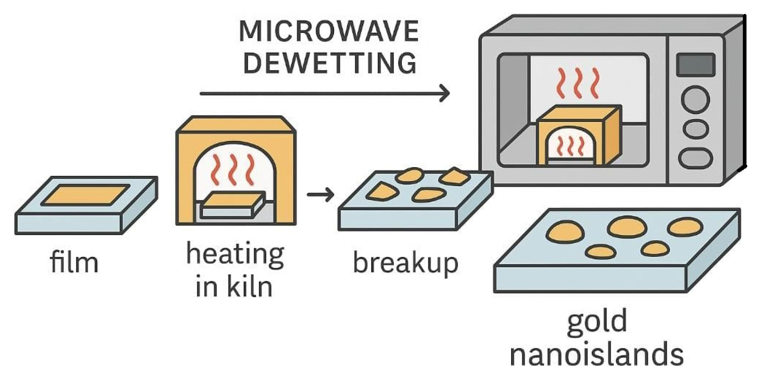
Two types of thin film samples were prepared for the fabrication of Au nanoislands, i.e., single layer and double layer, by using Au wire (AGE401-25, diameter 0.2 mm, Laborimpex, Forest, Belgium). The substrate type of choice was BK7 glass, 25 mm × 25 mm, 1.1 mm thick (Fisher Scientific, Waltham, MA, USA). The first type of sample consisted of Au layers of various thicknesses: 4, 5, 6, 8, and 10 nm. The second type of sample consisted of two layers: a 3 nm thick chromium layer (Cr, pellets) grown on BK7 glass substrates to improve Au adhesion. All samples were deposited on BK7 glass substrates by a thermal evaporator (model E306 Auto, from Edwards Ltd., Burgess Hill, UK) under vacuum (1.3 × 10⁻⁵ Pa), which ensured oxidation-free conditions. The Cr and the Au were placed separately in tungsten boats for thermal evaporation under vacuum. An electric current passed through each of the two boats, causing the metals to melt and evaporate, which were then collected as films on the glass substrates at the top of the evaporator. The thickness of the Au and Cr layers was monitored and controlled by a quartz crystal microbalance (QCM) during the evaporation process. The next step in nanoisland formation is annealing. The material was heated to a specific temperature and then slowly cooled in a classic oven or microwave over a period of time to form the nanoislands. The traditional annealing technique is thermal annealing, which involves placing the single Au layer sample in an oven for 10 h at a temperature of 550 °C. The two layers of Cr and Au were placed in the oven for 10 h at a temperature of 480 °C. The required temperature difference between the single layer and the two layers is due to the different chemical and physical properties of Au and Cr, i.e., these differences affect the surface energy and thus the adhesion process, increasing the stability of the Au on the glass substrate [34,40]. After the annealing, the samples were left for 2 h inside the microwave for each sample to form the desired nanoislands (Figure 2). Further details on the procedure are available elsewhere [41]. The microwave oven used was a conventional General Electric Countertop Microwave Oven, 700 Watts, model 2,440,640 (Fischer Scientific, Waltham, MA, USA). Microwave kilns are suitable for small projects and allow metal to be melted or annealed without special tools. A microwave kiln reaches its maximum temperature in 5 to 15 min, depending on its size and power. In general, microwave kilns can reach a maximum temperature of approximately 900 °C. After melting or annealing the material, the kiln must be cooled for at least 20 min before opening to avoid damaging the sample. Figure 2 illustrates the container kiln, consisting of two parts, the base and the hood, in which the sample was installed. All samples were subjected to the same preparation and manufacturing conditions in terms of heating and cooling. Placed inside the microwave kiln for 13 min, the samples reached the annealing temperature of 550 °C.
The kiln acted as a furnace to keep the required temperature, and the samples were left inside for 2 h to obtain the desired nanoislands. This temperature remained stable throughout the sample annealing time in the oven/microwave, monitored by using an infrared thermometer (12:1 optics, single laser, range up to 750 °C). The difference in using annealing temperatures between a single and two layers is that the Cr layer adhered completely and strongly to the glass during the evaporation stage, as well as to the gold layer on top of it, while Au alone required a high temperature to interfere with the glass and to gain great stability and form the nanoisland. Figure 2 schematically shows the full procedure of a rapid method for fabricating the gold nanoisland using a microwave kiln. A scanning electron microscope (SEM), atomic force microscopy (AFM), and X-ray diffraction XRD were used to accurately characterize the surface properties of the gold nanoisland, and clear results were obtained. Finally, UV-Vis spectra in the range of 400–800 nm were analyzed by a UV-Vis spectrophotometer to assess the ability of Au nanoislands for LSPR.

3. Results and Discussion

Microwave annealing of pure gold thin films (4–10 nm) on BK7 glass substrates typically leads to dewetting and the formation of discontinuous gold nanoislands due to weak adhesion between gold and glass. The low interfacial energy promotes rapid morphological changes, resulting in poor film stability. In contrast, when a 3 nm chromium adhesion layer is introduced beneath the gold film, the interface bonding is significantly enhanced. This stronger adhesion delays or suppresses dewetting during microwave annealing, maintaining better film continuity. The Cr layer also modifies the stress relaxation pathways, leading to more controlled surface morphology. However, the presence of Cr may slightly alter the optical and electronic properties compared with pure Au films. Overall, microwave annealing highlights the crucial role of adhesion layers in determining the thermal stability and morphology of ultrathin gold films.
The nanoislands presented in this work have been generated by microwave annealing of thin Au layers, previously deposited by vacuum thermal evaporation on BK7 glass substrates. SEM was used to measure the average particle size and average interparticle distance to correlate them with their properties and performance. The technological parameters for manufacturing the nanoislands for optimal performance were also determined, as will be further presented and discussed in this section.

3.1. Morphological and Structural Analysis: SEM, AFM, and XRD

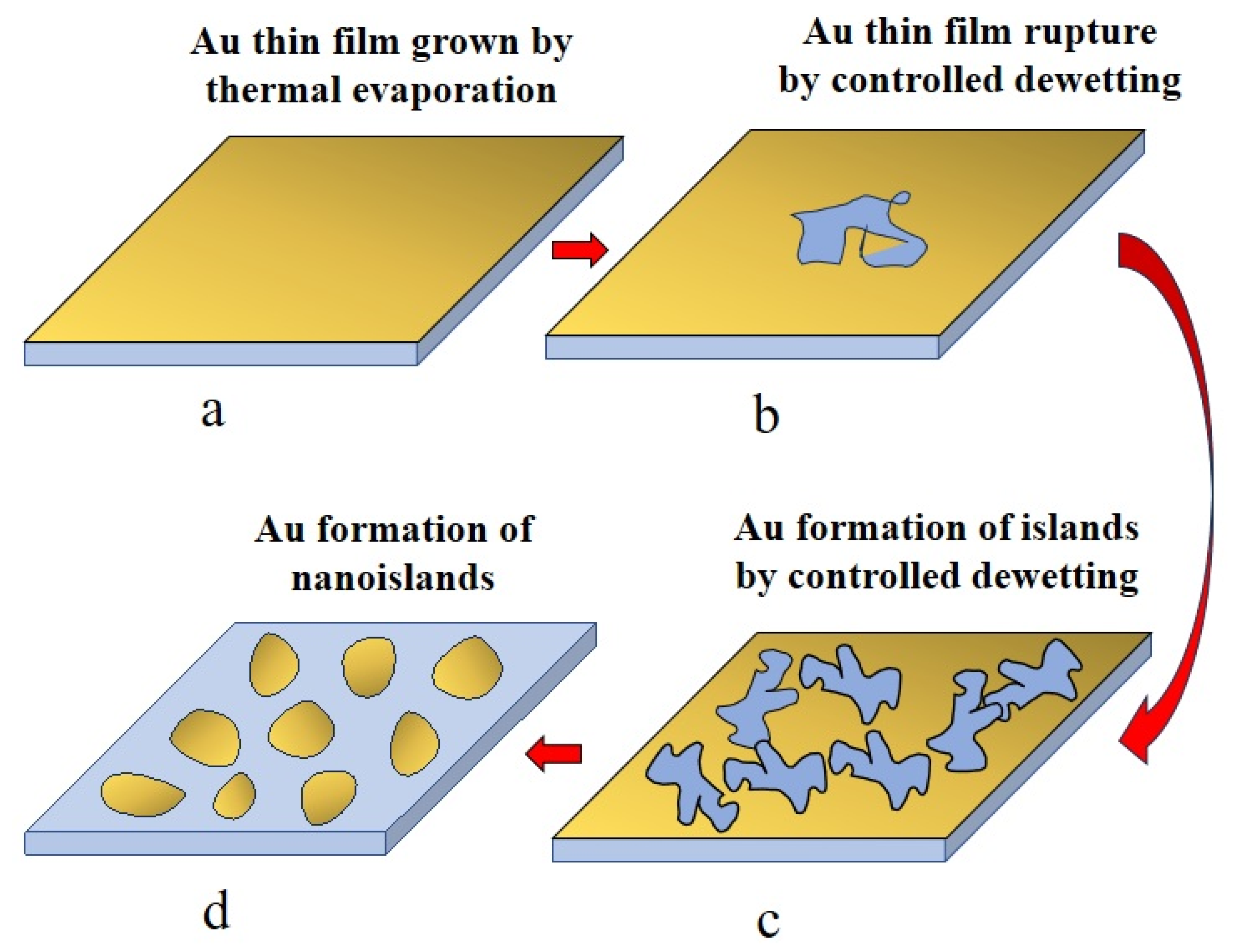
Figure 3 shows the steps of the nanostructure dewetting development with the temperature change by the kiln annealing procedure for a thin film of 5 nm. The Au thin film transforms into nanoislands gradually with time and with increasing temperature.
This sequential process involved in the formation of gold nanoislands from an ultrathin film through controlled dewetting under microwave-assisted annealing takes place as follows. In the first stage (a), a continuous Au thin film is deposited by thermal evaporation, forming a metastable layer on the glass substrate. Upon microwave heating, the film becomes unstable and ruptures (b), initiating the dewetting process as driven by minimization of surface and interfacial energies. As the process advances (c), the ruptured film reorganizes into irregular island-like fragments, reflecting the competition between surface diffusion and capillary instabilities. With continued microwave annealing, these fragments undergo further coarsening and spheroidization, ultimately producing well-defined, thermodynamically stable nanoislands (d). Compared with conventional furnace heating, microwave-assisted annealing accelerates this pathway due to rapid and volumetric energy transfer, enhancing atomic mobility while reducing the time window for undesirable substrate flow or excessive coalescence. As a result, this technique promotes the reproducible formation of uniform and optically active gold nanoislands, whose localized surface plasmon resonance properties can be finely tuned by the initial film thickness and the precise control of the annealing process.
The AFM images of the 5 nm gold thin film deposited on glass without an adhesion layer clearly illustrate the morphological transformation induced by thermal annealing in the kiln/microwave setup, as presented in Figure 4. Prior to annealing (a), the surface appears relatively smooth, with only small height variations corresponding to the continuous but unstable thin film morphology and/or substrate surface quality. After microwave annealing for 13 min (b), the film undergoes complete dewetting, breaking up into a dense array of well-defined nanoislands with average heights on the order of several tens of nanometers, as evidenced by the pronounced topographical contrast.
The high density and uniform distribution of islands indicate efficient dewetting promoted by the rapid and homogeneous microwave heating, which accelerates atomic diffusion and minimizes coalescence into excessively large structures. Such a transformation directly correlates with the emergence of a strong and well-resolved LSPR band in the optical spectra, absent in the as-deposited continuous film (see Section 3.2).
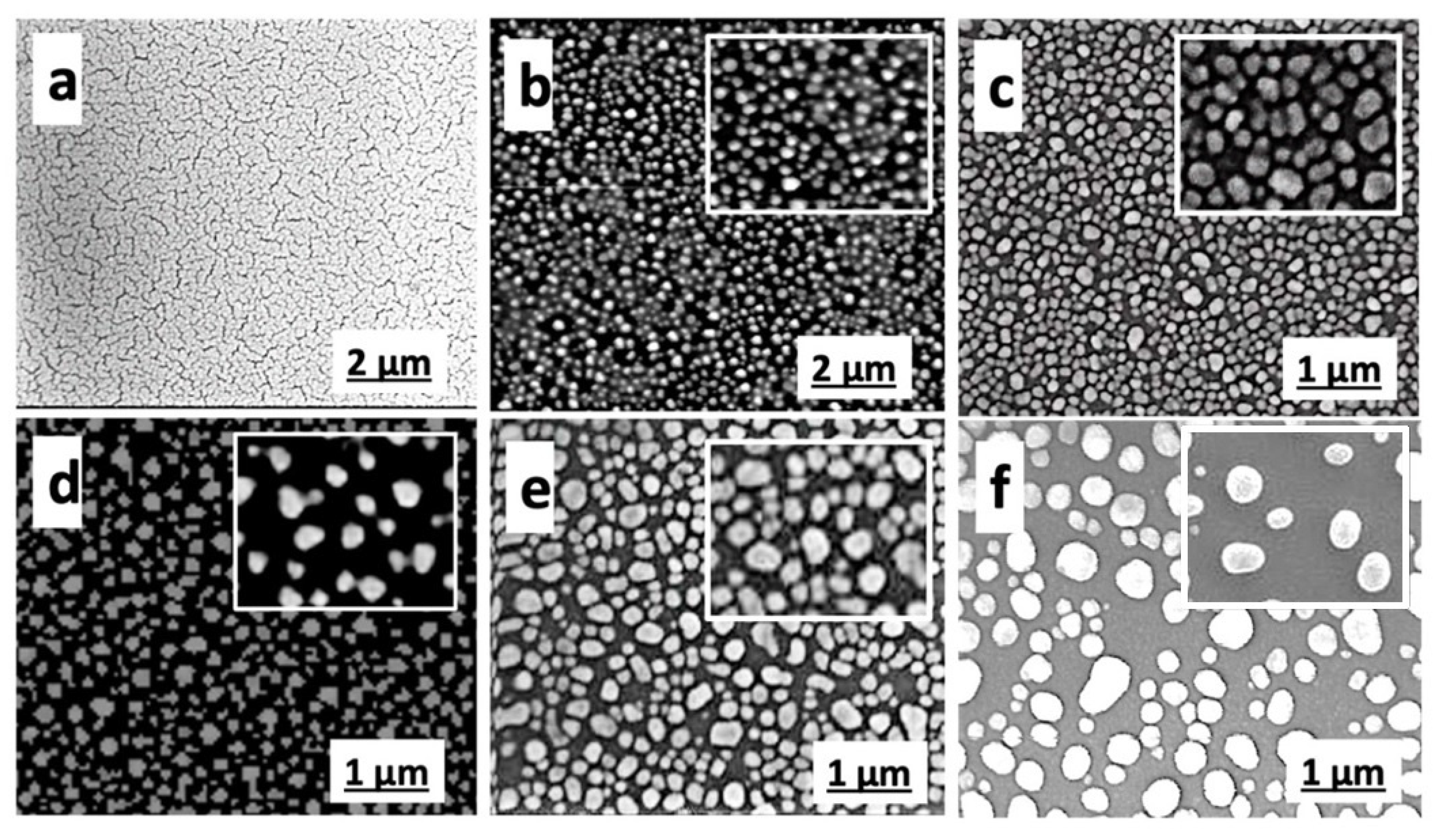
These results highlight the effectiveness of microwave annealing for producing reproducible plasmonic nanostructures from ultrathin gold films without the need for adhesion layers. The AFM data not only highlight the morphological transformation of the 5 nm Au film upon annealing but also reveal a significant increase in surface roughness. Before annealing (a), the root-mean-square (RMS) roughness is expected to be low, typically in the range of 1–2 nm, consistent with a nearly continuous ultrathin film with only minor height fluctuations. After annealing at 550 °C for 13 min (b), the RMS roughness increases significantly, reaching values of the order of 10–20 nm, reflecting the formation of discrete nanoislands with heights approaching several tens of nanometers, as seen in the AFM height scale. This sharp increase in roughness provides quantitative confirmation of the dewetting process and correlates directly with the emergence of pronounced LSPR features in the optical spectra. Thus, RMS roughness evolution offers a simple but powerful descriptor linking the nanoscale topography of the film to its plasmonic functionality. The kiln/microwave-based technique created Au nanoislands similar to those achieved by the conventional oven technique [41,42]. Microwave annealing results in the development of distinct Au nanoislands, which are characterized by an LSPR band in UV-vis absorption spectra at about 600 nm as shown in the AFM and SEM images. Figure 5 shows scanning electron microscope images of the first image of a Au thin film before annealing and others with thicknesses of 4, 5, 6, 8, and 10 nm of the Au film after annealing, revealing the progressive morphological evolution of gold thin films upon annealing in a kiln/microwave setup, as a function of their initial thickness. This will be properly correlated with the optical properties of the samples in Section 3.2.
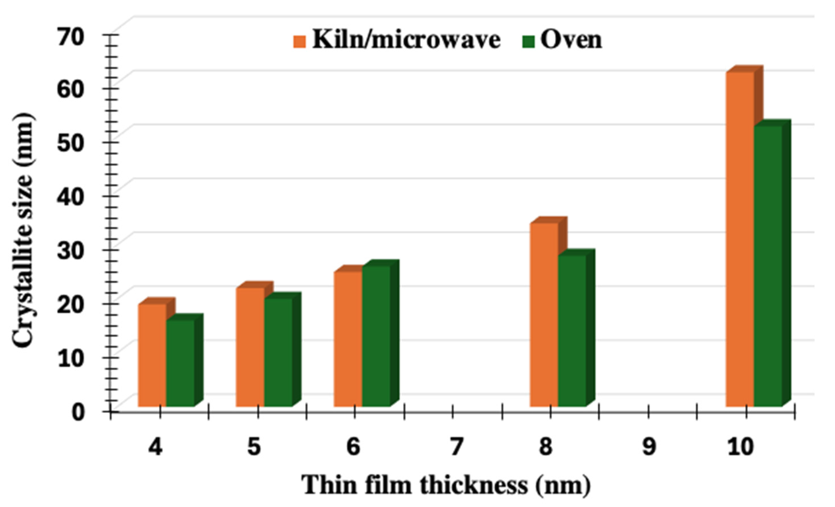
The XRD-derived crystallite size analysis highlights clear differences between gold nanoislands formed by kiln/microwave annealing and those produced by conventional oven treatment. Figure 6 compares the crystal size of our previous results from the oven-based technique [32,39] and the current results from the kiln/microwave-based technique. For all investigated thicknesses, the crystallite size increases systematically with the initial film thickness, reflecting the natural progression of coalescence and growth during the dewetting process. However, a consistent distinction is observed between the two annealing methods: microwave annealing yields slightly larger crystallite sizes compared with oven annealing, with the difference becoming more pronounced at higher thicknesses, particularly for the 8 and 10 nm films. This behavior can be attributed to the rapid and volumetric heating associated with microwaves, which accelerates atomic mobility and promotes more efficient grain coarsening within a shorter processing time. In contrast, oven annealing, governed by slower conductive heat transfer, results in more moderate crystallite growth under comparable conditions [43]. The implication of this structural difference is significant for optical performance since larger crystallite sizes generally reduce grain boundary scattering and contribute to sharper and more defined plasmonic resonances. Furthermore, the enhanced efficiency of microwave annealing not only improves structural quality but also offers advantages in terms of reduced processing time and energy consumption, reinforcing its suitability as a superior method for controlled fabrication of plasmonic gold nanostructures.

3.2. Optical Properties of the Nanoislands

The optical properties of gold nanoislands are governed by their LSPR behavior, which strongly depends on the initial thickness of the deposited gold film and the subsequent dewetting process. Thin films close to the percolation threshold (e.g., 3–5 nm) typically dewet into small, densely packed islands, resulting in broad and relatively intense absorption bands in the visible range, often centered near 520–550 nm. As the initial film thickness increases (6–10 nm or more), the resulting islands grow larger and become more widely spaced, which leads to a red-shift of the absorption maximum toward 550–600 nm, accompanied by narrower but weaker plasmonic features. This shift arises from both the increased particle size, which enhances dipolar plasmon coupling, and the reduced surface density of islands, which alters near-field interactions. In addition, thicker starting films favor partial coalescence and a greater degree of island faceting, further contributing to spectral red-shift and peak broadening. Thus, the initial gold film thickness acts as a key tuning parameter for controlling the plasmonic resonance of gold nanoislands, enabling the design of nanostructured films with tailored optical responses for sensing, photonic, and catalytic applications.
UV-vis spectra in the range of 400–800 nm demonstrate the ability of Au nanoislands to absorb the light within 500–650 nm wavelength, i.e., revealing that the frequency changes according to the structures of the nanoislands with different thicknesses of 4, 5, 6, 8, and 10 nm, as shown in the Figure 7. Both sets of spectra display a distinctive LSPR band in the 550–600 nm range, characteristic of gold nanoislands formed after film dewetting. Panel (a) corresponds to conventional oven annealing, while panel (b) corresponds to kiln/microwave annealing. In both cases, thinner films (e.g., 4 nm) exhibit stronger and broader absorption peaks due to enhanced plasmonic coupling and smaller, denser nanoislands, while thicker films (8–10 nm) show weaker and red-shifted LSPR bands, consistent with larger island sizes and reduced plasmonic response. Compared with oven annealing (a), microwave-assisted annealing (b) produces slightly sharper and more intense LSPR features, suggesting more efficient nanoisland formation and improved uniformity due to rapid and volumetric heating. This highlights the influence of the annealing technique on the plasmonic behavior of gold nanostructured thin films. The band of the LSPR ranges from 550 to 650 nm. These peaks change depending on the thickness of the initial Au thin film [44]. To properly correlate these optical results with the SEM investigations presented in Figure 5, we will explain further and in detail. The as-deposited 5 nm film (a) displays a continuous, labyrinth-like structure characteristic of a just-percolated metallic layer, with no distinct plasmonic features expected. After annealing, the 4 nm film (b) undergoes complete dewetting into a dense array of small and relatively uniform nanoislands, typically 10–20 nm in diameter, which correlates with a sharp and intense absorption peak in the 520–540 nm range due to strong dipolar plasmon coupling. Increasing the initial thickness to 5 nm (c) results in slightly larger islands, around 20–30 nm, with reduced density, producing a red-shift of the plasmon band toward 540–560 nm and a moderate broadening of the absorption profile. At 6 nm (d), more distinct and faceted islands appear, in the range of 30–40 nm, further red-shifting the resonance to 550–570 nm. For the 8 nm film (e), the nanoislands grow to 40–60 nm, with less uniformity and larger interparticle spacing, leading to an absorption peak near 560–580 nm of lower intensity, as near-field coupling diminishes and multipolar contributions emerge. The thickest film, 10 nm (f), produces large, widely spaced islands exceeding 70 nm in diameter, yielding a broad and weak resonance shifted toward 580–600 nm, strongly affected by radiative damping and size polydispersity. These observations confirm a clear correlation between initial film thickness, final island size distribution, and the optical response: thinner films give rise to dense, small islands with strong, blue-shifted plasmon bands, whereas thicker films generate progressively larger and sparser islands, resulting in red-shifted, broadened, and weakened absorption features. The sequential evolution from a continuous Au thin film to well-defined nanoislands under microwave-assisted annealing directly correlates with the changes observed in the UV–vis spectra. The as-deposited film shows only weak, featureless absorption, while initial rupture and fragmentation introduce broad plasmonic features. As the film further dewets and reorganizes into discrete islands, a distinct localized surface plasmon resonance emerges, whose peak position and intensity depend on the final island size and spacing. Thus, the structural pathway of controlled dewetting, from film rupture to nanoisland formation, underpins the optical response, with microwave annealing offering superior control and efficiency in tailoring plasmonic properties. Many groups report that when ultrathin Au films on soda-lime/BK7 glass are annealed in a classic manner near or above the glass transition, the Au dewets into islands, and the glass itself forms a circumferential “rim” (meniscus) around each island as it partially embeds [39,44,45,46]. The rising of these rims depends on the thickness of the thin film, and the rims’ height should be higher at 10 nm thickness than for 8, 6, 5, or 4 nm in our gold films [45]. Indeed, when ultrathin gold films are annealed on BK7 glass via conventional furnace heating near or above the glass transition temperature (≈557 °C), the softened substrate tends to flow around the dewetted gold nanoislands, forming circumferential “rims” and partially embedding the islands. While this eases adhesion, it adversely affects the LSPR by altering the dielectric environment and introducing spectral broadening and shifts. By contrast, microwave-assisted dewetting within a kiln offers substantial benefits: its volumetric heating mechanism ensures rapid and uniform energy delivery directly to both film and substrate, thus swiftly reaching the dewetting threshold without extended exposure of the glass to temperatures near the glass transition temperature (Tg). This minimizes substrate flow; suppresses rim formation; preserves the morphological integrity of the gold nanoislands; and retains sharper, more consistent optical characteristics. Additionally, microwave annealing is prominently more energy-efficient and time-effective than conventional thermal methods, making it both higher in quality and more cost-effective for scalable fabrication of plasmonic nanostructures.

4. Conclusions

The formation of Au nanoislands by using a microwave/oven annealing process has yielded remarkable and surprising results. Manufacturing time was reduced from 10 h to only 13 min compared with traditional techniques. The microwave oven stores thermal energy throughout the annealing process, giving the Au nanoislands superior specifications compared with those obtained with traditional methods. Annealing by using a traditional oven differs from a convection oven, which uses a fan to distribute air inside. The traditional oven is controlled by a thermostat to reach the desired temperature (550 °C for 10 h) to obtain nanoislands from thin films of different thicknesses (4, 5, 6, 8, and 10 nm). The XRD results corroborate the AFM, SEM, and optical data by showing that the crystallite size increases with the initial Au film thickness and that microwave annealing consistently produces larger crystallites than conventional oven treatment. For thinner films (4–5 nm), the smaller crystallites (15–25 nm) correspond to sharp and intense LSPR peaks in the 520–550 nm range. With increasing thickness, particularly at 8–10 nm, crystallite sizes exceed 60 nm under microwave annealing, which explains the observed red-shift of the absorption band toward 580–600 nm and its progressive broadening. The larger and better-defined crystallites generated by microwave annealing reduce grain boundary scattering, thereby preserving sharper plasmonic resonances compared with oven annealing, which yields smaller grains and a more damped optical response. This direct structural–optical correlation highlights the advantage of microwave annealing for producing plasmonically superior nanoislands. Altogether, microwave annealing in a kiln emerges as a state-of-the-art approach for fabricating plasmonic nanostructures, combining precise morphological control with enhanced optical quality, reduced processing times, and superior cost efficiency compared with conventional thermal methods.

Author Contributions

Conceptualization, A.G.G.A.-R., A.A., K.R.M., and C.-D.C.; methodology, A.G.G.A.-R., A.A., K.R.M., and C.-D.C.; validation, A.G.G.A.-R., A.A., K.R.M., and C.-D.C.; formal analysis, A.G.G.A.-R., A.A., and K.R.M.; investigation, A.G.G.A.-R., A.A., K.R.M., and C.-D.C.; resources, A.G.G.A.-R., A.A., K.R.M., and C.-D.C.; data curation, A.A. and C.-D.C.; writing—original draft preparation, A.G.G.A.-R., A.A., K.R.M., and C.-D.C.; writing—review and editing, A.G.G.A.-R., A.A., K.R.M., and C.-D.C.; visualization, A.A. and C.-D.C.; supervision, A.A. and C.-D.C.; project administration, A.A. and C.-D.C.; funding acquisition, A.A. and C.-D.C. All authors have read and agreed to the published version of the manuscript.

Funding

Alaa Alasadi gratefully recognizes the research mobility scholarship he received from the French government via the Embassy of France in Baghdad, Iraq, and Campus France (https://www.campusfrance.org, N° Dossier: 172919N), as a six-month fellowship grant in collaboration with Catalin-Daniel Constantinescu, through an agreement between the CNRS (DR12) and the LP3 laboratory (UMR 7341 CNRS AMU) in Marseille.

Data Availability Statement

The original contributions presented in this study are included in the article. Further inquiries can be directed to the corresponding authors.

Acknowledgments

The authors acknowledge the Iraqi Cultural attaché in London for funding this project and the Sheffield Hallam University for assistance. Some experiments have been conducted by using the “Lasers & Micro-Procédés” (LaMP) facilities at LP3, and also the “Plateforme de nano et micro-fabrication” (PLANETE) facilities at CINaM (UMR 7325 CNRS AMU).

Conflicts of Interest

The authors declare no conflicts of interest.

References

  1. Schmidbaur, H.; Cihonski, J.L. Noble Metals (Chemistry). In Encyclopedia of Physical Science and Technology, 3rd ed.; Academic Press: Cambridge, MA, USA, 2003; pp. 463–492. [Google Scholar] [CrossRef]
  2. Bernardo-Gavito, R.; Serrano, A.; García, M.A.; Miranda, R.; Granados, D. Local characterization of the optical properties of annealed Au films on glass substrates. J. Appl. Phys. 2013, 114, 164312. [Google Scholar] [CrossRef]
  3. Piwnuan, C.; Muangphat, C.; Wootthikanokkhan, J. Enhancing NIR Shielding Properties of Au/CsWO3 Composite via Physical Mixing and Solvothermal Processes. Materials 2024, 17, 2746. [Google Scholar] [CrossRef] [PubMed]
  4. Freestone, I.; Meeks, N.; Sax, M.; Higgitt, C. The Lycurgus Cup—A Roman nanotechnology. Gold Bull. 2007, 40, 270–277. [Google Scholar] [CrossRef]
  5. Hammami, I.; Alabdallah, N.M.; Al-Jomaa, A.; Kamoun, M. Gold nanoparticles: Synthesis properties and applications. J. King Saud Univ.-Sci. 2021, 33, 101560. [Google Scholar] [CrossRef]
  6. Oddy, W.A. Gilding: An outline of the technological history of the plating of gold on to silver or copper in the Old World. Endeavour 1991, 15, 29–33. [Google Scholar] [CrossRef]
  7. Hanawa, T. Overview of metals and applications. In Metals for Biomedical Devices; Niinomi, M., Ed.; Woodhead Publishing Limited: Cambridge, UK, 2010; ISBN 978-1-84569-434-0. [Google Scholar] [CrossRef]
  8. Wang, H.H.; Su, C.H.; Wu, Y.J.; Lin, C.A.J.; Lee, C.H.; Shen, J.L.; Chan, W.H.; Chang, W.H.; Yeh, H.I. Application of Gold in Biomedicine: Past, Present and Future. Int. J. Gerontol. 2012, 6, 1–4. [Google Scholar] [CrossRef]
  9. Balfourier, A.; Kolosnjaj-Tabi, J.; Luciani, N.; Gazeau, F. Gold-based therapy: From past to present. Proc. Natl. Acad. Sci. USA 2020, 117, 22639–22648. [Google Scholar] [CrossRef]
  10. Schatz, G.C.; Van Duyne, R.P. Electromagnetic Mechanism of Surface-Enhanced Spectroscopy; John Wiley & Sons, Ltd.: New York, NY, USA, 2002. [Google Scholar] [CrossRef]
  11. Badilescu, S.; Raju, D.; Bathini, S.; Packirisamy, M. Gold Nano-Island Platforms for Localized Surface Plasmon Resonance Sensing: A Short Review. Molecules 2020, 25, 4661. [Google Scholar] [CrossRef]
  12. Madou, M. Fundamentals of Microfabrication: The Science of Miniaturization, 2nd ed.; CRC Press: Boca Raton, FL, USA, 2002; Available online: https://books.google.co.in/books?id=9bk3gJeQKBYC (accessed on 24 May 2025).
  13. Kim, T.J.; Jung, Y.H.; Zhang, H.; Kim, K.; Lee, J.; Ma, Z. Photolithography-Based Nanopatterning Using Re-entrant Photoresist Profile. ACS Appl. Mater. Interfaces 2018, 10, 8117–8123. [Google Scholar] [CrossRef]
  14. Qin, D.; Xia, Y.; Whitesides, G.M. Soft lithography for micro- and nanoscale patterning. Nat. Protoc. 2010, 5, 491–502. [Google Scholar] [CrossRef]
  15. Alasadi, A.; Claeyssens, F.; Allwood, D.A. Rapid Fabrication of 2-D Magnetic Microstructures by Laser Direct Writing (LDW). IEEE Trans. Magn. 2021, 57, 1–6. [Google Scholar] [CrossRef]
  16. Haes, A.J.; Haynes, C.L.; Van Duyne, R.P. Nanosphere Lithography: Self-Assembled Photonic and Magnetic Materials. MRS Online Proc. Libr. 2000, 636, 481. [Google Scholar] [CrossRef]
  17. Bercea, A.I. Periodic Multi-Scale Structuring of Surfaces by Self-Assembly of Colloidal Masks: Shape-Size Coupling Effects. Ph.D. Thesis, Université de Limoges, Limoges, France, 2021. Available online: https://theses.fr/2021LIMO0103 (accessed on 24 May 2025).
  18. Jambhulkar, S.; Ravichandran, D.; Zhu, Y.; Thippanna, V.; Ramanathan, A.; Patil, D.; Fonseca, N.; Thummalapalli, S.V.; Sundaravadivelan, B.; Sun, A.; et al. Nanoparticle Assembly: From Self-Organization to Controlled Micropatterning for Enhanced Functionalities. Small 2024, 20, e2306394. [Google Scholar] [CrossRef]
  19. Schaaf, P.; Constantinescu, C.; Matei, A. Preface on laser material interactions: From basic science to industrial applications (LaserMaterInter2020). Appl. Surf. Sci. Adv. 2021, 6, 100133. [Google Scholar] [CrossRef]
  20. Constantinescu, C.; Kallepalli, L.N.D.; Delaporte, P.; Utéza, O.; Grojo, D. Laser processing of metal thin films using transparent microsphere arrays. Appl. Surf. Sci. 2015, 336, 112–117. [Google Scholar] [CrossRef]
  21. Schaaf, P.; Constantinescu, C.; Matei, A. Laser material processing: From fundamental interactions to innovative applications (E-MRS). Appl. Surf. Sci. Adv. 2024, 21, 100592. [Google Scholar] [CrossRef]
  22. Nedyalkov, N.; Nikov, R.; Koleva, M.; Atanasov, P.A.; Constantinescu, C.; Delaporte, P.; Grojo, D. Nanoparticle-decorated ceramic as substrate in surface enhanced Raman spectroscopy. Appl. Surf. Sci. 2015, 336, 16–20. [Google Scholar] [CrossRef]
  23. Constantinescu, C.; Kallepalli, L.N.D.; Delaporte, P.; Utéza, O.; Grojo, D. Arrays of metallic micro-/nano-structures by means of colloidal lithography and laser dewetting. Appl. Surf. Sci. 2016, 374, 124–131. [Google Scholar] [CrossRef]
  24. Yang, L.; Wei, J.; Ma, Z.; Song, P.; Ma, J.; Zhao, Y.; Huang, Z.; Zhang, M.; Yang, F.; Wang, X. The Fabrication of Micro/Nano Structures by Laser Machining. Nanomaterials 2019, 9, 1789. [Google Scholar] [CrossRef]
  25. Itina, T.E. Understanding mono- and bi-metallic Au and Ni nanoparticle responses to fast heating. Nanoscale Adv. 2024, 6, 5451–5463. [Google Scholar] [CrossRef] [PubMed]
  26. Mølhave, K.; Madsen, D.N.; Rasmussen, A.M.; Carlsson, A.; Appel, C.C.; Brorson, M.; Bøggild, P. Solid gold nanostructures fabricated by electron beam deposition. Nano Lett. 2003, 3, 1499–1503. [Google Scholar] [CrossRef]
  27. Theerthagiri, J.; Lee, S.J.; Karuppasamy, K.; Park, J.; Yu, Y.; Kumari, M.A.; Choi, M.Y. Fabrication strategies and surface tuning of hierarchical gold nanostructures for electrochemical detection and removal of toxic pollutants. J. Hazard. Mater. 2021, 420, 126648. [Google Scholar] [CrossRef]
  28. Losic, D.; Mitchell, J.G.; Voelcker, N.H. Fabrication of gold nanostructures by templating from porous diatom frustules. New J. Chem. 2006, 30, 908–914. [Google Scholar] [CrossRef]
  29. Hsu, M.-S.; Chen, Y.-L.; Lee, C.-Y.; Chiu, H.-T. Gold nanostructures on flexible substrates as electrochemical dopamine sensors. ACS Appl. Mater. Interfaces 2012, 4, 5570–5575. [Google Scholar] [CrossRef]
  30. Kim, J.-H.; Kang, T.; Yoo, S.M.; Lee, S.Y.; Kim, B.; Choi, Y.-K. A well-ordered flower-like gold nanostructure for integrated sensors via surface-enhanced Raman scattering. Nanotechnology 2009, 20, 235302. [Google Scholar] [CrossRef]
  31. Xue, M.; Ma, X.; Xie, Z.; Duan, L.; Jiang, Y.; Zhang, M.; Cao, T. Fabrication of Gold-Directed Conducting Polymer Nanoarrays for High-Performance Gas Sensor. Chem.-Asian J. 2010, 5, 2266–2270. [Google Scholar] [CrossRef]
  32. Al-Rubaye, A.G.G. Development of Optical Biosensors Based on Metal Nanostructures for Pollution (Mycotoxins) Detection. Ph.D. Thesis, Sheffield Hallam University, Sheffield, UK, 2019. [Google Scholar] [CrossRef]
  33. Jin, L.; Liu, B.; Louis, M.E.; Li, G.; He, J. Highly crystalline mesoporous titania loaded with monodispersed gold nanoparticles: Controllable metal–support interaction in porous materials. ACS Appl. Mater. Interfaces 2020, 12, 9617–9627. [Google Scholar] [CrossRef] [PubMed]
  34. Karakouz, T.; Tesler, A.B.; Bendikov, T.A.; Vaskevich, A.; Rubinstein, I. Highly stable localized plasmon transducers obtained by thermal embedding of gold island films on glass. Adv. Mater. 2008, 20, 3893–3899. [Google Scholar] [CrossRef]
  35. Vigil, E.; Saadoun, L.; Ayllón, J.A.; Domènech, X.; Zumeta, I.; Rodríguez-Clemente, R. TiO2 thin film deposition from solution using microwave heating. Thin Solid Film. 2000, 365, 12–18. [Google Scholar] [CrossRef]
  36. Jun, T.; Song, K.; Jeong, Y.; Woo, K.; Kim, D.; Bae, C.; Moon, J. High-performance low-temperature solution-processable ZnO thin film transistors by microwave-assisted annealing. J. Mater. Chem. 2010, 21, 1102–1108. [Google Scholar] [CrossRef]
  37. Al-Gaashani, R.; Radiman, S.; Tabet, N.; Daud, A.R. Effect of microwave power on the morphology and optical property of zinc oxide nano-structures prepared via a microwave-assisted aqueous solution method. Mater. Chem. Phys. 2011, 125, 846–852. [Google Scholar] [CrossRef]
  38. Bertrand, E.; Blake, T.; De Coninck, J. Dynamics of dewetting. Colloids Surf. A Physicochem. Eng. Asp. 2010, 369, 141–147. [Google Scholar] [CrossRef]
  39. Hsu, C.-Y.; Huang, J.-W.; Gwo, S.; Lin, K.-J. The facile fabrication of tunable plasmonic gold nanostructure arrays using microwave plasma. Nanotechnology 2009, 21, 35302. [Google Scholar] [CrossRef]
  40. Tesler, A.B.; Chuntonov, L.; Karakouz, T.; Bendikov, T.A.; Haran, G.; Vaskevich, A.; Rubinstein, I. Tunable localized plasmon transducers prepared by thermal dewetting of percolated evaporated gold films. J. Phys. Chem. C 2011, 115, 24642–24652. [Google Scholar] [CrossRef]
  41. Al-Rubaye, A.G.; Nabok, A.; Tsargorodska, A. LSPR Biosensor Based on Nanostructured Gold Films: Detection of Mycotoxins. Procedia Technol. 2017, 27, 131–132. [Google Scholar] [CrossRef]
  42. Al-Rubaye, A.G.; Nabok, A.; Tsargorodska, A. Spectroscopic ellipsometry study of gold nanostructures for LSPR bio-sensing applications. Sens. Bio-Sens. Res. 2017, 12, 30–35. [Google Scholar] [CrossRef]
  43. Tsuji, M.; Hashimoto, M.; Nishizawa, Y.; Kubokawa, M.; Tsuji, T. Microwave-assisted synthesis of metallic nanostructures in solution. Chem.-A Eur. J. 2005, 11, 440–452. [Google Scholar] [CrossRef] [PubMed]
  44. Tesler, A.B.; Maoz, B.M.; Feldman, Y.; Vaskevich, A.; Rubinstein, I. Solid-state thermal dewetting of just-percolated gold films evaporated on glass: Development of the morphology and optical properties. J. Phys. Chem. C 2013, 117, 11337–11346. [Google Scholar] [CrossRef]
  45. Thompson, C.V. Solid-State Dewetting of Thin Films. Annu. Rev. Mater. Res. 2012, 42, 399–434. [Google Scholar] [CrossRef]
  46. Sundaresan, S.; Liu, X.; Campbell, R.; Caride, B.; Look, D.C.; Myers-Ward, R.L.; Gaskill, D.K.; Hite, J.; Eddy, C.; Caldwell, J.D.; et al. Comparison of Solid-State Microwave Annealing with Conventional Furnace Annealing of Ion-Implanted SiC. J. Appl. Phys. 2021, 129, 135701. [Google Scholar] [CrossRef]
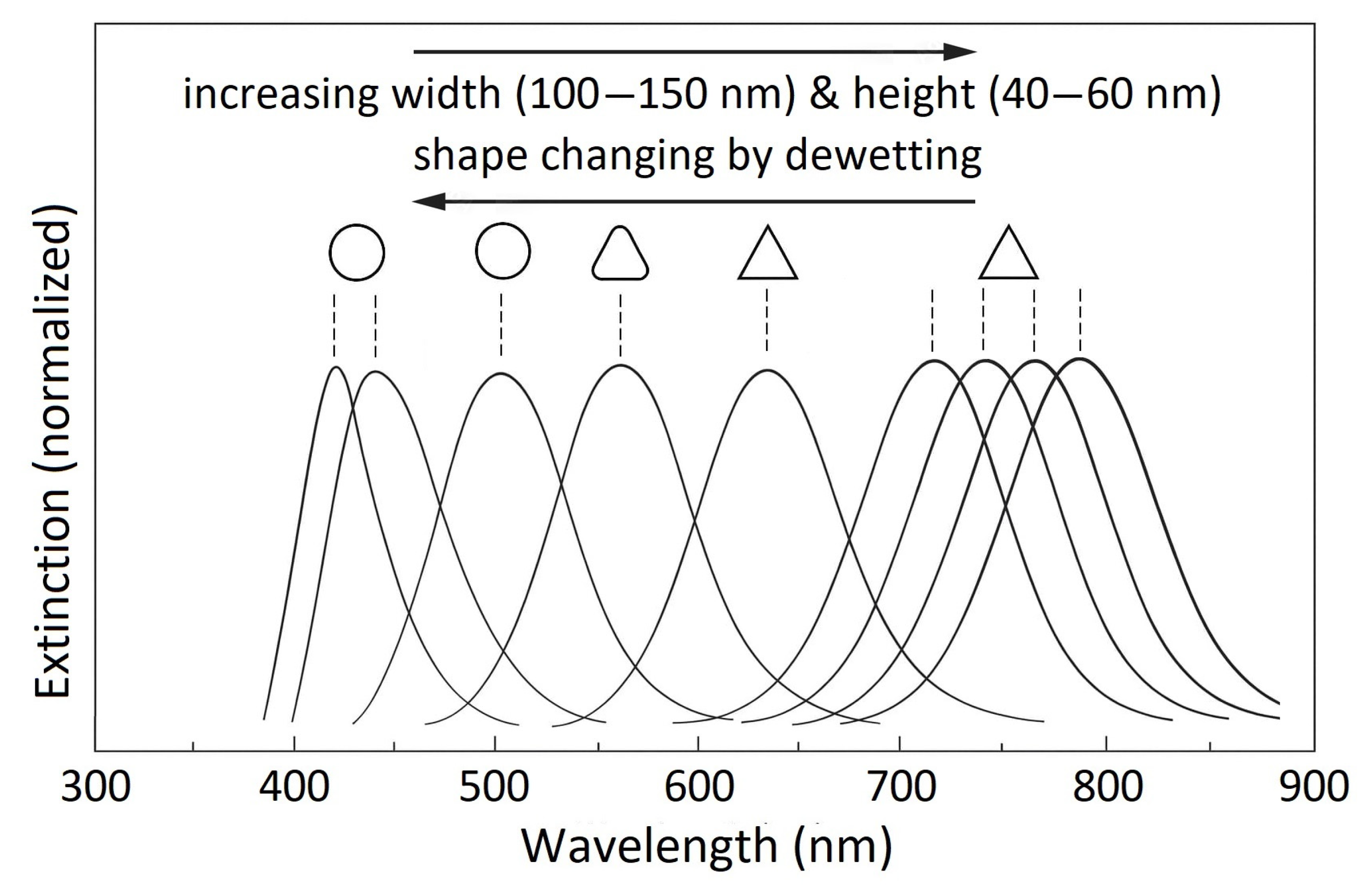
Figure 1. The localized surface plasmon resonance (LSPR) results in strong absorption or scattering of light at specific wavelengths, depending on the nanostructure’s size, shape, and type of materials—e.g., Au, Ag, Ir, etc. Image adapted from Ref. [17], for a series of Au resonator plots made by colloidal lithography mask combined with pulsed laser deposition and subsequent controlled dewetting.
Metals 15 01030 g001
Figure 2. The schematic for fabricating gold nanoislands by microwave annealing/dewetting.
Metals 15 01030 g002
Figure 3. The formation of Au nanostructures by controlled dewetting, using a microwave-assisted technique of annealing: Au thin film deposition in (a), Au thin film rupture by controlled dewetting in (b), Au formation of islands by further dewettting effects in (c), and Au nanoisland formation as the final step, in (d).
Metals 15 01030 g003
Figure 4. AFM images of 5 nm gold thin film without adhesion layer: (a) before annealing and (b) after thermal annealing in a kiln/microwave at 550 °C for 13 min.
Metals 15 01030 g004
Figure 5. SEM image of a 5 nm Au thin film, as deposed/before annealing, in (a) SEM images of nanoislands formation after annealing in the kiln/microwave setup and the effect of increasing thin film thickness: (b) 4 nm, (c) 5 nm, (d) 6 nm, (e) 8 nm, and (f) 10 nm.
Metals 15 01030 g005
Figure 6. XRD measurements and comparison of the crystallite size of Au nanoislands, for microwave annealing vs. a traditional oven.
Metals 15 01030 g006
Figure 7. UV–vis absorption spectra of 4–10 nm Au thin films after (a) oven annealing and (b) kiln/microwave annealing. The films exhibit a localized surface plasmon resonance (LSPR) band at 550–600 nm, with thinner films showing stronger absorption. Microwave annealing produces sharper and more intense LSPR peaks, indicating more efficient nanoisland formation compared with conventional oven annealing.
Metals 15 01030 g007
Disclaimer/Publisher’s Note: The statements, opinions and data contained in all publications are solely those of the individual author(s) and contributor(s) and not of MDPI and/or the editor(s). MDPI and/or the editor(s) disclaim responsibility for any injury to people or property resulting from any ideas, methods, instructions or products referred to in the content.