Article

The Role of Pores in the Cavitation Erosion of Additively Manufactured Metal: An In Situ Study

1
School of Mechanical Engineering, Yanshan University, Qinhuangdao 066004, China
2
Shanghai Engineering Research Center of Hot Manufacturing, Shanghai Dianji University, Shanghai 201306, China
*
Author to whom correspondence should be addressed.
Metals 2025, 15(7), 787; https://doi.org/10.3390/met15070787
Submission received: 24 May 2025 / Revised: 7 July 2025 / Accepted: 10 July 2025 / Published: 11 July 2025
(This article belongs to the Section Metal Failure Analysis)

Abstract

Additively manufactured (AM) parts have been applied in many areas with the risk of cavitation erosion (CE), and pores are common defects in AM metals. However, the role of pores in CE is still unclear, and a systematic investigation is needed. In this study, 316L stainless steel was selected as a model material and produced using laser powder bed fusion; the porosity was 6.4%. The morphological evolution of various pores during CE was investigated via electron backscatter diffraction and scanning electron microscopy. It was found that material removal easily occurred around large polygonal pores. The critical size for large polygonal pores was estimated to be between 13 and 20 μm. For narrow pores, concavity first appeared around the pores during CE, and then the narrow pores closed. Small spherical pores with sizes of 3–9 μm showed strong resistance to CE, and no damage occurred within the 60 min CE period. The main reason that different pores played different roles in CE was analyzed. Finally, factors for improving the CE resistance of AM metals were suggested. The research results are helpful for understanding the CE behaviors of AM metals and porous materials.

1. Introduction

Pores are common defects in additively manufactured (AM) metals [1,2]. According to their formation mechanism, pores are categorized as lack-of-fusion, keyhole, and gas pores [3,4,5]. Different types of pores have various shapes and sizes. An insufficient energy density results in the incomplete fusion of new material to previously deposited powder, thus resulting in lack-of-fusion pores. This type of pore is generally large and polygonal [6,7]. The sudden closure of the laser keyhole leaves a pore in the molten pool. When the pore is caught by the solidification front, a keyhole pore appears [8,9,10]. Compared with lack-of-fusion pores, keyhole pores are smaller and more regular. Gas pores are formed because of various gases, such as hydrogen and argon [11]. These gases can become entrapped by fluctuations on the surface of the molten pool, be introduced by powders, or precipitate from the molten metal during solidification [11,12]. Gas pores are small and spherical. In most cases, pores are detrimental to mechanical properties [13,14,15,16].
Cavitation erosion (CE) is a general form of damage to parts, such as pumps, propellers, valves, hydraulic turbines, and pipes, in liquid environments [17,18,19,20]. CE even occurs in high-speed gears and bearings under liquid lubrication conditions [21,22]. The formation and collapse of bubbles lead to CE. When a high-speed liquid flows through a solid surface or the flow rate suddenly changes, bubbles form inside the liquid owing to the decrease in pressure. The collapse of bubbles generates strong shock waves and micro-jets. Under repeated impact, the solid undergoes deformation, material removal, and even fracture [23,24,25,26]. Persistent slip lines and striations on eroded materials indicate the fatigue nature of CE [27,28,29].
In recent years, AM parts have been applied in many areas with the risk of CE, and their CE behavior is receiving increasing attention. Compared with parts prepared via traditional methods, AM parts possess higher residual stress, higher hardness, and finer grain size. These factors are beneficial for CE resistance [30]. However, the surface roughness of AM parts is poor, and if they are used directly, the weight loss rate in the initial stage of CE is high [31]. Grain morphology affects CE resistance. For AM 316L stainless steel, the equiaxed crystal structure exhibits lower CE resistance than the columnar crystal structure because the former contains more grain boundaries [32]. In addition to the roughness and grain morphology, pores also affect the CE resistance of AM metals. Pores are usually the potential initiation sites of cracking during CE [33,34].
However, not all pores have a negative effect on the CE resistance of metals. It has been reported that pore closure occurs in 316L stainless steel during CE [35]. This is contrary to the general phenomenon reported in the literature [34]. A possible reason is that pores with different sizes and shapes may have different effects on CE resistance [36,37]. CE is a special fatigue process, and phenomena in the common fatigue process may help explain CE. In the common fatigue of AM Inconel 718, there is a size effect of the pores on fatigue. In a structure with an average grain size of 48 μm, the critical size of a harmful pore is 20 μm [38]. Interestingly, for AM 316L stainless steel with many pores, the fatigue strength is similar to that of conventionally made steel [39]. These results indicate the complex roles of pores in fatigue.
In this study, 316L stainless steel was selected as the model material. Its microstructure is simple and uniform, which is conducive to analyzing the shape and size effects of pores in CE. In addition, 316L stainless steel has been widely used in hydraulic systems with risks of CE because of its good mechanical properties and corrosion resistance [32,34]. The morphologies of various pores in AM 316L stainless steel were observed before and after CE. The research results are helpful for understanding the CE behaviors of AM metals and porous materials. The novelty of this study lies in the use of in situ observation to systematically study the evolution process of polygonal narrow holes and spherical pores during CE, and it provides the reasons for their damage, closure, and preservation.

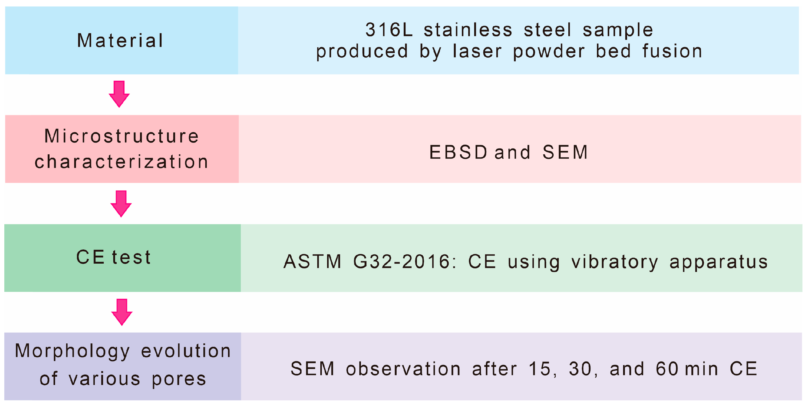
2. Materials and Methods

In this study, 316L stainless steel samples (Φ15 mm × 100 mm) were manufactured via laser powder bed fusion using an ED 3028 machine (Shanghai ESU Laser Technology Co., Ltd., Shanghai, China) in an argon atmosphere. The powder particle sizes were 10–50 μm. The process parameters were a laser power of 350 W, a scanning speed of 1800 mm/s, a hatch spacing of 90 μm, and a layer thickness of 60 μm. The aim of using these parameters is to obtain certain pores. The chemical composition was (wt%) 0.03 C, 17.17 Cr, 11.65 Ni, 2.82 Mo, 0.91 Si, 0.74 Mn, and balanced Fe. Because heat treatment can cause pore cracking [40], the as-built metal was directly tested without heat treatment in this study. The hardness of the AM 316L stainless steel used in this study is 37 HRC.
The morphology of pores in AM 316L stainless steel was observed at the steel’s cross-section (perpendicular to the build direction) using a Hitachi S4800 scanning electron microscope (SEM) (Hitachi High-Tech Corporation, Tokyo, Japan). Typical pores are shown in Figure 1, and the porosity is 6.4%. The shapes and sizes of the pores vary. Polygonal pores are large, and their side lengths are tens of micrometers, as indicated by the blue arrows. They are usually lack-of-fusion pores. The pores in the bottom left corner of Figure 1 are narrow in width and long in length. They are referred to as narrow pores in this study. Smooth edges indicate that they are pores rather than cracks. The purple arrow indicates a pore with a partially melted powder particle. There are also some spherical pores, as indicated by white arrows. Considering their shape and small size, it is believed that they are gas pores. This study systematically characterizes the morphological evolution of these four types of pores in CE.
Before CE, the as-built microstructure was examined via electron backscatter diffraction (EBSD). Specimens (Φ10 mm × 3 mm) were cut from the as-built bar. After grinding with 4000-grade SiC paper, the EBSD specimens were finally polished using Ar ions with an 19520 CP cross-section polisher (JEOL Ltd., Tokyo, Japan). EBSD analysis was performed with a ZEISS Gemini 460 SEM (ZEISS Group, Oberkochen, Germany) and OIM analysis 8.6 software. The scanning regions were selected randomly. Points with confidence index values of less than 0.2, usually corresponding to pores, were filtered. In the EBSD results, the high-angle grain boundaries, low-angle grain boundaries, and twin boundaries are shown as black, gray, and white lines, respectively.
After EBSD analysis, the specimens were subjected to cavitation erosion using an XOQS-2500 machine (Nanjing Xianou Instrument Manufacturing Co., Ltd., Nanjing, China), in accordance with ASTM G32-2016 [41] and the method in our previous study [24]. The CE specimen was installed on a holder and immersed in 25 °C pure water. The eroded surface was oriented perpendicular to the build direction. The distance between the horn tip and the specimen was 0.5 mm. The frequency, peak-to-peak displacement amplitude, and power were 20,000 Hz, 50 μm, and 1500 W, respectively. Specimens were subjected to cavitation erosion for 15, 30, and 60 min. At each test interval, the eroded surfaces were observed via SEM to characterize the changes in their pores. Figure 2 shows the experimental flowchart of this study.

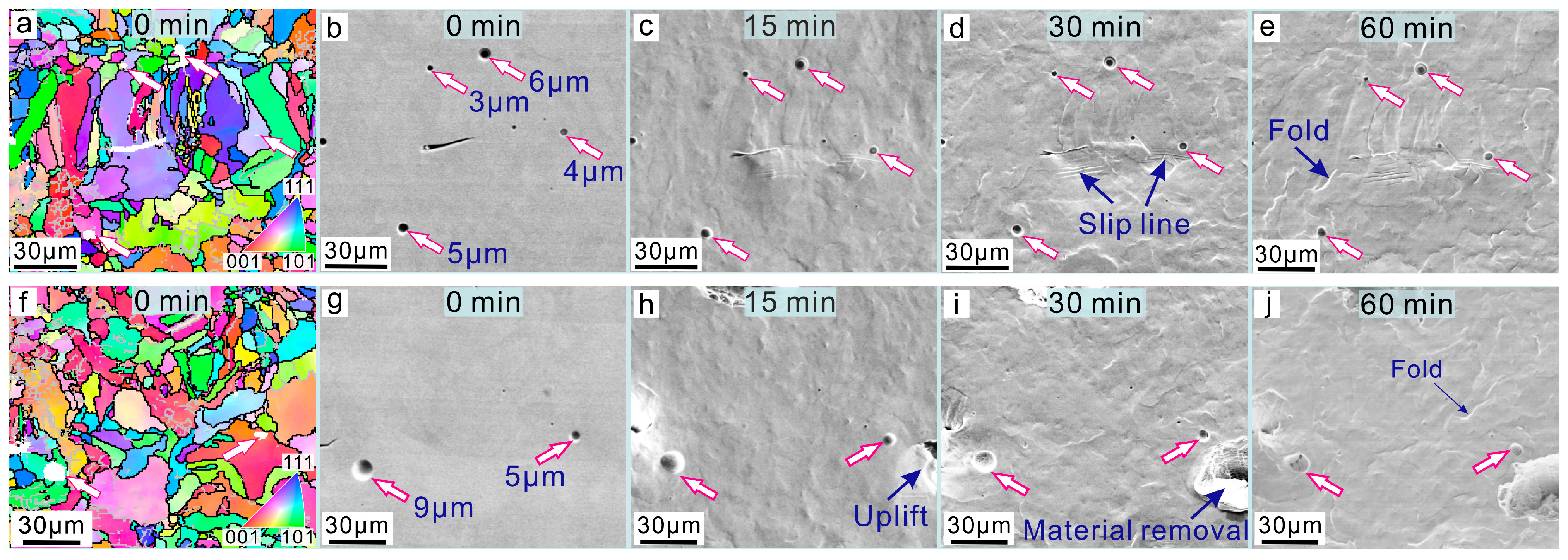
3. Results and Discussion

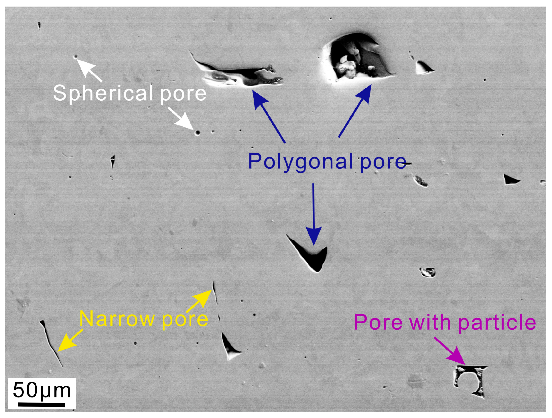
3.1. Role of Polygonal Pores in CE

An EBSD image of a polygonal pore (white) before CE is shown in Figure 3a. The overall dimension of the polygonal pore were ~50 μm. The grains around this pore were heterogeneous. The equiaxed grains were fine and 2–10 μm in diameter, and the columnar grains were 30–50 μm in length. Low-angle grain boundaries were present in some grains. Interestingly, no twin boundaries were found. Before CE, the metal around the pore was flat and smooth (Figure 3b). After 15 min of CE, an uplift appeared at the bottom of the pore (Figure 3c). The dashed line indicates the original position of the pore edge before CE. The uplift was thick and appeared to be above 30 μm. Based on Figure 3a and c, the surface area of the uplift covered more than 50 grains. Considering the thickness, there were hundreds of grains in the uplift. After 30 min of CE, the uplift in Figure 3c fell off (Figure 3d). The material that was removal propagated downward after 60 min of CE (Figure 3e). An enlarged image of the blue dashed region is shown in Figure 3f. The pore wall was smooth but uneven, likely representing the free surface formed during solidification. Slip lines appeared in front of the material removed. An enlarged image of the yellow dashed region in Figure 3e is shown in Figure 3g. Numerous striations indicate the fatigue nature of CE.
The overall dimensions of the other two polygonal pores were between 20 and 27 μm (Figure 4a,b). After 15 min of CE, a large portion of the metal near the upper pore was falling off (Figure 4c). Material removal occurred next to the lower pore. After 30 min of CE (Figure 4d), the portion falling off in Figure 4c had fallen off. It appears that these two pores in Figure 4b comprise one pore that is connected below the sample’s surface. Under cavitation exposure, the thin material layer above the pore was removed, revealing the pore volume below. When the CE time was 60 min, material removal was more severe (Figure 4e). Many striations can be observed in Figure 4f, and slip lines can be observed in Figure 4g. From the results in Figure 3 and Figure 4, it can be concluded that material removal easily occurred around the polygonal pores.

3.2. Role of Narrow Pores in CE

The morphological evolution of a narrow pore in CE is shown in Figure 5a–e. The length of the pore was 47 μm, and its largest width was a mere 4 μm (Figure 5b). The lower tip of this pore ended in a grain (Figure 5a). In the middle and at the bottom of this pore, the misorientation angles between the two sides were measured to be 3° and 4°, respectively, as highlighted by the cubes in Figure 5b. After 15 min of CE, concavity appeared to the left of the narrow pore. The metal on two sides of the pore pushed together (Figure 5c). Interestingly, after 30 min of CE, the pore closed (Figure 5d). When the CE time was 60 min, the complete closure of the pore occurred (Figure 5e). Concavity appeared before the closure of the narrow pore. This indicates that the closure was the result of large plastic deformations.
Another two narrow pores are shown in Figure 5f,g. The largest width of the left pore was 13 μm. The right tip of this pore was very narrow, with a misorientation angle of only 3° between the two sides. The largest width of the right pore was 7 μm. After 15 min of CE, concavity appeared. The pore morphology after 30 min of CE was similar to that after 15 min of CE (Figure 5h,i). When the CE time was 60 min, the pore was almost completely closed (Figure 5j). The largest width of the fourth narrow pore was 11 μm (Figure 5k,l). After 15 and 30 min of CE (Figure 5m,n), concavities and slip lines appeared next to the pore, similarly to the cases shown in Figure 5c,h. When the CE time was 60 min, complete closure of the pore also occurred (Figure 5o). Clearly, the behavior of the narrow pores in CE was entirely different from that of the polygonal pores. The narrow pores closed even though their width was 13 μm, and the polygonal pores showed substantial material removal.

3.3. Role of Spherical Pores in CE

Four spherical pores with diameters of 3–6 μm were separately distributed in Figure 6a,b. The 4 μm pore was located in the grain’s interior, and the other pores were located at the grain boundaries. A small narrow pore can also be observed at the image’s center. After 15 min of CE (Figure 6c), these four spherical pores maintained their shapes and sizes, even though the small narrow pore closed. When the CE time was 30 and 60 min, slip lines and folds appeared on the metal, but these four spherical pores did not change (Figure 6d,e).
The morphological evolution of another two spherical pores under CE is shown in Figure 6f–j. One spherical pore was 9 μm in diameter, and another was 5 μm in diameter. The pores were all located at grain boundaries. After 15 min of CE, the metal to the right of the 5 μm pore was uplifted (Figure 6h). However, this pore was free of deformation. When the CE time was 30 and 60 min, material removal occurred next to the 5 μm pore. Interestingly, this pore remained free of damage (Figure 6i,j). Thus, spherical pores exhibited strong resistance to CE.

3.4. Role of Pores with Powder Particles in CE

The morphological evolution of pores with partially melted powder particles in CE is shown in Figure 7. Figure 7a–c show the same region, and Figure 7d–f show another region. Three pores containing partially melted powder particles are shown in Figure 7b. The particle in the lower left corner of Figure 7b was fused with the matrix at its lower side. For the particle in the lower right corner of Figure 7b, both its upper and lower sides are connected with the matrix. Regular grains between the particles and matrix can be observed in the IPF map (Figure 7a). However, after 15 min of CE, all these particles had fallen off (Figure 7c). A similar situation is shown in Figure 7d–f. After 15 min of CE, one particle had completely fallen off, and another particle had partially fallen off (Figure 7f).

3.5. Size and Shape Effects of the Pores

The behaviors of various pores were different in the same CE environment (Figure 3, Figure 4, Figure 5, Figure 6 and Figure 7), and whether a pore is damaged depends on its shape and size. In general, large polygonal pores and small spherical pores are lack-of-fusion and gas pores, respectively [6,7,11,12]. In the present study, the pores were not classified by their formation mechanism. This is because, during the layer-by-layer AM process, the holes formed in the previous layer may change in the subsequent layer [42]. Thus, classifying the pores by their formation mechanism will not be accurate. Based on the above phenomena, the behaviors of the different pores in CE are proposed, as illustrated schematically in Figure 8. Large polygonal pores, narrow pores, and small spherical pores were subjected to a CE environment. Cavitation bubbles are usually several hundred micrometers in diameter [17,43]. They are unlikely to enter the interior of large polygonal pores and collapse. However, it is believed that a micro-jet carrying remnant bubbles can enter the interior of a large polygonal pore. In addition, remnant bubbles can further collapse and induce impact [44]. As a result, the inner pressure of a large polygonal pore suddenly increases. The edge of the pore will generate uplift (Figure 3 and Figure 4). Once uplift occurs, the stress state becomes more severe, and material removal readily occurs. The critical size for large polygonal pores was estimated to be between 13 and 20 μm in this study.
For narrow and small spherical pores, the micro-jet cannot enter the pore interior owing to their small sizes. The impact of the micro-jet only affects the specimen’s surface. Under compressive stress, the edge of a narrow pore starts to deform, which results in concavity formation. When the deformation reaches a certain level, the narrow pore closes. After concavity formation and pore closure, erosion is unlikely to cause further damage.
Spherical pores have strong resistance to CE because of their small size and spherical shape. From a shape perspective, spherical pores do not cause stress concentration. It is worth noting that small pores with sizes of approximately 1 μm in 316L stainless steel closed after 45 min of CE in Ref. [35]. However, in this study, pores with sizes of 3–9 μm were free of deformation after 60 min of CE. Upon careful observation, it was found that the pores in Ref. [35] were caused by inclusion or precipitation, and they were deep circular holes rather than spherical pores. The difference in the shape may result in different CE resistance. When designing porous materials that face the risk of CE, the shape of the pores should be controlled based on the research results of this study.

3.6. Improving the CE Resistance of AM Metals

Pores may not be completely eliminated in industrial production, and the inhibition of large polygonal pores is important to improve the CE resistance of AM parts. It should be pointed out that the formation and collapse of bubbles, as well as the size of the micro-jets, vary between different liquids and temperatures [45]. Moreover, the critical size of large polygonal pores depends on the liquid and temperature.
Furthermore, based on the research findings of this study, it is speculated that an increase in plasticity also enhances the CE resistance of AM metals. This is because the closure of narrow pores requires large plastic deformation. If plasticity is low, cracking may occur during concavity formation. Optimizing plasticity through annealing may result in decreases in the dislocation density and an increase in the grain size of AM metals, all of which are unfavorable. In this study, no twin boundaries were found in the as-built metal. After annealing, twin boundaries, which are potential sites of erosion initiation, may form in face-centered cubic materials [46,47]. Therefore, developing methods to improve the plasticity of AM metals remains a major challenge.
A smooth surface is also important to inhibit the CE of AM parts. As shown in Figure 3, once a portion of the metal uplifts, a further 15 min of CE will lead to material removal. One piece of removed metal may contain more than one hundred grains. Any portion elevated from the metal will lead to a stress-state transformation from simple compression to complex multiaxial loading. If the surface roughness is poor, the material loss rate of AM parts during the initial stage of CE will be very high [48]. Therefore, polishing AM metals is important for industrial applications.
Cracks are another type of defect in AM metals [1]. Although cracks were not considered in this study, the behaviors of narrow pores and pores with particles provide indications of how cracks will behave in CE. If the crack is perpendicular to the specimen surface, its behavior will be similar to that of a narrow pore, and it will likely close. If the crack is inclined to the surface, the portion above the crack—that is, the portion next to the specimen’s surface—will easily fall off, similarly to pores with partially melted powder particles.
In this study, we investigated the role of pores during CE in pure water, specifically pores destroyed the continuity of the passive film. The role of pores during CE in corrosive liquids, such as seawater and acid, should be investigated in future studies.

4. Conclusions

In this study, we investigated the evolution of various pores in AM 316L steel during CE using EBSD and SEM. The following conclusions were drawn:
(1) The behaviors of large polygonal pores, narrow pores, and small spherical pores in CE are proposed, and the main reason that different pores play different roles in CE is discussed.
(2) For large polygonal pores, the edge portion of the pores easily generates uplift, which may include hundreds of grains, in the early stages of CE. Material removal then occurs around the polygonal pores. The critical size for large polygonal pores was estimated to be between 13 and 20 μm in this study.
(3) For narrow pores, concavity first appears around the pores in CE. The narrow pores then close because of plastic deformation. Spherical pores with sizes of 3–9 μm have strong resistance to CE, and no damage occurs during the 60 min CE period because of their small size and spherical shape.
(4) When pores with partially melted powder particles are placed in a CE environment, powder particles easily fall off.

Author Contributions

Conceptualization, Z.W. and Y.S.; methodology, Y.S. and B.M.; investigation, Z.W., Y.S. and B.M.; writing—original draft, Y.S.; writing—review and editing, Z.W.; project administration, Z.W. All authors have read and agreed to the published version of the manuscript.

Funding

This research was funded by the Shanghai Engineering Research Center for Hot Manufacturing Program, grant number 18DZ2253400.

Data Availability Statement

The raw/processed data required to reproduce these findings cannot be shared at this time as the data also forms part of an ongoing study.

Conflicts of Interest

The authors declare no conflicts of interest.

Abbreviations

The following abbreviations are used in this manuscript:
AMAdditively manufactured
CECavitation erosion
SEMScanning electron microscope
EBSDElectron backscatter diffraction

References

  1. Mukherjee, T.; Elmer, J.W.; Wei, H.L.; Lienert, T.J.; Zhang, W.; Kou, S.; DebRoy, T. Control of grain structure, phases, and defects in additive manufacturing of high-performance metallic components. Prog. Mater. Sci. 2023, 138, 101153. [Google Scholar] [CrossRef]
  2. Sanaei, N.; Fatemi, A. Defects in additive manufactured metals and their effect on fatigue performance: A state-of-the-art review. Prog. Mater. Sci. 2021, 117, 100724. [Google Scholar] [CrossRef]
  3. Ren, Z.; Gao, L.; Clark, S.J.; Fezzaa, K.; Shevchenko, P.; Choi, A.; Everhart, W.; Rollett, A.D.; Chen, L.; Sun, T. Machine learning-aided real-time detection of keyhole pore generation in laser powder bed fusion. Science 2023, 379, 89–93. [Google Scholar] [CrossRef] [PubMed]
  4. Laleh, M.; Hughes, A.E.; Yang, S.; Wang, J.; Li, J.; Glenn, A.M.; Xu, W.; Tan, M.Y. A critical insight into lack-of-fusion pore structures in AM stainless steel. Addit. Manuf. 2021, 38, 101762. [Google Scholar]
  5. Li, E.; Zhou, Z.; Wang, L.; Zou, R.; Yu, A. Numerical studies of melt pool and gas bubble dynamics in laser powder bed fusion process. Addit. Manuf. 2022, 56, 102913. [Google Scholar] [CrossRef]
  6. Vandecasteele, M.; Heylen, R.; Iuso, D.; Thanki, A.; Philips, W.; Witvrouw, A.; Verhees, D.; Booth, B.G. Towards materials and process agnostic features for the classification of pore types in metal additive manufacturing. Mater. Des. 2023, 227, 111757. [Google Scholar] [CrossRef]
  7. Fan, F.; Jiang, M.; Wang, P.; Liu, C.; Liu, Z.; Chen, Z. Defect-associated microstructure evolution and deformation heterogeneities in additively manufactured 316L stainless steel. Mater. Sci. Eng. A 2022, 861, 144287. [Google Scholar] [CrossRef]
  8. Chen, L.; Yao, X.; Tan, C.; He, W.; Su, J.; Weng, F.; Chew, Y.; Ng, N.P.H.; Moon, S.K. In-situ crack and keyhole pore detection in laser directed energy deposition through acoustic signal and deep learning. Addit. Manuf. 2023, 69, 103547. [Google Scholar] [CrossRef]
  9. Zhao, C.; Parab, N.D.; Li, X.; Fezzaa, K.; Tan, W.; Rollett, A.D.; Sun, T. Critical instability at moving keyhole tip generates porosity in laser melting. Science 2020, 370, 1080–1086. [Google Scholar] [CrossRef]
  10. Yu, T.; Zhao, J. Quantifying the mechanisms of keyhole pore evolution and the role of metal-vapor condensation in laser powder bed fusion. Addit. Manuf. 2023, 72, 103642. [Google Scholar] [CrossRef]
  11. Stopka, K.S.; Desrosiers, A.; Nicodemus, T.; Krutz, N.; Andreaco, A.; Sangid, M.D. Intentionally seeding pores in AM alloy 718: Process parameters, microstructure, defects, and fatigue. Addit. Manuf. 2023, 66, 103450. [Google Scholar]
  12. Fiegl, T.; Franke, M.; Körner, C. Correlation of powder degradation, energy absorption and gas pore formation in laser-based powder bed fusion process of AlSi10Mg0.4. Addit. Manuf. 2022, 56, 102917. [Google Scholar] [CrossRef]
  13. Mostafaei, A.; Ghiaasiaan, R.; Ho, I.T.; Strayer, S.; Chang, K.C.; Shamsaei, N.; Shao, S.; Paul, S.; Yeh, A.C.; Tin, S.; et al. Additive manufacturing of nickel-based superalloys: A state-of-art review on process-structure-defect-property relationship. Prog. Mater. Sci. 2023, 136, 101108. [Google Scholar] [CrossRef]
  14. Zhang, R.; Li, W.; Jiao, Y.; Paniagua, C.; Ren, Y.; Lu, H. Porosity evolution under increasing tension in wire-arc additively manufactured aluminum using in-situ micro-computed tomography and convolutional neural network. Scr. Mater. 2023, 225, 115172. [Google Scholar] [CrossRef]
  15. Luo, Y.W.; Zhang, B.; Feng, X.; Song, Z.M.; Qi, X.B.; Li, C.P.; Chen, G.F.; Zhang, G.P. Pore-affected fatigue life scattering and prediction of additively manufactured Inconel 718: An investigation based on miniature specimen testing and machine learning approach. Mater. Sci. Eng. A 2021, 802, 140693. [Google Scholar] [CrossRef]
  16. Yang, Y.; Hu, L.; Huang, J. Effect of microstructure and printed pores on the adiabatic shearing behavior of Ti6Al4V titanium alloy manufactured by selective electron beam melting. Mater. Charact. 2023, 196, 112642. [Google Scholar] [CrossRef]
  17. Gonzalez-Avila, S.R.; Nguyen, D.M.; Arunachalam, S.; Domingues, E.M.; Mishra, H.; Ohl, C.D. Mitigating cavitation erosion using biomimetic gas-entrapping microtextured surfaces (GEMS). Sci. Adv. 2020, 6, eaax6192. [Google Scholar] [CrossRef]
  18. McClintock, D.A.; Liu, Y.; Bruce, D.R.; Winder, D.E.; Schwartz, R.G.; Kyte, M.; Blokland, W.; Sangrey, R.L.; Carroll, T.M.; Long, C.D.; et al. Small-bubble gas injection to mitigate cavitation-induced erosion damage and reduce strain in target vessels at the spallation neutron source. Mater. Des. 2022, 221, 110937. [Google Scholar] [CrossRef]
  19. Reuter, F.; Deiter, C.; Ohl, C.D. Cavitation erosion by shockwave self-focusing of a single bubble. Ultrason. Sonochem. 2022, 90, 106131. [Google Scholar] [CrossRef]
  20. Dong, F.; Li, X.; Zhang, L.; Ma, L.; Li, R. Cavitation erosion mechanism of titanium alloy radiation rods in aluminum melt. Ultrason. Sonochem. 2016, 31, 150–156. [Google Scholar] [CrossRef]
  21. Mo, X.; Wang, J.; Cheng, L.; Ouyang, T. Numerical prediction of vibration-induced cavitation erosion in high-speed gears using erosion risk indicators. Tribol. Int. 2023, 179, 108122. [Google Scholar] [CrossRef]
  22. Cheng, F.; Wu, F.; Wang, S.; Peng, X.; Cao, Y.; Yang, S. A prediction model for suction cavitation erosion in a journal bearing. Tribol. Int. 2023, 184, 108424. [Google Scholar] [CrossRef]
  23. Sreedhar, B.K.; Albert, S.K.; Pandit, A.B. Cavitation damage: Theory and measurements—A review. Wear 2017, 372−373, 177–196. [Google Scholar] [CrossRef]
  24. Wang, Z.; Wang, Y. Cavitation erosion and its effect on corrosion resistance of nuclear-grade Z3CN20-09M stainless steel. Mater. Des. 2022, 221, 110978. [Google Scholar] [CrossRef]
  25. Hou, G.; Ren, Y.; Zhang, X.; Dong, F.; An, Y.; Zhao, X.; Zhou, H.; Chen, J. Cavitation erosion mechanisms in Co-based coating exposed to seawater. Ultrason. Sonochem. 2020, 60, 104799. [Google Scholar] [CrossRef]
  26. Chen, S.Y.; Xu, W.L.; Luo, J.; Li, J.B.; Zhai, Y.W. Experimental study on the mesoscale causes of the effect of sediment size and concentration on material cavitation erosion in sandy water. Wear 2022, 488–489, 204114. [Google Scholar] [CrossRef]
  27. Cheng, J.; Wu, Y.; Hong, S.; Cheng, J.; Qiao, L.; Wang, Y.; Zhu, S. Cavitation-erosion behavior and mechanism of high-velocity oxygen-fuel sprayed CuAlNiTiSi medium-entropy alloy coating. Surf. Coat. Technol. 2022, 432, 128096. [Google Scholar] [CrossRef]
  28. Wang, Z.; Wang, Y. Hydrogen participates in cavitation erosion in water. Wear 2022, 504–505, 204435. [Google Scholar] [CrossRef]
  29. Qiao, Y.; Chen, J.; Zhou, H.; Wang, Y.; Song, Q.; Li, H.; Zheng, Z. Effect of solution treatment on cavitation erosion behavior of high-nitrogen austenitic stainless steel. Wear 2019, 424–425, 70–77. [Google Scholar] [CrossRef]
  30. Girelli, L.; Tocci, M.; Montesano, L.; Gelfi, M.; Pola, A. Investigation of cavitation erosion resistance of AlSi10Mg alloy for additive manufacturing. Wear 2018, 402–403, 124–136. [Google Scholar] [CrossRef]
  31. Guse, F.; Voshage, M.; Schleifenbaum, J.H.; Schmitz, K. Cavitation erosion resistance of additive manufactured materials. Chem. Eng. Technol. 2023, 46, 37–44. [Google Scholar] [CrossRef]
  32. Ding, H.; Tang, Q.; Zhu, Y.; Zhang, C.; Yang, H. Cavitation erosion resistance of 316L stainless steel fabricated using selective laser melting. Friction 2021, 9, 1580–1598. [Google Scholar] [CrossRef]
  33. Sasaki, H.; Takeo, F.; Soyama, H. Cavitation erosion resistance of the titanium alloy Ti-6Al-4V manufactured through additive manufacturing with various peeing methods. Wear 2020, 462–463, 203518. [Google Scholar] [CrossRef]
  34. Hardes, C.; Pöhl, F.; Röttger, A.; Thiele, M.; Theisen, W.; Esen, C. Cavitation erosion resistance of 316L austenitic steel processed by selective laser melting (SLM). Addit. Manuf. 2019, 29, 100786. [Google Scholar] [CrossRef]
  35. Tian, Y.; Zhao, H.; Yang, R.; Liu, X.; Chen, X.; Qin, J.; McDonald, A.; Li, H. In-situ SEM investigation on stress-induced microstructure evolution of austenitic stainless steel subjected to cavitation erosion and cavitation erosion-corrosion. Mater. Des. 2022, 213, 110314. [Google Scholar] [CrossRef]
  36. Wilson-Heid, A.E.; Beese, A.M. Combined effects of porosity and stress state on the failure behavior of laser powder bed fusion stainless steel 316L. Addit. Manuf. 2021, 39, 101862. [Google Scholar] [CrossRef]
  37. Plessis, A.; Yadroitsava, I.; Yadroitsev, I. Effects of defects on mechanical properties in metal additive manufacturing: A review focusing on X-ray tomography insights. Mater. Des. 2020, 187, 108385. [Google Scholar] [CrossRef]
  38. Prithivirajan, V.; Sangid, M.D. The role of defects and critical pore size analysis in the fatigue response of additively manufactured IN718 via crystal plasticity. Mater. Des. 2018, 150, 139–153. [Google Scholar] [CrossRef]
  39. Kumar, P.; Jayaraj, R.; Suryawanshi, J.; Satwik, U.R.; McKinnell, J.; Ramamurty, U. Fatigue strength of additively manufactured 316L austenitic stainless steel. Acta Mater. 2020, 199, 225–239. [Google Scholar] [CrossRef]
  40. Laleh, M.; Sadeghi, E.; Revilla, R.I.; Chao, Q.; Haghdadi, N.; Hughes, A.E.; Xu, W.; De Graeve, I.; Qian, M.; Gibson, I.; et al. Heat treatment for metal additive manufacturing. Prog. Mater. Sci. 2023, 133, 101051. [Google Scholar] [CrossRef]
  41. ASTM G32-16; Standard Test Method for Cavitation Erosion Using Vibratory Apparatus. ASTM International: West Conshohocken, PA, USA, 2016. [CrossRef]
  42. Sinclair, L.; Leung, C.L.A.; Marussi, S.; Clark, S.J.; Chen, Y.; Olbinado, M.P.; Rack, A.; Gardy, J.; Baxter, G.J.; Lee, P.D. In situ radiographic and ex situ tomographic analysis of pore interactions during multilayer builds in laser powder bed fusion. Addit. Manuf. 2020, 36, 101512. [Google Scholar] [CrossRef]
  43. Dular, M.; Ohl, C.D. Bulk material influence on the aggressiveness of cavitation—Questioning the microject impact influence and suggesting a possible way to erosion mitigation. Wear 2023, 530–531, 205061. [Google Scholar] [CrossRef]
  44. Gonzalez-Parra, J.C.; Robles, V.; Devia-Cruz, L.F.; Rodriguez-Beltran, R.I.; Cuando-Espitia, N.; Camacho-Lopez, S.; Aguilar, G. Mitigation of cavitation erosion using laser-induced periodic surface structures. Surf. Interfaces 2022, 29, 101692. [Google Scholar] [CrossRef]
  45. France, J.P.; Michel, J.M. Fundamentals of Cavitation, 1st ed.; Kluwer Academic Publishers: New York, NY, USA, 2005. [Google Scholar] [CrossRef]
  46. Zhang, W.; Chabok, A.; Kooi, B.J.; Pei, Y. Additive manufacturing high entropy alloys: A review of the microstructure and properties. Mater. Des. 2022, 220, 110875. [Google Scholar] [CrossRef]
  47. Wang, Z.; Zhang, B. Cavitation erosion behavior of high-nitrogen austenitic stainless steel: Effect and design of grain-boundary characteristics. Mater. Des. 2021, 201, 109496. [Google Scholar] [CrossRef]
  48. Krella, A.; Marchewicz, A. Effect of mechanical properties of CrN/CrCN coatings and uncoated 1.402 stainless steel on the evolution of degradation and surface roughness in cavitation erosion. Tribol. Int. 2023, 177, 107991. [Google Scholar] [CrossRef]
Figure 1. Typical pores in AM 316L stainless steel.
Metals 15 00787 g001
Figure 2. Experimental flowchart.
Metals 15 00787 g002
Figure 3. The morphological evolution of a polygonal pore in CE. Panels (a,b) show the inverse pore figure (IPF) and SEM images of a polygonal pore before CE. (ce) SEM images after 15, 30, and 60 min CE, respectively. Panels (f,g) show enlarged images of the blue and yellow dashed regions in (e).
Metals 15 00787 g003
Figure 4. The morphological evolution of another two polygonal pores in CE. Panels (a,b) show IPF and SEM images before CE. (ce) SEM images after 15, 30, and 60 min CE, respectively. Panels (f,g) show enlarged images of the blue and yellow dashed regions in (e).
Metals 15 00787 g004
Figure 5. The morphological evolution of narrow pores in CE. (a,b) show IPF and SEM images of a narrow pore before CE. (ce) SEM images after 15, 30, and 60 min CE, respectively. (f,g) show IPF and SEM images of another two narrow pores before CE. (hj) SEM images after 15, 30, and 60 min CE, respectively. (k,l) show IPF and SEM images of the fourth narrow pore before CE. (mo) SEM images after 15, 30, and 60 min CE, respectively.
Metals 15 00787 g005
Figure 6. The morphological evolution of spherical pores in CE. (a,b) show IPF and SEM images of four spherical pores before CE. (ce) SEM images after 15, 30, and 60 min CE, respectively. (f,g) show IPF and SEM images of another two spherical pores before CE. (hj) SEM images after 15, 30, and 60 min CE, respectively.
Metals 15 00787 g006
Figure 7. The morphological evolution of pores with partially melted powder particles in CE. (ac) Region 1. (a,b) show IPF and SEM images before CE. (c) SEM image after 15 min CE. (df) Region 2. (d,e) show IPF and SEM images before CE. (f) SEM image after 15 min CE.
Metals 15 00787 g007
Figure 8. Schematic illustration of the behaviors of various pores in CE.
Metals 15 00787 g008
Disclaimer/Publisher’s Note: The statements, opinions and data contained in all publications are solely those of the individual author(s) and contributor(s) and not of MDPI and/or the editor(s). MDPI and/or the editor(s) disclaim responsibility for any injury to people or property resulting from any ideas, methods, instructions or products referred to in the content.

