Article

Room-Temperature Superplasticity in a Biodegradable Zn-0.1Mg Alloy

1
Institute of Materials, Faculty of Materials, Metallurgy and Recycling, Technical University of Košice, Letná 1/9, 042 00 Košice, Slovakia
2
Institute of Materials Research of SAS, Slovak Academy of Sciences, Watsonova 47, 040 01 Košice, Slovakia
3
Institute of Physics, Faculty of Science, Pavol Jozef Šafárik University, Park Angelinum 9, 041 54 Košice, Slovakia
4
Faculty of Electrical Engineering and Informatics, Technical University of Košice, Letná 1/9, 042 00 Košice, Slovakia
*
Author to whom correspondence should be addressed.
Metals 2025, 15(10), 1161; https://doi.org/10.3390/met15101161
Submission received: 27 August 2025 / Revised: 13 October 2025 / Accepted: 20 October 2025 / Published: 21 October 2025
(This article belongs to the Special Issue The Forming Behaviour and Plasticity of Metallic Alloys)

Abstract

Biodegradable zinc-based alloys have recently emerged as promising candidates for temporary biomedical implants due to their favorable biocompatibility, appropriate degradation rate, and relatively simple processing. In this study, the Zn-0.1Mg alloy was investigated after being processed by means of a two-step equal-channel angular pressing (ECAP) route, consisting of the first pass at 150 °C followed by a second pass at room temperature. The mechanical properties were evaluated using uniaxial tensile tests at different strain rates, while the microstructure and phase composition were analyzed using synchrotron hard X-ray diffraction and transmission electron microscopy (TEM). The processed alloy exhibited a remarkable enhancement in both strength and ductility compared to the annealed state. At the lowest applied strain rate, a fracture elongation of up to 240% was achieved at room temperature, representing a unique manifestation of superplasticity under ambient conditions. Diffraction analysis confirmed the stability of the supersaturated Zn matrix with minor Mg2Zn11 intermetallic phase. TEM observations revealed an ultrafine-grained microstructure and activation of non-basal slip systems, which enabled efficient plastic flow. These findings demonstrate that controlled severe plastic deformation provides an effective pathway for tailoring Zn-Mg alloys, opening opportunities for their use in the next generation of bioresorbable low-to-moderate load orthopedic fixation devices, e.g., plates, screws, suture anchors and craniofacial miniplates.

1. Introduction

In contemporary orthopedic and trauma surgery, more than two-thirds of all implanted medical devices are metallic [1,2]. Among them, the most widely used are stainless steel 316L, cobalt-chromium (Co-Cr) alloys, and titanium and its alloys (e.g., Ti-6Al-4V). In cardiovascular interventions, thin-walled stents made of CoCr alloys or nitinol predominate, whereas in dental implantology, titanium and its alloys dominate, accounting for more than 90% due to their excellent biocompatibility and high corrosion resistance. However, these materials exhibit a significantly higher density (4.5–9 g·cm−3) compared to human bone (0.27–1.8 g·cm−3) [3], as well as an elastic modulus (190–200 GPa for stainless steel, 210–240 GPa for Co-Cr alloys, and 90–110 GPa for titanium alloys) is significantly higher than that of bone (3–30 GPa).
The implantation of permanent biomedical devices is frequently associated with inflammatory responses, thrombus formation, decalcification, osteopenia, and subsequent osteoporosis in the vicinity of the implant, resulting from load transfer from bone to implant, known as “stress shielding” [4]. Other complications include long-term implant migration [5], incompatibility with standard CT and MRI imaging, and restrictions in new tissue growth in younger patients [6]. A further burden for patients is the need for secondary surgery to remove temporary implants made from such materials.
In vascular therapy, a persistent problem of permanent metallic stents is late thrombosis and restenosis [7,8]. An ideal vascular scaffold should provide sufficient radial support to prevent elastic recoil during healing after angioplasty and subsequently degrade at a rate matching vessel recovery, thereby restoring normal reactivity [9].
The long-term presence of permanent implants may also induce local and systemic complications. One of the most critical is the release of metal ions and wear debris from implant surfaces, particularly at modular junctions or sliding interfaces. This wear results in the diffusion of cobalt, chromium, nickel, vanadium, and titanium ions into surrounding tissues and the bloodstream, provoking local inflammation and aseptic osteolysis [10,11,12]. Another major issue is biofilm formation on implant surfaces, which promotes bacterial adhesion and periprosthetic infection. Such infections are resistant to conventional antibiotics and require complex surgical management. The incidence of infections following primary implantation is estimated at 1–3% but can exceed 10% after revision surgery [13]. Systemic consequences of metal particle release are especially associated with hip implants made of CoCrMo alloys. Elevated cobalt and chromium concentrations may cause neurological symptoms, cardiomyopathy, and thyroid or renal dysfunction [13,14]. Nanoparticles from these alloys may also accumulate in the liver, spleen, and central nervous system [15]. These complications not only reduce quality of life but also necessitate costly revision surgeries, whose success rate remains only about 60–80% [16].
In this context, replacing permanent orthopedic and vascular implants with bioresorbable alternatives is highly desirable. These materials are composed solely of elements naturally present in the human body. They degrade at a controlled rate, providing mechanical support only during the healing phase, after which they are fully resorbed. Moreover, their degradation products (Zn, Ca, Mg ions) contribute to healing and regeneration of surrounding soft tissues [17,18,19]. For clinical applicability, implants must fulfill general requirements summarized in Table 1, which were first systematically introduced by Heiden et al. [20].
Beyond mechanical performance, biodegradable metallic implants must be biocompatible and avoid adverse local or systemic responses. In degradable systems, excessive hydrogen evolution during corrosion can lead to gas pocket formation (classically observed in Mg-based implants). At the same time, it must provide sufficient mechanical reinforcement of the tissue throughout the entire healing period. For hard tissues, the elastic modulus should be as close as possible to that of natural bone (10–20 GPa) to minimize complications associated with stress shielding. Both tensile strength and yield strength must exceed those of the supported tissue. For cortical bone, the yield strength ranges from 80 to 120 MPa and the ultimate tensile strength ranges from 130 to 180 MPa [3,21]. Surface characteristics—such as chemistry, roughness, and topography should be optimized to enhance cell adhesion and proliferation. The degradation rate must be controlled so that the implant is fully resorbed by the body within an appropriate time frame (from six months to several years) without the formation of large particles or toxic products.
Until recently, research on bioresorbable alloys has been primarily focused on Mg-, Ca-, and Fe-based systems [22,23,24]. However, increasing attention is now devoted to zinc alloys due to their relatively low degradation rate, good biocompatibility, and simple preparation [25,26,27,28,29]. To enhance their mechanical properties, which still limit broader clinical use, alloying combined with plastic deformation is commonly employed [30,31,32]. For biomedical applications, several Zn-based biodegradable alloys have been investigated, particularly Zn-Mg, Zn-Cu, Zn-Li, and Zn-Mn systems [25]. Among these, Zn-Mg is the most extensively studied binary system [33,34]. Previous studies [35,36], have shown that increasing Mg content can significantly improve mechanical strength due to the formation of the secondary Mg2Zn11 phase. However, this strengthening effect is often accompanied by reduced ductility and corrosion resistance. The resulting mechanical properties are strongly influenced by processing, including severe plastic deformation (SPD) techniques such as ECAP and HPT, which effectively refine the microstructure and improve both strength and ductility [37].
In this context, the study by Ye et al. [32] is particularly noteworthy. The authors demonstrated that in a low-alloyed Zn-0.1Mg system (wt.%) subjected to room-temperature rotary-die equal-channel angular pressing (ECAP), it was possible to achieve a yield strength of 329 MPa, ultimate tensile strength of 383 MPa, excellent ductility of 45.6%, and a relatively low degradation rate in Hank’s solution (0.014 mm/year). In terms of both strength and ductility, this biodegradable material considerably exceeds the requirements for vascular and orthopedic scaffolds (see Table 1).
The aim of this work was therefore to explore approaches for achieving the required mechanical properties in Zn-Mg alloys by employing a modified, single-pass ECAP route that is operationally simpler than multi-pass procedures. To activate non-basal slip systems (prismatic and pyramidal), a two-step strategy was applied: the first pass at 150 °C followed by a second pass at room temperature, with the second pass performed via route Bc (90° sample rotation between passes). The resulting microstructure and phase composition were analyzed via high-energy synchrotron X-ray diffraction and transmission electron microscopy (TEM). The Zn-0.1Mg alloy processed in this manner was further evaluated via uniaxial tensile testing to establish the fundamental mechanical characteristics of the material.
Superplastic flow in metals is classically associated with (i) very fine grains (typically <10 μm and often sub-micrometer), (ii) low strain rates (~10−3–10−4 s−1), and (iii) sufficiently high temperatures (conventionally ≥ 0.5 Tₘ) that facilitate grain-boundary-mediated deformation (grain-boundary sliding accommodated by diffusion and intragranular slip). While elongation > 200% is a pragmatic benchmark, strain-rate sensitivity is a key criterion; in superplastic regimes, the m-value
m   =   𝜕 ln σ 𝜕   l n   ϵ ˙ ε ,   T
is typically m ≳ 0.3–0.5 [38,39,40]. Reports on hcp alloys (Ti-6Al-4V, Mg systems) and Zn-based alloys (e.g., Zn-Al, Zn-Ag, Zn-0.033 Mg) show that achieving such behavior generally requires ultrafine grains and careful control of rate and temperature [38,39,40,41].
Severe plastic deformation (SPD) routes—especially equal-channel angular pressing (ECAP)—are effective in producing ultrafine-grained (UFG) microstructures with a high fraction of high-angle boundaries, modified textures, and activation of non-basal slip in hcp Zn (prismatic/pyramidal), all of which improve room-temperature formability and delay strain localization [32,37,42]. In low-alloyed Zn-Mg systems, ECAP (and hybrid extrusion-ECAP) has delivered simultaneous gains in strength and ductility and, under appropriate rate/temperature conditions, very high elongations consistent with superplastic-like ductility [32,39,40,43]. Motivated by these advances, we examine whether a two-step ECAP schedule can tailor Zn-0.1 Mg to exhibit large plastic reserve at room temperature, and discuss the results in the context of microstructure and texture.

2. Materials and Methods

2.1. Material Preparation

The Zn-0.1Mg alloy (wt.%) was prepared by melting high-purity Zn (99.998%) and Mg (99.985%) in a resistance-heated furnace at 600 °C under a protective argon atmosphere (99.999% purity). The melt was gravity-cast into graphite molds with a diameter of 20 mm and a length of 70 mm. The resulting chemical composition of the alloy corresponded to the nominal values. After surface machining, the samples underwent homogenization annealing at 380 °C for 2 h to dissolve intermetallic phases, followed by water quenching. This condition is hereafter referred to as the ‘annealed’ state. From the annealed alloy, smooth cylindrical specimens with a diameter of 8 mm were machined and encapsulated in a 1 mm thick copper sleeve (Figure 1c). The encapsulated samples were then deformed in an ECAP die with an internal channel angle φ = 90°, having a circular cross-section of 10 mm diameter, at a ram speed of 8 mm·min−1. The first pass was conducted at 150 °C, while the second pass was performed at room temperature via route BC (with a 90° sample rotation between passes). The material processed by this combination of two ECAP passes hereafter referred to as ‘150 °C/RT’.

2.2. Mechanical Properties Analysis

Specimens for the tensile test were machined from the alloy in both the annealed and 150 °C/RT states (Figure 1a), with dimensions shown in Figure 1b, and subjected to uniaxial tensile testing. The tensile tests were carried out using the electromechanical testing machine H300KU Tinius Olsen (Tinius Olsen Testing Machine Company, Horsham, PA, USA) under constant crosshead speed, adjusted so that the nominal initial engineering strain rates were: 0.001 s−1, 0.00025 s−1, and 0.0001 s−1. All experiments were performed at room temperature. For each strain rate, at least three specimens were tested to ensure reproducibility.

2.3. X-Ray Diffraction Experiment

Half of the fractured tensile specimen processed via the 150 °C/RT ECAP route was subjected to transmission X-ray diffraction measurements using high-energy synchrotron radiation at the I12 beamline of the Diamond Light Source [44]. The experimental conditions were as follows:
The synchrotron radiation emitted from a superconducting undulator was monochromated by a pair of Si (111) crystals in Laue geometry, yielding a photon energy of E = 87.368 keV, corresponding to a wavelength of λ = 0.14191 Å. The brilliance of this radiation was approximately 1011 photons·s−1·mm−2 per 0.1% bandwidth. The selected photon energy was sufficient to fully penetrate the specimen, allowing bulk structural information to be obtained. The incident beam cross-section was 0.5 × 0.5 mm. The specimen was mounted on a vertical translation stage for scanning. As illustrated in Figure 2, the specimen was first aligned at position 0 (near the threads, 18 mm from the fracture surface) and then translated downward in 0.5 mm steps towards the fracture zone, resulting in a total of 36 measurements along the gauge length. The exposure and data acquisition time at each position was 1 s. At each step, a 2D diffraction pattern was recorded using a Pilatus 2M CdTe detector (DECTRIS Ltd., Baden, Switzerland) with a resolution of 1475 × 1679 pixels and a pixel size of 172 µm × 172 µm. The 2D diffraction images were subsequently radially integrated and analyzed using the GSAS-II software package version 5799 [45].

2.4. Structure Analysis

The structure of the alloy after tensile testing was examined via high-resolution scanning transmission electron microscopy (STEM) using a JEOL JEM-2100F (JEOL Ltd., Tokyo, Japan), equipped with a Schottky field emission gun and operated at an accelerating voltage of 200 kV. Phase identification within the alloy structure was carried out by selected area electron diffraction (SAED) in TEM mode. Thin foil specimens for TEM were prepared by conventional metallographic procedures, including sectioning, mechanical grinding, and polishing to a thickness of ~50 μm, followed by ion milling using a JEOL Ion Slicer EM-09100IS (JEOL, Japan). Ar+ ion thinning was carried out at 6 kV, followed by a final low-energy polish at 3 kV. Specimens were extracted from two regions:
(i)
Position 0, minimally affected by deformation;
(ii)
Position 36, just beneath the fracture surface (see Figure 2).
The documented TEM foil surface was aligned parallel to the specimen’s longitudinal axis after tensile testing.

3. Results

3.1. Mechanical Testing Results

Figure 3 shows the engineering stress-strain curves for the annealed specimen (v = 0.00025 s−1) and the ECAP-processed alloy (150 °C/RT) at various strain rates. The mechanical properties derived from these tests are summarized in Table 2, including yield strength (YS), ultimate tensile strength (UTS) and fracture elongation (FE) at various strain rates. As can be seen, the material in the annealed state exhibits relatively low YS and UTS (70 and 74 MPa, respectively) and extremely poor ductility, with FE = 1.8%. Fracture occurred by brittle transgranular failure, as illustrated in Figure 3b. In contrast, the alloy processed by ECAP at 150 °C/RT shows a pronounced increase in both strength and ductility, with stress-strain behavior characteristic of ultrafine-grained materials. These typically display low uniform elongation on the order of 5% (from YS to UTS), followed by large non-uniform elongation (from UTS to fracture), where the flow stress gradually decreases. High plasticity is evidenced by the reduction in area (RA = 96%), as shown in Figure 3c.
The mechanical response of the 150 °C/RT material was strongly strain-rate sensitive: as strain rate decreased, YS and UTS declined, while ductility increased markedly. At the lowest strain rate tested (0.0001 s−1), the alloy exhibited FE values as high as 240%, which can be classified as a superplastic state.
In general, superplasticity is observed under the following conditions: (i) low strain rates of 0.001–0.0001 s−1; (ii) elevated deformation temperatures of ~0.5 Tm; and (iii) a fine-grained microstructure. A material is commonly considered superplastic when elongation exceeds 200% (or true strain εt > 1). Superplasticity has been reported in hcp alloys such as Ti-6Al-4V [38], Mg alloys (e.g., AZ31), Zn-Al and Zn-Ag alloys [39], and Zn-0.33Mg (wt.%) alloy [40], most of which were processed by severe plastic deformation techniques such as ECAP or HPT to obtain refined microstructures. However, their superplastic behavior was achieved only at elevated temperatures in the range of 200–800 °C. In the present case, the superplastic state was measured at room temperature (20 °C), which is unique.

3.2. XRD Structural Analysis

Figure 4a presents the X-ray diffraction patterns obtained after radial integration of the 2D Debye-Scherrer images into the 2θ-I diffraction space. At all measurement positions—from the region near the threads to the fracture zone—the dominant phase was identified as the hexagonal close-packed (hcp) Zn phase with space group symmetry P63/mmc (No. 194). In addition, minor intermetallic phases were detected, namely Mg2Zn11 with space group Pm-3m (No. 200), and possibly MgZn2 (P63/mmc), as illustrated in Figure 4b.
Figure 5 shows the variations in structural parameters obtained during scanning of the tensile specimen from the threaded region (position 0) towards the fracture zone (position 36). Based on this comparison, it can be stated that the lattice parameter a does not vary by more than 0.04% across the scanned range, whereas the parameter c changes by up to 0.14%. Thus, the overall volumetric expansion of the unit cell in the fracture region is primarily attributed to the increase in the c parameter. The crystallite size remains approximately constant (~1 μm) throughout the scanned interval, while the second-order elastic microstrains, determined from the broadening of Bragg peaks using the Williamson-Hall method [45], systematically decrease towards the fracture. This reduction is most likely a consequence of dislocation relaxation through slip along basal, prismatic, and pyramidal systems activated by the preceding ECAP plastic deformation. The lowest value of elastic microstrain is observed in the fracture zone, which can be attributed to stress release due to the presence of a free surface behind the fracture. The preferred orientation of crystallites, expressed by the texture index J (Figure 5d), increases gradually from the specimen grip region towards the fracture zone.

3.3. Analysis of First-Order Elastic Residual Strains and Stresses

Residual elastic strains and associated (Type I) residual stresses may arise from severe plastic deformation (ECAP) and subsequent processing, and they can strongly influence yield behavior, strain localization, damage initiation, and dimensional stability. Diffraction methods probe lattice strains via Bragg-peak positions; by comparing measured d-spacings with a stress-free reference d0 and using suitable elastic constants, one obtains residual elastic strains and the corresponding macrostresses. In the present work, we apply this approach to assess the extent to which the two-pass ECAP route induces or relaxes internal stresses in Zn-0.1Mg and to relate these to texture and ultrafine-grained microstructure. For methodology and interpretation, see [41,47,48].
The GSAS-II software package enables the determination of first-order residual elastic strains based on the ellipticity of Debye-Scherrer rings obtained from 2D XRD images, using the method described by B. B. He [49]. Using this capability, residual strains were determined along the tensile specimen from the threaded region to the fracture zone. Figure 6a shows one of the 2D XRD images, while Figure 6b presents the upper-right quadrant of the same image with the crystallographic indices of the corresponding Debye-Scherrer rings indicated. Figure 6c illustrates the variations of the normal strain tensor components (ε11, ε22) and the shear component (ε12) along the observed part of the fractured specimen. The calculation was carried out using the (112) diffraction ring, which is recommended for hcp systems such as Ti, Mg, and Zn. Based on the obtained strain values, the residual internal stresses can subsequently be calculated using the following relations:
For normal stresses:
σ 11 = E 1 v 2 ε 11 + v ε 22
σ 22 = E 1 v 2 ε 22 + v ε 11
For shear stresses:
σ 12 = G ε 12
where E is Young’s modulus, which for Zn is 108 GPa; υ is Poisson’s ratio, which for Zn is 0.25; and G is the shear modulus, calculated as G = E 2 1 + v .
Based on Figure 6c and the application of the corresponding relations for calculating the stress tensor components, it was possible to localize the region with the highest internal stress. This region is located approximately 1 mm from the fracture surface and exhibits tensile stresses of σ11 = 59 MPa and σ22 = 63 MPa. The maximum shear stress σ12 reaches a value of 11 MPa.

3.4. Structure Analysis

STEM in transmission mode documented the structure after tensile testing, and SAED confirmed the presence of individual phases (Figure 7). The structure of the alloy was composed of Zn solid solution features and particles of the Mg2Zn11 phase embedded within the solid solution, as confirmed by the diffraction patterns in Figure 7. In the pattern of the Zn solid solution, strong reflections of the major Zn phase are observed. Less intense reflections from the minor Mg2Zn11 phase are also present.
The structure and substructure of the Zn-0.1Mg alloy after tensile testing at position 0 (near the threads) is documented by STEM in Figure 8a–d. The structure consisted of regions containing polyhedral grains of the solid solution with equilibrium grain boundaries and lower dislocation density (Figure 8a), as well as regions with elongated grains within deformation bands exhibiting higher dislocation density (Figure 8b). The average size of equiaxed solid solution grains was ~580 ± 156 nm (grain size was determined from N = 32 grains), while the average width and length of elongated grains were ~510 ± 116 nm and ~1100 ± 230 nm, respectively, (N = 9). Within the solid solution, regions with a polygonized dislocation structure were observed (Figure 8c), along with dislocation interactions with particles embedded in the solid solution (Figure 8d). The relatively low dislocation density in the solid solution grains with predominantly high-angle equilibrium boundaries indicates that the structure of this state is recovered and, in some cases, recrystallized.
Figure 9a–d illustrate the structure and substructure of the Zn-0.1Mg alloy after tensile testing at position 36 (fracture zone of the specimen). The structure consisted of regions with polyhedral solid solution grains exhibiting equilibrium grain boundaries and lower dislocation density (Figure 9a), as well as regions with elongated grains and the formation of subgrains within deformed grains characterized by higher dislocation density (Figure 9b). The average size of equiaxed solid solution grains was ~480 ± 136 nm (N = 18), while the average width and length of elongated grains were ~470 ± 164 nm and ~990 ± 308 nm (N = 11), respectively, values slightly smaller compared with those in the minimally deformed region (position 0). However, in the region beneath the fracture surface, coarser elongated grains were observed, with average widths exceeding 1 μm and lengths above 2 μm, within which finer subgrains had developed.
Within the solid solution, regions with polygonized dislocation arrangements were observed (Figure 9c), together with dislocation-particle interactions (Figure 9d). The morphology of the solid solution structure at position 36 of the tensile-tested specimen shows only minor differences compared with that observed at position 0. Nevertheless, an increased amount of polygonized dislocation networks was identified, indicating a more extensive degree of structure recovery.

4. Discussion

Ye et al. in their scientific publication [32] presented a method to transform the originally brittle hcp Zn-0.1Mg alloy with low strength into a strong yet ductile material. For processing, they employed eight passes of rotary-die equal-channel angular pressing (ECAP) conducted at room temperature. The application of severe plastic deformation led to significant grain refinement from the initial size of >30 μm down to ~0.5 μm (width) × ~0.9 μm (length) dimensions. Simultaneously, in the hexagonal lattice, not only basal slip systems {0001}<11 2 ¯ 0> but also prismatic {10 1 ¯ 0}<11 2 ¯ 0> and, most importantly, pyramidal slip systems {10 1 ¯ 1}<11 2 ¯ 0>, {10 1 ¯ 1}<11 2 ¯ 3>, {11 2 ¯ 2}< 1 ¯ 1 ¯ 23> were activated. Without ECAP, plastic deformation in the hcp Zn-0.1Mg occurs almost exclusively through dislocation slip along basal planes, which are limited in number (only three), resulting in poor ductility and brittle fracture. Schmid factor analysis of EBSD images, however, demonstrated that after ECAP processing there is a pronounced increase in the number of activated pyramidal slip systems of the first order {10 1 ¯ 1}<11 2 ¯ 3> (12 variants) and of the second order {11 2 ¯ 2}< 1 ¯ 1 ¯ 23> (6 variants). Their activation enables more efficient dislocation motion along diagonal directions of the hcp lattice, thereby substantially improving ductility from the original < 2% up to 45.6%.
The aim of this work was to explore new approaches for achieving highly ductile to superplastic properties in Zn-Mg alloys using a modified ECAP processing route. The ECAP method applied in this study consisted of only two passes: the first conducted at 150 °C and the second at room temperature. Such a procedure is considered technologically simpler, suitable for processing larger volumes of material, and promising for future automation. A further objective was to explore whether this alloy can achieve very high uniform ductility at room temperature (we use ε > 200% as a pragmatic benchmark). We emphasize that elongation alone does not establish superplastic flow, which is commonly associated with a high strain-rate sensitivity (m ≳ 0.3–0.5) and grain-boundary-sliding. In the absence of rate-jump measurements of m in this work, we therefore refer to superplastic-like ductility [43].
As demonstrated by the tensile test results of specimens processed using the ECAP method (150 °C/RT) at a low strain rate of v = 0.0001 s−1, an elongation to fracture of up to 240% was achieved. This exceptional result indicates a high plastic reserve of the material, which can be further enhanced either by microalloying (e.g., with Li, Mn, and Ca) or by additional plastic deformation. In fine-grained Zn-based alloys, trace Li can promote microstructural refinement and nano-precipitation (Zn-Li), thereby increasing the strain-hardening capacity and delaying diffuse necking, concurrent increases in strength and elongation have been reported in Zn-Li and Zn-(Al)-Li alloys. Mn additions further refine grains and reduce the harmful effect of Fe by forming (Fe,Mn)-Zn intermetallics, which improves intergranular cohesion and ductility. Controlled Ca additions introduce fine CaZn13 dispersoids that stabilize sub-micrometer grains (Zener pinning) and raise work-hardening; however, excessive or coarse CaZn13 can degrade ductility, underscoring the need for dispersion control. These microalloying routes are synergistic with SPD (e.g., ECAP), which sustains the grain sizes that favor grain-boundary-mediated deformation at room temperature.
Another objective of this study was the analysis of the phase composition and structure of the material after fracture using high-energy X-ray radiation. In contrast to conventional laboratory measurements performed in Bragg-Brentano reflection geometry, which provide surface-sensitive information, these measurements were carried out in transmission mode using Debye-Scherrer geometry, thus delivering bulk information from the analyzed specimen. The experiments were conducted by scanning the specimen from a region located 18 mm away from the fracture surface up to the fracture itself. The results revealed that the specimen across its entire profile consisted of a major hcp-Zn phase and minor intermetallic phases Mg2Zn11 and possibly also the MgZn2. It was confirmed that neither ECAP processing nor severe plastic deformation during tensile loading induced any transformation of the major Zn phase, which remained hcp (space group P63/mmc, No. 194). However, an increase in the unit cell volume of approximately 0.16% was observed in the region near the fracture. This volumetric expansion was primarily caused by elongation of the unit cell along the <0001> direction (i.e., along the c-axis).
An interesting observation is the linear decrease of second-order microstrains towards the necking region and subsequently towards the fracture of the specimen. Since second-order microstrains are an indicator of dislocation density, this suggests that their fraction decreases with increasing plastic deformation. This phenomenon can be explained by the activation of pyramidal slip systems, which facilitate the motion of dislocations from the grain interiors towards grain boundaries or the free surface, while the low density of precipitates in the lattice is insufficient to effectively pin them. Such dislocation motion may also result in the annihilation of pre-existing dislocations present in the structure prior to tensile loading.
The XRD line-profile-derived coherence length (‘crystallite’/domain size) remains approximately 1 µm before and after tensile deformation; any change lies within the experimental uncertainty. TEM shows an ultrafine-grained microstructure (e.g., equiaxed grains ~0.58 µm and elongated grains with widths ~0.47–0.51 µm and lengths ~0.99–1.10 µm). Taken together, this indicates that ECAP produced the principal refinement (from >30 µm in the annealed state to sub-micrometer grains), whereas the subsequent tensile test did not appreciably alter the coherently diffracting domain size. Because the XRD domain size measures coherence length and is influenced by low-angle boundaries and dislocation substructure, it is related to—but not identical to—the TEM grain size. The texture of the material, expressed by the preferred orientation of crystallites, becomes more pronounced towards the necking region and the fracture. As also reported by Ye et al. [32], the Zn-0.1Mg alloy exhibits a sharp texture in which the basal planes (0001) are oriented perpendicular to the direction of ECAP deformation (⟨0001⟩∥ED).
Macroscopic first-order residual stresses reach a maximum at approximately 1 mm from the fracture surface, with the values of normal stresses attaining ~30% of the yield strength of this material.
From the comparison of the structure of the homogenized Zn-0.1Mg alloy after double ECAP processing (150 °C/RT) and subsequent tensile testing at the site of maximum contraction, it can be concluded that the structures differ only slightly. After two ECAP passes, the alloy structure consisted of equiaxed grains (~0.6 μm) and elongated grains (~0.5 μm in width and ~1.1 μm in length), or subgrains of the Mg solid solution in Zn. Within the solid solution grains, dislocations were mostly arranged in networks. These dislocation networks acted as nuclei of low-angle subgrain boundaries formed during slip deformation in the course of ECAP processing.
Grain refinement induced by severe plastic deformation during ECAP, together with preferential slip deformation during the second ECAP pass, likely contributed to dynamic recovery and, in some cases, recrystallization of the alloy matrix. The migration of grain boundaries and dislocations was further hindered by fine Mg2Zn11 phase particles (of size ~ 60 nm), which were present in the alloy and likely precipitated during the first ECAP pass at 150 °C. The interaction of dislocations with such particles is evident in Figure 8d and Figure 9d.
A similarly high elongation of ~240% at the same strain rate was also achieved by the authors of Ref. [42] for a Zn-0.5Mn alloy, whose ultrafine-grained structure was prepared by multiple-pass hot extrusion. In that alloy, severe extrusion led to refinement of the Zn solid solution grains down to ~0.35 μm and also to refinement of the intermetallic MnZn13 particles, which effectively hindered dislocation and grain boundary migration. In contrast, the strong fibrous texture was weakened into a random texture. Such changes in grain orientation and texture may result from grain rearrangement or grain rotation during the extrusion process. Evidence of preferential alloy deformation by dislocation slip is the absence of deformation twins in the solid solution grains. Dislocation slip occurs predominantly in pyramidal planes of the solid solution at high levels of plastic strain, while the contribution of twinning and basal slip decreases [50].
Significant microstructural refinement via ECAP (4p-Bc-RT) was also confirmed by the authors of Ref. [43] in a Zn-0.1Mg-0.05Li alloy. In that study, ultrafine solid solution grains with an average size of ~1.47 μm were formed, accompanied by an increased dispersion of Mg2Zn11 particles. EBSD analysis revealed an increased fraction of deformed grains and a decrease in the fraction of recrystallized grains. The fraction of low-angle grain boundaries and the Kernel average misorientation also increased, indicating a higher proportion of deformed grains. The basal texture of the ECAP-processed solid solution was removed, and the share of multiple texture components increased. The alloy simultaneously exhibited high strength and ductility (YS: 349 MPa; UTS: 457 MPa; elongation: 67.6%). The structure of the Mg solid solution in Zn analyzed in this study, even after tensile testing near the fracture zone, contained equiaxed grains (~0.48 μm) and elongated grains (~0.47 μm in width and ~1 μm in length), or subgrains showing signs of alignment in the tensile loading direction. Their average dimensions were slightly smaller compared to the ECAP state, and they exhibited a higher dislocation density. Dislocations were frequently arranged in networks and polygons, forming smaller subgrains within larger grains. These observations indicate that during tensile testing, dynamic recovery and recrystallization occurred, along with further refinement of solid solution grains or subgrains, which led to the achievement of high elongation (~240%) at a low strain rate of 0.0001 s−1 and room temperature. However, this was accompanied by relatively low strength values (YS: 147 MPa; UTS: 173 MPa). At higher strain rates (0.0001 s−1), increased strength characteristics were measured (YS: 219 MPa; UTS: 258 MPa), but at the expense of reduced elongation (62%), which is close to the value reported in Ref. [43]. Grain refinement and the reduced mobility of grain boundaries or dislocations were also supported by the pinning effect of Mg2Zn11 particles, whose interactions with dislocations were observed both after tensile testing and after ECAP processing. With increasing deformation intensity, the c lattice parameter, the unit cell volume, and the texture index also increased. A similar increase in the 10 1 ¯ 1 fiber texture index was reported by the authors of Ref. [42] in a Zn-1Mg alloy subjected to multiple-pass hot extrusion. Such a texture is favorable for enhancing plasticity in hcp metals and reduces their tendency towards plastic strain localization and brittle fracture. The authors of Ref. [51] also analyzed the structure of specimens after tensile testing. They observed further grain refinement of the solid solution down to ~1.23 μm, a significant increase in the fraction of recrystallized grains, and a corresponding decrease in the fraction of deformed grains. The structure consisted predominantly of fine equiaxed grains with a smaller fraction of coarser elongated grains of the solid solution. EBSD analysis confirmed the increased fraction of recrystallized grains by a pronounced decrease in the proportion of low-angle grain boundaries and a reduction in the Kernel average misorientation. All these findings demonstrate that dynamic recrystallization of the solute-containing Zn solid solution also occurs during tensile testing, where strain softening is reflected in the tensile curves by the continuous stress decrease after reaching the ultimate tensile strength and by relatively high elongation values. The additional deformation energy during tensile loading leads to further multiplication of dislocations and activates recrystallization processes in Zn with low solute content, owing to its low recrystallization temperature.
Processing of Zn and other materials with an hcp lattice strongly affects their texture. An important structural parameter influencing the texture of these materials is the Schmid factor, which defines the potential for activation of deformation modes with different values of the critical resolved shear stress (CRSS) for slip or twinning [52]. Due to the low CRSS value for basal slip in the hcp Zn lattice, a weak basal texture is unfavorable for improving the strength of Zn, but advantageous for increasing its ductility. In addition to basal slip, non-basal slip modes should also be activated to enable homogeneous deformation of Zn. Grain refinement below a critical size (typically ~0.1 μm) can significantly suppress deformation by twinning [53,54]. In the present study, the alloy after ECAP was refined to an average grain size of ~1.47 μm, and therefore twinning during ECAP deformation was more difficult to activate. Among the non-basal slip systems, slip along pyramidal planes is thus the most likely to be activated, whereas the probability of prismatic slip activation remains low.

5. Conclusions

The long-term presence of permanent implants in the human body is frequently associated with serious complications such as inflammation, release of metal ions, or the necessity of revision surgery. Bioresorbable alloys, which gradually degrade in vivo while providing sufficient mechanical support during the healing process, represent a promising alternative to permanent implant materials. Among all currently investigated bioresorbable alloys, Zn-Mg alloys are particularly attractive due to their low degradation rate and favorable biocompatibility. The aim of this study was therefore to demonstrate how a modified ECAP technique can be employed to achieve a combination of strength, ductility, and even superplasticity at room temperature in the Zn-0.1Mg alloy.

Main Findings

  • Relative to the annealed baseline (380 °C/2 h + water quench), characterized by a coarse-grained hcp Zn solid solution with grain size > 30 µm, low dislocation density, and minor Mg2Zn11 (see Table 2), the two-step ECAP route (150 °C/RT) produced a concurrent increase in strength and ductility at room temperature.
  • At the lowest applied strain rate (0.0001 s−1), an elongation of up to 240% was achieved, representing an exceptional manifestation of superplasticity at room temperature.
  • Transmission (Debye-Scherrer) X-ray diffraction using high-energy synchrotron radiation confirmed the presence of the hcp supersaturated Zn solid solution together with minor intermetallic phases Mg2Zn11 and possibly also MgZn2. No transformation of the matrix hcp-Zn phase (space group P63/mmc, No. 194) was detected under severe deformation.
  • The ECAP-processed alloy exhibits an ultrafine microstructure with TEM grain size ≈ 0.5–1.0 μm. The XRD line-profile-derived domain (‘crystallite’) size is ≈1 μm and remains unchanged within uncertainty before vs. after tensile testing. Thus, the microstructural refinement is attributed to ECAP, not to the subsequent tensile deformation.
  • The activation of non-basal slip systems (prismatic and pyramidal) significantly contributed to the enhanced ductility in the hcp lattice of the supersaturated Zn solid solution.
  • The two-step ECAP route is operationally simpler than multi-pass processing and is amenable to larger material volumes. The present mechanical and microstructural results indicate that Zn-Mg alloys are promising candidates for certain bioresorbable applications; however, translational potential remains contingent on targeted degradation and biocompatibility studies and fatigue/fretting-corrosion evaluation.

6. Patents

KOČIŠKO, R.; PETROUŠEK, P.; LUPTÁK, M.; SAKSL, K. Method of processing zinc alloys to achieve superplastic properties. Slovakia: Technical University of Košice. Patent application no. 100-2025, filed 7 August 2025.

Author Contributions

Conceptualization, K.S., R.K., P.P., M.M., M.F., Z.M. and I.C.; Methodology, K.S., R.K., P.P., M.M., M.F., D.C., Z.M., I.C. and A.L.; Software, M.L. (Maksym Lisnichuk), M.L. (Miloslav Luptak) and A.L.; Validation, R.K., P.P., M.M. and M.F.; Formal analysis, K.S., R.K., P.P., M.M., M.F., Z.M., I.C. and A.L.; Investigation, K.S., R.K., P.P., M.M., D.C., Z.M., B.B., I.C. and M.L. (Miloslav Luptak); Resources, K.S., K.G. and M.L. (Maksym Lisnichuk); Data curation, M.M., D.C., B.B., M.L. and A.L.; Writing—original draft, K.S.; Writing—review & editing, R.K., P.P., M.M., M.F., Z.M., I.C. and M.L. (Maksym Lisnichuk); Visualization, K.S., R.K., M.M. and M.F.; Supervision, K.S., M.F. and A.L.; Project administration, K.G.; Funding acquisition, K.S. and K.G. All authors have read and agreed to the published version of the manuscript.

Funding

This work was supported by the Slovak Research and Development Agency under Contract no. APVV-23-0030, APVV-20-0205, and VV-MPV-24-0264, VEGA projects No. 1/0122/25, 2/0039/23, and KEGA project No. 011TUKE-4/2025. This research was funded in part by the international project M-ERA.NET 3/2022/235/H2MobilHydride.

Data Availability Statement

The original contributions presented in this study are included in the article. Further inquiries can be directed to the corresponding author.

Acknowledgments

We acknowledge Diamond Light Source for access to beamline I12 under proposals MG38389-1.

Conflicts of Interest

The authors declare no conflicts of interest.

Abbreviations

The following abbreviations are used in this manuscript:
ECAPEqual-Channel Angular Pressing
XRDX-ray Diffraction
2D XRDTwo-Dimensional X-ray Diffraction
hcpHexagonal close-packed
EDExtrusion Direction
EBSDElectron Backscatter Diffraction
YSYield strength
UTSUltimate tensile strength
FEFracture elongation
CTComputed Tomography
NMRNuclear Magnetic Resonance

References

  1. Abd-Elaziem, W.; Darwish, M.A.; Hamada, A.; Daoush, W.M. Titanium-Based Alloys and Composites for Orthopedic Implants Applications: A Comprehensive Review. Mater. Des. 2024, 241, 112850. [Google Scholar] [CrossRef]
  2. Abd-elrhman, Y.; Gepreel, M.A.H.; Abdel-Moniem, A.; Kobayashi, S. Compatibility Assessment of New V-Free Low-Cost Ti-4.7Mo-4.5Fe Alloy for Some Biomedical Applications. Mater. Des. 2016, 97, 445–453. [Google Scholar] [CrossRef]
  3. Ziaie, B.; Khalili, S.M.R. Evaluation of Fatigue Life for Dental Implants Using FEM Analysis. Prosthesis 2021, 3, 300–313. [Google Scholar] [CrossRef]
  4. Hofmann, G.O.; Wagner, F.D. New Implant Designs for Bioresorbable Devices in Orthopaedic Surgery. Clin. Mater. 1993, 14, 207–215. [Google Scholar] [CrossRef]
  5. Viceconti, M.; Muccini, R.; Bernakiewicz, M.; Baleani, M.; Cristofolini, L. Large-Sliding Contact Elements Accurately Predict Levels of Bone-Implant Micromotion Relevant to Osseointegration. J. Biomech. 2000, 33, 1611–1618. [Google Scholar] [CrossRef] [PubMed]
  6. Pietrzak, W.S.; Sarver, D.; Verstynen, M. Bioresorbable Implants—Practical Considerations. Bone 1996, 19, S109–S119. [Google Scholar] [CrossRef]
  7. Waksman, R. Update on Bioabsorbable Stents: From Bench to Clinical. J. Interv. Cardiol. 2006, 19, 414–421. [Google Scholar] [CrossRef]
  8. Bonan, R.; Asgar, A.W. Biodegradable Stents—Where Are We in 2009? US Cardiol. Rev. 2009, 6, 81–84. [Google Scholar] [CrossRef]
  9. Fischman, D.L.; Leon, M.B.; Baim, D.S.; Schatz, R.A.; Savage, M.P.; Penn, I.; Detre, K.; Veltri, L.; Ricci, D.; Nobuyoshi, M.; et al. A Randomized Comparison of Coronary-Stent Placement and Balloon Angioplasty in the Treatment of Coronary Artery Disease. N. Engl. J. Med. 1994, 331, 496–501. [Google Scholar] [CrossRef]
  10. Caicedo, M.S.; Pennekamp, P.H.; McAllister, K.; Jacobs, J.J.; Hallab, N.J. Soluble Ions More than Particulate Cobalt-Alloy Implant Debris Induce Monocyte Costimulatory Molecule Expression and Release of Proinflammatory Cytokines Critical to Metal-Induced Lymphocyte Reactivity. J. Biomed. Mater. Res. A 2010, 93, 1312–1321. [Google Scholar] [CrossRef]
  11. Urban, R.M.; Jacobs, J.J.; Tomlinson, M.J.; Gavrilovic, J.; Black, J.; Peoc’h, M. Dissemination of Wear Particles to the Liver, Spleen, and Abdominal Lymph Nodes of Patients with Hip or Knee Replacement. J. Bone Jt. Surg. 2000, 82, 457–477. [Google Scholar] [CrossRef]
  12. Kiran, M.; Boscainos, P.J. Adverse Reactions to Metal Debris in Metal-on-Polyethylene Total Hip Arthroplasty Using a Titanium-Molybdenum-Zirconium-Iron Alloy Stem. J. Arthroplast. 2015, 30, 277–281. [Google Scholar] [CrossRef] [PubMed]
  13. Tande, A.J.; Patel, R. Prosthetic Joint Infection. Clin. Microbiol. Rev. 2014, 27, 302–345. [Google Scholar] [CrossRef]
  14. Stephen, S. Arthroprosthetic Cobaltism Associated with Metal on Metal Hip Implants. BMJ 2012, 344, 344–430. [Google Scholar] [CrossRef] [PubMed]
  15. Langkamer, V.G.; Case, C.P.; Heap, P.; Taylor, A.; Collins, C.; Pearse, M.; Solomon, L. Systemic Distribution of Wear Debris after Hip Replacement. A Cause for Concern? J. Bone Jt. Surg.-Ser. B 1992, 74, 831–839. [Google Scholar] [CrossRef] [PubMed]
  16. Sharkey, P.F.; Lichstein, P.M.; Shen, C.; Tokarski, A.T.; Parvizi, J. Why Are Total Knee Arthroplasties Failing Today-Has Anything Changed after 10 Years? J. Arthroplast. 2013, 29, 1774–1778. [Google Scholar] [CrossRef]
  17. Kabir, H.; Munir, K.; Wen, C.; Li, Y. Recent Research and Progress of Biodegradable Zinc Alloys and Composites for Biomedical Applications: Biomechanical and Biocorrosion Perspectives. Bioact. Mater. 2021, 6, 836–879. [Google Scholar] [CrossRef]
  18. Zheng, Y.F.; Gu, X.N.; Witte, F. Biodegradable Metals. Mater. Sci. Eng. R Rep. 2014, 77, 1–34. [Google Scholar] [CrossRef]
  19. Tahmasebifar, A.; Kayhan, S.M.; Evis, Z.; Tezcaner, A.; Çinici, H.; Koç, M. Mechanical, Electrochemical and Biocompatibility Evaluation of AZ91D Magnesium Alloy as a Biomaterial. J. Alloys Compd. 2016, 687, 906–919. [Google Scholar] [CrossRef]
  20. Heiden, M.; Walker, E.; Stanciu, L. Magnesium, Iron and Zinc Alloys, the Trifecta of Bioresorbable Orthopaedic and Vascular Implantation—A Review. J. Biotechnol. Biomater. 2015, 5, 1–9. [Google Scholar] [CrossRef]
  21. Reilly, D.T.; Burstein, A.H. The Elastic and Ultimate Properties of Compact Bone Tissue. J. Biomech. 1975, 8, 393–405. [Google Scholar] [CrossRef] [PubMed]
  22. Gu, X.N.; Zheng, Y.F. A Review on Magnesium Alloys as Biodegradable Materials. Front. Mater. Sci. China 2010, 4, 111–115. [Google Scholar] [CrossRef]
  23. Wang, Z.; Song, J.; Peng, Y. New Insights and Perspectives into Biodegradable Metals in Cardiovascular Stents: A Mini Review. J. Alloys Compd. 2024, 1002, 175313. [Google Scholar] [CrossRef]
  24. Kraus, T.; Moszner, F.; Fischerauer, S.; Fiedler, M.; Martinelli, E.; Eichler, J.; Witte, F.; Willbold, E.; Schinhammer, M.; Meischel, M.; et al. Biodegradable Fe-Based Alloys for Use in Osteosynthesis: Outcome of an in Vivo Study after 52 Weeks. Acta Biomater. 2014, 10, 3346–3353. [Google Scholar] [CrossRef]
  25. Yang, H.; Jia, B.; Zhang, Z.; Qu, X.; Li, G.; Lin, W.; Zhu, D.; Dai, K.; Zheng, Y. Alloying Design of Biodegradable Zinc as Promising Bone Implants for Load-Bearing Applications. Nat. Commun. 2020, 11, 401. [Google Scholar] [CrossRef]
  26. Lu, C.; Song, C.; Yu, Y.; Yang, L.; Zheng, W.; Luo, F.; Xiao, Y.; Luo, J.; Xu, J. Biodegradable Zinc Alloys with High Strength and Suitable Mechanical Integrity as Bone Repair Metals. Sci. Rep. 2024, 14, 30558. [Google Scholar] [CrossRef]
  27. Li, H.F.; Shi, Z.Z.; Wang, L.N. Opportunities and Challenges of Biodegradable Zn-Based Alloys. J. Mater. Sci. Technol. 2020, 46, 136–138. [Google Scholar] [CrossRef]
  28. Kubásek, J.; Dvorský, D.; Čapek, J.; Marketa, S.; Klara, H.; Vojtěch, D. Zinc Alloys as Prospective Materials for Biodegradable Medical Devices. Manuf. Technol. 2020, 20, 779–784. [Google Scholar] [CrossRef]
  29. Young, J.; Reddy, R.G. Synthesis, Mechanical Properties, and in Vitro Corrosion Behavior of Biodegradable Zn-Li-Cu Alloys. J. Alloys Compd. 2020, 844, 156257. [Google Scholar] [CrossRef]
  30. Duan, J.; Li, L.; Liu, C.; Suo, Y.; Wang, X.; Yang, Y. Novel Zn-2Cu-0.2Mn-XLi (x = 0, 0.1 and 0.38) Alloys Developed for Potential Biodegradable Implant Applications. J. Alloys Compd. 2022, 916, 165478. [Google Scholar] [CrossRef]
  31. Naik, M.; Narasaiah, N.; Chakravarthy, P.; Kumar, R.A. Hot-Extrusion Behavior of Biodegradable Zn-Mg Alloys. Mater. Today Proc. 2022, 56, 1432–1439. [Google Scholar] [CrossRef]
  32. Ye, L.; Liu, H.; Sun, C.; Zhuo, X.; Ju, J.; Xue, F.; Bai, J.; Jiang, J.; Xin, Y. Achieving High Strength, Excellent Ductility, and Suitable Biodegradability in a Zn-0.1Mg Alloy Using Room-Temperature ECAP. J. Alloys Compd. 2022, 926, 166906. [Google Scholar] [CrossRef]
  33. Ji, C.; Ma, A.; Jiang, J.; Song, D.; Liu, H.; Guo, S. Research Status and Future Prospects of Biodegradable Zn-Mg Alloys. J. Alloys Compd. 2024, 993, 174669. [Google Scholar] [CrossRef]
  34. Pachla, W.; Przybysz, S.; Jarzębska, A.; Bieda, M.; Sztwiertnia, K.; Kulczyk, M.; Skiba, J. Structural and Mechanical Aspects of Hypoeutectic Zn-Mg Binary Alloys for Biodegradable Vascular Stent Applications. Bioact. Mater. 2021, 6, 26–44. [Google Scholar] [CrossRef]
  35. Xiao, C.; Wang, L.; Ren, Y.; Sun, S.; Zhang, E.; Yan, C.; Liu, Q.; Sun, X.; Shou, F.; Duan, J.; et al. Indirectly Extruded Biodegradable Zn-0.05wt%Mg Alloy with Improved Strength and Ductility: In Vitro and in Vivo Studies. J. Mater. Sci. Technol. 2018, 34, 1618–1627. [Google Scholar] [CrossRef]
  36. Jarzȩbska, A.; Bieda, M.; Kawałko, J.; Koprowski, P.; Rogal, L.; Chulist, R.; Kania, B.; Sztwiertnia, K.; Pachla, W.; Kulczyk, M. Synergistic Effect of Mg Addition and Hydrostatic Extrusion on Microstructure and Texture of Biodegradable Low-Alloyed Zinc. IOP Conf. Ser. Mater. Sci. Eng. 2018, 375, 012008. [Google Scholar] [CrossRef]
  37. Kočiško, R.; Kvačkaj, T.; Kováčová, A.; Zemko, M. The Influence of ECAP Geometry on the Effective Strain Distribution. Adv. Mater. Res. 2015, 1127, 135–141. [Google Scholar] [CrossRef]
  38. Zhang, W.; Ding, H.; Cai, M.; Yang, W.; Li, J. Low-Temperature Superplastic Deformation Mechanism in Ti-6Al-4V Alloy Processed by Friction Stir Processing. Mater. Sci. Eng. A 2019, 764, 138261. [Google Scholar] [CrossRef]
  39. Nazeer, F.; Long, J.; Yang, Z.; Li, C. Superplastic Deformation Behavior of Mg Alloys: A-Review. J. Magnes. Alloys 2022, 10, 97–109. [Google Scholar] [CrossRef]
  40. Wang, X.; Meng, B.; Han, J.; Wan, M. Effect of Grain Size on Superplastic Deformation Behavior of Zn-0.033 Mg Alloy. Mater. Sci. Eng. A 2023, 870, 144877. [Google Scholar] [CrossRef]
  41. Withers, P.J.; Bhadeshia, H.K.D.H. Residual Stress Part 2—Nature and Origins. Mater. Sci. Technol. 2001, 17, 366–375. [Google Scholar] [CrossRef]
  42. Jarzębska, A.; Bieda, M.; Maj, Ł.; Chulist, R.; Wojtas, D.; Strąg, M.; Sułkowski, B.; Przybysz, S.; Pachla, W.; Sztwiertnia, K. Controlled Grain Refinement of Biodegradable Zn-Mg Alloy: The Effect of Magnesium Alloying and Multi-Pass Hydrostatic Extrusion Preceded by Hot Extrusion. Metall. Mater. Trans. A Phys. Metall. Mater. Sci. 2020, 51, 6784–6796. [Google Scholar] [CrossRef]
  43. Nieh, T.G.; Wadsworth, J.; Sherby, O.D. Superplasticity in Metals and Ceramics; Proceedings of the Riso International Symposium on Metallurgy and Materials Science; Cambridge University Press: Cambridge, UK, 1997; pp. 207–231. [Google Scholar] [CrossRef]
  44. Drakopoulos, M.; Connolley, T.; Reinhard, C.; Atwood, R.; Magdysyuk, O.; Vo, N.; Hart, M.; Connor, L.; Humphreys, B.; Howell, G.; et al. I12: The Joint Engineering, Environment and Processing (JEEP) Beamline at Diamond Light Source. Synchrotron Radiat. 2015, 22, 828–838. [Google Scholar] [CrossRef] [PubMed]
  45. Toby, B.H.; Von Dreele, R.B. GSAS-II: The Genesis of a Modern Open-Source All Purpose Crystallography Software Package. Appl. Crystallogr. 2013, 46, 544–549. [Google Scholar] [CrossRef]
  46. Rietveld, H.M. A Profile Refinement Method for Nuclear and Magnetic Structures. Appl. Crystallogr. 1969, 2, 65–71. [Google Scholar] [CrossRef]
  47. Withers, P.J.; Bhadeshia, H.K.D.H. Residual Stress Part 1—Measurement Techniques. Mater. Sci. Technol. 2001, 17, 355–365. [Google Scholar] [CrossRef]
  48. Fitzpatrick, M.E.; Lodini, A. Analysis of Residual Stress by Diffraction Using Neutron and Synchrotron Radiation; CRC Press: Boca Raton, FL, USA, 2003. [Google Scholar] [CrossRef]
  49. He, B.B. Two-Dimensional X-Ray Diffraction; John Wiley & Sons, Inc.: Hoboken, NJ, USA, 2009; pp. 1–426. [Google Scholar] [CrossRef]
  50. Guo, P.; Li, F.; Yang, L.; Bagheri, R.; Zhang, Q.; Li, B.Q.; Cho, K.; Song, Z.; Sun, W.; Liu, H. Ultra-Fine-Grained Zn-0.5Mn Alloy Processed by Multi-Pass Hot Extrusion: Grain Refinement Mechanism and Room-Temperature Superplasticity. Mater. Sci. Eng. A 2019, 748, 262–266. [Google Scholar] [CrossRef]
  51. Cao, F.; Li, L.; Suo, Y.; Wang, X.; Yang, Y.; Duan, J. High-Strength and High-Ductility Low-Alloy Biodegradable Zn-Mg-Li Alloy via Hybrid Extrusion-ECAP Processing: In Vitro and in Vivo Study. J. Alloys Compd. 2025, 1038, 182884. [Google Scholar] [CrossRef]
  52. Nan, X.L.; Wang, H.Y.; Zhang, L.; Li, J.B.; Jiang, Q.C. Calculation of Schmid Factors in Magnesium: Analysis of Deformation Behaviors. Scr. Mater. 2012, 67, 443–446. [Google Scholar] [CrossRef]
  53. Cepeda-Jiménez, C.M.; Molina-Aldareguia, J.M.; Pérez-Prado, M.T. Origin of the Twinning to Slip Transition with Grain Size Refinement, with Decreasing Strain Rate and with Increasing Temperature in Magnesium. Acta Mater. 2015, 88, 232–244. [Google Scholar] [CrossRef]
  54. Wątroba, M.; Bednarczyk, W.; Kawałko, J.; Bała, P. Effect of Zirconium Microaddition on the Microstructure and Mechanical Properties of Zn-Zr Alloys. Mater. Charact. 2018, 142, 187–194. [Google Scholar] [CrossRef]
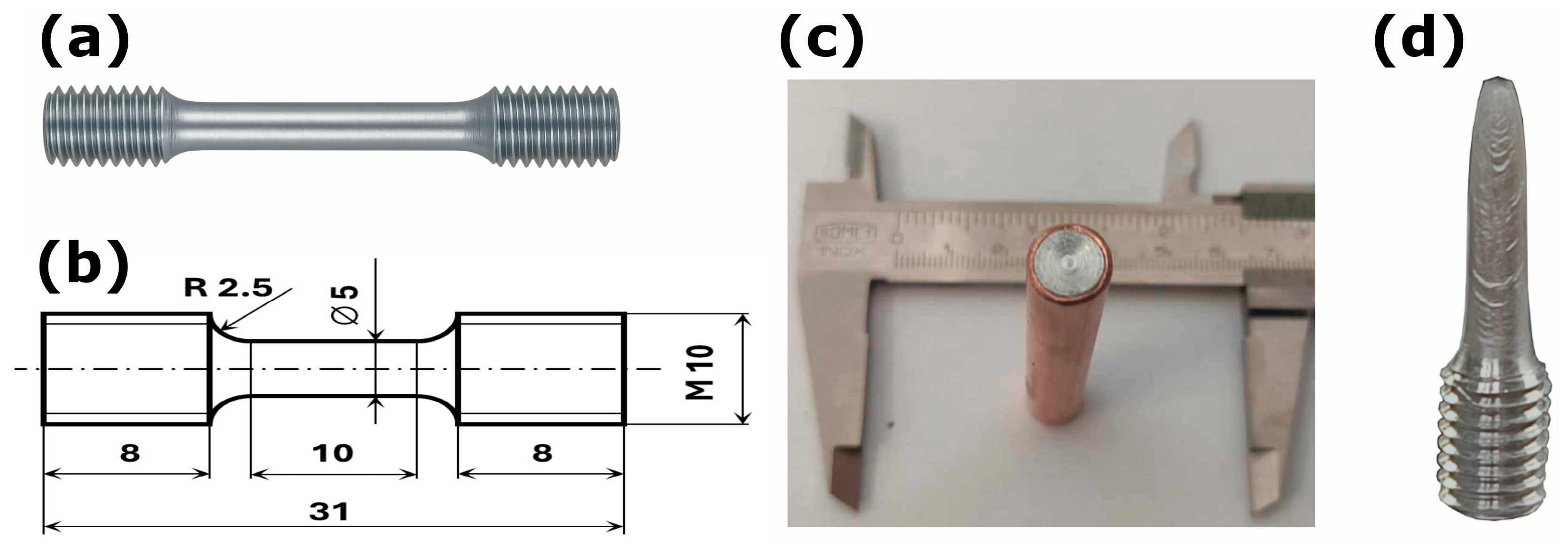
Figure 1. (a) Photograph of the cylindrical tensile specimen used for uniaxial testing; (b) dimensions of the tensile specimen; (c) sample of annealed Zn-0.1Mg alloy encapsulated in a copper sleeve; (d) half of a tensile specimen after fracture of the alloy processed by the ECAP 150 °C/RT route.
Metals 15 01161 g001
Figure 2. Schematic workflow of synchrotron X-ray powder diffraction (XRD) data acquisition for Zn-0.1Mg tensile specimens: specimen layout and gauge region; diffraction geometry and beamline setup; two-dimensional Debye-Scherrer rings recorded on the area detector; azimuthal integration to obtain 1D I(2θ) profiles.
Metals 15 01161 g002
Figure 3. (a) Engineering stress-strain curves of the Zn-0.1Mg alloy in the annealed state and after ECAP processing at 150 °C/RT; (b) fracture surface of the annealed material; (c) fracture surface of the 150 °C/RT material.
Metals 15 01161 g003
Figure 4. (a) X-ray diffraction patterns obtained from the transmission experiment of the Zn-0.1Mg tensile specimen after fracture (position 0 near the thread, 36 at the fracture zone); (b) detail of the XRD patterns in the 2θ range of 1–3°, showing Bragg reflections of the intermetallic phases Mg2Zn11 and MgZn2; (c) example of a diffraction pattern subjected to Rietveld refinement [46], including the refinement of structural parameters of the major hcp Zn phase.
Metals 15 01161 g004
Figure 5. Refined structural parameters obtained from Rietveld fitting of the X-ray diffraction patterns: (ac) lattice parameters and unit cell volume of hcp Zn; (df) microstrain, crystallite size, and texture index J representing the preferred crystallite orientation (texture).
Metals 15 01161 g005
Figure 6. Residual stress analysis of the fractured Zn-0.1Mg tensile specimen: (a) two-dimensional XRD pattern; (b) upper-right quadrant of the same image with selected Debye-Scherrer rings indexed; (c) variation of ε11, ε12, and ε12 as a function of position along the specimen.
Metals 15 01161 g006
Figure 7. Phase analysis results obtained by SAED in STEM, confirming the presence of phases: Zn solid solution and Mg2Zn11 particles within the Zn solid solution.
Metals 15 01161 g007
Figure 8. Structure of the Zn-0.1Mg alloy at position 0 of the tensile-tested specimen: (a) equiaxed solid solution grains—STEM; (b) elongated solid solution grains—STEM; (c) dislocation arrangements within the solid solution—STEM; (d) dislocation-particle interactions—TEM.
Metals 15 01161 g008
Figure 9. Structure of the Zn-0.1Mg alloy at position 36 of the tensile-tested specimen: (a) equiaxed solid solution grains—STEM; (b) elongated solid solution grains with subgrain formation—STEM; (c) dislocation arrangements within the solid solution—STEM; (d) dislocation-particle interactions—TEM.
Metals 15 01161 g009
Table 1. General requirements for biodegradable metallic implants (following Heiden et al. [20]).
PropertyVascular ScaffoldsOrthopedic Scaffolds
Cell responseSupport adhesion of vascular endothelial cellsSupport bone growth (osteoblasts, osteoclasts)
Mechanical integrity>8 months>6 months
Yield strength>200 MPa>230 MPa
Tensile strength>300 MPa>300 MPa
Fracture elongation>15–18%>15–18%
Elastic modulusLow, flexible (for vessel bending)Close to cortical bone (10–20 GPa)
Fatigue limit (107 cycles)>256 MPa>256 MPa
Elastic recoil after expansion<4%N/A
Hydrogen evolution<10 μL/cm2/day<10 μL/cm2/day
Table 2. Tensile test results: YS—Yield strength; UTS—Ultimate tensile strength; FE—Fracture elongation. Variability ≤ 5% for all metrics.
SampleYS [MPa]UTS [MPa]FE [%]
Annealed, v = 0.00025 s−170 ± 2.474 ± 2.91.8 ± 0.07
150 °C/RT, v = 0.001 s−1219 ± 8258 ± 962 ± 2.5
150 °C/RT, v = 0.00025 s−1165 ± 6212 ± 7150 ± 6
150 °C/RT, v = 0.0001 s−1147 ± 5173 ± 6240 ± 10
Disclaimer/Publisher’s Note: The statements, opinions and data contained in all publications are solely those of the individual author(s) and contributor(s) and not of MDPI and/or the editor(s). MDPI and/or the editor(s) disclaim responsibility for any injury to people or property resulting from any ideas, methods, instructions or products referred to in the content.