Article

Formation and Characterization of Ti-Al Intermetallic and Oxide Layers on Ti6Al4V as Interlayers for Hydroxyapatite Coatings

Department of Materials Science and Engineering, National University of Science and Technology Politehnica of Bucharest, 060042 Bucharest, Romania
*
Author to whom correspondence should be addressed.
Metals 2025, 15(10), 1159; https://doi.org/10.3390/met15101159
Submission received: 18 September 2025 / Revised: 15 October 2025 / Accepted: 18 October 2025 / Published: 21 October 2025
(This article belongs to the Special Issue Light Alloy and Its Application (3rd Edition))

Abstract

This study explores a novel approach to enhance the surface properties of Ti-Al alloys for biomedical applications by creating a compositional gradient layer through aluminum deposition using Electrical Discharge Machining (EDM). The primary goal was to develop a metallurgically bonded intermetallic zone that supports strong adhesion and improved compatibility for subsequent hydroxyapatite (HA) deposition. Aluminum was deposited onto a Ti6Al4V substrate via EDM under controlled conditions, followed by thermal and thermochemical treatments to induce diffusion and intermetallic phase formation. Comprehensive analyses using optical and electron microscopy (SEM), energy-dispersive spectroscopy (EDS), and X-ray diffraction (XRD) revealed the formation of well-adhered layers composed of complex Ti-Al intermetallics such as TiAl2 and TiAl3, along with oxide phases including TiO2 and Al2O3. Thermal and thermochemical treatments further improved surface hardness, reaching up to 1057 HV, and influenced the diffusion behavior of aluminum, titanium, and vanadium. Adhesion tests confirmed that the untreated and thermochemically treated layers exhibited superior mechanical stability, while thermal treatment alone led to brittleness and delamination. These findings demonstrate that a properly engineered intermediate aluminide layer can significantly improve the performance of bioceramic coatings, particularly HA, by providing enhanced structural integrity and biocompatibility.

1. Introduction

Titanium and its alloys, particularly Ti6Al4V, are widely used in biomedical implants owing to their exceptional combination of low density, high mechanical strength, corrosion resistance, and biocompatibility [1,2]. Ti6Al4V is extensively applied in orthopedic and dental implants due to its ability to withstand physiological environments and mechanical loads. However, one of the major limitations of titanium alloys lies in their relatively bioinert nature, which can hinder direct bone integration and reduce long-term implant stability [3,4,5]. To overcome this, surface modification techniques have been developed to enhance biological responses and improve osteointegration.
Hydroxyapatite (HA), a calcium phosphate compound chemically similar to human bone mineral, is one of the most effective bioactive coatings for titanium-based implants [6,7]. It promotes early-stage bone bonding, facilitates mineralization, and supports long-term osseointegration. However, the adhesion strength between HA and metallic substrates often remains suboptimal due to the thermal expansion mismatch, poor chemical bonding, and differences in mechanical properties [8,9]. These interface-related issues can lead to delamination, crack propagation, or loss of functionality, particularly under cyclic mechanical loading [10].
One promising strategy to address these challenges involves introducing a functionally graded interlayer that provides both chemical affinity and mechanical compatibility between the metallic substrate and the HA coating [11,12]. Titanium aluminides (Ti-Al intermetallic compounds) have emerged as suitable candidates for this role due to their high hardness, thermal and chemical stability, low density, and good oxidation and corrosion resistance [13,14]. When formed as a transitional layer, Ti-Al phases such as TiAl, TiAl2, and TiAl3 can provide strong metallurgical bonding with the substrate and improve HA coating adhesion while simultaneously acting as a diffusion barrier [15]. Furthermore, the formation of oxide phases such as TiO2 and Al2O3 during post-treatment enhances biocompatibility by promoting protein adsorption and apatite nucleation in biological environments [16,17,18]. Nevertheless, the formation of such intermetallic and oxide layers is non-trivial due to challenges in achieving controlled diffusion and phase stability. Conventional methods like pack cementation, thermal spraying, and sol–gel coating often involve high-temperature processing or complex chemical handling, which can introduce residual stresses or degrade the substrate [19,20,21]. In recent years, electrospark deposition (ESD), a micro-arc pulsed deposition technique, has garnered increasing attention for surface alloying applications. ESD enables localized and controlled deposition of alloying elements through high-frequency electrical discharges, offering several advantages such as minimal heat-affected zones, strong metallurgical bonding, and fine microstructural control [22,23,24].
In this study, aluminum was deposited onto Ti6Al4V substrates using Electrical Discharge Machining (EDM), a variant of ESD, under controlled conditions to create a Ti–Al rich surface layer. Subsequent thermal and thermochemical treatments were employed to induce the formation of intermetallic compounds and oxide layers. This approach aims to fabricate a compositionally graded interface layer that improves adhesion and supports subsequent HA coating. The selection of EDM as a deposition method offers precision and control not easily achievable through conventional thermal processes, especially in biomedical applications where dimensional integrity and substrate preservation are critical [25,26]. The subsequent phase following deposition involves thermotreatment and thermochemical treatment to enhance the layer’s properties. Heat treatments, such as solution treatment and aging, are crucial for altering the microstructure of Ti6Al4V [27]. By controlling the quantity and distribution of α and β phases, the mechanical properties of Ti6Al4V can be effectively managed. Different heat treatment methods can produce distinct microstructures, including equiaxed, bimodal, or lamellar α + β structures, each offering a unique set of properties [28]. Aging processes can further increase hardness, while annealing can enhance impact toughness without diminishing tensile strength. The specific parameters of annealing, such as temperature and cooling rate, determine the resulting microstructure and mechanical characteristics [29]. When Ti6Al4V undergoes heat treatment in an air environment, particularly at temperatures exceeding 600 °C, an “alpha case” (a hard, brittle layer enriched with oxygen) and TiO2 oxide may form on the surface. This can influence surface properties and should be considered in process design or subsequent surface removal [30].
Thermochemical nitriding is another surface engineering process designed to significantly enhance the hardness, wear resistance, and corrosion resistance of Ti6Al4V [31]. This process typically results in a hard outer compound zone, primarily composed of titanium nitride (TiN) and titanium sub-nitride (Ti2N), along with a deeper diffusion zone where nitrogen is in solid solution within the titanium lattice [32]. Gas nitriding is a widely used method in which Ti6Al4V components are heated in a furnace with a nitrogen-rich atmosphere, often a mixture of nitrogen and an inert gas like argon. Ammonia (NH3) can also be employed, as it dissociates at high temperatures to provide nascent nitrogen. The typical temperature range is from 700 °C to 950 °C. Higher temperatures increase the diffusion rate of nitrogen, leading to thicker nitride layers, but can also cause undesirable grain growth in the bulk material [33]. The formation of hard ceramic phases like TiN and Ti2N results in a harder, wear-resistant, and corrosion-resistant surface, significantly enhancing the durability and performance of components operating in challenging environments [31,34]. The surface layers were characterized in detail using optical and scanning electron microscopy (SEM), energy-dispersive spectroscopy (EDS), and X-ray diffraction (XRD). Mechanical performance was evaluated via microhardness testing and adhesion studies. Special attention was given to the formation and evolution of Ti-Al intermetallics (e.g., TiAl2, TiAl3), the development of oxide phases (e.g., TiO2, Al2O3), and their influence on layer integrity and functionality. By engineering this intermediate zone, the study aims to enhance both the structural and biological performance of HA coatings, contributing to more reliable and long-lasting biomedical implants.
The present study aims to develop and characterize Ti-Al intermetallic/oxide surface layers on Ti6Al4V alloy using the EDM process and subsequent thermal/thermochemical treatments. These modified layers are designed as intermediate interlayers to enhance compatibility with hydroxyapatite (HAp) coatings, rather than to demonstrate direct HAp deposition. The investigation focuses on structural, chemical, and mechanical aspects that determine the suitability of such surfaces for future biofunctionalization. Experimental evaluation of HAp adhesion will be the subject of subsequent work.

2. Materials and Methods

2.1. Substrate Preparation

The substrate material used in this study was Grade 5 titanium alloy (Ti6Al4V), known for its widespread application in biomedical implants due to its strength, corrosion resistance, and biocompatibility. Samples were prepared by cutting bulk Ti6Al4V alloy into rectangular specimens with dimensions of 25 × 39 × 33.2 mm using a Buehler Abrasimet M precision abrasive cutting machine (Buehler, Lake Bluff, IL, USA). The resulting samples were subjected to a surface grinding procedure to ensure flatness and remove any residual surface defects, utilizing abrasive papers with grit sizes of 120, 600, and 800 in sequential steps.

2.2. Aluminum Deposition via EDM

Aluminum was deposited onto the Ti6Al4V substrate using a controlled Electrical Discharge Machining (EDM) process, implemented through a customized setup. In this process, a cylindrical aluminum electrode (34 mm in length, 3.01 mm in diameter) was rotated at a constant speed to maintain uniform deposition. The EDM process operated at a constant power of 5 W, with air used as the dielectric medium.
To maintain process repeatability and spatial control, a CNC 3018 Pro system (Figure 1) was employed to automate and direct the electrode motion. The deposition path was programmed using the laserGRBL software V 2.3.0, with G-code instructions setting the treatment pattern over a 20 × 13 mm rectangular field. The thickness of the deposited Al layer was controlled by adjusting the specific energy input and the number of discharge passes. All samples were processed under constant power (5 W), electrode rotation speed (200 rpm), traverse speed (120 mm s−1), and 20 repeated criss-cross passes, corresponding to a specific processing time of approximately 17.5 s cm−2. A calibration curve of layer thickness versus number of passes, obtained from cross-sectional SEM measurements and mass gain data, was used to verify reproducibility. The resulting layer thickness before heat treatment was 12.0 ± 0.6 µm.

2.3. Post-Deposition Treatments

Following EDM deposition, samples were subjected to two types of post-processing treatments to enhance surface diffusion and intermetallic phase development: (1) Thermal Treatment: Specimens were heat-treated in an electric muffle furnace at 600 °C for 3 h, followed by controlled furnace cooling over a 12 h period. Samples were placed face-down in a bed of aluminum oxide (Al2O3) powder to minimize oxidation and support uniform heat distribution; (2) Thermochemical Treatment: A separate batch of samples underwent a thermochemical surface doping process. Specimens were embedded in a solid reactive medium composed of 30 g urea (CON2H4), 3.4 g ammonium chloride (NH4Cl), 40 g graphite, and 26.5 g aluminum oxide. These samples were then processed under the same thermal regime (600 °C for 3 h), followed by controlled cooling. This treatment was designed to induce nitrogen and carbon diffusion, supporting the formation of nitrides and carbides in addition to aluminides.

2.4. Characterization Techniques

The treated and untreated specimens were subjected to comprehensive characterization to evaluate their surface morphology, microstructure, phase composition, mechanical properties, and adhesion performance. Surface morphology and layer uniformity were initially examined using optical microscopy (Nikon, Tokyo, Japan) following etching with Kroll’s reagent, which revealed microstructural features of both the substrate and the modified layers. Detailed surface and cross-sectional microstructures were further analyzed using a scanning electron microscope (Thermo Scientific Quattro S SEM, Waltham, MA, USA). To investigate the elemental composition and distribution across the treated regions, energy-dispersive X-ray spectroscopy (EDS) mapping was conducted in conjunction with SEM. Phase identification and crystallographic analysis were carried out using X-ray diffraction (XRD), enabling the detection of intermetallic compounds, oxide phases, and potential nitride formations induced by thermal and thermochemical treatments. Mechanical evaluation included Vickers microhardness testing using a Falcon 503 microhardness tester (INNOVATEST, Maastricht, The Netherlands). Measurements were performed on both surface and cross-sectional areas under a load of 10 g, with four replicates per sample to ensure statistical consistency. To assess coating adhesion, scratch testing was conducted using a UMT-TriboLab system (Bruker, Ettlingen, Germany) under progressive loading conditions ranging from 0 to 50 N over 60 s. The procedure followed the EN 1071-3:2005 standard [35], and coating failure modes were subsequently analyzed via SEM to evaluate adhesion integrity and failure mechanisms

3. Results and Interpretations

3.1. Optical Microscopy Analysis

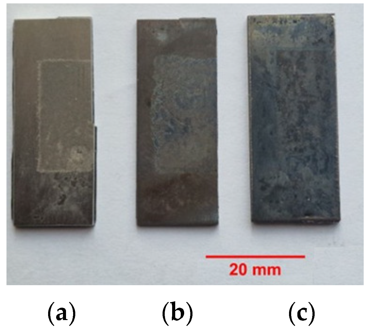
Initial surface observations were conducted using optical microscopy to examine the visual and structural differences among untreated, thermally treated, and thermochemically treated Ti6Al4V samples following aluminum deposition. Figure 2 presents comparative surface images. For the untreated sample (Figure 2a), a relatively uniform layer was observed, with no significant oxidation or phase separation visible to the naked eye. The thermally treated sample (Figure 2b) shows distinct color variations emerged, indicating the formation of different oxide layers. A yellow substrate tone was overlaid by bluish surface regions, suggesting the presence of aluminum oxide (Al2O3). The thermochemically treated sample (Figure 2c) presents a darkened, nearly black surface layer was evident, likely caused by the formation of carbon-rich compounds or graphene residues due to the graphite-rich doping medium. These observations were reinforced through etching with Kroll’s reagent, which revealed changes in the microstructure and suggested the successful formation of intermetallic and oxide phases following each treatment.
As shown in Figure 3a, the integration between the layer and substrate was confirmed to be effective. Additionally, changes in the crystallization of the substrate post-treatment are observed. The untreated sample, as shown in Figure 3a, exhibits an equiaxed α + β structure near the surface. In contrast, Figure 3b displays columnar prior β grains with aligned α platelets oriented perpendicular to the surface. Figure 3c shows the dendritic structure with new α colonies within the β phase.

3.2. Scanning Electron Microscopy (SEM) and EDS Analysis

SEM analysis provided detailed insight into the surface morphology and structural integrity of the coatings. In terms of surface morphology (Figure 4), the untreated sample showed a uniform but shallow deposited layer. The thermally treated sample exhibited signs of microcracking and fine porosity, indicative of thermal expansion and contraction. The thermochemically treated sample presented a thicker, more continuous layer with visible microcracks attributed to thermal stress and rapid phase transformations.
The cross-sectional morphology (Figure 5) analysis shows the thicknesses of the deposited layers that were measured as approximately 11.97 μm (untreated), 8.21 μm (thermally treated), and 13.07 μm (thermochemically treated). Despite the presence of microcracks, which are normal for this type of deposition, all layers displayed good adhesion to the substrate.
The slightly lower thickness observed after heat treatment (~8.2 µm) compared to the as-deposited layer (~12.0 µm) can be attributed to several phenomena: (i) mutual diffusion of Ti and Al at 600 °C, leading to the formation of dense Ti-Al intermetallics and the consumption of the Al-rich outer zone; (ii) oxidation and densification of the surface, which form a compact TiO2/Al2O3 layer and reduce the measurable metallic thickness; and (iii) partial removal of a porous rim during polishing. Therefore, the reported value refers to the continuous, adherent intermetallic zone rather than any loose or discontinuous surface fragments.
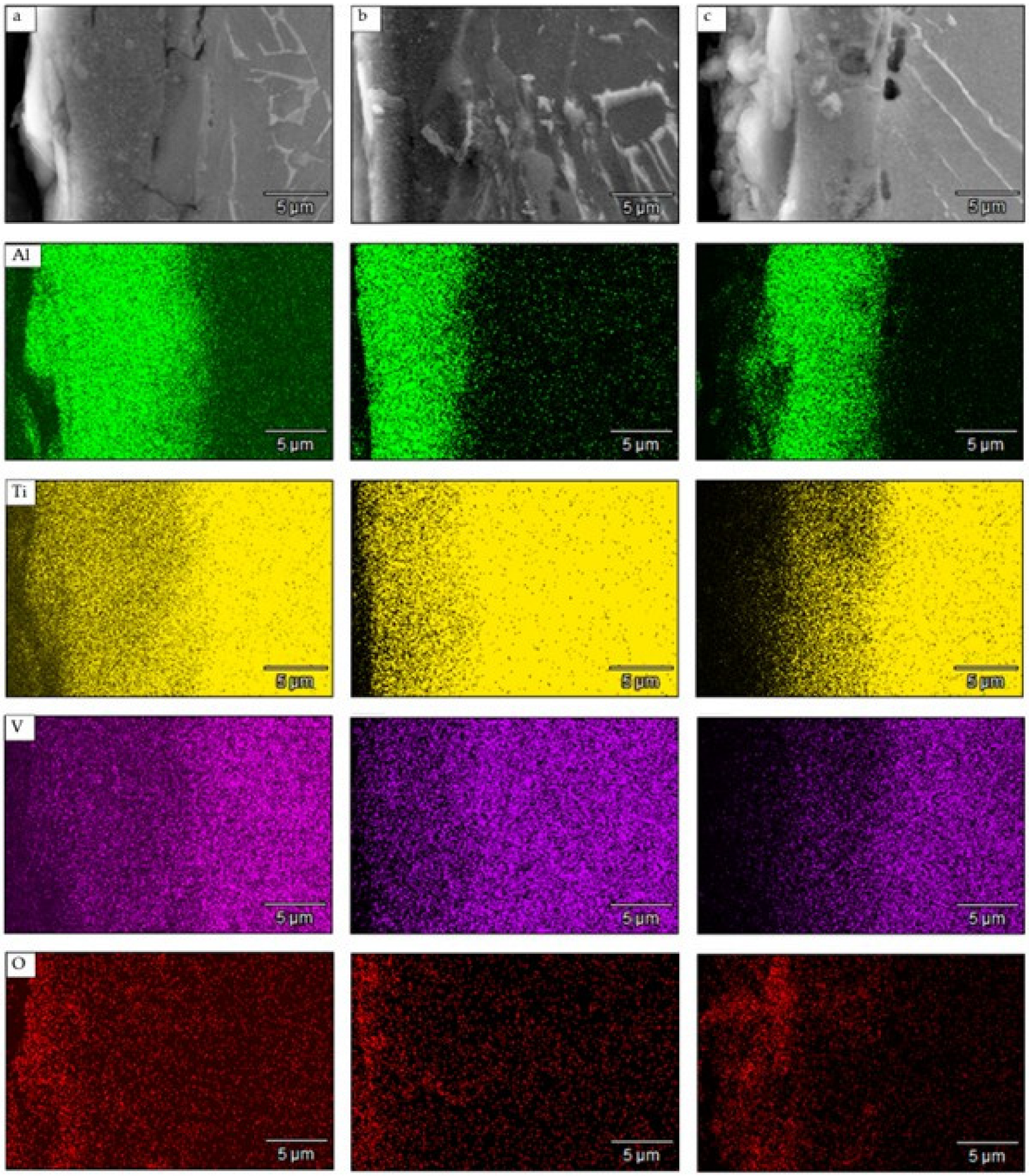
Elemental Composition (EDS, Figure 6) shows that the aluminum concentration was highest at the surface, confirming successful deposition. Titanium and vanadium were detected in the coating layer, indicating diffusion from the substrate into the aluminum layer. Oxygen was also present across all treatments due to atmospheric exposure, but it likely contributed beneficially to oxide layer formation.
Among all samples, the thermochemically treated one showed the most complex elemental gradient and the highest aluminum penetration, confirming that the reactive environment enhanced diffusion.
The detection of oxygen across all the samples was attributed to processing in the absence of an inert atmosphere. However, in this context, the presence of oxygen is not expected to negatively affect the performance of the layer because it may contribute to oxide layer formation, which can be beneficial in certain applications.
The enrichment of Ti near the surface after the 600 °C treatment (Figure 6) results from the unequal diffusion rates of Ti and Al at this temperature. Ti exhibits a higher diffusivity along grain boundaries and defects in the growing intermetallic layer and can migrate outward under the oxygen potential gradient present during oxidation. Concurrently, the formation of TiO2 at the surface consumes Ti and promotes its flux toward the surface, producing a classical Kirkendall-type effect. This mechanism explains the Ti concentration increase observed in the heat-treated layer.

3.3. X-Ray Diffraction (XRD) Analysis

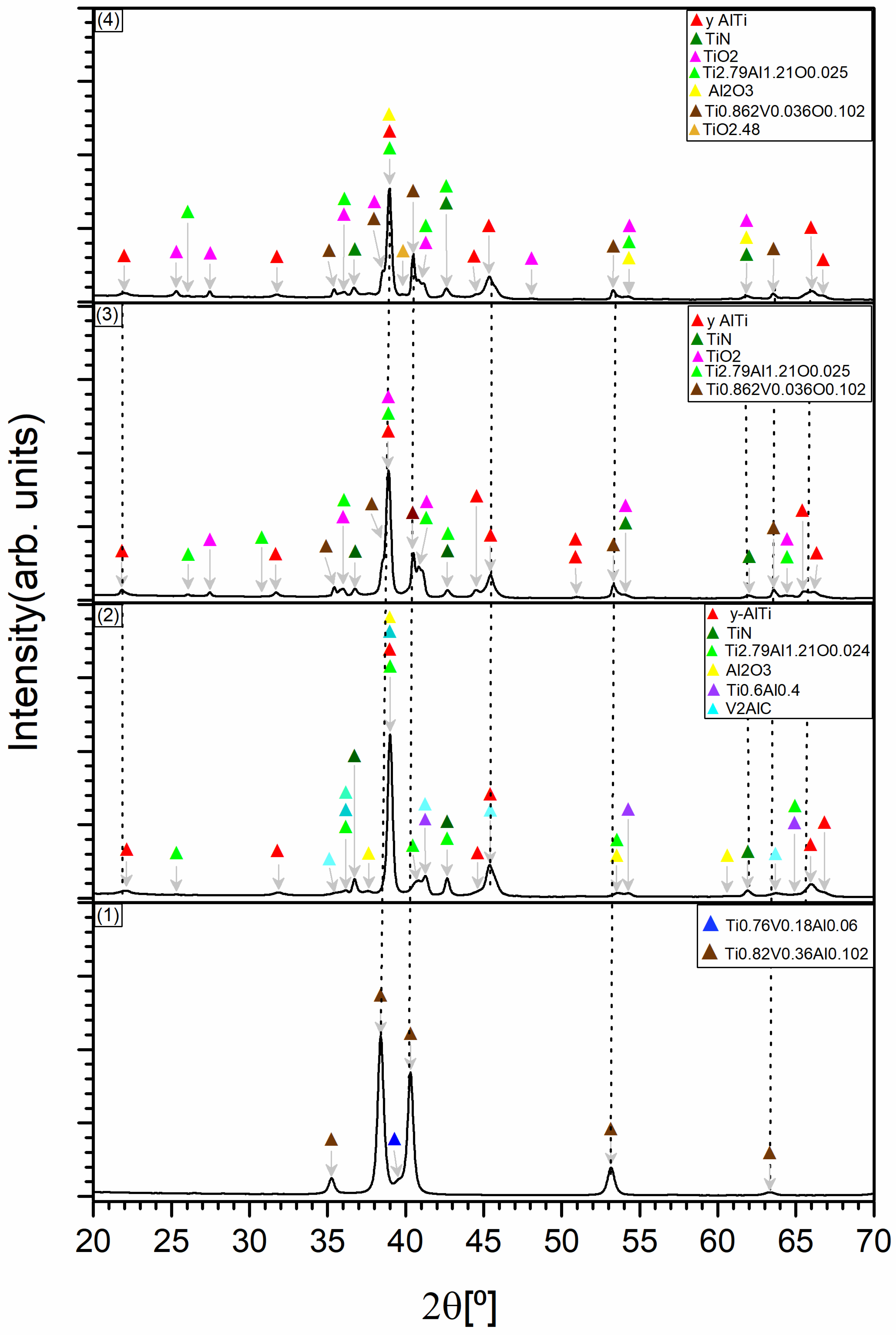
X-ray diffraction (XRD) analysis (Figure 7) was conducted to assess phase evolution during each stage of surface treatment applied to Ti6Al4V alloy samples. The diffraction pattern of the untreated specimen (Pattern 1) displayed sharp and well-defined peaks corresponding to both α-phase (hexagonal close-packed, HCP) and β-phase (body-centered cubic, BCC) titanium. These peaks were indexed to solid solution compositions of Ti0.76V0.18Al0.06 and Ti0.82V0.36Al0.102, confirming the expected biphasic structure of the alloy with vanadium and aluminum incorporated within the titanium lattice.
After aluminum deposition via electrical discharge machining (Pattern 2), new peaks emerged that were assigned to aluminum oxide (Al2O3), titanium nitride (TiN), and a complex ternary compound Ti2.79Al1.21O0.024. These findings indicate that oxidation and nitridation processes had already initiated, likely due to localized thermal effects during EDM and exposure to the ambient atmosphere. Importantly, the appearance of a V2AlC MAX phase points to initial interfacial diffusion between vanadium in the substrate and the deposited aluminum layer. MAX phases are known for their high thermal stability and favorable mechanical properties, which could contribute to stress distribution and coating performance. The early formation of Ti2.79Al1.21O0.024 suggests the onset of oxygen-rich intermetallic or oxynitride phases with potential functional relevance.
Following thermal treatment at 600 °C (Pattern 3), notable changes in peak intensity and sharpness were observed. Peaks corresponding to Al2O3 and TiO2 became more intense and defined, indicating crystallization and growth of oxide phases. The continued presence of the Ti2.79Al1.21O0.025 phase, with slightly altered stoichiometry, supports its thermodynamic stability and possible role as a transitional bonding layer. The emergence of crystalline TiO2—likely in anatase or rutile form—is particularly significant for biomedical applications, as these oxide forms are associated with enhanced osteointegration and surface bioactivity. While the α- and β-Ti phases were still present, their diminished intensity suggested progressive transformation toward a more complex surface enriched in oxides and intermetallics.
In the thermochemically treated sample (Pattern 4), dominant TiN peaks confirmed successful surface nitriding under nitrogen- and carbon-rich conditions. The detection of γ-TiAl intermetallics provides compelling evidence of Al–Ti interdiffusion and ordered phase formation, reinforcing the metallurgical integrity of the interface. The absence of Ti2N peaks suggests that nitriding remained confined to the surface, avoiding excessive nitrogen uptake that could compromise ductility. Oxide phases such as TiO2 and Al2O3 persisted with high crystallinity, and the ternary Ti2.79Al1.21O0.025 compound remained stable throughout processing, further emphasizing its resilience and likely functional importance in the modified surface architecture.
Overall, the XRD results clearly demonstrate a progressive increase in phase complexity—from solid solution and intermetallic formation to oxide and nitride crystallization—resulting from each successive treatment. The combined presence of hard intermetallics, stable oxides, and surface nitrides is expected to significantly enhance surface hardness, corrosion resistance, and bioceramic compatibility. This tailored surface structure offers an ideal platform for hydroxyapatite deposition, supporting improved mechanical anchoring and biological integration in implant applications.
Based on the combination of XRD, EDS, and cross-sectional analyses, the layer structure can be schematically described as follows: After Al deposition (EDM): a thin oxide/nitride film (Al2O3, TiN traces), an Al-rich modified zone with initial Ti–Al intermixing, a Ti–Al diffusion zone, and the Ti6Al4V substrate; After heat treatment (600 °C/3 h): a compact surface oxide layer composed of TiO2 and Al2O3, an intermetallic zone containing TiAl3, TiAl2, and γ-TiAl phases, followed by a Ti-rich diffusion zone and the substrate; After thermochemical treatment: a TiN-containing top layer together with TiO2/Al2O3, underlain by γ-TiAl and TiAl2/TiAl3 phases and a gradual transition to the substrate.
This stratified architecture indicates the progressive transformation from pure Al deposits to complex oxide/intermetallic systems with diffusion-bonded interfaces.
The observed sequence reflects (i) chemical potentials of O and N at the surface (air vs. N-releasing pack), (ii) diffusion kinetics of Ti and Al (promoting intermetallic formation: TiAl3 → TiAl2 → γ-TiAl with increasing Ti activity), and (iii) parabolic oxidation of Ti to TiO2 in air. Thus, sample (2) develops a TiO2/Al2O3-capped intermetallic architecture with only trace TiN, whereas sample (3) exhibits a TiN-containing top layer over Ti-Al intermetallics, forming a duplex ceramic/metallic gradient conducive to hardness and wear resistance.

3.4. Microhardness Evaluation

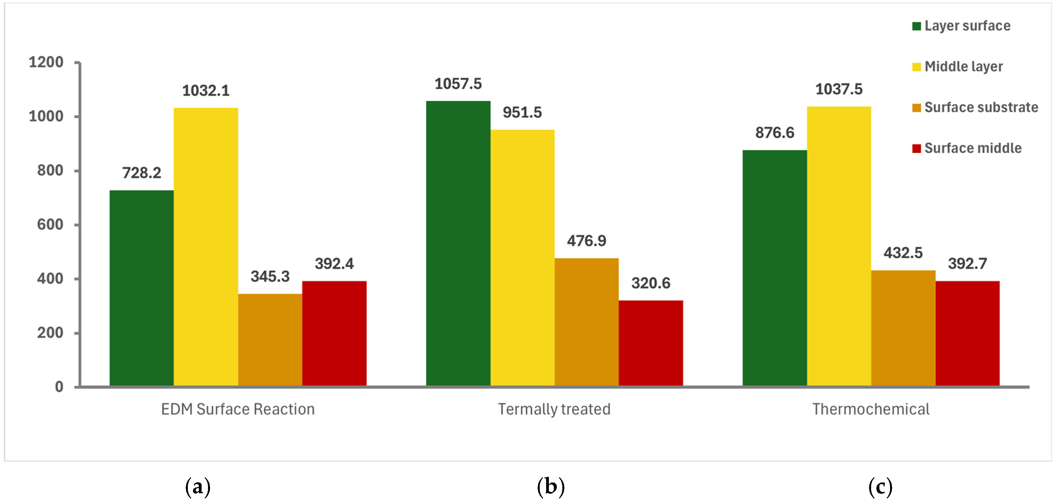
Microhardness measurements revealed substantial improvements in surface and subsurface hardness following the applied treatments. The untreated Ti6Al4V substrate exhibited a baseline surface hardness of approximately 345.3 HV. After thermal treatment, the surface hardness increased markedly to 476.9 HV, while the thermochemically treated sample reached 432.5 HV, indicating the formation of mechanically reinforced surface layers (Figure 8).
Further analysis within the depth profile of the coatings revealed that the thermochemically treated samples exhibited peak hardness values up to 1034 HV in the intermediate region, with slight local reductions (e.g., down to 951.5 HV) attributed to microstructural relaxation or the presence of thermal-induced cracks. The highest hardness values were recorded at the outermost surface, reaching up to 1057.5 HV for both the thermally and thermochemically treated samples—nearly three times higher than that of the untreated alloy (~360 HV).
These results confirm the formation of hard intermetallic compounds (e.g., γ-TiAl, TiAl2, TiAl3), along with oxide (TiO2, Al2O3) and nitride (TiN) phases, which collectively contribute to the enhanced mechanical robustness and wear resistance of the treated surfaces. Such improvements are critical for load-bearing biomedical applications where long-term mechanical durability and resistance to surface degradation are essential.

3.5. Adhesion Testing

The adhesion performance of the modified surfaces was evaluated using progressive-load scratch testing. The untreated sample exhibited the highest critical load, approximately 56.5 N (Figure 9a), indicative of strong mechanical integrity and ductility at the substrate-coating interface. In contrast, the thermally treated sample demonstrated a significantly lower critical load of approximately 42.7 N (Figure 9b), likely due to the formation of brittle oxide and intermetallic phases that reduced the coating’s resistance to mechanical stress.
The thermochemically treated sample showed intermediate adhesion behavior, with a critical load of approximately 53.8 N (Figure 9c).
This suggests that the surface nitriding introduced during thermochemical treatment enhanced hardness while still maintaining sufficient interfacial bonding strength.
Scanning electron microscopy (SEM) analysis of the scratch tracks (Figure 10) provided further insight into failure modes. The untreated sample showed minimal material loss along the scratch path, confirming good cohesion. In contrast, the thermally treated sample exhibited significant delamination and surface fragmentation, correlating with its lower adhesion threshold. The thermochemically treated surface displayed variable performance—some regions showed minimal degradation, while others revealed partial coating detachment—highlighting local heterogeneity and underscoring the need for optimization of process parameters to ensure uniform coating behavior.
These results demonstrate that surface treatments significantly influence coating adhesion, and that thermochemical processing offers a promising balance between mechanical enhancement and interfacial stability, pending further refinement.

4. Discussion

Unlike conventional coating techniques such as plasma spraying, chemical vapor deposition (CVD), sol–gel methods, or hot-dip aluminizing, the EDM-based process enables localized surface alloying while minimizing thermal exposure to the bulk material. Previous research has demonstrated the limitations of traditional approaches in terms of coating delamination, thermal mismatch, or process complexity. For example, Otsuka et al. [10] reported excellent biocompatibility of plasma-sprayed hydroxyapatite (HA) coatings on Ti alloys, yet identified delamination under cyclic loading due to thermal expansion mismatch with the substrate. In contrast, the present EDM process provides a controllable, contactless deposition method that supports strong metallurgical bonding and localized interdiffusion.
Mechanical alloying and high-energy ball milling offer broader diffusion capabilities but often produce surface irregularities that are unsuitable for biomedical interfaces. The EDM technique used here circumvents these issues by producing a clean, patterned surface through spark-induced melting and rapid solidification. Following deposition, solid-state post-treatments at 600 °C promoted the formation of titanium–aluminum intermetallics (TiAl2, TiAl3, γ-TiAl), stable oxide phases (Al2O3, TiO2), and nitrides (TiN), all of which are critical to improving both surface hardness and chemical stability.
The formation of TiN in particular is notable, as it has been linked to improved osteoblast adhesion and antimicrobial behavior, according to recent findings by Li et al. [33]. The presence of Al2O3 and TiO2 further supports enhanced corrosion resistance and serves as a diffusion barrier that improves the long-term stability of overlying HA coatings. XRD analysis also revealed the persistence of a ternary phase, Ti2.79Al1.21O0.025, across all post-treatment conditions. The thermodynamic stability and chemical inertness of TiN under physiological and tribological environments, together with its role as a diffusion-barrier interlayer, highlight its potential to anchor subsequent ceramic coatings (e.g., HAp) and to contribute to surface passivation and corrosion/wear resistance.
In addition to its scientific merits, this method offers practical advantages. It is scalable, cost-effective, and avoids the high-vacuum or inert-gas requirements typical of many advanced deposition techniques. Furthermore, by restricting thermal processing to the surface zone, it preserves the bulk mechanical properties of Ti6Al4V—a critical requirement for orthopedic and dental implants. Unlike hot-dip aluminizing, which can adversely affect the alloy’s core strength, the approach used here maintains structural continuity from the substrate through to the engineered surface.
The observed surface hardness values, reaching up to approximately 1057 HV, align with the hardness range needed for load-bearing implant surfaces. Meanwhile, scratch testing confirmed that thermochemically treated surfaces exhibit sufficient adhesion strength to support stable coatings under mechanical load. This combination of hardness and interfacial bonding is difficult to achieve using conventional techniques. While plasma-based approaches, such as those explored by Tsui et al. [19], can deliver high hardness, they often suffer from limited uniformity and poor adhesion, particularly on complex geometries.
The design developed in this study aligns closely with current trends in biomaterials research. Recent reviews, such as the one by Li et al. [33], emphasize the need for customizable, layered surface architectures that combine mechanical resilience with tailored biological response. The engineered surface presented here satisfies these criteria by providing a tunable interlayer suitable for bioactive coatings, mechanical stability under physiological stresses, and compatibility with manufacturing processes for next-generation implants.
Looking forward, further development should include detailed in vitro and in vivo assessments to confirm the biocompatibility and long-term performance of the modified surfaces. Optimizing the thermochemical composition may also enable targeted phase formation, including MAX phases for improved wear resistance or bioactive oxynitrides. Additional studies should explore HA deposition using electrochemical or sol–gel techniques to evaluate interfacial bonding with the engineered layer. Finally, comprehensive mechanical testing—including fatigue, wear, and corrosion resistance in simulated body fluid—will be essential to validate the clinical readiness of this surface engineering strategy.
Literature reports indicate that oxide-rich Ti surfaces, particularly those containing TiO2 and Al2O3 phases, exhibit enhanced wettability and facilitate apatite nucleation when exposed to simulated body fluids or Hank’s solution [34]. The intermetallic/oxide layers produced in this work, therefore, create a favorable chemical and structural foundation for subsequent HAp coating, although direct adhesion testing was beyond the current study’s scope.
The functional properties of the modified surfaces can be directly related to their microstructural constituents. The outer TiO2 and Al2O3 films are responsible for high chemical stability and serve as bioactive sites capable of hydroxylation, which facilitates protein adsorption and subsequent apatite nucleation in physiological environments [34]. The underlying Ti-Al intermetallic phases (γ-TiAl, TiAl2, TiAl3) contribute to the significant increase in hardness (up to ~1057 HV) and wear resistance, while maintaining metallic bonding continuity with the Ti6Al4V substrate. In the thermochemically treated variant, the presence of TiN further enhances hardness and abrasion resistance. Together, these phases provide a graded mechanical and chemical transition from oxide/ceramic top layers to the ductile Ti substrate, which is essential for durable performance and long-term biocompatibility in load-bearing biomedical applications.

5. Conclusions

This study demonstrates the feasibility of using Electrical Discharge Machining (EDM) for the deposition of aluminum onto Ti6Al4V substrates, followed by post-deposition thermal and thermochemical treatments to create a compositionally graded interface suitable for hydroxyapatite (HA) coating. The resulting multilayer structures exhibited significant changes in microstructure, phase composition, and mechanical performance.
Microscopic and spectroscopic analyses confirmed the successful formation of aluminum-rich intermetallic compounds (such as TiAl2 and TiAl3), oxides (Al2O3, TiO2), and, in thermochemically treated samples, nitrides (TiN). These phases contributed to increased surface hardness, reaching values over 1000 HV, while maintaining acceptable adhesion to the underlying titanium alloy. Among the tested approaches, thermochemical treatment produced the most promising results in terms of structural complexity and surface hardness, indicating effective diffusion and surface alloying. However, variability in adhesion performance and local structural integrity suggests that further optimization is needed to ensure consistent performance.
Overall, the results validate the concept of using EDM and post-treatment strategies to engineer intermediate bonding layers that support the adhesion and functionality of hydroxyapatite coatings. This multilayer approach holds strong potential for biomedical applications where mechanical robustness, biocompatibility, and coating adhesion are critical. Future work will focus on optimizing processing parameters and evaluating the biological performance of the coated structures in vitro and in vivo.

Author Contributions

Conceptualization, I.A.; Methodology, S.A.L., M.M. and F.M.; Validation, I.A. and F.M.; Formal analysis, M.M.; Investigation, S.A.L., D.E. and F.M.; Resources, I.A.; Data curation, D.E.; Writing—original draft, S.A.L. and M.M.; Writing—review & editing, F.M.; Supervision, M.M. All authors have read and agreed to the published version of the manuscript.

Funding

This research received no external funding.

Data Availability Statement

The original contributions presented in this study are included in the article. Further inquiries can be directed to the corresponding author.

Acknowledgments

The authors thank Dr. George E. Stan, Head of Laboratory 20 “Complex Heterostructures and Multifunctional Materials” of National Institute of Materials Physics, Atomistilor 405A, 077125, Magurele, Romania, for the XRD results.

Conflicts of Interest

The authors declare no conflicts of interest.

References

  1. Long, M.; Rack, H.J. Titanium alloys in total joint replacement—A materials science perspective. Biomaterials 1998, 19, 1621–1639. [Google Scholar] [CrossRef]
  2. Geetha, M.; Singh, A.K.; Asokamani, R.; Gogia, A.K. Ti based biomaterials, the ultimate choice for orthopaedic implants—A review. Prog. Mater. Sci. 2009, 54, 397–425. [Google Scholar] [CrossRef]
  3. Balla, V.K.; Bodhak, S.; Bose, S.; Bandyopadhyay, A. Porous tantalum structures for bone implants: Fabrication, mechanical and in vitro biological properties. Acta Biomater. 2010, 6, 3349–3359. [Google Scholar] [CrossRef]
  4. Liu, X.; Chu, P.K.; Ding, C. Surface modification of titanium, titanium alloys, and related materials for biomedical applications. Mater. Sci. Eng. R Rep. 2004, 47, 49–121. [Google Scholar] [CrossRef]
  5. Crăciunescu, E.; Sinescu, C.; Negruțiu, M.L.; Pop, D.M.; Lauer, H.C.; Rominu, M. Shear Bond Strength Tests of Zirconia Veneering Ceramics after Chipping Repair. J. Adhes. Sci. Technol. 2016, 30, 666–676. [Google Scholar] [CrossRef]
  6. Xia, W.; Lindahl, C.; Lausmaa, J.; Engqvist, H. Biomimetic Hydroxyapatite Deposition on Titanium Oxide Surfaces for Biomedical Application. In Advances in Biomimetics; Cavrak, M., Ed.; InTech: Rijeka, Croatia, 2011; pp. 427–443. [Google Scholar] [CrossRef]
  7. Tsui, Y.C.; Doyle, C.; Clyne, T.W. Plasma Sprayed Hydroxyapatite Coatings on Titanium Substrates. Part 2: Optimisation of Coating Properties. Biomaterials 1998, 19, 2031–2043. [Google Scholar] [CrossRef]
  8. Kitsugi, T.; Nakamura, T.; Oka, M.; Yan, W.Q.; Goto, T.; Shibuya, T.; Kokubo, T.; Miyaji, S. Bone-Bonding Behavior of Titanium and Its Alloys When Coated with Titanium Oxide (TiO2) and Titanium Silicate (Ti5Si3). J. Biomed. Mater. Res. 1996, 32, 149–156. [Google Scholar] [CrossRef]
  9. Rausch, M.A.; Shokoohi-Tabrizi, H.; Wehner, C.; Pippenger, B.E.; Wagner, R.S.; Ulm, C.; Moritz, A.; Chen, J.; Andrukhov, O. Impact of Implant Surface Material and Microscale Roughness on Initial Attachment and Proliferation of Primary Human Gingival Fibroblasts. Biology 2021, 10, 356. [Google Scholar] [CrossRef] [PubMed]
  10. Otsuka, Y.; Kawaguchi, H.; Mutoh, Y. Cyclic Delamination Behavior of Plasma-Sprayed Hydroxyapatite Coating on Ti–6Al–4V Substrates in Simulated Body Fluid. Mater. Sci. Eng. C 2016, 67, 533–541. [Google Scholar] [CrossRef] [PubMed]
  11. Matei, A.A.; Pencea, I.; Stanciu, S.G.; Hristu, R.; Antoniac, I.; Ciovica, E.; Sfat, C.E. Structural Characterization and Adhesion Appraisal of TiN and TiCN Coatings Deposited by CAE-PVD on a Carbide Composite Tool. J. Adhes. Sci. Technol. 2015, 29, 2576–2589. [Google Scholar] [CrossRef]
  12. Habibovic, P.; Barrere, F.; van Blitterswijk, C.A.; de Groot, K. Biomimetic Hydroxyapatite Coating on Metal Implants. J. Am. Ceram. Soc. 2002, 85, 517–522. [Google Scholar] [CrossRef]
  13. Appel, F.; Paul, J.D.H.; Oehring, M. Gamma Titanium Aluminide Alloys: Science and Technology; Wiley-VCH: Weinheim, Germany, 2011. [Google Scholar] [CrossRef]
  14. Dawod, N.; Miculescu, M.; Antoniac, I.V.; Miculescu, F.; Agop-Forna, D. Metal–Ceramic Compatibility in Dental Restorations According to the Metallic Component Manufacturing Procedure. Materials 2023, 16, 5556. [Google Scholar] [CrossRef]
  15. Clemens, H.; Mayer, S. Design, Processing, Microstructure, Properties, and Applications of Advanced Intermetallic TiAl Alloys. Adv. Eng. Mater. 2013, 15, 191–215. [Google Scholar] [CrossRef]
  16. Liu, R.; Dang, X.; Gao, Y.; Wu, T.; Zhu, Y. The Wear Behavior of the Laser-Cladded Ti-Al-Si Composite Coatings on Ti-6Al-4V Alloy with Additional TiC. Materials 2021, 14, 4567. [Google Scholar] [CrossRef]
  17. Hanawa, T. Research and Development of Metals for Medical Devices Based on Clinical Needs. Sci. Technol. Adv. Mater. 2012, 13, 064102. [Google Scholar] [CrossRef]
  18. Yao, C.; Slamovich, E.B.; Webster, T.J. Enhanced Osteoblast Functions on Anodized Titanium with Nanotube-like Structures. J. Biomed. Mater. Res. A 2008, 85A, 157–166. [Google Scholar] [CrossRef]
  19. Liu, D.M.; Troczynski, T.; Tseng, W.J. Water-Based Sol–Gel Synthesis of Hydroxyapatite: Process Development. Biomaterials 2001, 22, 1721–1730. [Google Scholar] [CrossRef]
  20. Chirca, O.; Biclesanu, C.; Florescu, A.; Stoia, D.I.; Pangica, A.M.; Burcea, A. Adhesive-Ceramic Interface Behavior in Dental Restorations: FEM Study and SEM Investigation. Materials 2021, 14, 5048. [Google Scholar] [CrossRef]
  21. Xie, B.; Gao, K. Research Progress of Surface Treatment Technologies on Titanium Alloys: A Mini Review. Coatings 2023, 13, 1486. [Google Scholar] [CrossRef]
  22. Singh, A.; Ghosh, A. A Thermo-Electric Model of Material Removal during Electric Discharge Machining. Int. J. Mach. Tools Manuf. 1999, 39, 669–682. [Google Scholar] [CrossRef]
  23. Kunieda, M.; Lauwers, B.; Rajurkar, K.P.; Schumacher, B.M. Advancing EDM through Fundamental Insight into the Process. CIRP Ann. Manuf. Technol. 2005, 54, 64–87. [Google Scholar] [CrossRef]
  24. Kumar, S.S.; Varol, T.; Canakci, A.; Kumaran, S.T.; Uthayakumar, M. A Review on the Performance of Materials by Surface Modification through EDM. Int. J. Lightweight Mater. Manuf. 2021, 4, 127–144. [Google Scholar] [CrossRef]
  25. Rajurkar, K.P.; Levy, G.; Malshe, A.; Sundaram, M.M.; McGeough, J.; Hu, X.; Resnick, R.; DeSilva, A. Micro and Nano Machining by Electro Physical and Chemical Processes. CIRP Ann. Manuf. Technol. 2006, 55, 643–666. [Google Scholar] [CrossRef]
  26. Yadav, S.; Kumar, V.; Misra, J.P.; Singh, R.K.; Upadhyay, V. Surface Modification by Electrical Discharge Machining: A Systematic Literature Review and Bibliometric Analysis. Proc. Inst. Mech. Eng. E J. Process Mech. Eng. 2023, 239, 3045–3061. [Google Scholar] [CrossRef]
  27. Ahmed, T.; Rack, H.J. Phase Transformations during Cooling in α+β Titanium Alloys. Mater. Sci. Eng. A 1998, 243, 206–211. [Google Scholar] [CrossRef]
  28. Boyer, R.R.; Welsch, G.; Collings, E.W. Materials Properties Handbook: Titanium Alloys; ASM International: Materials Park, OH, USA, 1994; ISBN 9780871703908. [Google Scholar]
  29. Uwanyuze, R.S.; Kanyo, J.; Myrick, S.F.; Schafföner, S. A Review on Alpha-Case Formation and Modeling of Mass Transfer during Investment Casting of Titanium Alloys. J. Alloys Compd. 2021, 865, 158558. [Google Scholar] [CrossRef]
  30. Borgioli, F.; Galvanetto, E.; Iozzelli, F.; Pradelli, G. Improvement of Wear Resistance of Ti–6Al–4V Alloy by Means of Thermal Oxidation. Mater. Lett. 2005, 59, 2159–2162. [Google Scholar] [CrossRef]
  31. Hörling, A.; Hultman, L.; Odén, M.; Sjölén, J.; Karlsson, L. Mechanical Properties and Machining Performance of Ti1–xAlxN-Coated Cutting Tools. Surf. Coat. Technol. 2005, 191, 384–392. [Google Scholar] [CrossRef]
  32. Sheng, X.; Wang, A.; Wang, Z.; Liu, H.; Wang, J.; Li, C. Advanced Surface Modification for 3D Printed Titanium Alloy Implant Interface Functionalization. Front. Bioeng. Biotechnol. 2022, 10, 850110. [Google Scholar] [CrossRef] [PubMed]
  33. Li, G.; Ma, F.; Liu, P.; Qi, S.; Li, W.; Zhang, K.; Chen, X. Review of Micro Arc Oxidation of Titanium Alloys: Mechanism, Properties and Applications. J. Alloys Compd. 2023, 948, 169773. [Google Scholar] [CrossRef]
  34. Zhang, Y.; Wang, T.; Lv, Y.; Guo, S. Superhydrophilic surface on Ti6Al4V with good HA-inducing ability prepared via an eco-friendly two-step method. Vacuum 2022, 205, 111390. [Google Scholar] [CrossRef]
  35. EN 1071-3:2005; Advanced Technical Ceramics—Methods of Test for Ceramic Coatings Part 3: Determination of Adhesion and Other Mechanical Failure Modes by a Scratch Test. European Committee for Standardization: Brussels, Belgium, 2005.
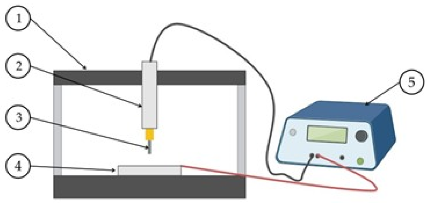
Figure 1. Deposition system (1) CNC 3018 Pro (2) Application, (3) Electrode, (4) Anode, (5) Custom-made deposition machine Mpi702Er.
Metals 15 01159 g001
Figure 2. Image of the samples: (a) untreated, (b) thermally treated, (c) thermochemically treated.
Metals 15 01159 g002
Figure 3. Optical microscopy images of the obtained layer (a) untreated, (b) thermally treated, (c) thermochemically treated.
Metals 15 01159 g003
Figure 4. SEM images on the sample surfaces: (a) untreated, (b) thermally treated, (c) thermochemically treated.
Metals 15 01159 g004
Figure 5. Samples SEM images in cross-section (a) untreated, (b) thermally treated, (c) thermochemically treated.
Metals 15 01159 g005
Figure 6. EDS analysis results cross-section (a) untreated, (b) thermally treated, (c) thermochemically treated.
Metals 15 01159 g006
Figure 7. X-ray diffraction (XRD) patterns of the Ti-6Al-4V alloy at different stages of surface modification: (1) the untreated substrate, (2) after aluminum deposition, (3) after thermal treatment, and (4) following thermochemical treatment in a nitrogen-rich atmosphere.
Metals 15 01159 g007
Figure 8. Micro-hardness evolution of the samples (a) untreated, (b) thermally treated, (c) thermochemically treated.
Metals 15 01159 g008
Figure 9. Recorded results of the adhesion test (a) untreated, (b) thermally treated, (c) thermochemically treated.
Metals 15 01159 g009
Figure 10. SEM images of the adhesion testing lines (a) untreated, (b) thermally treated, (c) thermochemically treated.
Metals 15 01159 g010
Disclaimer/Publisher’s Note: The statements, opinions and data contained in all publications are solely those of the individual author(s) and contributor(s) and not of MDPI and/or the editor(s). MDPI and/or the editor(s) disclaim responsibility for any injury to people or property resulting from any ideas, methods, instructions or products referred to in the content.