Article

Thermally Induced Gradient of Properties on a Superhydrophobic Magnesium Alloy Surface

by
Kirill A. Emelyanenko
1,
Alexander G. Domantovsky
1,2,
Elizaveta V. Chulkova
1,
Alexandre M. Emelyanenko
1 and
Ludmila B. Boinovich
1,*
1
A.N. Frumkin Institute of Physical Chemistry and Electrochemistry, Leninsky Prospect 31 bldg. 4, 119071 Moscow, Russia
2
N.S. Kurnakov Institute of General and Inorganic Chemistry, Leninsky Prospect 31, 119071 Moscow, Russia
*
Author to whom correspondence should be addressed.
Metals 2021, 11(1), 41; https://doi.org/10.3390/met11010041
Submission received: 26 November 2020 / Revised: 18 December 2020 / Accepted: 23 December 2020 / Published: 27 December 2020
(This article belongs to the Section Corrosion and Protection)

Abstract

:
Fabrication of superhydrophobic coatings for magnesium alloys is in high demand for various industrial applications. Such coatings not only extend the service life of metal structures, but also impart additional useful functional properties to the coated surface. In this study, we show that nanosecond laser processing of long, thin stripes of magnesium alloys followed by the deposition of a hydrophobic agent onto the magnesium oxide layer is a simple, convenient, and easily reproducible method for obtaining superhydrophobic surfaces with property gradient along the sample. The mechanism of the gradient in wettability and electrochemical properties of the magnesium alloy surface is discussed based on the high-temperature growth of magnesium oxide and its following degradation. The latter is related to the development of internal stresses and the formation of cracks and pores within the oxide layer at prolonged exposure to high temperatures during the interaction of a laser beam with the substrate. The effect of heating during laser processing of magnesium materials with limited sizes on the protective properties of the forming coatings is elucidated.

1. Introduction

The general approach to the design of superhydrophobic materials is based on the solution of three problems [1,2,3,4]. The first is related to the choice of an appropriate method of surface texturing to obtain the multimodal roughness on the treated surface. The second task is related to the selection of a suitable shape of textured elements, allows getting an increase in the local contact angle of the texture element due to the texture element curvature [4]. The final task, which is actual for the materials with initially high surface energy, is the reduction of the surface energy of the material by treating the surface with substances having low surface energy. In the following text, we will refer to such a substance as a hydrophobic agent. As it was clearly demonstrated in recent literature [5,6], laser surface texturing has emerged as powerful and versatile nanotechnology for producing surfaces with hierarchical roughness and high curvature of textural elements. Such roughness, required for obtaining the superhydrophobic state of materials, allows making an important step for the development of new materials for protection against icing and corrosion, fouling and bacterial contaminations [7,8,9,10,11,12,13,14,15,16].
Laser technology allows easily achieving the extreme water repellence for the diversity of materials. Besides, it was also shown that a combination of laser chemical modification and laser texturing followed by chemisorption of low surface energy compounds allows getting the superhydrophobic coatings with exceptional mechanical and chemical properties on top of different substrates [17,18,19,20]. Thus, providing breakthrough results in materials science [21,22,23,24,25,26,27,28], laser processing of materials has proven to be a quite simple and attractive method for surface modification.
Among the numerous areas of application of superhydrophobic coatings, a special place is occupied by the direction associated with the use of materials with gradient properties [29,30,31,32]. Such superhydrophobic coatings with gradient properties have already shown their efficiency for enhanced heat transfer [29], medical antiadhesion properties [30], oil-water separation [31], liquid transportation [32,33], fog harvesting [33], and in microreactor design [34].
The fabrication of superhydrophobic coatings on the surface of magnesium alloys in recent years caused particular interest [35,36,37,38,39,40], which is associated, on the one hand, with the initial low corrosion resistance of these alloys. On the other hand, magnesium and its alloys are in high demand in various applications [41,42]. For example, because of their vibration damping and vibration resistance, these alloys are used in a wide variety of vehicles. The low weight of products made of magnesium alloys is an important reason for the use of these alloys for the manufacture of various household items and equipment. Magnesium alloys surpass steel and aluminum in specific stiffness and, therefore, are actively used for the manufacture of parts subjected to bending loads (longitudinal and transverse) [41]. Magnesium alloys are non-magnetic, they do not produce sparks at all upon impact and friction, they are easily processed by cutting (6–7 times lighter than steel, 2–2.5 times lighter than aluminum), in addition, they have very high cold resistance [42]. The biodegradability of magnesium alloys makes them attractive materials for medical implants [43,44]. Thus, the superhydrophobic treatment of magnesium alloy along with the extension of the service life of metal structures allows imparting additional useful functional properties to its surface.
In this study, we will show that nanosecond laser processing of thin stripes of magnesium alloys is a simple, convenient, and easily reproducible method for obtaining surfaces with hierarchical roughness and property gradient along the sample. In addition, we will discuss for the first time the effect of heating during laser processing of magnesium samples with finite sizes on the protective properties of the forming coatings.

2. Materials and Methods

2.1. Sample Preparation

In this paper, we fabricated and studied the superhydrophobic coatings on the surface of MA8 magnesium alloy with the following chemical composition (in weight %): Mn 1.3–2.2, Ce 0.15–0.35, impurities <0.3, Mg balance. Flat sheets (2 mm thick) were cut on samples with a size of 20 × 80 mm2. For selecting an optimal mode of laser processing (Section 3.1), the areas of 20 × 20 mm2 at one end of the sample were subjected to laser treatment. At the next stage (Section 3.2), the laser treatment parameters, which have resulted in the best corrosion protection properties of test samples, were used to prepare the new sample with the whole sample area (that is, 20 × 80 mm2) processed by laser. Before the laser treatment, the samples were ground and polished with a set of SiC abrasive papers, then ultrasonically washed in deionized water and air-dried. To perform the laser processing of the samples, we used an Argent-M laser system (LTC, Saint-Petersburg, Russia) with an IR ytterbium fiber laser (wavelength of 1.064 μm, nominal power 20 W), which provides a wide choice of laser parameters, and a RAYLASE MS10 2-axis laser beam deflection unit (RAYLASE, Wessling, Germany). For this study, the beam waist was chosen with a diameter of 40 μm. All samples were textured by a single-pass parallel line pattern. The other parameters of laser beam raster scanning for different samples are presented in Table 1.
Laser treatment was performed in ambient conditions with humidity of 40–50% and a temperature of 20–25 °C. To perform the laser treatment, the samples were placed onto a thermally insulating ceramic tile with a thermal conductivity of 0.31 W·m−1·K−1. The laser processed samples were thoroughly washed with deionized water to remove surface micro- and nanoparticles weakly adhered to the sample.
Just after the laser processing, the samples were superhydrophilic, which was verified by complete water droplet spreading. To make the samples superhydrophobic, we have used chemical vapor deposition of a hydrophobic agent. For that, the samples were exposed to the vapors of fluorosilane CF3(CF2)7CH2O(CH2)3Si(OCH3)3 at a temperature of 105 °C inside the sealed cell. Right prior to hydrophobization, the samples were subjected to Bioforce UV cleaner treatment for 40 min to improve the adsorption of the hydrophobic agent. The samples obtained using different laser treatment modes were marked as listed in Table 1. A sample of bare MA8 alloy polished with #2500 SiC abrasive paper was used for the comparison and marked as the S0 sample.

2.2. Surface Characterization

The morphology and the chemical composition of samples were studied by field-emission scanning electron microscopy (FE-SEM) and energy-dispersive X-ray spectroscopy (EDX) on a FIB-SEM Nvision 40 workstation (Zeiss, Oberkochen, Germany) equipped with X-MAX energy-dispersive detector (Oxford Instruments, Tubney Wood, UK). The FE-SEM images were recorded in secondary electron detection mode at accelerating voltages of 2–5 kV. EDX microanalysis was performed at 10 kV accelerating voltage.
AFM measurements were carried out using a Nanoscope V multimode atomic force microscope (Veeco Instruments, Santa Barbara, CA, USA). Images were generated in tapping mode in the air with HQ:NSC15/NO AL silicon tips (Mikromasch, Sofia, Bulgaria) having a spring constant of 40 N/m and a radius of curvature of 8 nm. The scan rate was typically 1 Hz. Image processing and Ra parameter extraction were performed using the FemtoScan software (Advanced Technologies Center, Moscow, Russia).
The infrared spectra of the samples were investigated using Fourier-transform infrared (FTIR) spectrometry. All spectra were recorded on a Nicolet 6700 spectrometer (Thermo Scientific, Waltham, MA, USA) equipped with a mercury cadmium telluride (MCT) detector cooled with liquid N2 and a Smart SAGA (Specular Apertured Grazing Angle) accessory. To validate the presence of magnesium hydroxide on the laser textured surface and variation of its content along the gradient sample, we have measured the reflectance spectra at different locations of the sample. The angle of incidence was 80 degrees, and the diameter of the circular sampling area was 5 mm. The spectra were recorded at a resolution of 4 cm−1 in the spectral range of 650–4000 cm−1. All the spectra were derived from the result of an average of 128 scans and were recorded at room temperature. Baseline correction (OMNIC 8.0 TM) was applied to analyze the obtained spectra.
To characterize the wettability of the samples, we have measured contact and roll-off angles for a deionized water droplet. Contact angles were measured using the setup described in [45,46]. The droplets of the same volume 15 μL were used to measure the contact and roll-off angles, which were determined as an average for 5 different droplets on the chosen local area on top of a studied sample.
All electrochemical measurements were performed in 0.5 M aqueous solutions of NaCl at room temperature using an electrochemical workstation Elins P50x+FRA-24M (Elins, Moscow, Russia). For the electrochemical measurements, a three-electrode configuration was used, with a superhydrophobic sample as the working electrode (apparent area exposed to the electrolyte was 1 cm2), a platinum mesh as the counter electrode, and an Ag/AgCl electrode filled with saturated KCl solution as the reference electrode. The polarization curves were obtained at a sweep rate of 1 mV/s. The electrochemical impedance spectroscopy (EIS) measurements were conducted in a frequency range from 500 mHz to 100 kHz using a 10 mV amplitude.

3. Results

3.1. Selection of the Optimal Regime for Laser Processing

Contact and roll-off angles, measured for the samples obtained in this study by the laser processing followed by deposition and chemisorption of the hydrophobic agent are shown in Table 2.
The comparison of the wettability parameters indicated that the values of contact angles coincide for samples S1–S4 within the range of standard deviation of the experimental measurements; the roll-off angles slightly differed between the samples but did not exceed the value of 5°. Thus, every laser treatment regime used here allowed getting the samples in a superhydrophobic state. However, the main goal of the first stage of this study was to select the optimal parameters for laser treatment. The target parameter for such a selection was a corrosion resistance of the magnesium alloy in contact with a 0.5 M aqueous solution of NaCl. The corrosion resistance was estimated through the value of a corrosion current measured after 60 min of sample exposure to the electrolyte. We have measured the potentiodynamic curves and compared the properties of superhydrophobic coatings fabricated using a set of various regimes of laser treatment as listed in Table 1.
The polarization curves presented in Figure 1a indicated very high sensitivity of the corrosion current and corrosion potential to the pulse duration. As follows from the values of corrosion current, all laser-treated superhydrophobic samples demonstrated the improvement of corrosion resistance compared to the bare magnesium sample S0. However, the decrease in the pulse duration from 200 to 4 ns with a simultaneous rise of the repetition rate to 1 MHz, while keeping constant all other parameters (Table 1), allowed decreasing the corrosion current more than 6 orders of magnitude (Figure 1b, Table S1 in the Supplementary Information).
Such an impact of laser scanning parameters is evidently related to the fluence-dependent interplay of vaporization, melt-displacement and melt-ejection mechanisms under laser beam irradiation of metals. As it was shown in [47,48], cumulative heating affects surface morphology and matter removed as both vapor and liquid melt. Thus, at low to moderate fluences material removal occurs via vaporization and melt displacement, while at high fluences, the explosive ejection of liquid droplets from the melt pool is mainly responsible for material removal. The thermal diffusivity effectively enlarges the surface area subjected to laser ablation, and thus, the variation of pulse duration also causes variation in the size of the heat-affected zone. This zone extension is proportional to the square root of the product between the thermal diffusivity and pulse duration [49]. Besides, laser-treated surface temperature influences the rates of vaporization and melt ejection, thus being the determinative factor for the number of micro- and nanoparticles deposited back onto the material surface from the laser plume. The set of parameters chosen for the selection of an optimal regime of magnesium surface treatment allows considering regimes with dominating droplet ejection removal and with an essential contribution of metal vaporization removal. It is worth noting that magnesium has a high vapor pressure at temperatures even less than melting temperature [50,51] and demonstrates a much higher tendency to evaporate than aluminum, titanium, and iron at the same temperatures.
Interestingly that the appearance of fabricated samples was quite different; namely, sample S1 looked black-colored, while sample S4 was white-gray colored. Having in mind that magnesium oxide and/or magnesium hydroxide have white color, such different coloration of laser-treated surfaces is evidently related to the degree of the magnesium surface layer oxidation. The obtained coloration of the S4 sample indicates the presence of a notable amount of magnesium oxide and/or magnesium hydroxide in the very top surface layer. The second feature differentiating two samples is related to the light scattering from the surface allowed hypothesizing the higher roughness of sample S1 compared to S4. Since the microroughness of all samples is mainly defined by the line density, which was the same for all samples, we have studied the nanoroughness using AFM measurements. AFM images of samples obtained for the scanning area of 4 × 4 μm2 are presented in Figure S1 in the Supplementary Information. Arithmetical mean roughness (Ra) values defined for our samples S1 to S4 were 346 ± 36, 352 ± 36, 214 ± 42, and 190 ± 28 nm, respectively. Indeed, the nano roughness of the sample with a pulse duration of 4 ns and the fluence of 1.5 J/cm2 (sample S4) was nearly twice lower than that of the S1 sample (Table 1).
The analysis of the electrochemical data in Figure 1 indicates that the best corrosion protection properties were characteristic of the superhydrophobic sample S4. The corrosion resistance is defined by both a superhydrophobic state of the surface and by the corresponding composition of the surface layer [12]. In the following, we will discuss the influence of the temperature on the surface composition and the properties of the sample S4.

3.2. Mechanism of the Gradient Properties Formation

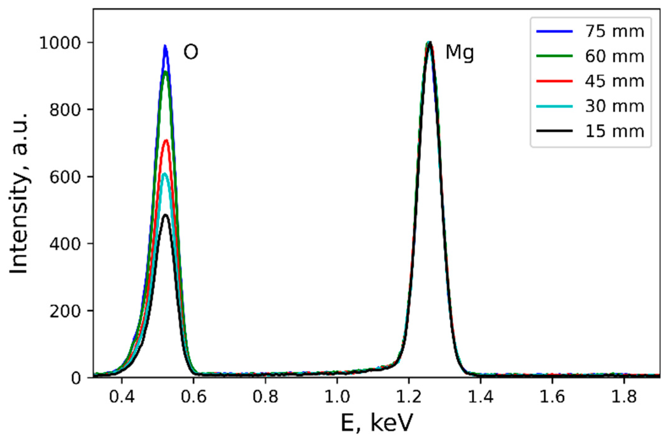
In the previous section, we have compared the properties of samples, for which the areas of 20 × 20 mm2 at one end of the sample were subjected to laser treatment. The treatment time for these samples was 27 min per sample. The wetting parameters of such samples were uniform along the whole textured area. At the next stage, we have studied the samples with the whole area processed by laser with the parameters of laser processing the same as those for the sample S4. The processing time for this sample was about 110 min. After sample washing with deionized water and drying in the oven at 100 °C for 10 min, the obtained sample appeared unevenly colorized with a smooth and continuous variation of color from dark gray on one end of the sample to white-gray on the other. To understand the variation in the sample coloration, we have studied the elemental composition and the morphology of the surface in different sample locations. In Figure 2, the EDX spectra are given for 5 different locations covering areas from dark gray, corresponding to the coordinate of 15 mm (black curve) from the beginning of texturing the sample to white-gray (blue curve) with the coordinate of 75 mm (i.e., 5 mm to the end of texturing).
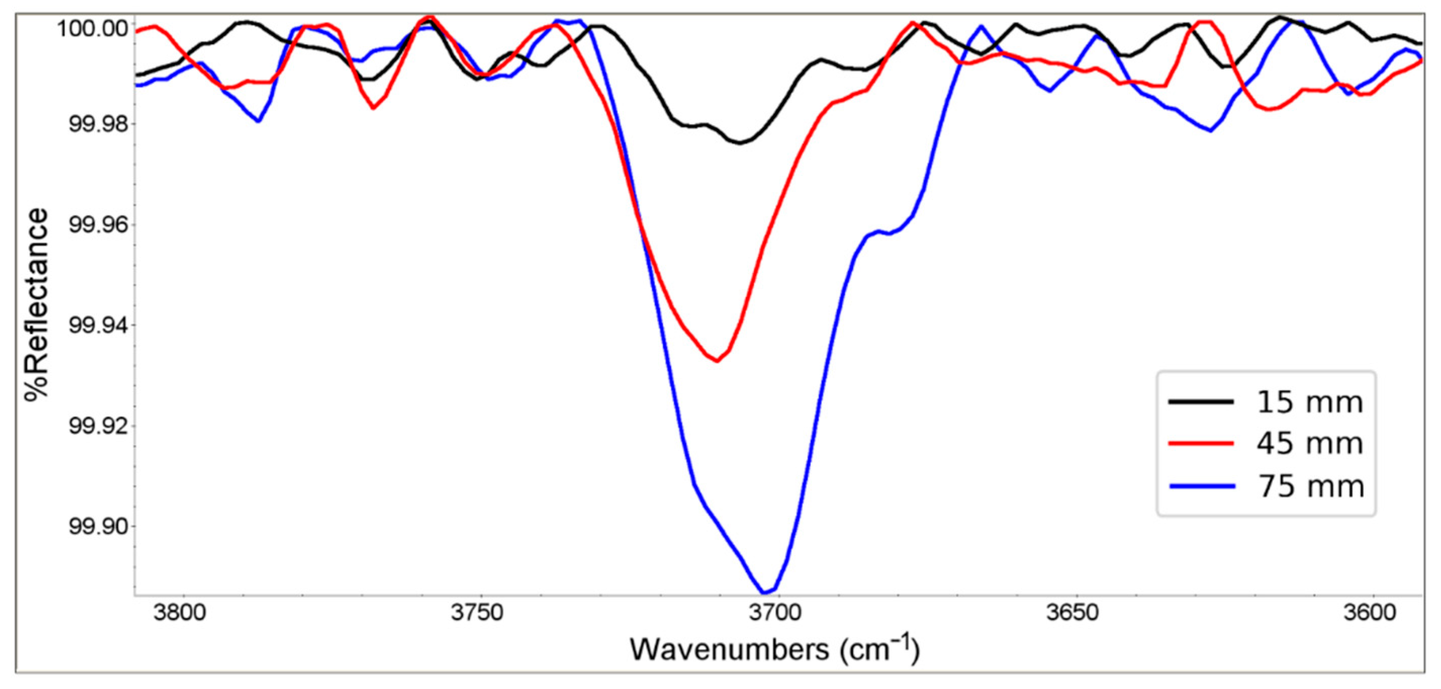
Analysis of EDX spectra shows a significant lateral gradient of oxygen content on the surface of the sample. For the white-gray area, the oxygen content is about twice as high as that characteristic of the dark-gray area, which may be related to the variation in the amount of both surface oxide and hydroxide along the sample. To differentiate between the contributions of these two oxygen-containing compounds, we have performed the FTIR spectroscopy study of the sample surface. Data of grazing angle reflectance spectra allow analyzing the very top surface layer. The sharp IR active band near 3700 cm−1 presented in Figure 3 is associated with a stretching vibration of a hydroxyl from magnesium hydroxide [52,53]. However, the obtained spectra show only trace amounts of hydroxides, which slightly increase from the dark-gray to white-gray areas. From these data, one can conclude that the oxygen content is mainly related to the presence of surface magnesium oxide, because of a low concentration of manganese and cerium in the considered alloy (not shown on the EDX spectra). It was repeatedly discussed in the literature that due to a high affinity to oxygen, magnesium alloys are easily oxidized when exposed to air at high temperatures [51,54,55,56].
During the laser treatment of the sample, heating is taking place due to absorption of laser irradiation and the energy released by the exothermic oxidation of the magnesium alloy, while heat removal from the sample is carried out by radiation, conduction, phase transitions (melting and evaporation) and convection. Taking into account the notable suppression of heat removal by conduction, which is a consequence of the location of the sample during laser treatment on a heat-insulating ceramic substrate, very strong heating of the sample can be expected.
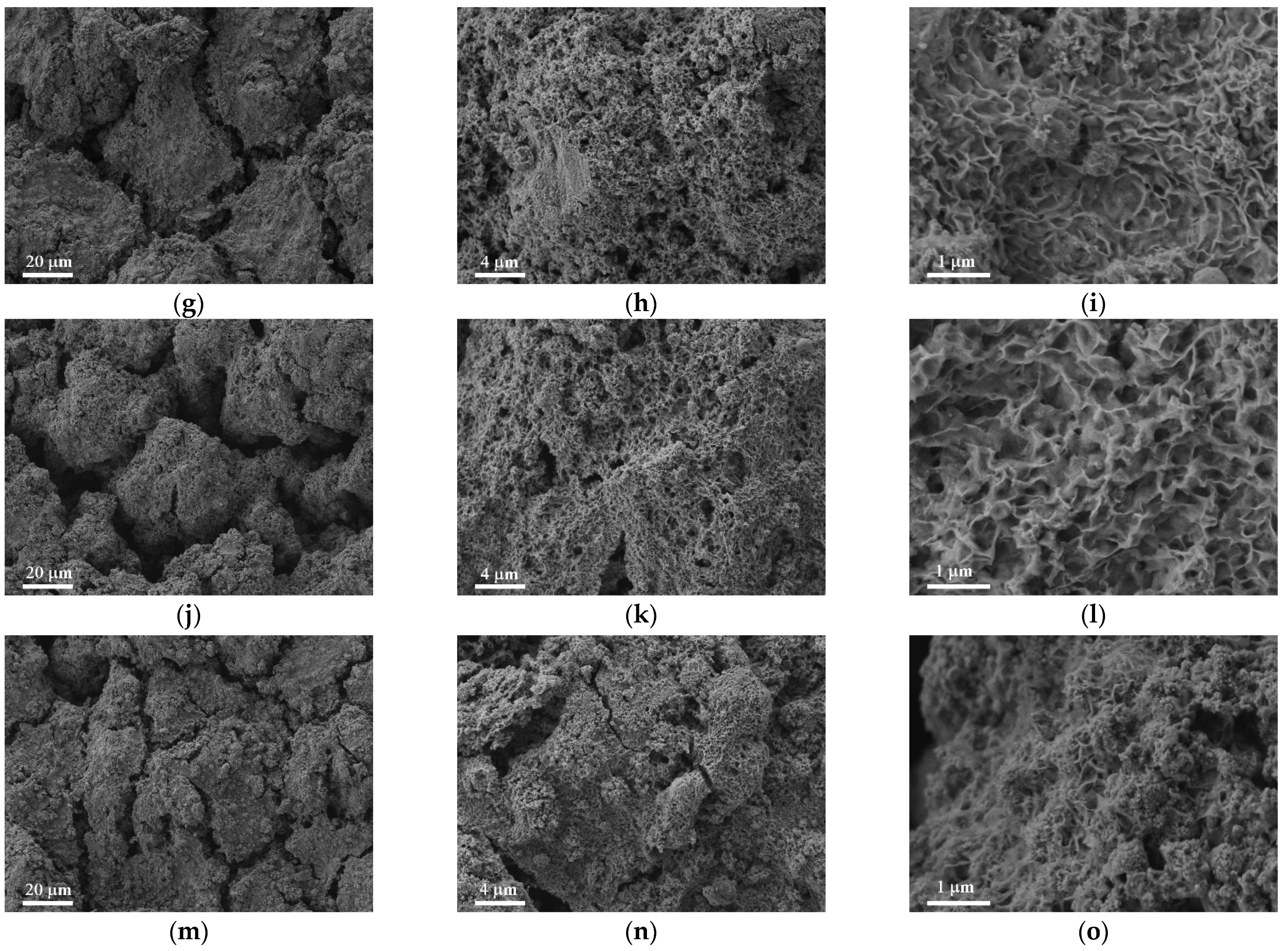
Several competing processes take place during laser treatment, such as oxidation, vigorous magnesium vaporization, local sample melting, and explosive ablation. Besides, the interaction of Mg2+ and droplets of molten Mg in the laser ablation plume with atmospheric O2 causes magnesium oxidation in the gaseous phase. Further partial deposition of micro- and nanoparticles of magnesium oxides from laser plume onto the heated/locally melted magnesium substrate leads to sintering of the oxide particles deposited from the plume with surface oxides. Growth and thickening of the magnesium oxide layer, accompanied by the evaporation of underlying heated magnesium substrate, simultaneously cause the development of internal stresses and the formation of cracks and pores within the oxide layer [51,55]. The density and sizes of such cracks and pores will be higher the more prolonged period the surface oxide layer is exposed to elevated temperatures. For the long samples processed in this study, a thick layer of surface oxide formed at the initial stage of laser processing (locations with coordinates close to zero) is maintained in a highly heated state for a much longer time than areas with coordinates close to 80 mm, textured at the end of the process. Thus, it was expected that the cracking and porosity of samples would gradually decrease from the initially textured areas to the opposite side of the sample. The structure of oxidized layers at different sample locations shown in Figure 4 for three different magnifications allows understanding the details of the surface texture both on micro- and nano levels. Indeed, from the images at all levels of magnification, we can conclude that the variation in the morphology goes smoothly from one end of the sample to the other, substantiating the above hypothesis.
For the sample areas with coordinates close to zero (Figure 4, top row) the porosity and the width of cracks are notably higher than for the areas at the opposite side of the sample (coordinates close to 80 mm, bottom row). Higher porosity and number of cracks result in a weaker adhesion of the textured layer to the magnesium alloy substrate and consequently lead to greater removal of the magnesium oxide layer from the surface upon sample washing after its laser processing. Such a scenario explains the increase in oxygen content of the surface layer when moving towards the areas with coordinates close to 80 mm.
Although the as-formed thick layer of high temperature oxide on top of magnesium alloy substrate is not protective against further oxidation and ion, charge, and water molecules transfer, the deposition of a hydrophobic agent layer atop of magnesium oxide allows providing such protective properties for the composite coating. Several mechanisms responsible for the enhancement of the oxide protection activity should be mentioned here. The deposited hydrophobic layer provides the water repellence of the surface, leading to the development of a high negative capillary pressure preventing the imbibition of the aggressive liquid into the oxide layer. Besides, the layer of the hydrophobic agent is responsible for negative charging of the surface in an aqueous medium and acts as a barrier for aggressive ions adsorption and transition across this layer [12]. To verify the protective action of the porous oxide layer with chemisorbed fluoro-oxysilane, we have studied the wettability characteristics and the corrosion resistance for the different locations along the sample.

3.3. The Gradient of the Protective Properties of the Fabricated Superhydrophobic Sample

The analysis of the contact and roll-off angles measured at different locations of the sample and presented in Figure 5a indicates the wettability gradient along the sample with the enhancement of water repellency toward the magnesium oxide enriched part of the sample. The variation in the roll-off angle from 2° to 12.6° is more notable than the corresponding variation in contact angle from 168.9° to 170.9°. This is related to the very high sensitivity of roll-off angles and their scattering across the surface to the peculiarities of the surface roughness, its nonuniformity, width, and depth of the cracks. For a better understanding of the variation of the characteristic sizes of pores and cracks, we have applied atomic force microscopy to study the nanomorphology of the gradient samples at locations corresponding to distances of 15, 30, 45, 60, and 75 mm from the starting edge of laser scanning.
AFM images presented in Figure S2 (Supplementary Information) indicate the decrease in the number of nanopores, their width and depth in the direction to the areas with coordinates close to 80 mm. Arithmetical mean roughness values defined for the presented locations indicate a notable decrease in the Ra value from Ra = (431 ± 32) nm at the distance of 15 mm to Ra = (245 ± 26) nm at the distance of 75 mm, substantiating the idea of the correlation between the surface roughness and the value and scattering of roll-off angles. However, it is worth noting that the superhydrophobic state with the extreme values of contact angles was still characteristic for all locations of the sample.
To study the ability of magnesium oxide formed at high temperatures and covered by a chemisorbed layer of the hydrophobic agent to protect against corrosion, we have measured electrochemical impedance spectra and potentiodynamic polarization curves at different locations of the sample, the same as those used to determine both the oxygen content and the wettability. The data presented in Figure 5b,c indicate the gradient of electrochemical properties of the fabricated coating. The transition from the dark-gray part of the sample to the white-gray enriched with the magnesium oxide is accompanied by the corrosion current decrease nearly three orders of magnitude. Simultaneously, the impedance modulus at low frequencies (f = 0.05 Hz) increases more than two orders of magnitude, reaching the value of 1.6 × 10⁸ Ω·cm2.
However, a reliable estimation of anticorrosion properties for the superhydrophobic sample requires the measurement of the time evolution of the corrosion current [12]. That is why we have analyzed the temporal behavior of corrosion currents, measured for two different locations of the freshly prepared gradient superhydrophobic sample. These data for the locations at distances of 15 and 75 mm from the starting edge of laser scanning are given in Table 3. It can be concluded that for the location of 75 mm enriched with the magnesium oxide, the corrosion current increases during the first hours and then tends to plateau value with the corrosion current value of Icor = 3.8 nA/cm2. Corrosion current for the location of 15 mm demonstrated similar temporal behavior with the plateau value of 1.6 μA/cm2 after 24 h of contact with 0.5 M NaCl solution. Thus, the corrosion current for any location atop of gradient sample even after a day of contact with corrosive medium retained the value orders of magnitude lower than that characteristic of the bare MA8 sample (Table S1), thus indicating the significant protective effect of a combination of the thick porous layer of magnesium oxide formed at elevated temperatures with the superhydrophobic state of the surface.
As mentioned above, the gradient properties detected in this study appear as a result of a laser heating-induced degradation of an oxide layer in the course of laser treatment of the subsequent surface areas. This concurrent to laser processing effect can be significant for thin stripe samples. It leads to an undesirable reduction in the corrosion protective effect and can be minimized by appropriate heat removal during the laser processing of the sample. Evidently, for high mass samples with a low surface to volume ratio, other effects related to fast laser heating and cooling, recrystallization and quenching, should contribute to the surface properties.

4. Conclusions

In this study, we have considered the nanosecond laser processing of magnesium alloy MA8 approach for the design of a surface layer with a hierarchical roughness. For all regimes of laser treatment used in this study, the subsequent deposition of fluorooxysilane molecules on the laser-textured surface resulted in establishing the superhydrophobic state with extremely high contact angles. Using the set of laser parameters providing the best water-repelling and anticorrosion properties for the small-scale test samples, we have analyzed for the first time the impact of sample heating if prolonged laser processing was used, on the electrochemical properties, morphology, wettability, and integrated elemental composition of surface layer for the metal stripes. For the samples in the form of long, thin stripes, the pronounced gradient of surface properties was detected. The observed phenomena are associated with the growth of high temperature magnesium oxide during interaction of laser beam with the substrate and following degradation of this oxide, induced by the development of internal stresses and the formation of cracks and pores within the oxide layer at the prolonged exposure to high temperatures.
In turn, high porosity and density of cracks developed in the oxide layer are responsible for low adhesion of magnesium oxide to the substrate, which causes easy partial removal of the oxide from the surface during the sample washing. It was found that the sample’s areas exposed to high temperature for a smaller time showed denser oxide layer and fewer number of pores. At the same time, the chemisorption of a hydrophobic agent onto the oxide layer allows getting the superhydrophobic state through the whole sample. It was detected that although the as-formed layer of high-temperature oxide on top of magnesium alloy substrate is not corrosion protective against further oxidation and ion, charge, and water molecule transfer, the deposition of a hydrophobic agent layer atop of magnesium oxide allows providing such protective properties for the obtained composite coating due to three different mechanisms, already discussed in the literature [12]. Namely, capillary repulsion of the corrosive medium from hydrophobic walls of pores and cracks minimize the contact of solid with liquid, negatively charged hydrophobic surface repulse chloride anions, fluorosilane molecules block the adsorption active sites of the surface leading to the inhibition of chloride anions adsorption.

Supplementary Materials

The following are available online at https://www.mdpi.com/2075-4701/11/1/41/s1, Table S1: Summary of electrochemical parameters of fabricated samples derived from the potentiodynamic polarization curves (Figure 1a); Figure S1: AFM images of the fabricated samples S1 to S4; Figure S2: AFM images of the gradient sample at different distances from the starting edge of laser scanning.

Author Contributions

Conceptualization, L.B.B.; methodology, L.B.B. and K.A.E.; software, K.A.E. and A.M.E.; validation, K.A.E., A.G.D., E.V.C. and L.B.B.; formal analysis, A.M.E.; investigation, A.G.D., E.V.C. and L.B.B.; resources, A.M.E.; writing—original draft preparation, K.A.E.; writing—review and editing, L.B.B.; visualization, A.M.E.; supervision, L.B.B.; project administration, A.M.E.; funding acquisition, A.M.E. and L.B.B. All authors have read and agreed to the published version of the manuscript.

Funding

This research was funded by RFBR, grant number 18-29-05008, and by the Ministry of Science and Higher Education of the Russian Federation.

Institutional Review Board Statement

Not applicable.

Informed Consent Statement

Not applicable.

Data Availability Statement

Data is contained within the article and Supplementary Materials.

Acknowledgments

The SEM and EDX studies were performed using shared experimental facilities supported by IGIC RAS state assignment. The authors are grateful to Natalia V. Kuzmina for her help with AFM imaging.

Conflicts of Interest

The authors declare no conflict of interest. The funders had no role in the design of the study; in the collection, analyses, or interpretation of data; in the writing of the manuscript, or in the decision to publish the results.

References

  1. Erbil, H.Y. Practical Applications of Superhydrophobic Materials and Coatings: Problems and Perspectives. Langmuir 2020, 36, 2493–2509. [Google Scholar] [CrossRef] [PubMed]
  2. Czyzyk, S.; Dotan, A.; Dodiuk, H.; Kenig, S. Easy-to-Clean Superhydrophobic Coatings Based on Sol-Gel Technology: A Critical Review. Rev. Adhes. Adhes. 2017, 5, 325–360. [Google Scholar] [CrossRef]
  3. Drelich, J.W.; Boinovich, L.; Chibowski, E.; Della Volpe, C.; Hołysz, L.; Marmur, A.; Siboni, S. Contact angles: History of over 200 years of open questions. Surf. Innov. 2020, 8, 3–27. [Google Scholar] [CrossRef] [Green Version]
  4. Boinovich, L.B.; Emelyanenko, A. The prediction of wettability of curved surfaces on the basis of the isotherms of the disjoining pressure. Colloids Surfaces A Physicochem. Eng. Asp. 2011, 383, 10–16. [Google Scholar] [CrossRef]
  5. Stratakis, E.; Bonse, J.; Heitz, J.; Siegel, J.; Tsibidis, G.; Skoulas, E.; Papadopoulos, A.; Mimidis, A.; Joel, A.-C.; Comanns, P.; et al. Laser engineering of biomimetic surfaces. Mater. Sci. Eng. R: Rep. 2020, 141, 100562. [Google Scholar] [CrossRef]
  6. Kumar, V.; Verma, R.; Kango, S.; Sharma, V.S. Recent progresses and applications in Laser-based surface texturing systems. Mater. Today Commun. 2020, 101736, 101736. [Google Scholar] [CrossRef]
  7. Darmanin, T.; Guittard, F. Recent advances in the potential applications of bioinspired superhydrophobic materials. J. Mater. Chem. A 2014, 2, 16319–16359. [Google Scholar] [CrossRef]
  8. Simpson, J.T.; Hunter, S.R.; Aytug, T. Superhydrophobic materials and coatings: A review. Rep. Prog. Phys. 2015, 78, 086501. [Google Scholar] [CrossRef]
  9. Conde, J.J.; Ferreira-Aparicio, P.; Chaparro, A.M. Anti-corrosion coating for metal surfaces based on superhydrophobic electrosprayed carbon layers. Appl. Mater. Today 2018, 13, 100–106. [Google Scholar] [CrossRef]
  10. Ma, L.; Wang, L.; Li, C.; Guo, J.; Shrotriya, P.; Deng, C.; Zhao, J. Hybrid Nanosecond Laser Processing and Heat Treatment for Rapid Preparation of Super-Hydrophobic Copper Surface. Metals 2019, 9, 668. [Google Scholar] [CrossRef] [Green Version]
  11. Li, X.; Shi, T.; Li, B.; Zhang, C.-W.; Zhong, B.; Lv, Y.; Zhang, Q. One-Step Preparation of Super-Hydrophobic Micro-Nano Dendrites on Al Alloy for Enhanced Corrosion Resistance. Metals 2018, 8, 960. [Google Scholar] [CrossRef] [Green Version]
  12. Boinovich, L.B.; Emelyanenko, A.M.; Modestov, A.D.; Domantovsky, A.G.; Emelyanenko, K.A. Not simply repel water: The diversified nature of corrosion protection by superhydrophobic coatings. Mendeleev Commun. 2017, 27, 254–256. [Google Scholar] [CrossRef]
  13. Ferrari, M.; Benedetti, A. Superhydrophobic surfaces for applications in seawater. Adv. Colloid Interface Sci. 2015, 222, 291–304. [Google Scholar] [CrossRef] [PubMed]
  14. Gule, N.P.; Begum, N.M.; Klumperman, B. Advances in biofouling mitigation: A review. Crit. Rev. Environ. Sci. Technol. 2016, 46, 535–555. [Google Scholar] [CrossRef]
  15. Esmeryan, K.D.; Avramova, I.A.; Castano, C.E.; Ivanova, I.A.; Mohammadi, R.; Radeva, E.I.; Stoyanova, D.S.; Vladkova, T.G. Early stage anti-bioadhesion behavior of superhydrophobic soot based coatings towards Pseudomonas putida. Mater. Des. 2018, 160, 395–404. [Google Scholar] [CrossRef]
  16. Emelyanenko, A.M.; Pytskii, I.S.; Kaminsky, V.V.; Chulkova, E.V.; Domantovsky, A.G.; Emelyanenko, K.A.; Sobolev, V.D.; Aleshkin, A.V.; Boinovich, L.B. Superhydrophobic copper in biological liquids: Antibacterial activity and microbiologically induced or inhibited corrosion. Colloids Surfaces B: Biointerfaces 2020, 185, 110622. [Google Scholar] [CrossRef] [PubMed]
  17. Boinovich, L.B.; Modin, E.B.; Sayfutdinova, A.R.; Emelyanenko, K.A.; Vasiliev, A.L.; Emelyanenko, A.M. Combination of Functional Nanoengineering and Nanosecond Laser Texturing for Design of Superhydrophobic Aluminum Alloy with Exceptional Mechanical and Chemical Properties. ACS Nano 2017, 11, 10113–10123. [Google Scholar] [CrossRef] [PubMed]
  18. Emelyanenko, A.M.; Boinovich, L.B.; Bezdomnikov, A.A.; Chulkova, E.V.; Emelyanenko, K.A. Reinforced Superhydrophobic Coating on Silicone Rubber for Longstanding Anti-Icing Performance in Severe Conditions. ACS Appl. Mater. Interfaces 2017, 9, 24210–24219. [Google Scholar] [CrossRef] [PubMed]
  19. Boinovich, L.B.; Emelyanenko, K.A.; Domantovsky, A.G.; Chulkova, E.V.; Shiryaev, A.A.; Emelyanenko, A.M. Pulsed Laser Induced Triple Layer Copper Oxide Structure for Durable Polyfunctionality of Superhydrophobic Coatings. Adv. Mater. Interfaces 2018, 5, 1801099. [Google Scholar] [CrossRef]
  20. Sataeva, N.E.; Boinovich, L.B.; Emelyanenko, K.A.; Domantovsky, A.G.; Emelyanenko, A.M. Laser-assisted processing of aluminum alloy for the fabrication of superhydrophobic coatings withstanding multiple degradation factors. Surf. Coatings Technol. 2020, 397, 125993. [Google Scholar] [CrossRef]
  21. Santos, E.C.; Shiomi, M.; Osakada, K.; Laoui, T. Rapid manufacturing of metal components by laser forming. Int. J. Mach. Tools Manuf. 2006, 46, 1459–1468. [Google Scholar] [CrossRef]
  22. Carter, L.; Martin, C.; Withers, P.J.; Attallah, M.M. The influence of the laser scan strategy on grain structure and cracking behaviour in SLM powder-bed fabricated nickel superalloy. J. Alloys Compd. 2014, 615, 338–347. [Google Scholar] [CrossRef]
  23. Son, Y.; Yeo, J.; Moon, H.; Lim, T.W.; Hong, S.; Nam, K.H.; Yoo, S.; Grigoropoulos, C.P.; Yang, D.-Y.; Ko, S.H. Nanoscale Electronics: Digital Fabrication by Direct Femtosecond Laser Processing of Metal Nanoparticles. Adv. Mater. 2011, 23, 3176–3181. [Google Scholar] [CrossRef]
  24. Yeo, J.; Kim, G.; Hong, S.; Kim, M.S.; Kim, D.; Lee, J.; Lee, H.B.; Kwon, J.; Suh, Y.D.; Kang, H.W.; et al. Flexible supercapacitor fabrication by room temperature rapid laser processing of roll-to-roll printed metal nanoparticle ink for wearable electronics application. J. Power Sources 2014, 246, 562–568. [Google Scholar] [CrossRef]
  25. Malinauskas, M.; Žukauskas, A.; Hasegawa, S.; Hayasaki, Y.; Mizeikis, V.; Buividas, R.; Juodkazis, S. Ultrafast laser processing of materials: From science to industry. Light. Sci. Appl. 2016, 5, e16133. [Google Scholar] [CrossRef] [PubMed] [Green Version]
  26. Boinovich, L.B.; Emelyanenko, A.M.; Emelyanenko, K.A.; Modin, E.B. Modus Operandi of Protective and Anti-icing Mechanisms Underlying the Design of Longstanding Outdoor Icephobic Coatings. ACS Nano 2019, 13, 4335–4346. [Google Scholar] [CrossRef] [PubMed]
  27. Ocaña, J.L.; Huerta-Murillo, D.; Lasagni, A.F.; Aguilar-Morales, A.I.; Alamri, S.; Cardoso, J.T.; García-Beltrán, A.; Cordovilla, F.; Angulo, I. Modification of Ti6Al4V surface properties by combined DLW-DLIP hierarchical micro-nano structuring. Adv. Opt. Technol. 2020, 9, 121–130. [Google Scholar] [CrossRef]
  28. Garcia-Giron, A.; Romano, J.-M.; Batal, A.; Dashtbozorg, B.; Dong, H.; Solanas, E.M.; Angos, D.U.; Walker, M.; Penchev, P.; Dimov, S.S. Durability and Wear Resistance of Laser-Textured Hardened Stainless Steel Surfaces with Hydrophobic Properties. Langmuir 2019, 35, 5353–5363. [Google Scholar] [CrossRef] [Green Version]
  29. Tokunaga, A.; Tsuruta, T. Enhancement of condensation heat transfer on a microstructured surface with wettability gradient. Int. J. Heat Mass Transf. 2020, 156, 119839. [Google Scholar] [CrossRef]
  30. Zhang, Q.; Dong, J.; Peng, M.; Yang, Z.; Wan, Y.; Yao, F.; Zhou, J.; Ouyang, C.; Deng, X.; Wan, Y. Laser-induced wettability gradient surface on NiTi alloy for improved hemocompatibility and flow resistance. Mater. Sci. Eng. C 2020, 111, 110847. [Google Scholar] [CrossRef]
  31. Ge, P.; Wang, S.; Zhang, J.; Yang, B. Micro-/nanostructures meet anisotropic wetting: From preparation methods to applications. Mater. Horiz. 2020, 7, 2566–2595. [Google Scholar] [CrossRef]
  32. Ju, J.; Zheng, Y.; Jiang, L. Bioinspired One-Dimensional Materials for Directional Liquid Transport. Accounts Chem. Res. 2014, 47, 2342–2352. [Google Scholar] [CrossRef] [PubMed]
  33. Chen, H.; Ran, T.; Gan, Y.; Zhou, J.; Zhang, Y.; Zhang, L.; Zhang, D.; Jiang, L. Ultrafast water harvesting and transport in hierarchical microchannels. Nat. Mater. 2018, 17, 935–942. [Google Scholar] [CrossRef] [PubMed]
  34. Wang, J.; Chao, P.H.-S.; Hanet, S.; Van Dam, R.M. Performing multi-step chemical reactions in microliter-sized droplets by leveraging a simple passive transport mechanism. Lab Chip 2017, 17, 4342–4355. [Google Scholar] [CrossRef]
  35. Gray-Munro, J.; Campbell, J. Mimicking the hierarchical surface topography and superhydrophobicity of the lotus leaf on magnesium alloy AZ31. Mater. Lett. 2017, 189, 271–274. [Google Scholar] [CrossRef]
  36. Ishizaki, T.; Shimada, Y.; Tsunakawa, M.; Lee, H.; Yokomizo, T.; Hisada, S.; Nakamura, K. Rapid Fabrication of a Crystalline Myristic Acid-Based Superhydrophobic Film with Corrosion Resistance on Magnesium Alloys by the Facile One-Step Immersion Process. ACS Omega 2017, 2, 7904–7915. [Google Scholar] [CrossRef]
  37. Boinovich, L.B.; Emelyanenko, A.M.; Pashinin, A.S.; Gnedenkov, S.V.; Egorkin, V.S.; Sinebryukhov, S.L. Mg alloy treatment for superhydrophobic anticorrosion coating formation. Surf. Innov. 2013, 1, 162–172. [Google Scholar] [CrossRef]
  38. Yeganeh, M.; Mohammadi, N. Superhydrophobic surface of Mg alloys: A review. J. Magnes. Alloy. 2018, 6, 59–70. [Google Scholar] [CrossRef]
  39. Joo, J.; Kim, D.; Moon, H.-S.; Kim, K.; Lee, J. Durable anti-corrosive oil-impregnated porous surface of magnesium alloy by plasma electrolytic oxidation with hydrothermal treatment. Appl. Surf. Sci. 2020, 509, 145361. [Google Scholar] [CrossRef]
  40. Ishizaki, T.; Kumagai, S.; Tsunakawa, M.; Furukawa, T.; Nakamura, K. Ultrafast fabrication of superhydrophobic surfaces on engineering light metals by single-step immersion process. Mater. Lett. 2017, 193, 42–45. [Google Scholar] [CrossRef]
  41. Gupta, M.; Sharon, N.M.L. Magnesium, Magnesium Alloys, and Magnesium Composites; Wiley: Hoboken, NJ, USA, 2010. [Google Scholar]
  42. Magnesium Technology; Springer Science and Business Media LLC: Berlin/Heidelberg, Germany, 2006.
  43. Seitz, J.-M.; Durisin, M.; Goldman, J.; Drelich, J.W. Recent Advances in Biodegradable Metals for Medical Sutures: A Critical Review. Adv. Heal. Mater. 2015, 4, 1915–1936. [Google Scholar] [CrossRef] [PubMed]
  44. Kamrani, S.; Fleck, C. Biodegradable magnesium alloys as temporary orthopaedic implants: A review. BioMetals 2019, 32, 185–193. [Google Scholar] [CrossRef] [PubMed]
  45. Emel’Yanenko, A.M.; Boinovich, L.B. Analysis of wetting as an efficient method for studying the characteristics of coatings and surfaces and the processes that occur on them: A review. Inorg. Mater. 2011, 47, 1667–1675. [Google Scholar] [CrossRef]
  46. Emelyanenko, A.; Boinovich, L. The role of discretization in video image processing of sessile and pendant drop profiles. Colloids Surfaces A: Physicochem. Eng. Asp. 2001, 189, 197–202. [Google Scholar] [CrossRef]
  47. Fishburn, J.; Withford, M.; Coutts, D.; Piper, J. Study of the fluence dependent interplay between laser induced material removal mechanisms in metals: Vaporization, melt displacement and melt ejection. Appl. Surf. Sci. 2006, 252, 5182–5188. [Google Scholar] [CrossRef]
  48. Fishburn, J.; Withford, M.; Coutts, D.W.; Piper, J.A. Study of the interplay of vaporisation, melt displacement and melt ejection mechanisms under multiple pulse irradiation of metals. Appl. Surf. Sci. 2006, 253, 662–667. [Google Scholar] [CrossRef]
  49. Stoian, R.; Colombier, J.-P. Advances in ultrafast laser structuring of materials at the nanoscale. Nanophotonics 2020, 9, 4665–4688. [Google Scholar] [CrossRef]
  50. Lide, D.R. CRC Handbook of Chemistry and Physics, 84th ed.; Lide, D.R., Ed.; CRC Press: Boca Raton, FL, USA, 2003. [Google Scholar]
  51. Tan, Q.; Yin, Y.; Mo, N.; Zhang, M.; Atrens, A. Recent understanding of the oxidation and burning of magnesium alloys. Surf. Innov. 2019, 7, 71–92. [Google Scholar] [CrossRef]
  52. Buchanan, R.A.; Caspers, H.H.; Murphy, J. Lattice Vibration Spectra of Mg (OH)_2 and Ca(OH)_2. Appl. Opt. 2008, 2, 1147–1150. [Google Scholar] [CrossRef]
  53. Hexter, R.M. On the Infrared Absorption Spectra Crystalline Brucite [Mg (OH)_2] and Portlandite [Ca(OH)_2]. J. Opt. Soc. Am. 2008, 48, 770–772. [Google Scholar] [CrossRef]
  54. Tan, Q.; Atrens, A.; Mo, N.; Zhang, M.-X. Oxidation of magnesium alloys at elevated temperatures in air: A review. Corros. Sci. 2016, 112, 734–759. [Google Scholar] [CrossRef] [Green Version]
  55. Czerwinski, F. Oxidation Characteristics of Magnesium Alloys. JOM 2012, 64, 1477–1483. [Google Scholar] [CrossRef]
  56. Ma, S.; Xing, F.; Ta, N.; Zhang, L. Kinetic modeling of high-temperature oxidation of pure Mg. J. Magnes. Alloy. 2020, 8, 819–831. [Google Scholar] [CrossRef]
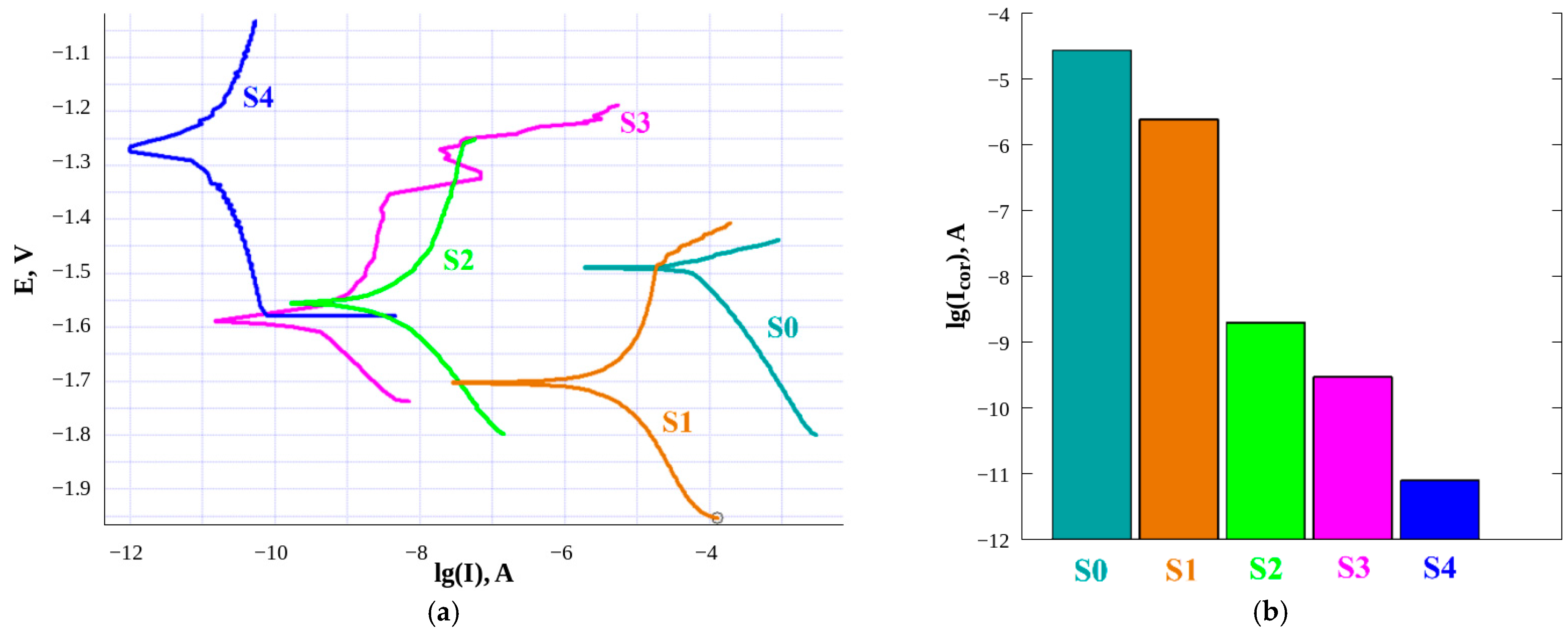
Figure 1. Polarization curves (a) and corrosion currents (b) registered in 0.5 M NaCl solution for bare Mg sample (S0) and superhydrophobic samples (S1 to S4) fabricated on magnesium alloy surface on the basis of laser treatment with different pulse durations.
Metals 11 00041 g001
Figure 2. The EDX spectra registered at different positions (indicated in the legend) along the 80 mm long superhydrophobic substrate S4.
Metals 11 00041 g002
Figure 3. The FTIR grazing angle reflectance spectra registered at different positions (indicated in the legend) along the 80 mm long superhydrophobic substrate S4.
Metals 11 00041 g003
Figure 4. SEM images of the sample S4 with a laser-processed area of 20 × 80 mm2. Sequential rows (top to bottom) correspond to distances of 15 (ac), 30 (df), 45 (gi), 60 (jl), and 75 (mo) mm from the starting edge of laser scanning; columns correspond to different magnification.
Metals 11 00041 g004aMetals 11 00041 g004b
Figure 5. Properties of the gradient sample. (a) Contact (blue triangles) and roll-off (red circles) angle as functions of position on the sample, measured from the place where laser processing began. Error bars indicate standard deviations for at least five independent measurements. Inset shows the image of the gradient sample; the laser raster scanning started from the left end, which appears darker, and proceeded to the right. (b) Variations of corrosion current (Icor, blue circles) and the ratio of Oxygen to Magnesium peak heights (red rhombi) in EDX spectra (presented in Figure 2) along the sample. (c) The impedance modulus spectra for different locations on the sample.
Metals 11 00041 g005
Table 1. Parameters of laser treatment for samples studied in this work.
Sample Line Density, mm−1Linear Scanning Rate, mm/sPulse Duration, nsRepetition Rate, kHzFluence, J/cm2
S14001002002076
S2400100142865.3
S340010085003.0
S4400100410001.5
Table 2. Wettability characteristics of fabricated samples.
Sample Contact Angle, °Roll-Off Angle, °
S043.1 ± 1.3No roll-off
S1170.7 ± 0.73.1 ± 0.6
S2171.2 ± 0.74.3 ± 1.1
S3171.5 ± 1.03.8 ± 0.5
S4170.9 ± 0.32.5 ± 0.5
Table 3. Time evolution of corrosion current at different locations, l, along the gradient sample.
Time, hCorrosion Current, A cm−2
l = 15 mml = 75 mm
15.0 × 10−91.0 × 10−11
161.2 × 10−75.2 × 10−10
201.5 × 10−63.4 × 10−9
241.7 × 10−63.8 × 10−9
Publisher’s Note: MDPI stays neutral with regard to jurisdictional claims in published maps and institutional affiliations.