Article

Mechanical Properties and Corrosion Resistance of Magnesium–Hydroxyapatite Composites Fabricated by Spark Plasma Sintering

1
Department of Materials Science and Engineering, Tokyo Institute of Technology, Tokyo 152–8550, Japan
2
Research Center for Structural Materials, National Institute for Materials Science, Tsukuba 305-0047, Japan
3
VSB-Technical University of Ostrava, 708 00 Ostrava-Poruba, Czech Republic
*
Author to whom correspondence should be addressed.
Metals 2020, 10(10), 1314; https://doi.org/10.3390/met10101314
Submission received: 5 August 2020 / Revised: 28 September 2020 / Accepted: 29 September 2020 / Published: 30 September 2020
(This article belongs to the Section Biobased and Biodegradable Metals)

Abstract

:
Recent studies indicate that biodegradable magnesium alloys and composites are attracting a great deal of attention in orthopedic applications. In this study, magnesium–hydroxyapatite (Mg–HAP) composites with different compositions and grain size were fabricated by a spark plasma sintering (SPS) method. Their mechanical properties and corrosion behavior in a pseudo-physiological environment were investigated by pH measurements and inductivity coupled plasma (ICP) elemental analysis after an immersion test using Hanks’ solution. The results clearly showed that the addition of HAP improved both the mechanical properties and corrosion resistance. The results also indicated that the finer grain size improved most of the properties that are needed in a material for an orthopedic implant. Furthermore, the authors reveal that there is a strong correlation between the compressive strength and the porosity. In order to achieve the same compressive strength as human bone using these fabrication conditions, it is revealed that the porosity should be lower than 1.9%.

1. Introduction

Magnesium and its alloys are considered innovative materials for orthopedic implants because of their many advantages which include biocompatibility and biodegradability [1,2,3]. Nowadays, other conventional metallic biomaterials such as titanium alloys and stainless steels are used in orthopedic applications [4,5,6,7,8]. However, they have two problems which are a high Young’s modulus and the necessity for a second surgery to remove implants after healing [9]. Young’s modulus misfit between implants and human bones causes in the lower bone density which is called a stress shielding effect. At the same time, the necessity of a second surgery is a non-negligible problem. There are a lot of difficulties when implants are removed, including screw breakage and buried implants [8,10,11]. Also, surgery cost is expensive and the cost cannot be ignored. To address these two points, magnesium and its alloys are widely mentioned as desirable materials for solving these problems [2,12,13]. The Young’s modulus of cortical bone is known to be approximately 20 to 30 GPa. On the other hand, Young’s modulus of pure magnesium is around 41 GPa while those of the conventional materials, such as titanium alloys and stainless steels are around 110 and 200 GPa, respectively [14]. Because of this reduced lack of fit, it has few possibilities to cause stress shielding effects. Also, magnesium is a biodegradable material which means it can dissolve in a human body [15]. Therefore, it doesn’t require implant removal after the healing process. Furthermore, since magnesium is an essential element for the human body, the element has no harmful characteristics itself [12,16].
However, magnesium and its alloys have several problems which includes high degradation rates and hydrogen evolution. The degradation rate of the materials is much faster than the healing rate of human bone [2]. Therefore, it is difficult to maintain the needed mechanical integrity during the whole healing process. Moreover, because of the fast degradation rate, the hydrogen evolution rate cannot be ignored as hydrogen gas may negatively affects the surrounding tissue. Thus, it is highly necessary to slow down the degradation rate so that magnesium and its alloys can be used for orthopedic implants.
Hydroxyapatite (HAP) is the best material to form a composite with magnesium in this application. HAP is the main component of human bones and has high biocompatibility and good mechanical properties. Moreover, HAP plays an unmatchable role in the regeneration of human bones [17]. Recent studies indicated that Mg–HAP composites or Mg alloys with HAP coatings are promising candidates for orthopedic implant applications in terms of those mechanical properties and corrosion resistance [18,19].
Recent studies of metal-based HAP composites fabricated by the spark plasma sintering (SPS) method have many different directions, including the selection of the metal element, evaluation of the processing methods and coating techniques. Pinc et al. [20,21] successfully fabricated zinc–hydroxyapatite (Zn–HAP) composites not only by extrusion but also by SPS. The composites fabricated by SPS showed adequate mechanical properties for potential scaffolding and augmentation of cancellous bone. The results of immersion tests also revealed that Zn–HAP composite is the promised material for the replacement of cancellous bones. Khodaei et al. [22] revealed that a 10 wt% addition of nanohydroxyapatite particles to magnesium, followed by SPS improved the compression strength by 27% compared with the conventional sintering method. Kubasek et al. [23] fabricated Mg-HAP composites with different preparation methods such as milling, stamping, extrusion and spark plasma sintering. The results indicated that the effects of the preparation method on the mechanical properties of Mg-HAP composites were even stronger than the volume fraction of reinforcement. Cao et al. [13] coated Mg matrix composites with a HAP layer using a chemical solution treatment. They tested the in vitro corrosion behavior and cell response by immersion, polarization test, and cell culture tests. The results indicated that composites with longer sintering times had better corrosion resistance and cell viability than those of composites with shorter sintering times.
Hanks’ solution is often used for analyzing and simulating the corrosion properties of metallic biomaterials in a human body since it is composed of inorganic salts and supplemented with glucose [13,24,25]. For the application of an orthopedic implant, this solution is one of the best solutions for investigating the corrosion properties as an in-vitro test comparing with other solutions.
In the present study, magnesium–hydroxyapatite (Mg–HAP) composites with different composition and different grain sizes were manufactured by SPS to investigate the improvement of mechanical properties and corrosion resistances. The microstructure of the composites was observed by optical microscopy (OM) and scanning electron microscopy (SEM) and analyzed by energy dispersive X-ray spectroscopy (EDS) and X–ray diffraction (XRD) measurements. The mechanical properties were evaluated by a compression test. Finally the corrosion behavior in a pseudo physiological environment was also investigated using Hanks’ solution.

2. Experimental Details

2.1. Sample Fabrication

Pure magnesium (Mg) powder with an average particle size of 180 µm and purity of 99.5% (Kojundo Chemical Laboratory, Saitama, Japan) and nanohydroxyapatite powder with a purity of 95% (Kishida Chemical, Osaka, Japan) were used as the starting powders in this study. The average crystallite size of the nanohydroxyapatite was calculated and is shown in the next section. Optical micrographs of these two starting powders are shown in Figure 1.
Mg powders have a random but still completely rounded shape with an average size of 180 µm. HAP powders were very fine and aggregated to each other. Two different powders were mixed with three different compositions (8, 10, 12 wt% of HAP, the rest being Mg). These compositions were coded as Mg–8HAP, Mg–10HAP, and Mg–12HAP, respectively. They were mixed in an argon atmosphere using a planetary micro ball mill (Pulverisette 7, Fritsch, Idar-Oberstein, Germany) with a rotation speed of 500 rpm for 10 min. For comparison, Mg–0HAP containing only pure Mg powder and Mg–10HAP106 containing 10 wt% of HAP and Mg powder which grain size is smaller than 106 µm were fabricated as well. All samples and their corresponding HAP compositions and Mg powder size are summarized in Table 1.
These five different conditions of mixed powders were compacted under 10 MPa for 1 min in a graphite die to form green compacts. Then, the green compacts were sintered under uniaxial pressure of 50 MPa for 10 min at 500 °C by spark plasma sintering (SPS) (SPS-511S, Fuji-SPS, Saitama, Japan). SPS is a method to sinter samples under a proper pressure in a vacuum by pulsed DC current which system is schematically drawn in Figure 2. Compared with conventional sintering methods this technique is able to sinter samples in shorter periods thanks to its high intensity, low-voltage, pulsed current [26]. Powder was compacted surrounded by the graphite die and punches and carbon papers were placed between the powders and them so as to remove sintered compacts easily after sintering. The graphite die and punches containing powder were sandwiched between graphite spacers in the vacuum chamber. This process is able to sinter the sample in a short process time and few processing steps compared with the conventional sintering method [27]. After the sintering process, the samples were cooled down to room temperature in the vacuum chamber.

2.2. Characterization

As-received pure Mg and pure HAP powders and mixed Mg–HAP powder were observed by OM (DMI3000M, Leica Microsystems, Wetzlar, Germany) which were already shown in Figure 1. After the mixing process, phases of all kind of powders were characterized by individual XRD measurements (RINT2100, Rigaku, Tokyo, Japan; CuKα radiation with 30 mA current and 40 kV voltage).
The microstructures of the sintered composites were observed by OM, SEM (JSM-7000F and JSM-7200F, JEOL, Tokyo, Japan) and EDS (JED-2300 Analysis Station Plus, JEOL, Tokyo, Japan). Those phases are characterized by XRD measurement with the same condition as above for the powders. To evaluate each condition of sintering process, porosities of the samples were measured from their weight and volume.

2.3. Mechanical Properties

As an investigation of the mechanical properties of the composites, a compression test was performed using an autograph (AG-1 1000kN, Shimadzu, Kyoto, Japan). Since orthopedic implants are placed under compression in practical application, a compression test could show more valuable results than a tensile test for these composites. In a compression test, the composites were machined into rectangularly shaped specimens of 4 × 4 × 8 mm3, and those samples were compressed using an initial strain rate of 10−4 s−1. For each composition, three samples were tested to assure its accuracy.

2.4. Corrosion Properties

To investigate the corrosion behavior in a pseudo-physiological environment, Hanks’ solution which simulates inorganic components of human body fluids was used. The combination ratio of reagents to make the Hanks’ solution is described in Table 2. Its pH immediately after preparation was 7.36. Three specimens for each different composition were machined into 4 × 4 × 8 mm3 rectangularly shape specimens, with only one surface exposed to the solution and the rest is covered by epoxy resin (EpoFix, Struers, Sarasota, FL, USA). The specimens were ground by the abrasive paper up to #600 grid and immersed in the Hanks’ solution at 37 °C for up to 5 days. Tests were conducted under air, whereas the samples and the solution were kept in closed containers. The corrosion properties were evaluated by both pH measurement and inductivity coupled plasma (ICP) elemental analysis (ICPS-7000 ver. 2, Shimadzu, Kyoto, Japan) at 1, 2 and 5 days of immersion time, respectively. The pH measurement was carried out after 5 days from the immersion. When magnesium or its alloys are immersed in the solution, the following reaction occurs:
Mg + 2 H 2 O Mg OH 2 + H 2
In the solution, some Mg(OH)2 is partially dissolved into its two ions according to the following reaction:
Mg OH 2 Mg 2 + + 2 OH
Since the existence of OH elevates its pH, pH values are a direct indicator of the amount of magnesium eluted from each sample. For the sake of background subtraction, the control solution, which contains only test solution without specimen, was also measured in the same way.

3. Results and Discussion

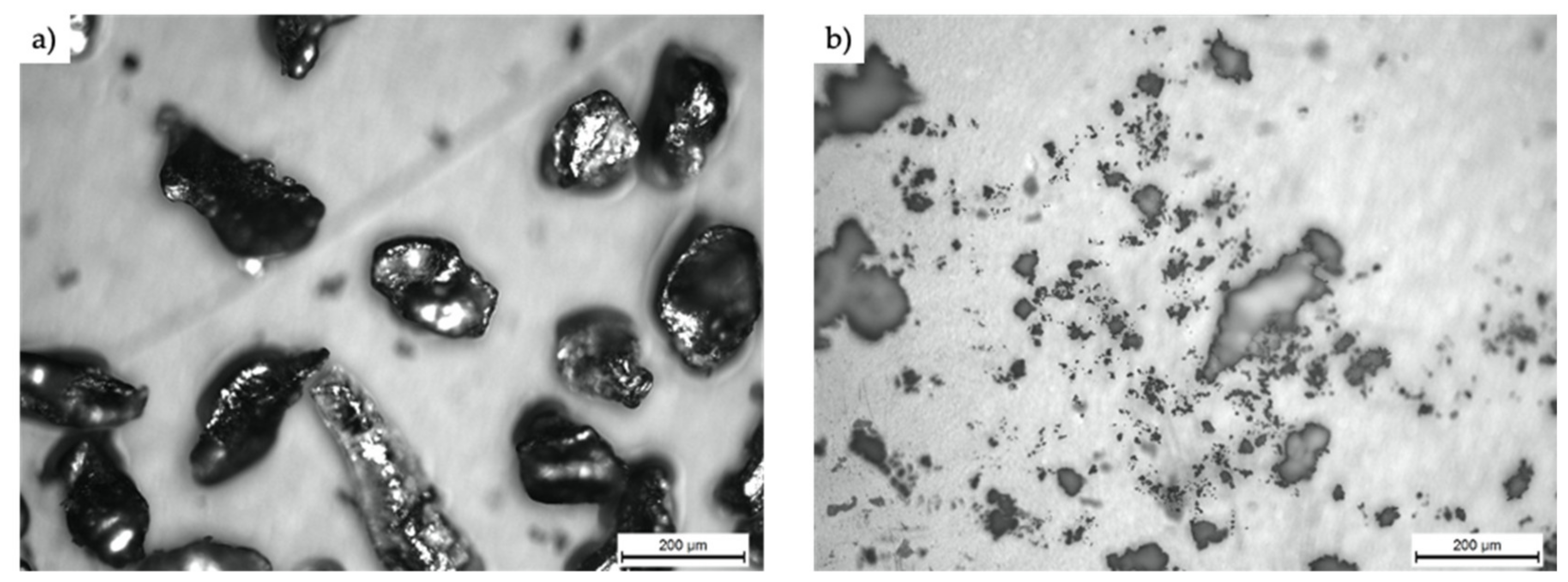
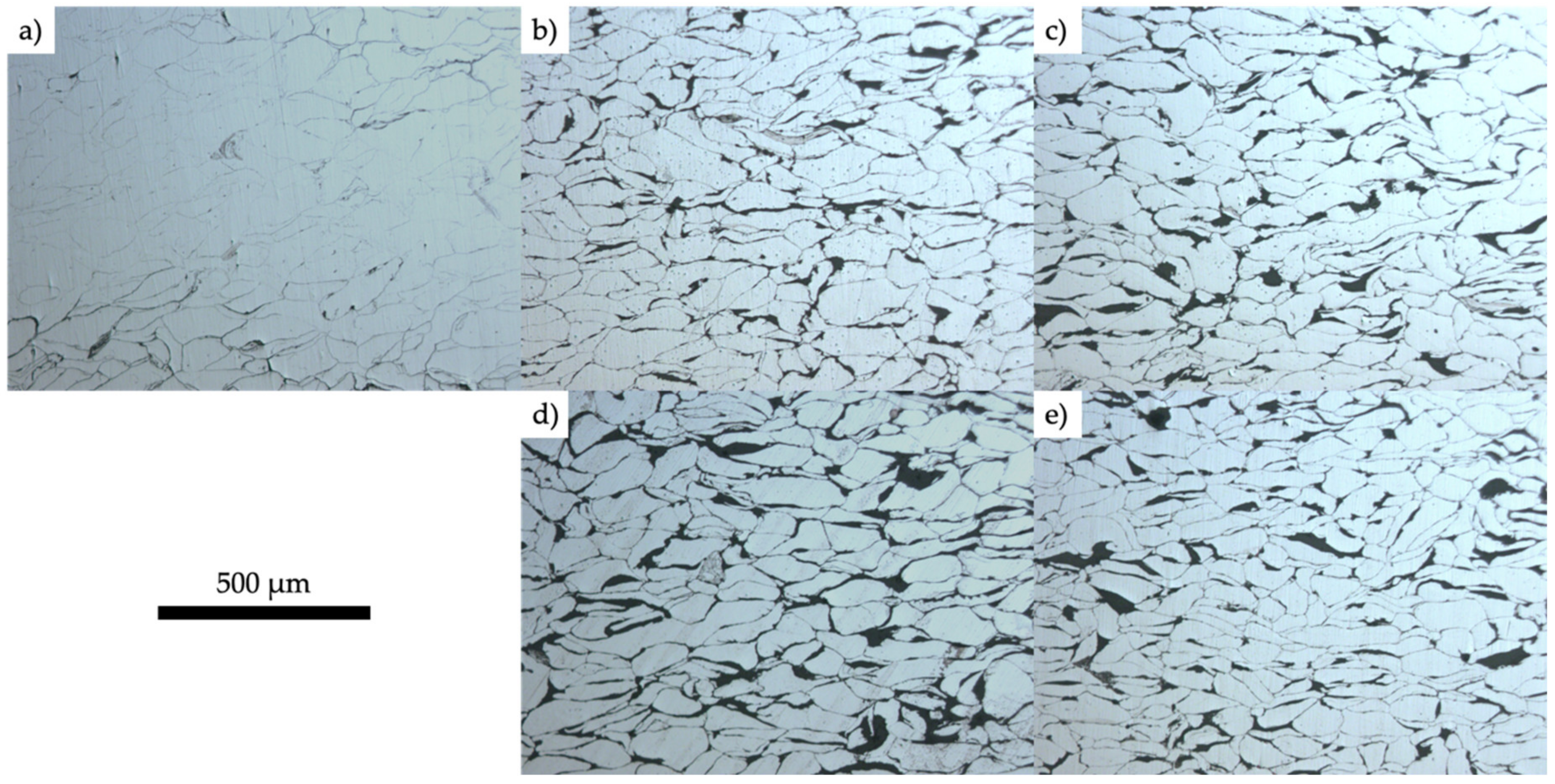
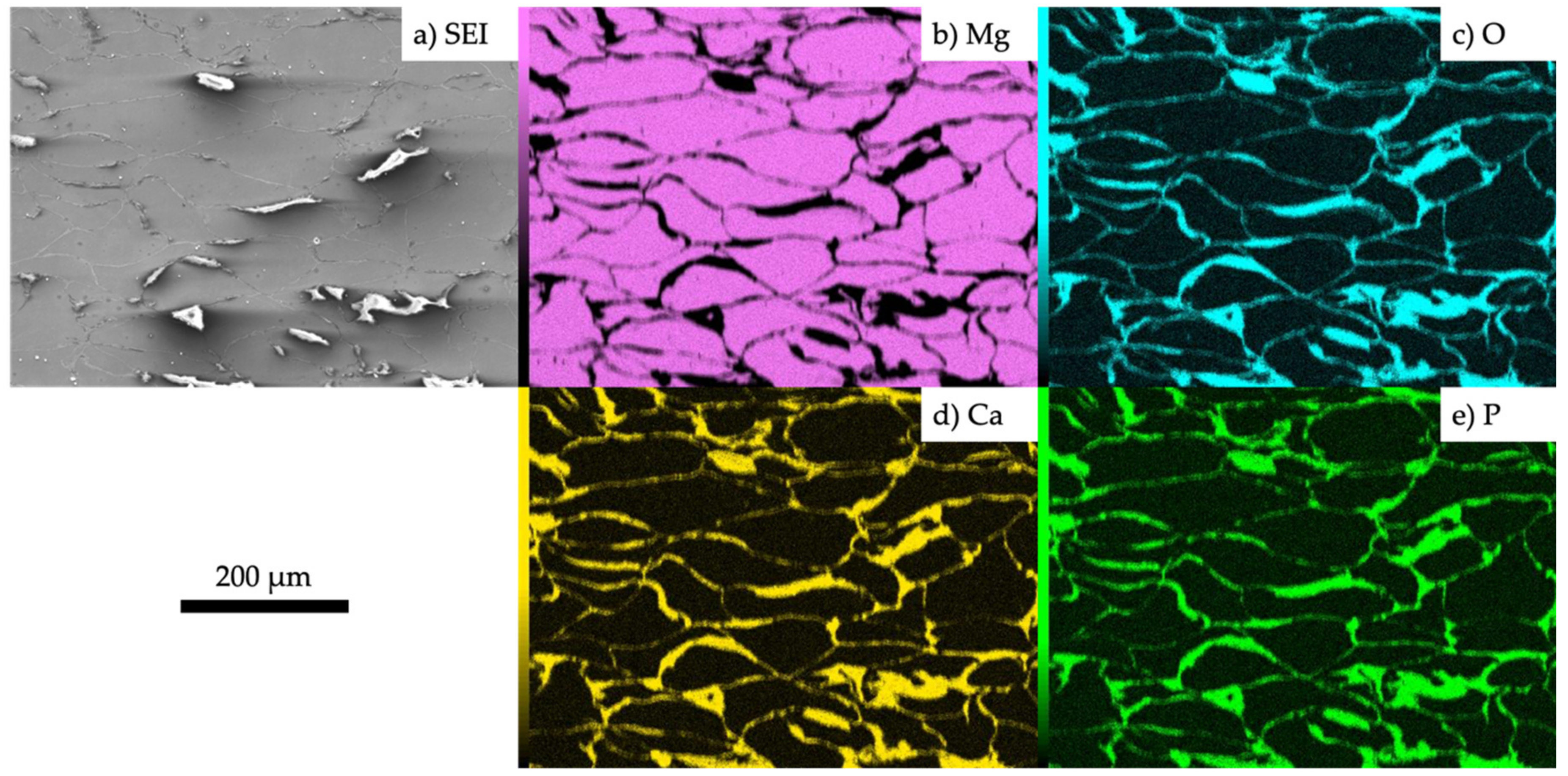
3.1. Characterization of the Fabricated Samples

The measured porosities of samples are indicated in Figure 3. Those were determined by the relative densities which were calculated from the samples’ weight and volume and the theoretical densities of pure Mg and HAP, 1.74 and 3.11 g/cm3, respectively. In order to understand the significant differences between each pair, t-tests were conducted. The p-values obtained from these statistical tests are shown in Figure 3. The sintering occurs more easily at the Mg powder–Mg powder interface compared with Mg powder–HAP powder interface since the difference between sintering temperature and the melting temperature of Mg is only around 200 °C, while the difference between the sintering temperature and that of HAP is much larger. From this point, it is the understandable result that the porosity of Mg–0HAP sample shows lower value than other compositions except Mg–10HAP106. In addition to that, it is also understandable that Mg–10HAP106 had lower porosity than Mg–10HAP since the smaller HAP powder can fill up gap between Mg powder particles.
Figure 4 shows optical micrographs of the sintered samples. Gray parts are elongated perpendicular to the compressive axis. Black regions might consist of pores other than the contents, but it is not easy to distinguish this in the OM observations. Figure 5 shows the EDS analysis results. The results indicated that, in addition to the white agglomerated white parts in secondary electron image (SEI) (Figure 5a), white lines between Mg parts are HAP. Comparing Figure 4 and Figure 5, it is confirmed that the gray parts in Figure 4 are Mg and the black parts in the same figure are HAP. From the secondary electron image (SEI) shown in Figure 6, white small nano-size particles were observed in the HAP phases. The existence of these separate HAP powders proves that the HAP hadn’t been fully sintered. To ensure that the effect of Mg powder size, the grain size of Mg–10HAP and Mg–10HAP106 were measured. As a result, the grain sizes of Mg–10HAP and Mg–10HAP106 were 56.82 ± 1.86 µm and 46.90 ± 1.22 µm respectively. This result confirms the effect of Mg powder size between these two different composites. Because the size of Mg grains observed by OM were much smaller than raw Mg powders, recrystallization seemed to be occurring during the SPS process.
Figure 7 shows the XRD measurement results. All samples after sintering exhibit not only Mg peaks, but also MgO peaks. This is because Mg was partially oxidized during the SPS process. Although the process was performed under vacuum conditions, they is enough air in the chamber and between powders in the die. Since the process was performed at high temperature, oxidation was much likely to occur compared with room temperature. Also, HAP was successfully detected in all samples containing more than 8% HAP. Since the amounts of HAP are small in all samples, they had very weak and blunt peaks, so they were not comparable for different compositions.
In order to determine the size of pure HAP powder, Scherrer’s equation was used:
τ = K λ β c o s θ
Here τ is the average size of the nanocrystallites, K is a function of the crystallite shape and is usually taken as 0.9 for HAP. λ is the X-ray wavelength, β is the peak half width, and θ is the Bragg angle. This equation is usually used for calculations of nanocrystallite size [28,29]. From this equation, it turned out that the size is around 32 nm.

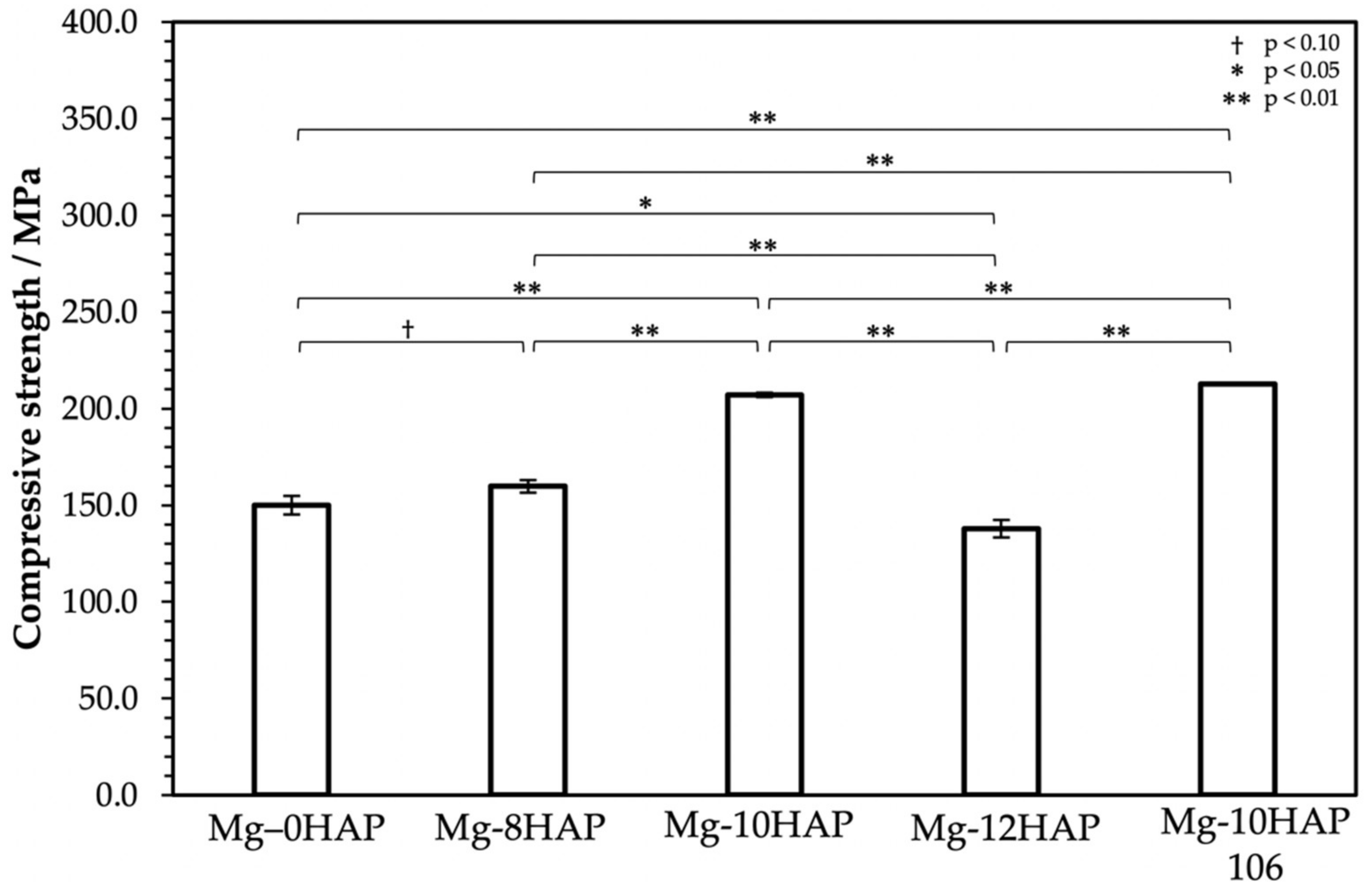
3.2. Mechanical Properties

Figure 8 shows the compressive strength of each sample which was taken from the compression test. The t-tests were performed as well as Figure 3 to understand the significant differences among each pair of compositions statistically. Among four samples having different composition of HAP except Mg–10HAP106 which was made of a different size of Mg powder, Mg–10HAP had the largest value (207.2 MPa) and Mg–12HAP had the smallest (137.8 MPa). For the samples containing 0 to 10% of HAP, the composite reinforcement mechanism can be applied. However, Mg–12HAP shows lower strength than other samples which have lower composition of HAP. There are two possible reasons for this result. One is the high porosity, as shown in Figure 3. High porosity causes lower density, especially for the sintered samples. The other reason is the weak bonding of HAP powders. Since the HAP cannot be fully sintered at this temperature, a high volume of HAP powder causes weaker bonding with HAP powders. While comparing the samples which had different grain size of Mg, it was revealed that smaller grains improve the mechanical properties since Mg–10HAP106 had around 5.7 MPa larger compressive strength than Mg–10HAP. This can be explained by the grain refinement as shown in Figure 4.
Figure 9 shows the relationship between the compressive strength and the porosity. Black marks and a white mark represent HAP-containing samples and pure Mg sample (Mg–0HAP), respectively. The trendline corresponds to only Mg–HAP samples not to Mg–0HAP since it is not a composite and its microstructure is quite different from that of composites. In fabricating composites by the SPS method, although the composition affects a lot of its mechanical properties and corrosion properties, the composition is not the only factor which affects them. Even with the same composition, the results would not be the same if the processing conditions were different. There are many factors including the raw powder size, the mixing condition of different powders, the sintering temperature and time, the sample size. One of the most promising ways to evaluate these samples fabricated by different conditions is the porosity. Apparently, the compressive strength and the porosity had a strong negative correlation in samples containing HAP and its correlation coefficient was about –0.95. In the metallic powder sintering like the present study, progress of sintering is affected by sintering conditions such as sintering temperature, sintering pressure and sintering time. In the case the sintering progressed well with desirable conditions, the connection of particles proceeds and the porosity of the sintered sample decreased in general. Decreasing the porosity means the stronger bonding at boundaries which also means that stronger compressive strength. These are the reasons why they had a such kind of correlation. Since the bone’s compressive strength is known to be around 250 MPa, none of the samples reached that high strength. From these perspectives, it is revealed that the porosity should be decreased to around 1.9% to achieve as high compressive strength like that of human bones.

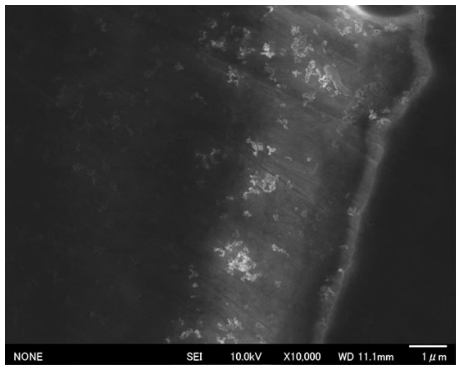
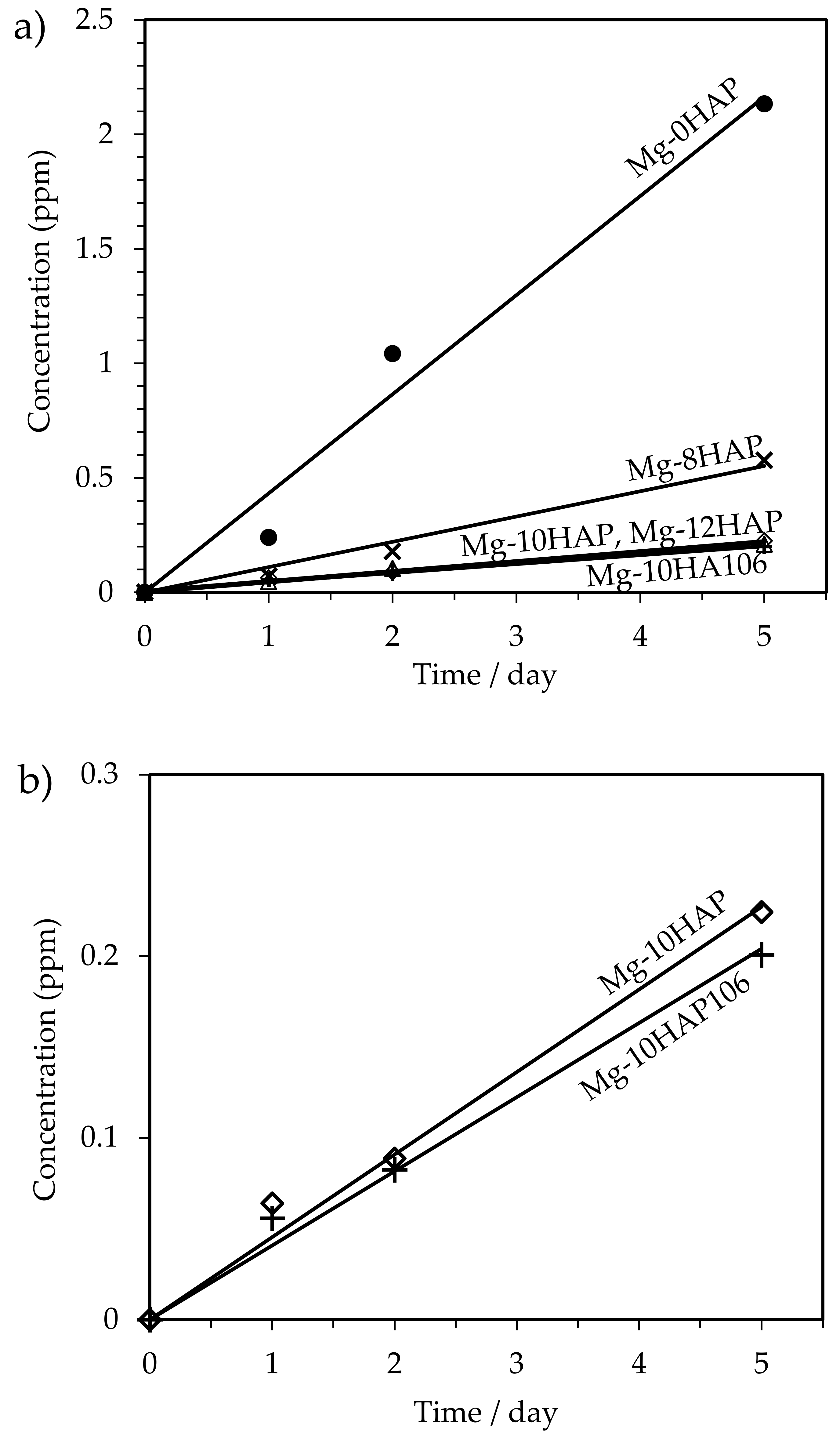
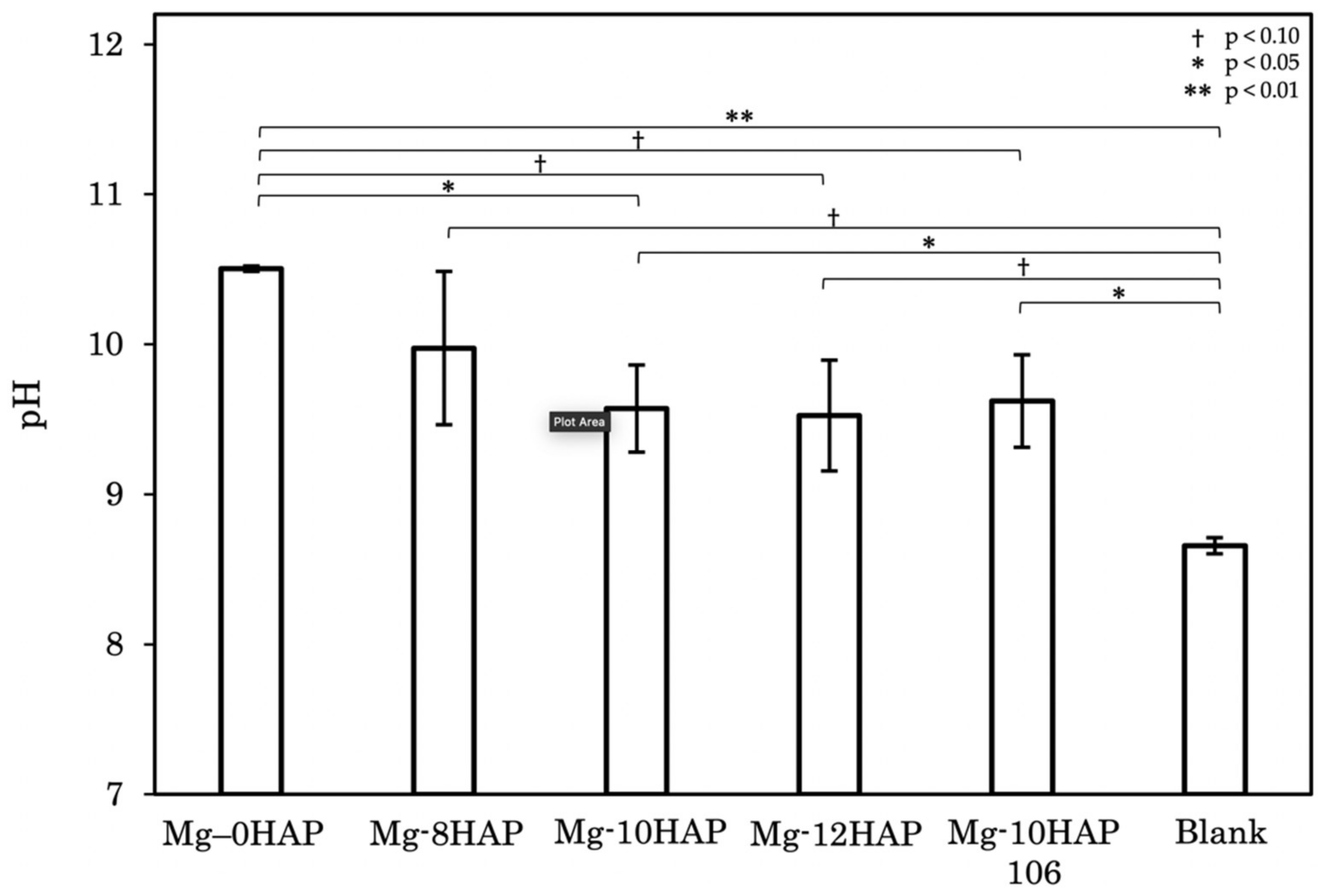
3.3. Corrosion Behavior

Figure 10 shows the Mg-ion concentration released from each sample after the immersion test measured by ICP analysis. As written in the experimental details section, a control which has no sample inside the container was measured in order to calculate the accurate value of released Mg ions. Mg–0HAP showed the highest concentration compared with others at all points. Therefore, it was confirmed that HAP addition improved the corrosion properties successfully. Moreover, Mg–8HAP apparently corroded faster than other samples which contain larger concentrations of HAP. Thus, it is suggested that Mg–10HAP and Mg–12HAP are the best Mg–HAP compositions for implants. Besides, as Figure 10b shows, Mg–10HAP106 corroded slightly slower than Mg–10HAP. Consequently, it was confirmed that finer Mg grains improve the corrosion behavior in agreement with other previous works [30,31,32].
Figure 11 shows the pH measurement results. t-Tests were conducted as well for Figure 3 and Figure 8 for figuring out the significant differences among each pair of all compositions and the control statistically. One of the most important factors to control the pH of Hanks’ solution is the amount of bicarbonate (HCO3) ion in the solution. Since the reactions below occurs easily under this temperature, it is not easy to maintain pH of the solution itself throughout the experiment:
CO 2 + H 2 O H 2 CO 3
H 2 CO 3 HCO 3 + H +
Therefore, even the pH of the control containers increases along the immersion time. For the average, results showed that the pH decreases as the amount of HAP increases.
In general, corrosion modes of magnesium alloys and composites change along the immersion time. At first, their corrosion proceeds in the general corrosion mode. The anodic and cathodic reactions in this mode are described as below, respectively.
Mg Mg 2 + + 2 e
H 2 O + e 1 2 H 2 + OH
Therefore, as these reactions proceed, pH of the solution increases. Since these reactions are randomly occurred at any places, a surface is equally corroded. As general corrosion proceeds, the corrosion mode gradually shifts to a localized corrosion mode. Because localized corrosion occurs at specific places corrosion at some places becomes more severe that at others as this mode proceeds. However, since localized corrosion occurs when the surface is not completely flat and has pores, it is assumed that not only general corrosion but also localized corrosion occurs in some samples which have a lot of pores. According to Figure 11, the errors are larger excluding Mg–0HAP which has the lowest porosity compared with others while other compositions show relatively higher porosity values. Consequently, for some HAP containing samples it is considered that both general corrosion and localized corrosion had proceeded while for other HAP-containing samples only general corrosion is considered to have proceeded. It is also suggested that each sample showed different corrosion behaviors even in each composition. As a result, it is indicated that HAP-containing samples have a lot of errors in this measurement.

4. Conclusions

Composites with different amounts of HAP or different Mg particle sizes were fabricated successfully using the SPS method and the Mg matrix grains were elongated perpendicular to the compressive axis. XRD results showed peaks of not only Mg and HAP which are elements of the starting powders, but also MgO which appeared because of the oxidation of Mg during the sintering process. Compression tests showed that finer grains strengthen the compressive strength which is understandable from the grain refinement. There was a strong negative correlation between the compressive strength and the porosity, with a correlation coefficient of about −0.95. This correlation indicated that the porosity should be decreased to around 1.9% to attain enough compressive strength for use as an orthopedic implant. ICP analysis showed that the addition of HAP significantly improved the corrosion resistance of Mg–HAP composites. Also, it showed that the samples which have finer grains have improved corrosion resistance. pH measurements indicated that there was a mixing of corrosion modes for each composition excluding Mg–0HAP. To summarize all the results, Mg–10HAP106 had the best properties as an orthopedic implant, although there is still some room for further improvement.

Author Contributions

Conceptualization and methodology, I.N. and E.K; sample manufacturing by spark plasma sintering, I.N.; characterization, I.N.; compression test, I.N.; inductivity coupled plasma elemental analysis and pH measurement, I.N. and Y.T.; writing—original draft preparation, I.N.; writing—review and editing, I.N., E.K. and T.Y. All authors have read and agreed to the published version of the manuscript.

Funding

This work was partially supported by Light Metal Educational Foundation, Inc.

Acknowledgments

The authors would like to appreciate M. Ueda and K. Kawamura, Tokyo Institute of Technology for providing the SPS machine and T. Hanawa, Tokyo Medical and Dental University for providing the equipment of the immersion test.

Conflicts of Interest

The authors declare no conflict of interest.

References

  1. Narita, K.; Kobayashi, E.; Sato, T. Sintering behavior and mechanical properties of magnesium/β-tricalcium phosphate composites sintered by spark plasma sintering. Mater. Trans. 2016, 57, 1620–1627. [Google Scholar] [CrossRef] [Green Version]
  2. Staiger, M.P.; Pietak, A.M.; Huadmai, J.; Dias, G. Magnesium and its alloys as orthopedic biomaterials: A review. Biomaterials 2006, 27, 1728–1734. [Google Scholar] [CrossRef] [PubMed]
  3. Zreiqat, H.; Howlett, C.R.; Zannettino, A.; Evans, P.; Schulze-Tanzil, G.; Knabe, C.; Shakibaei, M. Mechanisms of magnesium-stimulated adhesion of osteoblastic cells to commonly used orthopaedic implants. J. Biomed. Mater. Res. 2002, 62, 175–184. [Google Scholar] [CrossRef] [PubMed]
  4. Kazemi, M.; Ahangarani, S.; Esmailian, M.; Shanaghi, A. Investigation on the corrosion behavior and biocompatibility of Ti-6Al-4V implant coated with HA/TiN dual layer for medical applications. Surf. Coatings Technol. 2020, 397, 126044. [Google Scholar] [CrossRef]
  5. Dogan, H.; Findik, F.; Morgul, O. Friction and wear behaviour of implanted AISI 316L SS and comparison with a substrate. Mater. Des. 2002, 23, 605–610. [Google Scholar] [CrossRef]
  6. Shenhar, A.; Gotman, I.; Radin, S.; Ducheyne, P. Microstructure and fretting behavior of hard TiN-based coatings on surgical titanium alloys. Ceram. Int. 2000, 26, 709–713. [Google Scholar] [CrossRef]
  7. Zhong, Z.; Ma, J. Fabrication, characterization, and in vitro study of zinc substituted hydroxyapatite/silk fibroin composite coatings on titanium for biomedical applications. J. Biomater. Appl. 2017, 32, 399–409. [Google Scholar] [CrossRef]
  8. Ersen, O.; Tuzun, H.Y.; Ozsezen, A.M.; Bilekli, A.B.; Koca, K.; Kurklu, M. The procedure with less interest than it is done in orthopedic practice: Implant removal. Acta Medica Mediterr. 2019, 35, 825–828. [Google Scholar]
  9. Shibata, Y.; Tanimoto, Y.; Maruyama, N.; Nagakura, M. A review of improved fixation methods for dental implants. Part II: Biomechanical integrity at bone-implant interface. J. Prosthodont. 2015, 59, 84–95. [Google Scholar] [CrossRef]
  10. Hanson, B.; van der Werken, C.; Stengel, D. Surgeons’ beliefs and perceptions about removal of orthopaedic implants. BMC Musculoskelet. Disord. 2008, 9, 73. [Google Scholar] [CrossRef] [Green Version]
  11. Onche, I.I.; Osagie, O.E.; INuhu, S. Removal of orthopaedic implants: Indications, outcome and economic implications. J. West Afr. Coll. Surg. 2011, 1, 101–112. [Google Scholar] [PubMed]
  12. Cao, N.Q.; Pham, D.N.; Kai, N.; Dinh, H.V.; Hiromoto, S.; Kobayashi, E. In vitro corrosion properties of mg matrix in situ composites fabricated by spark plasma sintering. Metals 2017, 7, 358. [Google Scholar] [CrossRef] [Green Version]
  13. Cao, N.Q.; Le, H.M.; Pham, K.M.; Nguyen, N.V.; Hiromoto, S.; Kobayashi, E. In vitro corrosion and cell response of hydroxyapatite coated mg matrix in situ composites for biodegradable material applications. Materials 2019, 12, 3474. [Google Scholar] [CrossRef] [PubMed] [Green Version]
  14. Prasad, K.; Bazaka, O.; Chua, M.; Rochford, M.; Fedrick, L.; Spoor, J.; Symes, R.; Tieppo, M.; Collins, C.; Cao, A.; et al. Metallic biomaterials: Current challenges and opportunities. Materials 2017, 10, 884. [Google Scholar] [CrossRef] [PubMed]
  15. Xu, Z.; Linford, J.; Chen, S.; Smith, C.; Sankar, J. Preparation and characterization of porous magnesium alloys in biomedical applications. ASME Int. Mech. Eng. Congr. Expo. Proc. 2009, 14, 37–41. [Google Scholar]
  16. Jahnen-Dechent, W.; Ketteler, M. Magnesium basics. CKJ Clin. Kidney J. 2012, 5, i3–i14. [Google Scholar] [CrossRef] [PubMed] [Green Version]
  17. Canillas, M.; Pena, P.; Aza, A.H.D.; Rodríguez, M.A. Calcium phosphates for biomedical applications. Boletín la Soc. Española Cerámica y Vidr. 2017, 56, 91–112. [Google Scholar] [CrossRef]
  18. Corrosion behavior in hank’s solution of a magnesium—Hydroxyapatite composite processed by high-pressure torsion. Adv. Eng. Mater. 2020, 2000765. [CrossRef]
  19. Su, J.; Teng, J.; Xu, Z. Corrosion-wear behavior of a biocompatible magnesium matrix composite in simulated body fluid. Friction 2020, 10, 1–13. [Google Scholar] [CrossRef]
  20. Pinc, J.; Capek, J.; Kubasek, J.; Prusa, F.; Hybasek, V.; Vertat, P.; Sedlarova, I.; Vojtech, D. Characterization of a Zn-Ca5(PO4)3(OH) composite with a high content of the hydroxyapatite particles prepared by the spark plasma sintering process. Metals 2020, 10, 372. [Google Scholar] [CrossRef] [Green Version]
  21. Pinc, J.; Capek, J.; Hybasek, V.; Prusa, F.; Hosova, K.; Manak, J.; Vojtech, D. Characterization of newly developed zinc composite with the content of 8 wt. % of hydroxyapatite particles processed by extrusion. Materials 2020, 13, 1716. [Google Scholar] [CrossRef] [PubMed] [Green Version]
  22. Khodaei, M.; Nejatidanesh, F.; Shirani, M.J.; Iyenger, S.; Sina, H.; Savabi, O. Magnesium/nano-hydroxyapatite composite for bone reconstruction: The effect of processing method. J. Bionic Eng. 2020, 17, 92–99. [Google Scholar] [CrossRef]
  23. Kubasek, J.; Vojtech, D.; Maixner, J.; Dvorsky, D. The effect of hydroxyapatite reinforcement and preparation methods on the structure and mechanical properties of Mg-HA composites. Sci. Eng. Compos. Mater. 2017, 24, 297–307. [Google Scholar] [CrossRef]
  24. Agarwal, S.; Curtin, J.; Duffy, B.; Jaiswal, S. Biodegradable magnesium alloys for orthopaedic applications: A review on corrosion, biocompatibility and surface modifications. Mater. Sci. Eng. C 2016, 68, 948–963. [Google Scholar] [CrossRef] [PubMed] [Green Version]
  25. Kuwahara, H.; Al-Abdullat, Y.; Mazaki, N.; Tsutsumi, S.; Aizawa, T. Preparation of magnesium apatite on pure magnesium surface during immersing in hank’s solution. Mater. Trans. 2001, 42, 1317–1321. [Google Scholar] [CrossRef] [Green Version]
  26. Munir, Z.; Anselmi-Tamburini, U.; Ohyanagi, M. The effect of electric field and pressure on the synthesis and consolidation of materials: A review of the spark plasma sintering method. J. Mater. Sci. 2006, 41, 763–777. [Google Scholar] [CrossRef]
  27. Huang, J.L.; Nayak, P.K. Strengthening alumina ceramic matrix nanocomposites using spark plasma sintering. In Advances in Ceramic Matrix Composites; Low, I.M., Ed.; Woodhead Publishing: Cambridge, UK, 2014; pp. 218–234. [Google Scholar]
  28. Scherrer, P. Bestimmung der inneren Struktur und der Größe von Kolloidteilchen mittels Röntgenstrahlen. Nachr. Ges. Wiss. Göttingen 1912, 26, 98–100. [Google Scholar]
  29. Langford, J.I.; Wilson, A.J.C. Seherrer after sixty years: A survey and some new results in the determination of crystallite size. J. Appl. Cryst. 1978, 11, 102–113. [Google Scholar] [CrossRef]
  30. Alvarez-Lopez, M.; Pereda, M.D.; Del Valle, J.A.; Fernandez-Lorenzo, M.; Garcia-Alonso, M.C.; Ruano, O.A.; Escudero, M.L. Corrosion behaviour of AZ31 magnesium alloy with different grain sizes in simulated biological fluids. Acta Biomater. 2010, 6, 1763–1771. [Google Scholar] [CrossRef] [Green Version]
  31. Ge, Q.; Dellasega, D.; Demir, A.G.; Vedani, M. The processing of ultrafine-grained Mg tubes for biodegradable stents. Acta Biomater. 2013, 9, 8604–8610. [Google Scholar] [CrossRef]
  32. Argade, G.R.; Panigrahi, S.K.; Mishra, R.S. Effects of grain size on the corrosion resistance of wrought magnesium alloys containing neodymium. Corros. Sci. 2012, 58, 145–151. [Google Scholar] [CrossRef]
Figure 1. Optical micrographs of (a) the magnesium powder and (b) the hydroxyapatite (HAP) powder.
Metals 10 01314 g001
Figure 2. Schematic illustration of spark plasma sintering (SPS) system for manufacturing magnesium–hydroxyapatite composites.
Metals 10 01314 g002
Figure 3. The porosities of each composition showing no dependency on the HAP volume fraction. Comparing Mg–10HAP and Mg–10HAP106, it was found that the smaller magnesium grains are, the smaller the porosity is.
Metals 10 01314 g003
Figure 4. Optical micrographs of (a) Mg–0HAP, (b) Mg–8HAP, (c) Mg–10HAP, (d) Mg–12HAP, and (e) Mg–10HAP106 after sintering using SPS system.
Metals 10 01314 g004
Figure 5. The elemental analysis of Mg–10HAP. (a) Secondary electron image (SEI) of the same area as analyzed by X-ray spectroscopy (EDS). (be) are the results of EDS mapping analysis showing Mg, O, Ca, P, respectively. The results indicated that the white lines are HAP as well as agglomerated white parts in SEI.
Metals 10 01314 g005
Figure 6. SEI of a HAP part of Mg–10HAP after sintering using SPS system.
Metals 10 01314 g006
Figure 7. (a) XRD results of the raw magnesium and hydroxyapatite powders and the sintered magnesium–hydroxyapatite composites after sintering. Inset (b) is the magnified XRD pattern which shows the MgO peak of Mg–10HAP106 as an example.
Metals 10 01314 g007
Figure 8. Compressive strength of the sintered composites after sintering using SPS system obtained from the compression test.
Metals 10 01314 g008
Figure 9. Relationship between the compressive strength and the porosity showing the strong negative correlation in all samples containing HAP.
Metals 10 01314 g009
Figure 10. Mg concentration released from each sample after immersion test. (a) comparison of all samples and (b) enlarged view of data from samples Mg–10HA and Mg–10HA106.
Metals 10 01314 g010
Figure 11. pH of the solution after the immersion test for 5 days. Control which had no samples inside was measured as a comparison.
Metals 10 01314 g011
Table 1. Hydroxyapatite (HAP) composition and Mg powder size of each sample.
Sample NameMg–0HAPMg–8HAPMg–10HAPMg–12HAPMg–10HAP106
HAP composition0 wt%8 wt%10 wt%12 wt%10 wt%
Mg powder size~180 µm~180 µm~180 µm~180 µm≤106 µm
Table 2. Combination ratio of reagents for Hanks’ solution used for the immersion tests.
ReagentsNaClKClNa2HPO4·2H2OKH2PO4MgSO4·7H2ONaHCO3CaCl2
Concentration (g/L)8.000.400.060.060.200.350.14

