Article

Nuclear Transfer Perturbs Genomic Balance

Experimental Mitochondrial Genetics Group, School of Biomedicine, The University of Adelaide, Adelaide, SA 5005, Australia
*
Author to whom correspondence should be addressed.
Epigenomes 2026, 10(2), 38; https://doi.org/10.3390/epigenomes10020038
Submission received: 21 April 2026 / Revised: 22 May 2026 / Accepted: 2 June 2026 / Published: 5 June 2026

Abstract

Background: The transfer of a nucleus from one oocyte to another offers patients harbouring high levels of mitochondrial DNA mutation and sufferers of frequent fertilisation failure or early embryonic arrest the potential to have healthy children. However, a small amount of mtDNA is carried over with the nucleus as the transfer takes place. Consequently, we still need to distinguish between the effects of the carryover and the transfer of a nucleus itself from a mature oocyte. Methods: To overcome this, we analysed a series of hatching stage blastocysts generated using metaphase II spindle transfer and mitochondrial supplementation. The latter approach also introduces a small amount of mtDNA into the oocyte as fertilisation takes place. For both manipulations, an autologous approach was used to overcome the effects of third-party transfer. Results: We then compared the changes in global gene expression between the two groups. We found that the nuclear transfer process affected a number of gene networks and pathways. These included metabolic, cell cycle, inflammatory and immune, and epigenetic responses. A comparison with earlier stage blastocysts did not suggest that the cause was due to developmental delay. Conclusions: Critically, these changes could affect offspring health and well-being as is the case following somatic cell nuclear transfer.

1. Introduction

It is becoming increasingly evident that the nuclear and mitochondrial genomes need to communicate to enable effective cellular function. As a result, their interactions require modifications at any stage of a cell’s lifetime, a concept described as ‘genomic balance’ [1]. This is especially important throughout development when there are changes to the epigenome that regulate the expression of genes from the nuclear genome along with concomitant changes in mitochondrial DNA (mtDNA) copy number. This is highly apparent during oogenesis when the primordial germ cells, the precursor germ cells [2], initiate differentiation. This process is completed when the metaphase II (MII) stage is reached and oocytes have fertilisation potential. As a result, the highly DNA methylated primordial germ cells are subjected to a series of global DNA demethylation events mediated by TET-hydroxylation [3,4] until they reach the germinal vesicle stage when CpG methylation is reset. At the same time, mtDNA copy increases from 1400 [5] to ~300,000 [6] copies. CpG methylation then remains stable up to the MII stage [7] with mtDNA copy number being refined to >250,000 copies [6]. These processes lay the foundation for epigenetic inheritance from a maternal perspective [2] and the maternal-only inheritance of mtDNA [8].
Throughout preimplantation development, both sperm- and oocyte-driven methylation set the embryo’s methylome with at least three waves of DNA methylation prior to and including the blastocyst stage of development [7]. A key event is embryonic genome activation. This takes place between the 4- and 8-cell stages in larger mammals, namely, pigs and cattle. Similar patterns of DNA methylation are present in humans during early development [7]. Synchronously, there are changes in mtDNA copy number. Apart from a minor turnover event prior to the 4-cell stage, mtDNA copy number is reduced throughout development of the preimplantation embryo until the late morula/early blastocyst stage. At this stage, mtDNA replication commences [6,9]. However, it is restricted to the trophectodermal cells [9], which are the precursors of the placenta. The inner cell mass cells continue to dilute their mtDNA copy number until post-gastrulation. Replication of mtDNA then proceeds in a cell-specific manner. Nevertheless, the three waves of DNA methylation are complete by the late blastocyst stage [7].
Some of the more invasive assisted reproductive technologies are either being introduced into clinical medicine or are being investigated in readiness for implementation. However, they can distort the patterns of epigenetic and mtDNA inheritance [10,11]. Some of these technologies originate out of somatic cell nuclear transfer [12,13], for example metaphase II spindle transfer (MST) and pronuclear transfer (PNT) [14,15]. They have been proposed to help carriers of mtDNA disease or patients with repeated fertilisation or embryonic developmental failure to have children [16]. However, they are associated with delays in DNA methylation during preimplantation development [10] and altered gene expression profiles [11]. Consequently, the transfer of nuclei between oocytes or zygotes will necessitate the nucleus or nuclei to acclimatise to its/their new cytoplasmic environment. They will also need to interact with the resident cohort of mitochondrial genomes. This series of complex processes could lead to aberrant patterns of gene expression [11] or developmental delay [10]. To this end, we have shown that there were significant differences in gene expression and pathways between hatching blastocysts generated through MST and oocytes fertilised by intracytoplasmic sperm injection (ICSI) [11].
A confounding situation with the nuclear transfer technologies is that a small amount of mtDNA accompanies the nucleus as the somatic cell [17], spindle [18,19] or pronuclei [20,21] are transferred into the recipient oocyte or zygote, respectively. Indeed, it is evident that the introduction of a few copies of carried over mtDNA can alter the DNA methylation and gene expression patterns of blastocyst stage embryos [22]. This has been demonstrated in another assisted reproductive technology, namely mitochondrial supplementation as ICSI is performed (mICSI). Furthermore, it impacts on the DNA methylation and gene expression patterns in the tissues of resultant offspring [23]. Importantly, this issue has yet to be resolved for MST and PNT and results in offspring inheriting variable rates of carried over mtDNA [19,20]. Consequently, as two genomes are at play, it is difficult to determine which effects relate to the mitochondrial genome and which relate to the process of nuclear transfer.
The pig is an excellent model for the study of human development. A number of its organ systems and responses to physiological and pathophysiological stimuli are similar to those of humans [24,25]. Likewise, the pig’s embryology and early development are also representative of those in the human [3,7]. Here, we have used our pig model to analyse two sets of blastocyst stage embryos that have reached the hatching stage and are thus primed for implantation. The first was previously generated through MST where the MII spindle was removed and returned to its cytoplasm and, thus, possessed a small amount of mtDNA carryover and then fertilised by ICSI [11]. The second set was generated through mICSI where, again, a small amount of mtDNA was introduced into the oocyte as it was fertilised. Consequently, both sets of embryos possessed mtDNA introduced into the mature oocyte and were fertilised by ICSI. This, then, allowed us to focus specifically on the nuclear transfer procedure itself without the confounding complication of the carried over mtDNA. In other words, this allowed us to determine how the nuclear transfer process alone affects early global gene expression through RNA-seq and bioinformatics analyses. We further determined whether hatching blastocysts derived through MST exhibited developmental delay by analysing their global gene expression profiles with expanded blastocysts derived through mICSI. We report that nuclear transfer alone affects a large cohort of genes and a number of key developmental pathways, but they are more equivalent to mICSI-derived hatching blastocysts than their expanded counterparts.

2. Results

2.1. Comparison of MST with mICSI at the Hatching Blastocyst Stage

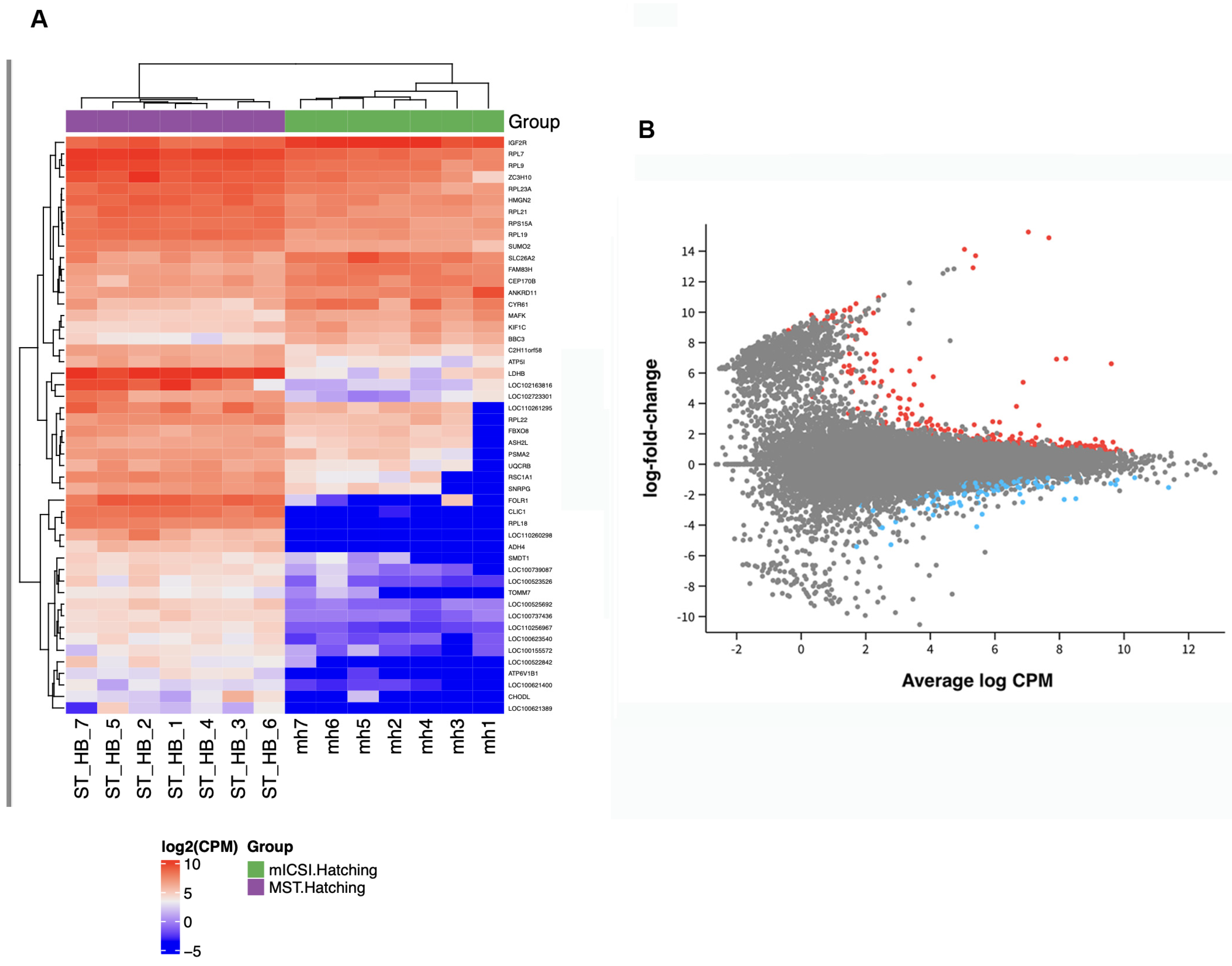
To determine how nuclear transfer alone affects global gene expression, we compared the gene expression profiles of hatching blastocysts generated through MST (MST-H) with those derived through mICSI (mICSI-H). We chose the hatching blastocyst stage as the three waves of DNA methylation would be complete by then and these embryos have implantation potential. In all, 423 gene transcripts (342 identifiable genes) were differentially expressed at FDR < 0.05 (Figure 1; Supplementary Table S1). The top 20 genes and their functions are listed in Table 1 and included genes associated with ribosomal proteins, metabolism, cell cycle and epigenetic regulation. We then performed KEGG pathway analysis and identified 32 affected pathways at p < 0.05 (Supplementary Table S2). The top 10 affected pathways (Table 2) included Ribosome-, Coronavirus-, Oxidative phosphorylation-, Endocytosis- and Oocyte meiosis-associated pathways. We then focused on pathways specifically upregulated (Table 2 and Table S3) and downregulated (Table 2 and Table S4). For the upregulated pathways, the five above-mentioned pathways were all upregulated whilst for the downregulated pathways those associated with inflammation (TNF signalling); metabolism (AMPK signalling); cells’ responses to stress, e.g., DNA damage (p53 signalling); and cell growth, proliferation and survival (PI3K-Akt signalling) were affected. The discordant expression of genes associated with Oxidative phosphorylation, metabolic signalling, cell growth and proliferation, and immune responses represent disruption to key early developmental events. This, in turn, suggests that the interactions between the nuclear and mitochondrial genomes were not fully reestablished.
We then analysed the RNA-seq data outputs in Reactome to determine if they had any significance to human outcomes and, specifically, disease (Table 3 and Table S5). Pathways and interactions associated with immune responses (interferon activity), transcription, cell growth and proliferation were all upregulated suggesting that there was a ‘recognition’ response to the transferred nucleus and that cell cycle regulation, growth and transcription were also affected.

2.2. Comparison of MST-H with mICSI-Derived Expanded (mICSI-E) Blastocysts

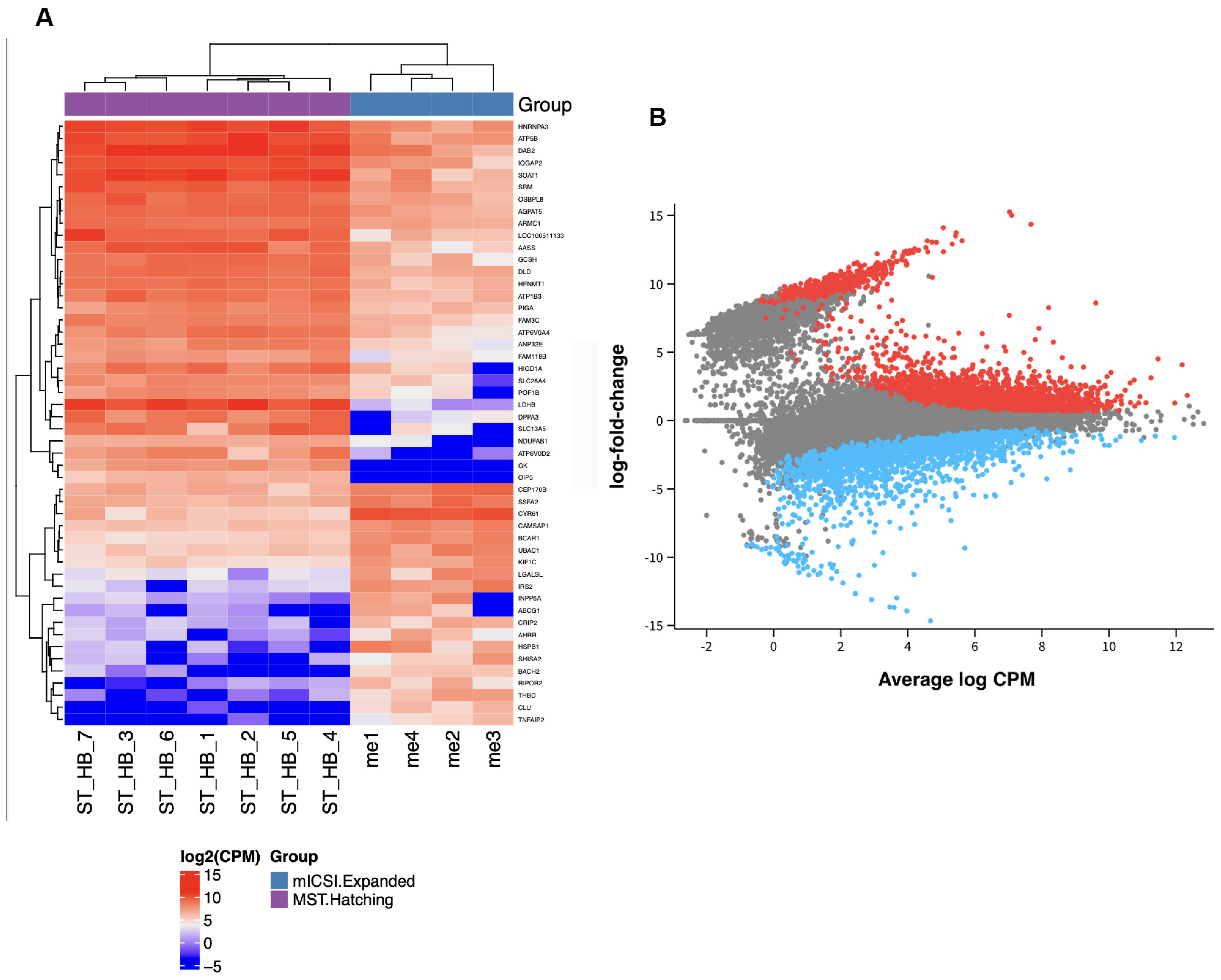
In order to ascertain if differential gene expression was due to developmental delay resulting from the process of nuclear transfer, as observed in somatic cell nuclear transfer [26], we compared the gene expression profiles of MST-H blastocysts with mICSI-E blastocysts. A total of 3937 genes were differentially expressed (3423 identifiable genes) (FDR < 0.05) (Figure 2; Supplementary Table S6) with the top 20 genes and their respective functions listed in Table 4. Again, this gene list comprised those associated with metabolism, epigenetic regulation, Ribosomes, cell cycle and inflammation. It also included factors associated with pluripotency. All the differentially expressed genes were assigned to 45 pathways when assessed by KEGG (Supplementary Table S2). Amongst the top 10 pathways were Oxidative phosphorylation, cell cycle, Cellular senescence, and Citrate cycle (TCA cycle) (Table 5). Those pathways that were upregulated included Oxidative phosphorylation, Metabolic pathways, cell cycle, Citrate cycle (TCA cycle), and DNA replication (Table 5 and Table S3) whilst the downregulated pathways included PI3K-Akt signalling, ECM-receptor interaction, signalling pathways regulating pluripotency of stem cells, and MAPK signalling (Table 5 and Table S4). Reactome pathway analysis again highlighted pathways associated with immune and inflammatory responses, metabolism, DNA synthesis and epigenetic regulation (Table 6 and Table S7). The larger number of differentially expressed genes between the MST-H and mICSI-E blastocysts suggest that the differences are largely due to developmental differences rather than the nuclear transfer process per se. Nevertheless, there were some similar differences that were observed in the MST-H and mICSI-H comparison. This included pathways and networks associated with Oxidative phosphorylation, cell cycle and immune responses.
A comparison of the commonly differentially expressed genes between the MST-H versus mICSI-H blastocysts and MST-H versus mICSI-E blastocysts revealed that 123 genes were uniquely expressed in the MST-H versus mICSI-H blastocyst comparison (Supplementary Table S8) and 3637 genes uniquely expressed in the MST-H versus mICSI-E blastocyst comparison (Supplementary Table S8). Conversely, a smaller number of differentially expressed genes were common to the two comparisons. When these two gene lists were analysed in Reactome, pathways and interactors associated with ribosomal activity and translation (Supplementary Table S9) and defective homologous recombination and cell cycle were affected (Supplementary Table S10), respectively. Consequently, there were fewer unique genes differentially expressed in the MST-H versus mICSI-H blastocyst comparison but a greater number of affected pathways and interactors.

3. Discussion

During oogenesis a number of genomic, epigenomic and cellular processes are established that enable a primordial germ cell to mature into a MII oocyte. These involve, for example, waves of DNA methylation/demethylation to the nuclear genome [5,7] and exponential replication and subsequent refinement of mtDNA copy number [6,27,28]. As a result, levels of maternal DNA methylation and mtDNA are set prior to fertilisation. Post-fertilisation, requisite levels of maternal and paternal DNA methylation are established by the late blastocyst stage [7]. In addition, mtDNA has been segregated to the two distinct cellular lineages that arise at this stage of development, namely the trophectoderm and in the inner cell mass [27]. We generated a series of hatching blastocysts through MST, as documented in [11], and mitochondrial supplementation, which both have introduced similar levels of mtDNA (350 to 780 copies) [11,22], and were fertilised by ICSI. This enabled us to determine the effects of the nuclear transfer process on gene expression in the mature oocyte at a key developmental stage in embryos with implantation potential. Consequently, as both sets of embryos were fertilised in a similar manner, we did not include a comparison with hatching blastocysts derived through non-manipulation. Indeed, our aim was to directly compare two sets of embryos where the only difference was the removal or otherwise of a MII spindle. In each case, autologous transfer was performed to circumvent a third-party effect at a nuclear DNA and mtDNA level [11] and the effects of ICSI on embryo development [29] were overcome. In this instance, autologous nuclear transfer refers to the removal of the spindle and its replacement within the same oocyte. This is distinct from a clinical protocol that would transfer the patient’s nucleus into a third-party oocyte.
By simply removing a MII spindle and returning it to its own cytoplasmic environment, we observed a host of genes that were differentially expressed by the hatching blastocyst stage. This suggests that the reconstructed oocyte is seeking to accommodate the returning nucleus and manage any damage that may have arisen through its extraction. The underlying question, then, is whether the process would cause a series of downstream events that could be harmful to the developing foetus and offspring or whether the effects would be benign. Importantly, we note the immune pathways were aberrantly regulated, as determined by KEGG and Reactome analyses. To this extent, the differentially regulated interferon responses are analogous to those associated with interactions with mtDNA [30] and resulting from cell stress [31].
In the case of mtDNA, it appears that the reintroduced nucleus is likely responding to the resident population of mtDNA in the oocyte rather than the very small population attached to the karyoplast. In essence, this suggests that the returning nucleus does not necessarily recognise the cytoplasm as being ‘self’ but rather a distinct entity, although it had previously interacted with this genome from the primordial germ cell stage onwards and carries a small population of mitochondria attached to its membrane. This may arise through the extraction process conditioning the attached mitochondria in a manner that causes the resident oocyte population to be deemed different. Nevertheless, the exposure, in our case, would be different to other forms of nuclear transfer where the karyoplast may undergo fusion, electrically or through virus, to an enucleated donor oocyte [32]. For cell stress, it appears that the mechanical processes of protrusion, extraction and replacement induce a sense of damage or, at least, significant change that instigates an immune chain response. Indeed, it has become increasingly evident that both the nuclear and mitochondrial genomes can elicit immune responses along with some of the epigenetic regulators [33]. For example, mitophagy, the process of mitochondrial autophagy, mediates the elimination of sperm mtDNA prior to embryonic genome activation in the newly formed embryo [34]. Likewise, more recently, it has been proposed as a mechanism for regulating the segregation of mtDNA at the zygote stage in mice whereby defective mtDNA are selected for elimination [35]. In our work, we identified mitophagy as an upregulated pathway following KEGG and Reactome analysis (Supplementary Tables S3 and S5). Furthermore, one factor associated with another form of paternal mtDNA regulation, namely, mitochondrial unfolded protein response (Supplementary Table S5), was also modulated. This has been associated with the accumulation of paternal mtDNA through an ALK-B1 mediated tRNA m1A epitranscriptomic modification [36].
Whilst cell growth and proliferation were downregulated, Oxidative phosphorylation was upregulated at the hatching blastocyst stage. These events are asynchronous with this stage of development. Cell proliferation would likely be upregulated in line with the need to generate sufficient cells to support the expansion of the embryo and transition to foetal development. Furthermore, cell proliferation normally utilises glycolysis or aerobic glycolysis as the predominant form of energy production [37]. This is a more efficient form of energy production as it provides lower levels of energy at a faster rate to support cell cycle activity and protein production [38]. On the other hand, cell growth is required to support the expansion of the trophectoderm to support the invasive trophoblasts that will give rise to the placenta [39], which in turn are reliant on growth factors modulated by innate and adaptive immune regulators [40].
In terms of the regulation of early development, we identified modulation of factors associated with the citric acid cycle, a metabolic pathway also present in the mitochondrion. The citric acid cycle donates electrons to the electron transfer chain to promote the production of ATP derived from Oxidative phosphorylation [38]. Nevertheless, by-products of the citric acid cycle are also mediators of DNA methylation. In this respect, they act as co-factors in the conversion of methylated DNA, 5-methylcytosine (5-mC), to its transient state, 5-hydroxy-methylcytosine (5-hmC) [41]. This process is key to the resetting of the embryonic genome during human preimplantation development [42] with depletion of 5-hmC affecting the activation of embryonic genes [43]. Indeed, other epigenetic regulators were also identified as being aberrantly regulated including histone lysine methyltransferase complex subunit ASH2-L and concomitant upregulation of a number of transcription factor pathways such as TFAP2 and RUNX1.
Our comparison of MST hatching blastocysts with mICSI expanded blastocysts highlighted a large number of differentially expressed genes including metabolic, steroidogenic and epigenetic regulators. However, the number was larger than for the comparison between the two hatching blastocyst cohorts, suggesting that the MST-derived hatching blastocysts were closer to their mICSI-derived counterparts. This indicates that the larger differences observed were a result of differences in stages of development rather developmental delays. Nevertheless, the aberrant patterns of reprogramming through the expression of epigenetic regulators, metabolism and cell cycle suggest an overlap and similarity to blastocysts derived through SCNT [26].
Whilst our autologous approach to nuclear transfer in this work would not be used in the clinical setting, it has clinical context. It clearly shows that the process of nuclear transfer through MST, and most likely PNT also, is mechanically disruptive to the oocyte and results in aberrant gene expression profiles. As a result, these anomalies could have implications for offspring health and well-being. From the limited number of large animals, primarily rhesus macaques, generated through MST, there is no evidence of serious abnormalities [18]. Nevertheless, insufficient, clinically relevant large animals have been generated or analysed to provide sufficient reassurance. Given that we observed errant metabolic, epigenetic and immune response pathways at the hatching blastocyst stage, it is important to establish whether associated disorders such as autoimmune, epigenetic or metabolic disorders would affect offspring. Indeed, from SCNT studies in large animal models [44,45], a large number of abnormalities have been reported that result from incomplete reprogramming and failure of the nucleus to adapt to its new cytoplasmic environment. These include birth-related defects, increased levels of post-natal loss, dysfunction of the immune system, failure of the kidneys and heart to function effectively, large offspring syndrome, circulatory-associated disorders, distress within the respiratory system, and movement and balance disorders, suggesting neurological dysfunction. These disorders add to the range of disorders that could occur when mtDNA carryover harbouring mutant mtDNA is selected for and potentially results in severe or even fatal mtDNA diseases [46]. Even the addition of extra copies of mtDNA introduced through mICSI can result in aberrant DNA methylation and gene expression profiles at the blastocyst stage [22]. Some of these effects are transmitted through to the offspring resulting in differential DNA methylation and gene expression profiles in their tissues [47]. It also leads to some key developmental milestones related to growth and biochemical and haematological parameters not being met or their delay in being established, as determined in offspring [23].
Perhaps Germinal Vesicle Transfer offers an opportunity to offset some of the epigenetic changes as the transfer of an earlier stage nucleus would allow time for its acclimatization to the ‘foreign’ cytoplasm. The maturing oocyte would then proceed through germinal vesicle breakdown and, following in vitro maturation, progress to MII in readiness for fertilisation [48]. However, a modified approach, namely ‘aggregated chromosomes transfer’, would allow similar processes to take place but considerably reduce the potential for mtDNA carryover [49]. Indeed, Australian reproductive law has made provision for further research to be conducted into Germinal Vesicle Transfer as an approach to overcome the transmission of mutant or deleted mtDNA from a carrier [50].
In conclusion, we have analysed a cohort of final stage preimplantation embryos that highlight the direct effects of the nuclear transfer approach through MST. By design, we have been able to exclude the effect of mtDNA carryover as shown in other models [22,23] and the effects of the fertilisation procedure (ICSI). Our data point to a large number of genes that are affected by nuclear transfer alone and influence key developmental pathways. However, these embryos appear to be nearer to their mICSI-counterparts than the earlier staged mICSI-expanded blastocysts. Nevertheless, there is commonality with SCNT-derived embryos suggesting that the removal and return of a nucleus alone induces a series of metabolic, cell cycle, inflammatory and immune, and epigenetic responses that could have downstream implications for offspring derived through such approaches. Consequently, it is imperative that a series of full-scale investigations are conducted in large animal models clinically relevant to humans before the nuclear transfer technologies undergo further clinical trialling or implementation.

4. Materials and Methods

4.1. Ethics Statement

The use of porcine oocytes and ovaries to generate embryos was registered with the University of Adelaide’s Animal Ethics Committee. However, formal ethics approval was not necessary as the material was collected from an abattoir after the material had been sourced for food production. As a result, no live animals were used and no animals were killed for research purposes.
All reagents and chemicals were purchased from Sigma-Aldrich (Bayswater, VIC, Australia) unless otherwise mentioned.

4.2. Oocyte Collection and In Vitro Maturation

Pairs of ovaries from gilts were obtained from a local abattoir, retained as pairs and transported to the laboratory in 0.9% NaCl solution (Baxter, Old Toongabbie, NSW, Australia) and maintained at 38.5 °C. Cumulus-oocyte-complexes (COCs) exhibiting diameters of 3–6 mm were aspirated from follicles of each pair of ovaries using an 18 G needle. Following isolation, the COCs from each pair were washed three times in handling media (medium 199-Hepes; (Thermo Fisher, Waltham, MA, USA)) which was supplemented with 10% filtered gilt follicular fluid (FF) and 5 µg/mL cycloheximide (CHX). They were then cultured in individual wells containing 500 μL CHX-supplemented in vitro maturation (IVM) media at 38.5 °C in a humidified incubator at 5% CO2 in air for 18–20 h. The IVM media comprised medium 199 (Thermo Fisher) supplemented with 0.88 M cysteamine, 5 μg/mL insulin, 0.61 mM L-glutamine, 0.80 mM Na-pyruvate, 10 IU/mL Folligon® (Pacific Vet Pty Ltd., Braeside, VIC, Australia), 10 IU/mL Chorulon® (Pacific Vet Pty Ltd.), and 0.10 μg/mL epidermal growth factor (EGF). COCs were then washed three times in IVM media minus CHX and cultured for another 25 h in IVM media minus CHX at 38.5 °C 5% CO2.

4.3. Collection of MII Oocytes

For the collection of MII oocytes, each individual well of 500 µL of expanded COCs received 10 µL of 25 mg/mL hyaluronidase to aid removal of the cumulus cells. The COCs were transferred to handling media for complete denudation using a narrow glass pipette. MII oocytes were then selected based on the presence of a polar body and retained in a 50 µL drop of handling media prior to microinjection. Additionally, only MII oocytes with a clear perivitelline space and a distinct separation between the oolemma and zona pellucida were used; otherwise, the oocytes were prone to lysis during enucleation.

4.4. MST

Just before the transfer of the spindle, MII oocytes were cultured for 5 min with 5 µg/mL cytochalasin B in handling media. Oocytes were then placed into individual 3 µL droplets comprising Porcine X media (PXM, Supplementary Table S11). Each oocyte was held by a holding pipette (H-30/120-30, ICSION Medical Pty. Ltd., Thebarton, SA, Australia) and its spindle observed via Oosight (Cambridge Research Instruments, Cambridge, UK). The spindle was then oriented to the 2–4 o’clock position. The spindle surrounded by minimal cytoplasm was aspirated with a 11 µm spike ICSI pipette (I-11-30, ICSION Medical Pty. Ltd.) under the Oosight filter. A 11 µm diameter spiked pipette minimises disruption to the metaphase plate, reduces damage to the chromosomes, and minimises mtDNA carryover of the accompanying cytoplasm [11]. Excess cytoplasm is further removed by expulsion of the spindle into the culture media followed by re-aspiration to reduce mtDNA carryover to 306 ± 49 copies [11]. Following enucleation, the spindle was returned to its original cytoplasm through the same break in the zona pellucida. Reconstructed oocytes were collected in a droplet of handling media.

4.5. Fertilisation

Spermatozoa in semen extender were provided by Terminal Mix (Sabor Ltd., Clare, SA, Australia). Initially, the extender underwent centrifugation at 300× g for 5 min and the supernatant was discarded. The spermatozoa were washed in sperm preincubation media (SPM) that had been pre-equilibrated and comprised medium 199 additionally containing 0.91 mM sodium pyruvate, 4.1 mM calcium lactate, 75.0 µg/mL streptomycin sulphate, 37.5 µg/mL penicillin-G, and 12% heat inactivated foetal bovine serum (Thermo Fisher); equilibrated for 4 h at 38.5 °C 5% CO2, and cooled to room temperature prior to the sperm pellet being resuspended. Spermatozoa were then centrifuged at 300× g for 5 min, resuspended in SPM and retained until use in the dark at room temperature.
Approximately 30 min prior to fertilisation, ICSI dishes were prepared with SpermSlow™ (Cooper Surgical Inc., Sydney, NSW, Australia), as described in the manufacturer’s instructions. To this extent, 2 × 10 µL droplets of SpermSlow™ and 5 µL droplets of handling media were loaded onto the lid of a 60 mm culture dish (Thermo Fisher). 1–2 µL of the sperm drop were added to the right of the leftmost SpermSlow™ drop and the two droplets merged. The droplets were covered with SAGE mineral oil (Cooper Surgical Inc.) and incubated at 38.5 °C 5% CO2 for 15 min. ICSI was performed with a 6 µm pipette (I-6.0-30; ICSION Medical Pty. Ltd.) and active spermatozoa selected. A single spermatozoon was mechanically immobilised and collected in the ICSI pipette containing SpermSlow™. Each reconstructed oocyte was clasped by a holding pipette with the reconstructed karyoplast positioned at 12 o’clock relative to the polar body. The ICSI pipette entered at 3 o’clock and the spermatozoon released in the direction of the 6 o’clock position to prevent injury to the karyoplast.

4.6. Isolation of Oocyte Mitochondria for mICSI

Fractions containing mitochondria were isolated from non-MII oocytes, as described in [22,23]. Non-MII oocytes were classified as those oocytes failing to mature after in vitro maturation. This is indicated by the absence of the first polar body during MII oocyte assessment. In all, 10 denuded non-MII oocytes from each ovary pair were washed in 0.1% PVP and placed in 5 mL of mitochondrial isolation buffer (20 mM Hepes pH 7.6, 220 mM mannitol, 70 mM sucrose, 1 mM EDTA) containing 2 mg/mL BSA. After 5 min, the oocytes underwent homogenization of 10 strokes with a Potter–Elvehjem tissue grinder set (VWR International, Radnor, PA, USA). The homogenate was then centrifuged at 800× g for 10 min and approximately 2.2 mL was transferred into two 1.7 mL Eppendorf tubes and centrifuged at 10,000× g for 20 min. The mitochondrial pellets from both tubes were resuspended in 1 mL of mitochondrial isolation buffer and centrifuged at 10,000× g for 20 min. The resultant mitochondrial pellet was reconstituted in 2 µL of mitochondrial isolation buffer prior to supplementing MII oocytes at the time of mICSI.

4.7. mICSI

After picking up the sperm, as described above for ICSI, 5 pl of mitochondrial isolate, which contained circa 780 copies of mtDNA [51], were drawn into the pipette and injected into an MII oocyte from the same ovary pair.

4.8. Embryo Culture

Reconstructed oocytes were cultured in porcine zygote media (PZM, Supplementary Table S11) at 38.5 °C 5% CO2, 5% O2. After 48 h (Day 2) and 120 h (Day 5), putative embryos were placed in fresh media. Once blastocysts reached the expanded or hatching blastocyst stage (Day 7 or 8), they were washed in sterile PBS without Ca2+ or Mg2+, collected and frozen individually in 2 µL PBS at −80° C in readiness for further analysis.

4.9. RNA Extraction, Library Preparation and Sequencing

The Arcturus PicoPure RNA Isolation Kit (Thermo Fisher) was used to extract RNA from hatching- and expanded-stage blastocysts, as described in [52,53]. This generated between 1 and 18 ng of input RNA. Once extracted, RNA was stored at −80 °C. The Australian Genome Research Facility undertook preparation of libraries using the whole extract and the Illumina SMART-Seq Ultra Low Input Kit (Illumina, San Diego, CA, USA), as described by the manufacturer, along with 16 cycles of amplification and ribosomal RNA depletion using Illumina Ribo-Zero Gold (Illumina). They also sequenced the RNA on a NovaSeq X Plus (Illumina) applying 150 bp paired-end chemistry.

4.10. RNAseq Data Analysis and Identification of Differentially Expressed Genes

The primary sequence data were produced with the Illumina DRAGEN BCL Convert 07.031.732.4.3.6 pipeline. They were then inspected for Illumina adapters, over-representation of sequences and contamination from other species. In all, this generated between 51 million and 109 million reads. The sequenced reads were then mapped to the Sus scrofa genome (GCF_000003025.6) with the STAR aligner V2.3.5a [54]. The transcripts were assembled with the StringTie Tool v2.1.4 by applying the reference annotation-based assembly option. As a result, 64 to 89% of the reads mapped to the reference genome. edgeR (version 4.2.2) was used to identify differentially expressed genes (https://bioconductor.org/packages/release/bioc/html/edgeR.html) using R 4.4.0 (accessed on 17 October–11 November 2025).

4.11. Functional Pathway Enrichment and Gene Network Analysis

Differentially expressed genes underwent KEGG pathway [55] over-representation analysis in edgeR. Reactome pathway analysis was performed using reactome.org and the gene analysis tool, PADOG (accessed 18–19 December 2025) [56]. Reactome analysis was carried out using official gene symbols and non-human identifiers projected to human. Pathways at p < 0.05 were regarded to be over-represented as a function of the number of genes present in each pathway for the KEGG analyses. Reactome pathways with an FDR of <0.05 were deemed to be over-represented in relation to the number of genes present in a given pathway. For KEGG, over-representation analyses included separate analyses of up- and downregulated genes. This approach provides the best statistical power for determining biologically significant pathways [57]. Upregulated and downregulated pathways were also combined in one table for the KEGG analyses to determine the overall total number of significant pathways.

4.12. Statistical Analysis

For differential gene expression analyses, the Bioconductor package edgeR v4.2.2 [58] was used within R v4.4.0 [59]. Within edgeR, the counts between blastocysts were normalised using the default trimmed mean of M values normalisation method. Differential expression between the groups was quantified using a generalised linear model to produce p-values. The false discovery rate (FDR) was generated by edgeR using the Benjamini–Hochberg method. FDR < 0.05 indicated over-representation for the number of genes identified in a given pathway.

Supplementary Materials

The following supporting information can be downloaded at: https://www.mdpi.com/article/10.3390/epigenomes10020038/s1, Table S1. Differentially expressed genes at FDR < 0.05 for the comparison between MST-hatching (MST-H) blastocysts and mICSI-hatching (mICSI-H) blastocysts. Table S2. KEGG Pathway Analysis for all differentially expressed genes from the comparisons between MST-hatching (MST-H) blastocysts and mICSI-hatching (mICSI-H) blastocysts; and MST-hatching (MST-H) blastocysts and mICSI-expanded (mICSI-E) blastocysts. Table S3. KEGG Pathway Analysis for upregulated differentially expressed genes from the comparisons between MST-hatching (MST-H) blastocysts and mICSI-hatching (mICSI-H) blastocysts; and MST-hatching (MST-H) blastocysts and mICSI-expanded (mICSI-E) blastocysts. Table S4. KEGG Pathway Analysis for downregulated differentially expressed genes from the comparisons between MST-hatching (MST-H) blastocysts and mICSI-hatching (mICSI-H) blastocysts; and MST-hatching (MST-H) blastocysts and mICSI-expanded (mICSI-E) blastocysts. Table S5. Reactome Pathway Analysis for all differentially expressed genes from the comparison between MST-hatching (MST-H) blastocysts and mICSI-hatching (mICSI-H) blastocysts. Table S6. Differentially expressed genes at FDR < 0.05 for the comparison between MST-hatching (MST-H) blastocysts and mICSI-expanded (mICSI-E) blastocysts. Table S7. Reactome Pathway Analysis for all differentially expressed genes from the comparison between MST-hatching (MST-H) blastocysts and mICSI-expanded (mICSI-E) blastocysts. Table S8. Commonly and uniquely expressed genes for the differentially expressed genes at FDR < 0.05 for the comparisons between MST-hatching (MST-H) blastocysts and mICSI-hatching (mICSI-H) blastocysts; and MST-hatching (MST-H) blastocysts and mICSI-expanded (mICSI-E) blastocysts. Table S9. Reactome Pathway Analysis for the uniquely differentially expressed genes from the comparison between MST-hatching (MST-H) blastocysts and mICSI-hatching (mICSI-H) blastocysts. Table S10. Reactome Pathway Analysis for the uniquely differentially expressed genes from the comparison between MST-hatching (MST-H) blastocysts and mICSI-expanded (mICSI-E) blastocysts. Table S11: Media composition for Porcine Zygotic Media (PZM) and Porcine X Medium (PXM).

Author Contributions

E.A.: Conceptualization, Formal analysis, Methodology, Writing—original draft, review and editing. J.C.S.J.: Conceptualization, Formal analysis, Methodology, Writing—original draft, review and editing, Supervision, Acquisition of funding. All authors have read and agreed to the published version of the manuscript.

Funding

This work project was funded by National Health and Medical Research Council Ideas Grant GNT2000723 (JSJ). Funding for open access charge: National Health and Medical Research Council.

Institutional Review Board Statement

The use of porcine oocytes and ovaries to generate embryos was registered with the University of Adelaide’s Animal Ethics Committee. However, formal ethics approval was not necessary as the material was collected from an abattoir after the material had been sourced for food production. As a result, no live animals were used and no animals were killed for research purposes.

Informed Consent Statement

Not applicable.

Data Availability Statement

Raw sequence data for gene expression are available under BioProjects PRJNA1139228 and 1380909.

Acknowledgments

We thank the Australian Genome Research Facility for preparing samples for RNA sequencing; and undertaking RNA sequencing, alignment of reads, and differential gene expression analyses.

Conflicts of Interest

The authors declare no conflicts of interest.

References

  1. St. John, J.C. Genomic Balance: Two Genomes Establishing Synchrony to Modulate Cellular Fate and Function. Cells 2019, 8, 1306. [Google Scholar] [CrossRef]
  2. Kobayashi, T.; Zhang, H.; Tang, W.W.C.; Irie, N.; Withey, S.; Klisch, D.; Sybirna, A.; Dietmann, S.; Contreras, D.A.; Webb, R.; et al. Principles of early human development and germ cell program from conserved model systems. Nature 2017, 546, 416–420. [Google Scholar] [CrossRef]
  3. Zhu, Q.; Sang, F.; Withey, S.; Tang, W.; Dietmann, S.; Klisch, D.; Ramos-Ibeas, P.; Zhang, H.; Requena, C.E.; Hajkova, P.; et al. Specification and epigenomic resetting of the pig germline exhibit conservation with the human lineage. Cell Rep. 2021, 34, 108735. [Google Scholar] [CrossRef]
  4. Gkountela, S.; Zhang, K.X.; Shafiq, T.A.; Liao, W.W.; Hargan-Calvopina, J.; Chen, P.Y.; Clark, A.T. DNA Demethylation Dynamics in the Human Prenatal Germline. Cell 2015, 161, 1425–1436. [Google Scholar] [CrossRef]
  5. Yu, B.; Dong, X.; Gravina, S.; Kartal, O.; Schimmel, T.; Cohen, J.; Tortoriello, D.; Zody, R.; Hawkins, R.D.; Vijg, J. Genome-wide, Single-Cell DNA Methylomics Reveals Increased Non-CpG Methylation during Human Oocyte Maturation. Stem Cell Rep. 2017, 9, 397–407. [Google Scholar] [CrossRef]
  6. Spikings, E.C.; Alderson, J.; St. John, J.C. Regulated mitochondrial DNA replication during oocyte maturation is essential for successful porcine embryonic development. Biol. Reprod. 2007, 76, 327–335. [Google Scholar] [CrossRef] [PubMed]
  7. Ivanova, E.; Canovas, S.; Garcia-Martinez, S.; Romar, R.; Lopes, J.S.; Rizos, D.; Sanchez-Calabuig, M.J.; Krueger, F.; Andrews, S.; Perez-Sanz, F.; et al. DNA methylation changes during preimplantation development reveal inter-species differences and reprogramming events at imprinted genes. Clin. Epigenetics 2020, 12, 64. [Google Scholar] [CrossRef] [PubMed]
  8. Giles, R.E.; Blanc, H.; Cann, H.M.; Wallace, D.C. Maternal inheritance of human mitochondrial DNA. Proc. Natl. Acad. Sci. USA 1980, 77, 6715–6719. [Google Scholar] [CrossRef] [PubMed]
  9. Hashimoto, S.; Morimoto, N.; Yamanaka, M.; Matsumoto, H.; Yamochi, T.; Goto, H.; Inoue, M.; Nakaoka, Y.; Shibahara, H.; Morimoto, Y. Quantitative and qualitative changes of mitochondria in human preimplantation embryos. J. Assist. Reprod. Genet. 2017, 34, 573–580. [Google Scholar] [CrossRef]
  10. Qi, S.; Wang, W.; Xue, X.; Lu, Z.; Yan, J.; Li, Y.; Zhang, Y.; Shu, M.; Song, C.; Wang, Q.; et al. Single-cell multiomics analyses of spindle-transferred human embryos suggest a mostly normal embryonic development. PLoS Biol. 2022, 20, e3001741. [Google Scholar] [CrossRef]
  11. Penn, A.; Andreas, E.; Okada, T.; St John, J.C. Nuclear transfer leads to aberrant cell cycle regulation and translation, as determined by gene expression, and selection of mitochondrial DNA in porcine blastocysts. NAR Mol. Med. 2024, 1, ugae018. [Google Scholar] [CrossRef] [PubMed]
  12. Gurdon, J.B.; Elsdale, T.R.; Fischberg, M. Sexually mature individuals of Xenopus laevis from the transplantation of single somatic nuclei. Nature 1958, 182, 64–65. [Google Scholar] [CrossRef]
  13. Wilmut, I.; Schnieke, A.E.; McWhir, J.; Kind, A.J.; Campbell, K.H. Viable offspring derived from fetal and adult mammalian cells. Nature 1997, 385, 810–813. [Google Scholar] [CrossRef]
  14. St John, J.C. The nuclear and mitochondrial genomes need to tango. How is their dance synchronized during development? NAR Mol. Med. 2026, 3, ugaf042. [Google Scholar] [CrossRef]
  15. St John, J.C.; Rodgers, R.J. The need to understand the underlying mechanisms associated with mitochondrial therapies in assisted reproduction before further clinical trials are performed. Hum. Reprod. 2026, 41, 469–478. [Google Scholar] [CrossRef]
  16. Subira, J.; Soriano, M.J.; Del Castillo, L.M.; de Los Santos, M.J. Mitochondrial replacement techniques to resolve mitochondrial dysfunction and ooplasmic deficiencies: Where are we now? Hum. Reprod. 2025, 40, 585–600. [Google Scholar] [CrossRef]
  17. Takeda, K.; Tasai, M.; Iwamoto, M.; Akita, T.; Tagami, T.; Nirasawa, K.; Hanada, H.; Onishi, A. Transmission of mitochondrial DNA in pigs and progeny derived from nuclear transfer of Meishan pig fibroblast cells. Mol. Reprod. Dev. 2006, 73, 306–312. [Google Scholar] [CrossRef]
  18. Ma, H.; Van Dyken, C.; Darby, H.; Mikhalchenko, A.; Marti-Gutierrez, N.; Koski, A.; Liang, D.; Li, Y.; Tippner-Hedges, R.; Kang, E.; et al. Germline transmission of donor, maternal and paternal mtDNA in primates. Hum. Reprod. 2021, 36, 493–505. [Google Scholar] [CrossRef] [PubMed]
  19. Costa-Borges, N.; Nikitos, E.; Spath, K.; Miguel-Escalada, I.; Ma, H.; Rink, K.; Coudereau, C.; Darby, H.; Koski, A.; Van Dyken, C.; et al. First pilot study of maternal spindle transfer for the treatment of repeated in vitro fertilization failures in couples with idiopathic infertility. Fertil. Steril. 2023, 119, 964–973. [Google Scholar] [CrossRef]
  20. McFarland, R.; Hyslop, L.A.; Feeney, C.; Pillai, R.N.; Blakely, E.L.; Moody, E.; Prior, M.; Devlin, A.; Taylor, R.W.; Herbert, M.; et al. Mitochondrial Donation in a Reproductive Care Pathway for mtDNA Disease. N. Engl. J. Med. 2025, 393, 461–468. [Google Scholar] [CrossRef] [PubMed]
  21. Meirelles, F.V.; Smith, L.C. Mitochondrial genotype segregation in a mouse heteroplasmic lineage produced by embryonic karyoplast transplantation. Genetics 1997, 145, 445–451. [Google Scholar] [CrossRef]
  22. Okada, T.; McIlfatrick, S.; Hin, N.; Aryamanesh, N.; Breen, J.; St John, J.C. Mitochondrial supplementation of Sus scrofa metaphase II oocytes alters DNA methylation and gene expression profiles of blastocysts. Epigenetics Chromatin 2022, 15, 12. [Google Scholar] [CrossRef] [PubMed]
  23. McIlfatrick, S.; O’Leary, S.; Okada, T.; Penn, A.; Nguyen, V.H.T.; McKenny, L.; Huang, S.Y.; Andreas, E.; Finnie, J.; Kirkwood, R.; et al. Does supplementation of oocytes with additional mtDNA influence developmental outcome? iScience 2023, 26, 105956. [Google Scholar] [CrossRef]
  24. Bode, G.; Clausing, P.; Gervais, F.; Loegsted, J.; Luft, J.; Nogues, V.; Sims, J. The utility of the minipig as an animal model in regulatory toxicology. J. Pharmacol. Toxicol. Methods 2010, 62, 196–220. [Google Scholar] [CrossRef] [PubMed]
  25. Larsen, M.O.; Rolin, B. Use of the Gottingen minipig as a model of diabetes, with special focus on type 1 diabetes research. ILAR J. 2004, 45, 303–313. [Google Scholar] [CrossRef]
  26. Gouveia, C.; Huyser, C.; Egli, D.; Pepper, M.S. Lessons Learned from Somatic Cell Nuclear Transfer. Int. J. Mol. Sci. 2020, 21, 2314. [Google Scholar] [CrossRef]
  27. Cree, L.M.; Samuels, D.C.; de Sousa Lopes, S.C.; Rajasimha, H.K.; Wonnapinij, P.; Mann, J.R.; Dahl, H.H.; Chinnery, P.F. A reduction of mitochondrial DNA molecules during embryogenesis explains the rapid segregation of genotypes. Nat. Genet. 2008, 40, 249–254. [Google Scholar] [CrossRef]
  28. Barritt, J.A.; Brenner, C.A.; Cohen, J.; Matt, D.W. Mitochondrial DNA rearrangements in human oocytes and embryos. Mol. Hum. Reprod. 1999, 5, 927–933. [Google Scholar] [CrossRef] [PubMed]
  29. Diemer, T.; Desjardins, C. Developmental and genetic disorders in spermatogenesis. Hum. Reprod. Update 1999, 5, 120–140. [Google Scholar] [CrossRef]
  30. West, A.P.; Khoury-Hanold, W.; Staron, M.; Tal, M.C.; Pineda, C.M.; Lang, S.M.; Bestwick, M.; Duguay, B.A.; Raimundo, N.; MacDuff, D.A.; et al. Mitochondrial DNA stress primes the antiviral innate immune response. Nature 2015, 520, 553–557. [Google Scholar] [CrossRef]
  31. Techer, H.; Gopaul, D.; Heuze, J.; Bouzalmad, N.; Leray, B.; Vernet, A.; Mettling, C.; Moreaux, J.; Pasero, P.; Lin, Y.L. MRE11 and TREX1 control senescence by coordinating replication stress and interferon signaling. Nat. Commun. 2024, 15, 5423. [Google Scholar] [CrossRef] [PubMed]
  32. Tachibana, M.; Sparman, M.; Sritanaudomchai, H.; Ma, H.; Clepper, L.; Woodward, J.; Li, Y.; Ramsey, C.; Kolotushkina, O.; Mitalipov, S. Mitochondrial gene replacement in primate offspring and embryonic stem cells. Nature 2009, 461, 367–372. [Google Scholar] [CrossRef] [PubMed]
  33. Riley, J.S.; Tait, S.W. Mitochondrial DNA in inflammation and immunity. EMBO Rep. 2020, 21, e49799. [Google Scholar] [CrossRef]
  34. Song, W.H.; Yi, Y.J.; Sutovsky, M.; Meyers, S.; Sutovsky, P. Autophagy and ubiquitin-proteasome system contribute to sperm mitophagy after mammalian fertilization. Proc. Natl. Acad. Sci. USA 2016, 113, E5261–E5270. [Google Scholar] [CrossRef]
  35. Frison, M.; Lockey, B.S.; Nie, Y.; Golder, Z.; Theiaspra, E.; Ryall, C.D.; Lyons, C.; Burr, S.P.; Prater, M.; Bozhilova, L.V.; et al. Ubiquitin-mediated mitophagy regulates the inheritance of mitochondrial DNA mutations. Science 2025, 390, 156–163. [Google Scholar] [CrossRef] [PubMed]
  36. Luo, Z.; Li, Y.; He, C.; Wu, D.; Dai, W.; Hu, L.; Yang, J.; Harry, B.L.; Ju, Z.; Tavernarakis, N.; et al. ALKB-1-dependent tRNA methylation is required for efficient paternal mitochondrial elimination. Nat. Commun. 2026, 17, 2144. [Google Scholar] [CrossRef] [PubMed]
  37. Kelly, R.D.; Mahmud, A.; McKenzie, M.; Trounce, I.A.; St John, J.C. Mitochondrial DNA copy number is regulated in a tissue specific manner by DNA methylation of the nuclear-encoded DNA polymerase gamma A. Nucleic Acids Res. 2012, 40, 10124–10138. [Google Scholar] [CrossRef]
  38. Pfeiffer, T.; Schuster, S.; Bonhoeffer, S. Cooperation and competition in the evolution of ATP-producing pathways. Science 2001, 292, 504–507. [Google Scholar] [CrossRef]
  39. Alarcon, V.B.; Marikawa, Y. Trophectoderm formation: Regulation of morphogenesis and gene expressions by RHO, ROCK, cell polarity, and HIPPO signaling. Reproduction 2022, 164, R75–R86. [Google Scholar] [CrossRef]
  40. Bros, M.; Haas, K.; Moll, L.; Grabbe, S. RhoA as a Key Regulator of Innate and Adaptive Immunity. Cells 2019, 8, 733. [Google Scholar] [CrossRef]
  41. Haseeb, A.; Makki, M.S.; Haqqi, T.M. Modulation of ten-eleven translocation 1 (TET1), Isocitrate Dehydrogenase (IDH) expression, alpha-Ketoglutarate (alpha-KG), and DNA hydroxymethylation levels by interleukin-1beta in primary human chondrocytes. J. Biol. Chem. 2014, 289, 6877–6885. [Google Scholar] [CrossRef] [PubMed]
  42. Montgomery, T.; Uh, K.; Lee, K. TET enzyme driven epigenetic reprogramming in early embryos and its implication on long-term health. Front. Cell Dev. Biol. 2024, 12, 1358649. [Google Scholar] [CrossRef] [PubMed]
  43. Liang, D.; Yan, R.; Long, X.; Ji, D.; Song, B.; Wang, M.; Zhang, F.; Cheng, X.; Sun, F.; Zhu, R.; et al. Distinct dynamics of parental 5-hydroxymethylcytosine during human preimplantation development regulate early lineage gene expression. Nat. Cell Biol. 2024, 26, 1458–1469. [Google Scholar] [CrossRef]
  44. Cibelli, J.B.; Campbell, K.H.; Seidel, G.E.; West, M.D.; Lanza, R.P. The health profile of cloned animals. Nat. Biotechnol. 2002, 20, 13–14. [Google Scholar] [CrossRef]
  45. Czernik, M.; Anzalone, D.A.; Palazzese, L.; Oikawa, M.; Loi, P. Somatic cell nuclear transfer: Failures, successes and the challenges ahead. Int. J. Dev. Biol. 2019, 63, 123–130. [Google Scholar] [CrossRef]
  46. McFarland, R.; Taylor, R.W.; Turnbull, D.M. Mitochondrial disease—Its impact, etiology, and pathology. Curr. Top. Dev. Biol. 2007, 77, 113–155. [Google Scholar] [CrossRef]
  47. St John, J.C.; Andreas, E.; Penn, A. Can the Supplementation of Oocytes with Extra Copies of mtDNA Impact Development Without Being Transmitted? A Molecular Account. Int. J. Mol. Sci. 2025, 26, 2746. [Google Scholar] [CrossRef]
  48. Liu, H.; Wang, C.W.; Grifo, J.A.; Krey, L.C.; Zhang, J. Reconstruction of mouse oocytes by germinal vesicle transfer: Maturity of host oocyte cytoplasm determines meiosis. Hum. Reprod. 1999, 14, 2357–2361. [Google Scholar] [CrossRef]
  49. Otsuki, J.; Nagai, Y.; Sankai, T. Aggregated chromosomes transfer in human oocytes. Reprod. BioMed. Online 2014, 28, 401–404. [Google Scholar] [CrossRef]
  50. Parliament of Australia. Mitochondrial Donation Law Reform (Maeve’s Law) Act 2022; Parliament of Australia: Canberra, Australia, 2022; Volume 26.
  51. Cagnone, G.L.; Tsai, T.S.; Makanji, Y.; Matthews, P.; Gould, J.; Bonkowski, M.S.; Elgass, K.D.; Wong, A.S.; Wu, L.E.; McKenzie, M.; et al. Restoration of normal embryogenesis by mitochondrial supplementation in pig oocytes exhibiting mitochondrial DNA deficiency. Sci. Rep. 2016, 6, 23229. [Google Scholar] [CrossRef] [PubMed]
  52. Okada, T.; McIlfatrick, S.; St. John, J.C. Mitochondrial DNA Deficiency and Supplementation in Sus scrofa Oocytes Influence Transcriptome Profiles in Oocytes and Blastocysts. Int. J. Mol. Sci. 2023, 24, 3783. [Google Scholar] [CrossRef]
  53. Okada, T.; Penn, A.; St. John, J.C. Mitochondrial DNA Supplementation of Oocytes Has Downstream Effects on the Transcriptional Profiles of Sus scrofa Adult Tissues with High mtDNA Copy Number. Int. J. Mol. Sci. 2023, 24, 7545. [Google Scholar] [CrossRef]
  54. Dobin, A.; Davis, C.A.; Schlesinger, F.; Drenkow, J.; Zaleski, C.; Jha, S.; Batut, P.; Chaisson, M.; Gingeras, T.R. STAR: Ultrafast universal RNA-seq aligner. Bioinformatics 2012, 29, 15–21. [Google Scholar] [CrossRef] [PubMed]
  55. Kanehisa, M.; Goto, S. KEGG: Kyoto encyclopedia of genes and genomes. Nucleic Acids Res. 2000, 28, 27–30. [Google Scholar] [CrossRef]
  56. Milacic, M.; Beavers, D.; Conley, P.; Gong, C.; Gillespie, M.; Griss, J.; Haw, R.; Jassal, B.; Matthews, L.; May, B.; et al. The Reactome Pathway Knowledgebase 2024. Nucleic Acids Res. 2024, 52, D672–D678. [Google Scholar] [CrossRef]
  57. Hong, G.; Zhang, W.; Li, H.; Shen, X.; Guo, Z. Separate enrichment analysis of pathways for up- and downregulated genes. J. R. Soc. Interface 2014, 11, 20130950. [Google Scholar] [CrossRef] [PubMed]
  58. Robinson, M.D.; McCarthy, D.J.; Smyth, G.K. edgeR: A Bioconductor package for differential expression analysis of digital gene expression data. Bioinformatics 2010, 26, 139–140. [Google Scholar] [CrossRef] [PubMed]
  59. R Core Team. R: A Language and Environment for Statistical Computing; R Foundation for Statistical Computing: Vienna, Austria, 2024; Available online: https://www.R-project.org/ (accessed on 11 November 2025).
Figure 1. Differential gene expression between MST- (n = 7) and mICSI-derived (n = 7) hatching blastocysts. Heat map representation of the top 50 most differentially expressed genes highlighting the clustering of each hatching blastocyst. A statistical cut off point of FDR < 0.05 was employed (A). Graphical representation of the distribution of genes expressed represented by log fold change (B). In each case, red indicates genes significantly upregulated and blue indicates those downregulated. Grey indicates presence of all other genes. CPM = counts per million.
Epigenomes 10 00038 g001
Figure 2. A comparison of the differential gene expression between MST-derived hatching blastocysts (n = 7) and mICSI-derived (n = 4) expanded blastocysts. Heat map highlighting the top 50 differentially expressed genes and the clustering of each blastocyst (FDR < 0.05) (A). The distribution of expressed genes represented as log fold change (B). Red highlights genes significantly upregulated and blue downregulated. Grey dots indicate all other genes. CPM = counts per million.
Epigenomes 10 00038 g002
Table 1. Top 20 differentially expressed genes as a result of nuclear transfer through the comparison of MST hatching blastocysts with mICSI hatching blastocysts. The full list of genes is available in Supplementary Table S1. Genes highlighted in red are associated with the preimplantation embryo.
Gene IDGene or Human EquivalentFunction
LDHBLactate dehydrogenase B Catalyses the interconversion of pyruvate and lactate
CLIC1Chloride Intracellular Channel 1Stabilisation of cell membrane potential
RPL18Ribosomal Protein L18Catalyses protein synthesis
FOLR1Folate Receptor AlphaLinked to membranes through glycosyl-phosphatidylinositol or exists as a soluble
LOC10052569260S ribosomal protein L7aInteracts with nuclear hormone receptors, e.g., thyroid hormone receptor
LOC102163816MALAT1Mediates cell cycle transition from G1 to S
LOC100737436Protein S100-A16 pseudogenePseudogene
LOC102723301RNA, 7SL, cytoplasmic 1Helps coordinate a global cellular response to stress
LOC110260298Unspecified
RSC1A1Regulator Of Solute Carriers 1Inhibits solute carrier family 5 (sodium/glucose cotransporter), member 1 gene (SLC5A1) expression and reduces SLC5A1 exocytosis
LOC100739087Large ribosomal subunit protein eL14Large ribonucleoprotein complex undertaking protein synthesis
LOC11025696740S ribosomal protein S17 pseudogenePseudogene
RPL19Ribosomal protein L19Catalyses protein synthesis
ASH2LASH2 like, histone lysine methyltransferase complex subunitEnables binding of beta-catenin activity and cis-regulatory region transcription. Regulates cell proliferation, stimulated by oestrogen, and involved in chromatin remodelling. Modulated by DNA damage
UQCRBUbiquinol-cytochrome c reductase binding proteinBinds to ubiquinone and is involved in electron transfer
FAM83Hfamily with sequence similarity 83 member HDevelops structure and calcification of tooth enamel
CYR61cysteine rich angiogenic inducer 61Inducer of growth factor
SNRPGSmall nuclear ribonucleoprotein polypeptide GA component of the U1, U2, U4, and U5 small nuclear ribonucleoprotein complexes, precursors of the spliceosome. The protein may also contribute to the U7 small nuclear ribonucleoprotein complex, which is involved in 3′ end of histone transcript processing.
ADH4Alcohol dehydrogenase 4 (class II)Metabolises products associated with ethanol, retinol, other aliphatic alcohols, hydroxysteroids, and lipid peroxidation.
LOC10052352640S ribosomal protein S23 pseudogenePseudogene
Table 2. KEGG pathway analysis for differentially expressed genes from the comparison between MST-H and mICSI-H blastocysts. Only the top 10 pathways are shown. The full list is available in Supplementary Tables S2–S4. Pathways highlighted in red are associated with the preimplantation embryo.
MST-H vs. mICSI-H (Overall)MST-H vs. mICSI-H (Upregulated)MST-H vs. mICSI-H (Downregulated)
RibosomeRibosomeLongevity regulating pathway
Coronavirus disease—COVID-19Coronavirus disease—COVID-19TNF signalling pathway
Huntington diseaseOxidative phosphorylationAMPK signalling pathway
ThermogenesisParkinson diseasep53 signalling pathway
Oxidative phosphorylationOocyte meiosisPI3K-Akt signalling pathway
EndocytosisHuntington diseaseABC transporters
Parkinson diseaseAmyotrophic lateral sclerosisToxoplasmosis
Amyotrophic lateral sclerosisAlzheimer diseasemTOR signalling pathway
Oocyte meiosisEndocytosisSmall cell lung cancer
Virion—HerpesvirusPrion diseaseFocal adhesion
Table 3. Reactome pathway and interactor analysis for the comparisons between MST-H and mICSI-H blastocysts. The full list of pathways and interactions is available in Supplementary Table S5. Pathways and interactors highlighted in red are associated with the preimplantation embryo.
MST-H vs. mICSI-HDirected Regulation
Antiviral mechanism by IFN-stimulated genesUp
Interactions of Rev with host cellular proteinsUp
Nuclear import of Rev proteinUp
Nuclear Receptor transcription pathwayUp
CD28 dependent PI3K/Akt signallingUp
RHO GTPases regulate CFTR traffickingUp
Defective CFTR causes cystic fibrosisUp
DeubiquitinationUp
Ub-specific processing proteasesUp
TP53 regulates transcription of additional cell cycle genes whose exact role in the p53 pathway remain uncertainUp
PI5P, PP2A and IER3 Regulate PI3K/AKT SignallingUp
Ethanol oxidationUp
Interferon gamma signallingUp
Cargo recognition for clathrin-mediated EndocytosisUp
Transcriptional regulation by the AP-2 (TFAP2) family of transcription factorsUp
TFAP2 (AP-2) family regulates transcription of growth factors and their receptorsUp
RUNX1 regulates oestrogen receptor mediated transcriptionUp
RUNX1 regulates transcription of genes involved in WNT signallingUp
RHOQ GTPase cycleUp
Defective OPLAH causes OPLAHDDown
MGMT-mediated DNA damage reversalDown
Table 4. Top 20 differentially expressed genes assessing the developmental potential of nuclear transfer through the comparison of MST hatching blastocysts with mICSI expanded blastocysts. The full list of genes is available in Supplementary Table S6. Genes highlighted in red are associated with the preimplantation embryo.
Gene IDGene or Human EquivalentFunction
CYR61Cysteine-rich angiogenic inducer 61 (CCN1)A promoter of cell adhesion, proliferation, and angiogenesis.
LOC100511133Developmental pluripotency-associated protein 3-likeTranscript variant.
AASSAlpha-aminoadipic semialdehyde synthaseInitiates the lysine degradation pathway in mammals.
ATP1B3Sodium/potassium-transporting ATPase subunit beta-3Regulates Na+ and K+ gradients in osmosis and electrical excitation.
BCAR1Breast cancer anti-oestrogen resistance protein 1Involved in cell motility, apoptosis, and cell cycle control.
OIP5Opa Interacting Protein 5Involved in packaging of telomere ends, cell cycle, and mitosis.
DLDDihydrolipoamide Dehydrogenase (mitochondrial)Present in multi-enzyme complexes regulating energy metabolism; is also a protease in its monomeric form.
IQGAP2IQ Motif Containing GTPase Activating Protein 2Regulates cell morphology and motility, acts as a tumour suppressor, and is involved in antiviral responses.
DPPA3Developmental pluripotency-associated protein 3Represses transcription, regulates cell division and pluripotency.
SOAT1Sterol O-Acyltransferase 1An endoplasmic reticulum factor that catalyses fatty acid-cholesterol ester formation.
GKGlycerol KinaseRegulates uptake of glycerol and metabolism. It catalyses glycerol phosphorylation through ATP, producing ADP and glycerol-3-phosphate.
HENMT1HEN Methyltransferase 1Promotes small RNA 2′-O-methyltransferase in RNA methylation.
HNRNPA3Heterogeneous Nuclear Ribonucleoprotein A3Promotes RNA binding activity and mRNA splicing.
SSFA2Sperm-specific antigen 2Promotes actin filament binding in the cytosol nucleoplasm and plasma membrane.
LDHBLactate dehydrogenase B Catalyses the interconversion of pyruvate and lactate.
ANP32EAcidic Nuclear Phosphoprotein 32 Family Member EEnables histone binding activity; histone chaperone activity; and protein folding chaperone.
IRS2Insulin Receptor Substrate 2Regulates insulin, insulin-like growth factor 1, through receptor tyrosine kinases and other downstream regulators.
CLUClusterinA chaperone secreted under stress conditions in the cytosol. Involved in cell death, tumour onset, and neurodegenerative disorders.
ABCG1ATP Binding Cassette Subfamily G Member 1Involved in transport of macrophage cholesterol and phospholipids.
CRIP2Cysteine Rich Protein 2A potential transcription factor possessing two LIM zinc-binding domains. It may mediate smooth muscle differentiation.
Table 5. KEGG pathway analysis for differentially expressed genes from the comparison between MST-H and mICSI-E blastocysts. Only the top 10 pathways are shown. The full list of pathways is available in Supplementary Tables S2–S4. Pathways highlighted in red are associated with the preimplantation embryo.
MST-H vs. mICSI-E (Overall)MST-H vs. mICSI-E (Upregulated)MST-H vs. mICSI-E (Downregulated)
Oxidative phosphorylationOxidative phosphorylationPathways in cancer
Non-alcoholic fatty liver diseaseMetabolic pathwaysHuman papillomavirus infection
Cell cycleCell cycleHippo signalling pathway
Diabetic cardiomyopathyNon-alcoholic fatty liver diseasePI3K-Akt signalling pathway
Cellular senescenceAmyotrophic lateral sclerosisECM-receptor interaction
Amyotrophic lateral sclerosisCitrate cycle (TCA cycle)Focal adhesion
Chemical carcinogenesis—reactive oxygen speciesParkinson’s diseaseSignalling pathways regulating pluripotency of stem cells
Parkinson diseaseDNA replicationMicroRNAs in cancer
Citrate cycle (TCA cycle)Diabetic cardiomyopathyMAPK signalling pathway
Human papillomavirus infectionCarbon metabolismInflammatory bowel disease
Table 6. Reactome pathway and interactor analysis for the comparison between MST-H and mICSI-E blastocysts. The full list of pathways and interactions is available in Supplementary Table S7. Pathways and interactors highlighted in red are associated with the preimplantation embryo.
MST-H vs. mICSI-EDirected Regulation
Cam-PDE 1 activationUp
Classical Kir channelsUp
Aerobic respiration and respiratory electron transportUp
Reverse Transcription of HIV RNAUp
Minus-strand DNA synthesisUp
Plus-strand DNA synthesisUp
2-LTR circle formationUp
Integration of viral DNA into host genomic DNAUp
Autointegration results in viral DNA circlesUp
APOBEC3G mediated resistance to HIV-1 infectionUp
Calcineurin activates NFATUp
HDMs demethylate histonesUp
Defective MTRR causes HMAEUp
Defective MTR causes HMAGUp
Defective MMADHC causes MMAHCDUp
Defective MMACHC causes MAHCCUp
CD28 dependent PI3K/Akt signallingUp
Alpha-oxidation of phytanateUp
Defective ABCB11 causes PFIC2 and BRIC2Up
Respiratory electron transportUp
TICAM1 deficiency Down
Disclaimer/Publisher’s Note: The statements, opinions and data contained in all publications are solely those of the individual author(s) and contributor(s) and not of MDPI and/or the editor(s). MDPI and/or the editor(s) disclaim responsibility for any injury to people or property resulting from any ideas, methods, instructions or products referred to in the content.