Review

Chikungunya Virus Mosquito Vectors: A Global Review of Field Surveillance and Laboratory Vector Competence (2020–2026)

1
Higher Institution Center of Excellence, Tropical Infectious Diseases Research and Education Centre (TIDREC), Universiti Malaya, Kuala Lumpur 50603, Malaysia
2
Department of Parasitology, Faculty of Medicine, Universiti Malaya, Kuala Lumpur 50603, Malaysia
*
Authors to whom correspondence should be addressed.
Insects 2026, 17(8), 796; https://doi.org/10.3390/insects17080796

Submission received: 24 June 2026 / Revised: 21 July 2026 / Accepted: 29 July 2026 / Published: 31 July 2026
(This article belongs to the Special Issue Challenges in Mosquito Surveillance and Control)

Simple Summary

The chikungunya virus (CHIKV) is an emerging alphavirus transmitted primarily by Aedes mosquitoes. This review synthesised evidence from 56 mosquito-based records (34 field surveillance records and 22 laboratory vector competence records) published from January 2020 to June 2026. Aedes aegypti was the most frequently detected species, confirmed as CHIKV-positive in 22 of the 29 field studies that examined it, followed by Aedes albopictus (positive in nine of 12).

Abstract

The chikungunya virus (CHIKV) is an emerging alphavirus transmitted primarily by Aedes mosquitoes. This narrative review synthesised evidence from 56 mosquito-based records (34 field surveillance records and 22 laboratory vector competence records) published from January 2020 to June 2026. Aedes aegypti was the most frequently detected species, confirmed as CHIKV-positive in 22 of the 29 field studies that examined it, followed by Aedes albopictus (positive in nine of 12). Both species demonstrated consistent laboratory competence. Descriptive laboratory analysis showed mean infection, dissemination, and transmission values of approximately 76%, 77%, and 37% for Ae. aegypti, and 79%, 80%, and 47% for Ae. albopictus, respectively; these values are descriptive summaries across heterogeneous laboratory protocols and inconsistently reported genotypes. East/Central/South African–Indian Ocean Lineage of the chikungunya virus (ECSA-IOL) showed the highest mean transmission efficiency among genotype groups. In addition, CHIKV RNA was also detected in Culex quinquefasciatus, Psorophora spp., Wyeomyia spp., and other culicids in field studies, but laboratory evidence does not support these species as confirmed competent vectors. Other Aedes species, including Aedes vittatus, Aedes polynesiensis, Aedes koreicus, and Aedes japonicus, may have regional relevance but require further validation. Key evidence gaps include incomplete genotype reporting, limited virus isolation from positive mosquito pools, and inconsistent transmission metrics. Future preparedness for CHIKV emergence and spread requires integrated surveillance combining field detection, virus isolation, genomic sequencing, and standardised vector competence assays within an integrated surveillance framework.

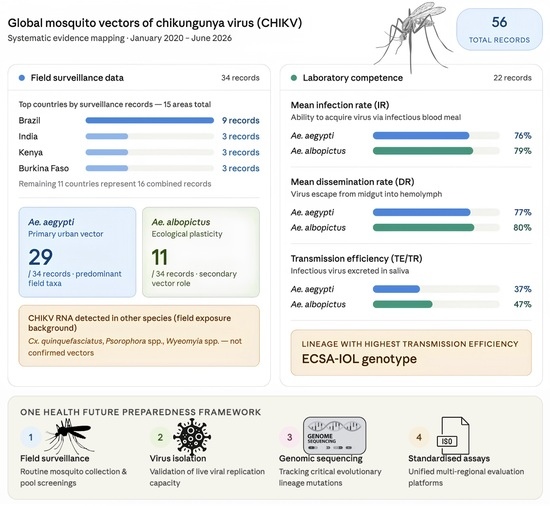
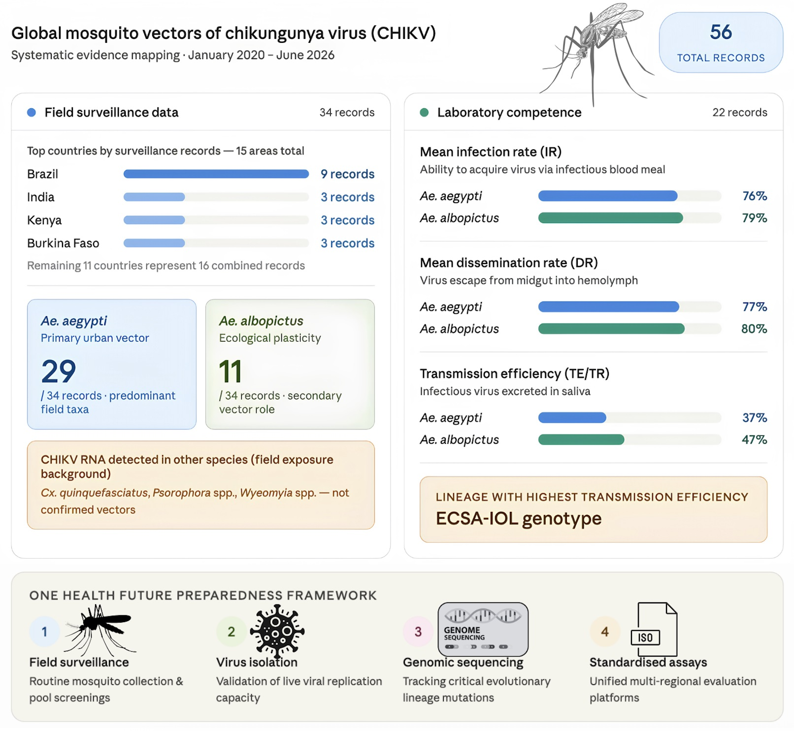
Graphical Abstract

1. Introduction

The chikungunya virus (CHIKV) is an alphavirus in the family Togaviridae and is an important cause of mosquito-borne febrile and arthritic disease. Human infection typically presents with acute fever, rash, headache, myalgia, and severe arthralgia. Although many cases resolve, persistent joint pain can last for months or even years, leading to long-term morbidity, reduced productivity, and substantial public health burden [1]. Over recent decades, CHIKV has triggered outbreaks across Africa, Asia, the Indian Ocean region, Europe, and the Americas, highlighting its capacity for geographic expansion and repeated re-emergence [2,3]. As of December 2024, local CHIKV transmission has been reported in 119 countries and territories [1], and global incidence rose from 0.28 to 11.13 per 100,000 population between 2004 and 2024, with the Americas bearing the highest burden [4].
Transmission dynamics are strongly influenced by mosquito ecology, especially the biology of Aedes aegypti (Linnaeus, 1762) and Aedes albopictus (Skuse, 1894) [1]. Aedes aegypti is highly adapted to urban settings, breeds readily in artificial containers, feeds on humans, and is closely linked to household-level transmission. These traits facilitate viral acquisition from viraemic hosts and rapid outbreak amplification in densely populated areas. On the other hand, Ae. albopictus exhibits greater ecological plasticity, thriving in all urban, peri-urban, rural, and vegetated environments. Its ongoing expansion into temperate regions has raised concerns about the potential for CHIKV establishment beyond historically tropical zones [5,6].
Multiple CHIKV genotypes and lineages have been identified, which include the Asian genotype, West African genotype, East/Central/South African genotype (ECSA), and the Indian Ocean lineage (IOL), which is derived from ECSA. These classifications are epidemiologically relevant, as the virus genotype may influence mosquito infection, dissemination, and transmission efficiency. ECSA and ECSA-IOL strains have been linked to major outbreaks and are frequently examined in laboratory vector competence studies [7,8,9].
A small number of adaptive mutations in the viral genome also shape CHIKV–vector interactions. The best characterised is the E1-A226V substitution in the E1 envelope glycoprotein, which arose independently within the Indian Ocean lineage (IOL) that emerged from the ECSA genotype. This single amino acid change enhances viral infectivity, replication, and dissemination in Ae. albopictus, shortening the extrinsic incubation period and improving transmission efficiency within the species; it is widely regarded as a key driver of the explosive IOL-associated outbreaks in the Indian Ocean region, India, and, subsequently, temperate regions where Ae. albopictus predominates [6,10,11]. Other substitutions, including the E2 mutations E2-L210Q and E2-K252Q, as well as additional E1 changes, can modulate midgut infection and dissemination, and may act epistatically with E1-A226V. These genotype-specific effects mean that a mosquito’s capacity to transmit CHIKV depends not just on the infecting virus strain but on the mosquito species as well.
Determining CHIKV vector status relies on both field and laboratory evidence. Field surveillance provides ecological evidence of mosquito-virus contact through the detection of CHIKV-positive specimens collected under natural conditions. Laboratory studies, in turn, assess whether mosquitoes can acquire the virus, support its dissemination, and transmit it via saliva. However, field RNA detection alone is insufficient to confirm vector competence, as it may reflect a recent infectious blood meal, non-infectious virus, abortive infection (in which the virus enters midgut cells but fails to establish a productive, disseminating infection), or contamination. Moreover, mosquito infection rate estimates are sensitive to sampling design, pool size, and detection probability. Thus, vector status should be assigned based on a combination of field detection, virus isolation, genomic confirmation, and laboratory evidence of transmission [12,13,14,15,16].
This review synthesises studies published between January 2020 and June 2026 on mosquito vectors of CHIKV, with an emphasis on field surveillance, laboratory vector competence, viral genotypes, country-level patterns, temporal trends, infection metrics, diagnostic approaches, and integrated surveillance considerations. The objective is to distinguish confirmed vectors from potential regional vectors and identify field-positive species that require further experimental validation.

2. Literature Search and Data Extraction

This is a narrative review; the structured search described below was undertaken to ensure transparency and reproducibility rather than to constitute a systematic or scoping review, and accordingly, no PRISMA flow diagram or formal risk-of-bias assessment was performed. A structured literature search was conducted using PubMed and Google Scholar to identify relevant studies published from 1 January 2020 to 18 June 2026. Two search strategies were applied to capture the two major evidence categories included in this review. The first search focused on field surveillance studies reporting CHIKV detection in field-collected mosquitoes, while the second search focused on laboratory vector competence studies evaluating experimental CHIKV infection and transmission in mosquitoes.
The literature search was designed to identify original mosquito-based CHIKV studies, including both field surveillance and laboratory vector competence investigations. Search terms combined CHIKV-related keywords with mosquito, surveillance, and vector competence terms, including “chikungunya”, “chikungunya virus”, “CHIKV”, “mosquito”, “Aedes”, “Culex”, “Anopheles”, “field-caught”, “field-collected”, “entomological surveillance”, “mosquito surveillance”, “arbovirus surveillance”, “minimum infection rate”, “MIR”, “virus isolation”, “RT-PCR”, “vector competence”, “oral infection”, “experimental infection”, “blood meal”, “extrinsic incubation”, “saliva”, “infection rate”, “dissemination rate”, and “transmission efficiency”.
Retrieved articles were screened first by title and abstract, followed by full-text assessment. Studies were included if they reported original data on CHIKV detection in field-collected mosquitoes or laboratory evaluations of mosquito vector competence for CHIKV. Reviews, systematic reviews, meta-analyses, case reports, vaccine-focused studies, human clinical studies without mosquito data, and studies lacking relevant entomological outcomes were excluded.
Countries are represented according to the published, indexed mosquito-based CHIKV studies that met the inclusion criteria rather than through selective inclusion. Duplicate records identified across both databases were removed manually, while the grey literature and non-indexed sources were not systematically searched.
Data were extracted into two standardised evidence tables: one for field surveillance studies and one for laboratory vector competence studies. Extracted variables included the authors, year of publication, country or mosquito origin, mosquito species, CHIKV strain or genotype, CHIKV strain group, sample size, infection rate (IR), minimum infection rate (MIR), dissemination rate (DR), transmission rate/transmission efficiency (TR/TE), extrinsic incubation period, incubation temperature, and detection method. Where applicable, CHIKV strains were grouped as Asian, West African, East/Central/South African (ECSA), ECSA–Indian Ocean lineage (ECSA-IOL), mixed Asian/ECSA-IOL, or not reported. Descriptive counts and means were calculated directly from the latest field and laboratory tables. Infection rate (IR) and dissemination rate (DR) were summarised using row-level mean values when multiple time points or populations were reported, while transmission efficiency/transmission rate (TE/TR) used the maximum reported value when no overall transmission estimate was available, which was chosen to capture the upper bound of demonstrated transmission potential rather than to represent a typical rate. These values should be interpreted as descriptive summaries rather than pooled estimates because mosquito infection rate and minimum infection rate (MIR) estimates can be biassed by pool size, sampling intensity, and detection probability.
All numerical comparisons in this manuscript are descriptive. Field-positive and MIR values were not pooled statistically because the studies differed in sampling design, pool size, outbreak timing, and detection method; laboratory rates were also summarised descriptively, as experiments differed in mosquito population, virus strain, infectious dose, incubation temperature, extrinsic incubation period, and saliva assay method.

3. Overall Summary of Mosquito-Based CHIKV Records

The review included 56 mosquito-based records from the latest dataset: 34 field surveillance records and 22 laboratory vector competence records. Field surveillance records documented CHIKV detection in naturally collected mosquitoes, whereas laboratory records evaluated experimental infection, dissemination, or transmission (Table 1 and Table 2). Of the 34 field surveillance records, 28 yielded confirmed CHIKV detection in at least one mosquito species, while the remaining six screened mosquitoes without detecting CHIKV (Table 1).
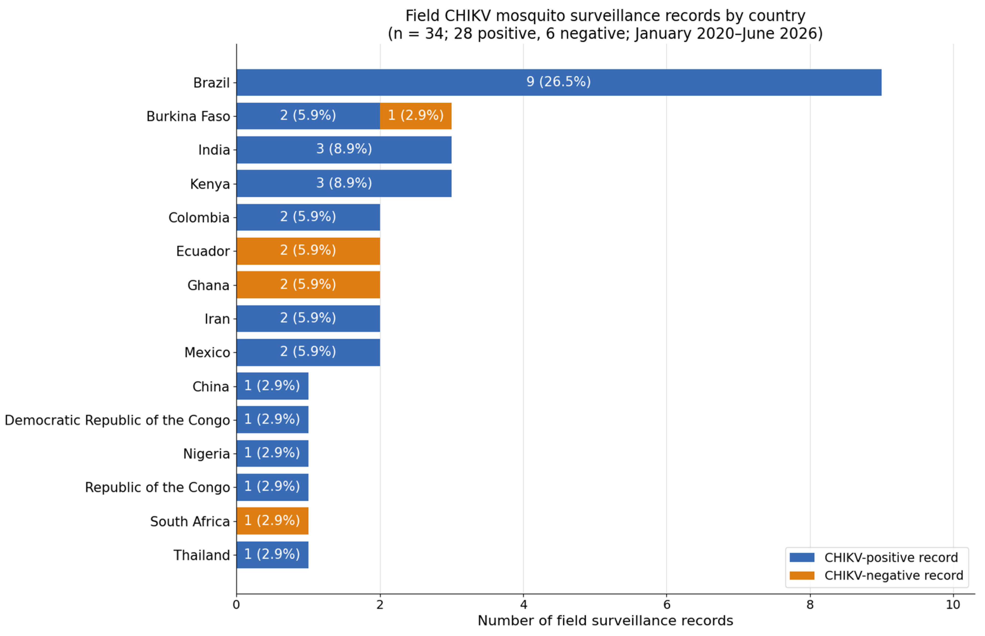
The field records were distributed across 15 countries or territories. Brazil contributed the highest number of records (9/34; 26.5%), followed by India, Kenya, and Burkina Faso (three records each). Ecuador, Iran, Colombia, Mexico, and Ghana contributed two records each, while the Republic of the Congo, the Democratic Republic of the Congo, Thailand, South Africa, China, and Nigeria each contributed one record (Table 1 and Figure 1). Surveillance in Ecuador, Ghana, and South Africa (five records in total) detected no CHIKV in any of the species screened, and one of the three Burkina Faso records was likewise negative; the remaining 28 field records each confirmed CHIKV in at least one mosquito species.
The field dataset contained a broader range of mosquito species than the laboratory dataset because field studies often collected mixed mosquito communities during outbreaks, endemic or ecological surveillance, and screened pools for viral RNA (Table 1). On the other hand, laboratory studies more commonly focused on known or suspected vector species and measured biological outcomes, such as infection, dissemination, and saliva-based transmission (Table 2).

3.1. Yearly Trend of Mosquito-Based CHIKV Records

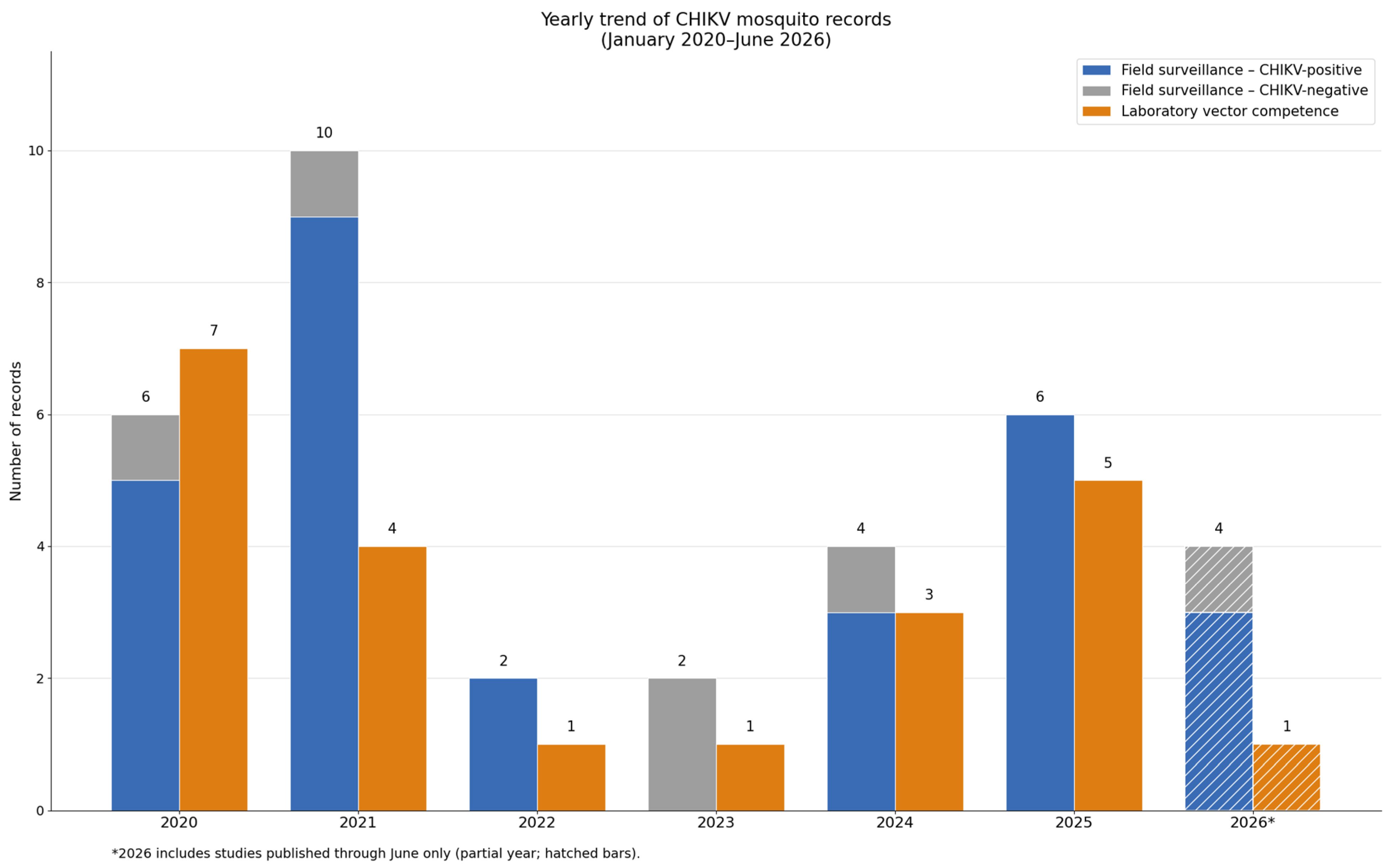
The yearly distribution of records showed that field surveillance and laboratory vector competence studies varied across the review period (Figure 2). Field surveillance records peaked in 2021 with 10 records, followed by 2025 with six records and 2020 with six records. Laboratory vector competence records were highest in 2020 with seven records, followed by 2025 with five records. Both evidence types were least represented in 2022 and 2023 (two field surveillance records and one laboratory record in each year).
Overall, there were more field surveillance studies than laboratory vector competence studies across most years, reflecting the larger contribution of mosquito screening studies to the dataset. The increase in both field and laboratory records in 2024 and 2025 suggests growing research attention to CHIKV mosquito surveillance and vector competence. However, the lower number of records in 2026 should be interpreted cautiously, as the review only included studies published up to June 2026.

3.2. Geographical Summary of CHIKV Mosquito Field Surveillance Records

3.2.1. The Americas

Field surveillance in the Americas was dominated by Aedes aegypti and concentrated in Brazil, which contributed the most records of any country (nine of 34; 26.5%) across several states, including Pará, Rio Grande do Norte, Bahia, Mato Grosso, Paraná, Minas Gerais, São Paulo, and Goiás. All nine Brazilian records were CHIKV-positive for Ae. aegypti, while several Brazilian studies also reported viral RNA in Ae. albopictus; Culex quinquefasciatus (Say, 1823); Aedes fluviatilis (Lutz, 1904); Wyeomyia bourrouli (Lutz, 1905); Psorophora ferox (Humboldt, 1819); Psorophora albigenu (Peryassú, 1908); and other culicids, and a dedicated review of CHIKV’s spatiotemporal distribution and genotype patterns in Brazil highlighted Ae. albopictus as a potential secondary vector in regions where the E1-A226V mutation is present [11]. Where genotype was reported, most Brazilian detections involved ECSA strains [17,18,19,20] while one record was classified as ECSA-IOL [21], with the data indicating sustained CHIKV circulation between 2020 and 2025.
The repeated detection of CHIKV in Ae. aegypti is consistent with its role in urban transmission, whereas its detection in Ae. albopictus suggests possible involvement in peri-urban or mixed ecological settings. Reports involving Culex, Psorophora, and Wyeomyia mosquitoes can only indicate that CHIKV RNA was present in field-collected specimens and cannot establish these species as competent vectors; Cx. quinquefasciatus has been examined in only two laboratory vector competence studies (Table 2), with low and inconsistent outcomes, while Psorophora and Wyeomyia have not been experimentally evaluated, so these detections are best read as ecological exposure rather than competence. The same pattern recurred elsewhere in the Americas. In Ecuador, surveillance focused on human-biting Ae. aegypti in both Quinindé [22] and in Santo Domingo de los Tsáchilas, with the latter also including some Ae. albopictus [23], but no CHIKV was detected in either study. In Colombia, CHIKV was detected in Ae. aegypti in Ibagué [24] and in both Ae. aegypti and Ae. albopictus in Cauca [25], indicating that both species may contribute to transmission and warrant surveillance. In Mexico, surveillance targeted Ae. aegypti exclusively, with natural infection of indoor females in Mérida [26] and an Asian-genotype detected in Ciudad Juárez [27]. Across the Americas, Ae. aegypti was consistently the principal field-detected vector, while Ae. albopictus emerged as a secondary peri-urban species, and non-Aedes detections reflected exposure rather than confirmed transmission.

3.2.2. Africa

African surveillance spanned East, West, and Central Africa, and repeatedly recovered Ae. aegypti alongside a wider range of Aedes and non-Aedes species. In Kenya, three studies involved Ae. aegypti, Aedes vittatus (Bigot, 1861), and Cx. quinquefasciatus, with two records classified as ECSA [15,28] and one genotype not reported [29]; CHIKV detection in Ae. aegypti is consistent with its role as a major vector, whereas RNA detection in Ae. vittatus and Cx. quinquefasciatus, as is the case for other species besides Ae. aegypti, documents outbreak-period exposure but does not establish competence by itself, which requires experimental confirmation (Section 4). In Burkina Faso, three studies conducted in Bobo-Dioulasso and Pouytenga mainly involved Ae. aegypti, wherein Gomgnimbou et al. sampled Ae. aegypti, Aedes furcifer (Edwards, 1913), and Ae. vittatus but detected no CHIKV [30]; meanwhile, Laouali et al. reported ECSA-classified CHIKV in Ae. aegypti pools from Bobo-Dioulasso [31], and Toé et al. reported its circulation in Ae. aegypti during the 2023 Pouytenga outbreak [32], together indicating a central role for Ae. aegypti in outbreak-related surveillance.
In Central Africa, single records from the Republic of the Congo (Ae. aegypti, Ae. albopictus, and Culex spp. [33]) and from Matadi in the Democratic Republic of the Congo (Ae. albopictus in an outbreak setting [34]) indicated ECSA-related circulation; of the species collected, CHIKV was confirmed in Ae. albopictus (a positive pool in the Republic of the Congo, and detections during the Matadi outbreak), whereas Ae. aegypti and Culex spp.’s pools tested negative (Table 1). West and South African surveillance added further breadth: Ghanaian studies contributed Aedes-focused arbovirus surveillance without detecting CHIKV [35,36]; a South African study screened diverse Aedes, Culex, Anopheles, and Mansonia species but detected no CHIKV [37]; and nationwide surveillance in Nigeria detected CHIKV in multiple Aedes species, including Ae. aegypti and Aedes luteocephalus (Newstead, 1907) [38]. Across Africa, Ae. aegypti again dominated the field evidence, while the recurrent detection of Ae. vittatus and other forest-associated or non-Aedes species underscores the ecological complexity of African settings and the need for experimental confirmation of any secondary vector roles.

3.2.3. Asia

Asian surveillance combined routine xenomonitoring with newer molecular approaches. India contributed three 2021 records that used molecular xenomonitoring of CHIKV and other arboviruses in Ae. aegypti and Ae. albopictus, with one ECSA-classified record [39] and two without genotypes [40,41], highlighting the value of integrated arbovirus surveillance where dengue, chikungunya, and Zika co-circulate in shared vector populations. Iran contributed two contrasting records: Bakhshi et al. detected the Asian-genotype CHIKV RNA in Culiseta longiareolata (Macquart, 1838), Culex tritaeniorhynchus (Giles, 1901), and Anopheles maculipennis (Meigen, 1818) s.l. [42]—as these are not classical CHIKV vectors, this should be interpreted as exposure rather than competence—while Abbasi et al. used metagenomic sequencing across Cx. quinquefasciatus; Culex pipiens Linnaeus, 1758; Ae. aegypti; Ae. albopictus; Anopheles stephensi (Liston, 1901); and Anopheles culicifacies (Giles, 1901), classifying the virus as ECSA with positivity in Ae. albopictus and Ae. aegypti [43]—here, confirmed positivity in the recognised Aedes vectors indicates active circulation, whereas Culex and Anopheles detections reflect exposure requiring experimental validation. Thailand contributed a single but instructive record in which CHIKV RNA was detected in field-collected Cx. quinquefasciatus from Bangkok; however, laboratory follow-up did not support productive replication, illustrating the value of distinguishing field detection from vector competence [16]. Finally, China reported whole-genome CHIKV detection during the Foshan outbreak (ECSA-IOL [44]); whole-genome sequencing from field-collected mosquitoes confirms an intact viral genome, enables accurate genotype assignment, and provides stronger molecular evidence of field infection than partial RNA detection alone. Together, the Asian records illustrate the growing application of whole-genome and metagenomic sequencing during 2020–2026; while RT-PCR and qPCR have been the mainstay of arbovirus detection since the early 2000s, many field studies still relied on molecular detection without sequence confirmation, which contributed to incomplete genotype reporting (Table 1).

3.2.4. Mediterranean and Europe

The search returned no indexed 2020–2026 field surveillance records reporting CHIKV detection in mosquitoes from mainland Europe. The European and Mediterranean evidence is therefore represented in this review through laboratory vector competence studies rather than field collections [9,45,46,47]. This gap most likely reflects publication timing rather than an absence of risk, and targeted European entomological surveillance is flagged as a priority.
Table 1. Published field surveillance studies reporting CHIKV detection in mosquitoes, 2020–2026.
No.AuthorsYearCountry/AreaLocalityMosquito SpeciesCHIKV GenotypeSample SizeInfection Rate/
Positive Pools (%)
MIR (%)Detection Method
1Bakhshi et al. [42]2020IranIran (North Khorasan, Mazandaran, and Fars provinces)Cs. longiareolata;
Cx. tritaeniorhynchus; An. maculipennis s.l.
AsianCs. longiareolata, 34;
Cx. tritaeniorhynchus, 129;
An. maculipennis s.l., 514
Cs. longiareolata: 5.9% (2/34);
Cx. tritaeniorhynchus: 1.6% (2/129);
An. maculipennis s.l.: 0.4% (2/514)
NRRT-PCR/qPCR
2Cruz et al. [17]2020BrazilXinguara, Pará, BrazilAe. aegypti;
Cx. quinquefasciatus
ECSA492 (36 pools)Ae. aegypti: 19.4%
(7/36 pools);
Cx. quinquefasciatus: 2.8% (1/36 pools)
NRRT-PCR/qPCR; IFA; virus isolation
3De Melo Ximenes et al. [21]2020BrazilNatal, Rio Grande do Norte, BrazilAe. aegypti;
Ae. albopictus
Ae. fluviatilis;
Wyeomyia bourrouli;
Hg. leucocelaenus;
Ae. serratus;
Ae. taeniorhynchus;
Ae. scapularis
ECSA-IOLTotal: 314 mosquitoes
Wy. bourrouli: 210,
Ae. albopictus: 73,
Ae. fluviatilis: 10,
Hg. leucocelaenus: 7,
Ae. scapularis: 7,
Ae. aegypti: 5,
Ae. serratus: 1,
Ae. taeniorhynchus: 1
CHIKV-positive species detected; infection rate by species not reported
(Ae. aegypti,
Ae. albopictus,
Ae. fluviatilis, Wy. bourrouli)
NRRT-PCR
4Vairo et al. [33]2020Republic of the CongoRepublic of the Congo (Kouilou and Pointe-Noire)Ae. aegypti;
Ae. albopictus;
Culex. spp.
ECSA-IOLAe. albopictus: Nkoungou, 18; Diosso, 61; Mengo, 8; Matombi, 16; Mabindou, 25, Mpita, 34; Tchiamba Nzassi, 47; Siafoumou, 10
Ae. aegypti: Mptiya, 20; Diosso, 2
Culicinae spp.: 12
Ae. albopictus:
1 positive pool;
Ae. aegypti: negative;
Culicinae spp.: negative
NRRT-PCR/qPCR
5Ortega-López et al. [22]2020EcuadorQuinindé, EcuadorAe. aegyptiNR429 individualsNegativeNegativeRT-PCR
6Heath et al. [29]2020KenyaKenya (Kisumu, Chulaimbo, Ukunda, Msambweni)Ae. aegyptiNR714 pools5.9%
(43/714 pools)
0.49%
(0.34–0.68%)
RT-PCR/qPCR
7Teixeira et al. [18]2021BrazilVitória da Conquista, Bahia State, BrazilAe. aegyptiECSA480 (450 larvae in 30 pools of 25, plus 30 individual larvae)13.3%
(4/30 pools)
4.3%RT-PCR/qPCR
8Chand et al. [39]2021IndiaJabalpur and Narsinghpur districts, Central IndiaAe. aegypti;
Ae. albopictus;
Ae. vittatus
ECSATotal mosquitoes: 2991
Ae. aegypti: 2831,
Ae. albopictus: 149,
Ae. vittatus: 11
Ae. aegypti: 1.27%
(5/394 pools)
Ae. aegypti: 0.36%RT-PCR/qPCR;
plaque assay
9Lutomiah et al. [15]2021KenyaMombasa, KenyaAe. aegypti;
Ae. vittatus;
Cx. quinquefasciatus
ECSATotal mosquitoes:
6899 adults;
Ae. aegypti: 911;
Ae. vittatus: 1137;
Cx. quinquefasciatus: 4492
Ae. aegypti: 0.87%
(1/115 pools);
Ae. vittatus: negative;
Cx. quinquefasciatus: 1.65%
(4/243 pools)
Ae. aegypti(F): 0.30%;
Cx. quinquefasciatus (F): 0.08%;
Cx. quinquefasciatus (M): 0.10%
RT-PCR/qPCR; virus isolation
10Weggheleire et al. [34]2021Democratic Republic of the CongoMatadi, Democratic Republic of the Congo (DRC)Ae. aegypti;
Ae. albopictus
ECSA11 adult mosquito pools; 15 larvae poolsAe. albopictus: 45.5% (5/11 adult pools); 13.3% (2/15 larval pools)0–1.31%RT-PCR/qPCR; ELISA
11Phumee et al. [16]2021ThailandBangkok, ThailandCx. quinquefasciatusECSA-IOL43 (16 males, 27 females)18.6%
(8/43 mosquitoes)
NRRT-PCR/qPCR; IFA; virus isolation
12Da Silva-Neves et al. [48]2022BrazilCuiabá and Várzea Grande, Mato Grosso, BrazilAe. aegypti;
Ae. albopictus;
Cx. quinquefasciatus;
Ps. albigenu;
Ps. ferox
NR5040 adults
(2873 males; 2167 females) in 398 pools
Ae. aegypti F: 6.52% (3/46);
Ae. aegypti M: 6.06% (2/33);
Ae. albopictus F: 13.79% (4/29);
Cx. quinquefasciatus F: 21.35% (19/89);
Cx. quinquefasciatus M: 13.13% (13/99);
Ps. albigenu F: 16.67% (2/12);
Ps. ferox F: 12.50% (2/16)
NRRT-PCR/qPCR
13Carrasquilla et al. [24]2021ColombiaIbagué, ColombiaAe. aegyptiNRAe. aegypti: 482 individuals (133 pools), 317 individuals;
Ae. albopictus: 7
Ae. aegypti: 0.8% 0.21%RT-PCR/qPCR
14Joannides et al. [35]2021GhanaMole Game Reserve and Larabanga, Northern GhanaAe. aegypti;
Ae. vittatus
NR1957 (75 pools)NegativeNART-PCR/qPCR
15Munivenkatappa
et al. [40]
2021IndiaKarnataka State, IndiaAe. aegyptiNR50 pools10%
(5/50 pools)
NRPCR
16Vikram et al. [41]2021IndiaDelhi, IndiaAe. aegyptiNR981 individuals13.0% NRRT-PCR/qPCR
17Kirstein et al. [26]2021MexicoMérida, Yucatán, MexicoAe. aegyptiNR2161 individuals6.0%
(129/2161 females)
NRRT-PCR/qPCR
18Leandro et al. [49]2022BrazilFoz do Iguaçu, BrazilAe. aegyptiNR109 individuals5.45% (3/55)NRRT-PCR/qPCR; plaque assay
19Akyea-Bobi et al. [36]2023GhanaAccra, Ghana (Madina, Achimota Forest)Ae. aegypti;
Ae. albopictus
NRAe. aegypti: 1493
(120 pools);
Ae. albopictus: 1 pool
NegativeNART-PCR/qPCR
20Guarido et al. [37]2023South AfricaSouth Africa (Gauteng, Limpopo, Northwest, Mpumalanga, KwaZulu-Natal)Ae. durbanensis;
Ae. mcintoshi;
Cx. pipiens s.l.;
Cx. univittatus;
Anopheles spp.;
Mansonia spp.
NR39,035 (1462 pools)NegativeNART-PCR/qPCR
21Almeida-Souza et al. [19]2024BrazilSalinas, Minas Gerais, BrazilAe. aegypti;
Cx. quinquefasciatus
ECSATotal: 421 individuals
Ae. aegypti: 31 pools;
Cx. quinquefasciatus:
26 pools
Ae. aegypti: 32.3% (10/31);
Cx. quinquefasciatus:
7.7% (2/26)
Ae. aegypti: 6.06%;
Cx. quinquefasciatus: 0.78%
RT-PCR/qPCR
22Banho et al. [20]2024BrazilSão José do Rio Preto, São Paulo, BrazilAe. aegypti;
Ae. albopictus;
Aedes spp.;
Culex spp.
ECSATotal: 1183
Ae. aegypti: 744;
Ae. albopictus: 11;
Aedes sp.: 1;
Culex spp.: 427
Ae. aegypti females: 33% (26/79);
Ae. aegypti males: 34% (27/79);
Ae. albopictus females: 2.5% (2/79);
Culex spp. females: 21.5% (17/79);
Culex spp. males: 8.8% (7/79)
NRRT-PCR/qPCR
23Silva et al. [50]2024BrazilGoiânia, Goiás, Brazil Ae. aegypti NR1570 (157 pools)1.3%
(2/157 pools)
0.127%RT-PCR/qPCR
24Gomgnimbou et al. [30]2024Burkina FasoBobo-Dioulasso, Burkina FasoAe. aegypti;
Ae. furcifer;
Ae. vittatus
NRTotal: 976 individuals
Ae. aegypti: 959;
Ae. furcifer: 6;
Ae. vittatus: 11
NegativeNART-PCR/qPCR
25Hernández-Acosta et al. [27]2025MexicoCiudad Juárez, Chihuahua, MexicoAe. aegyptiAsian328 individuals65.65%26.0%RT-PCR/qPCR
26Banho et al. [51]2025BrazilSão José do Rio Preto, São Paulo, BrazilAe. aegypti;
Ae. albopictus;
Culex spp.
ECSA1914 individualsTotal: 5.6% (107/1914)
Ae. aegypti F: 32.7% (35/107);
Ae. aegypti M: 28.0% (30/107);
Ae. albopictus F: 1.9% (2/107;
Culex spp. F: 26.2% (28/107);
Culex spp. M: 11.2% (12/107)
NRRT-PCR/qPCR; virus isolation
27Musili et al. [28]2025KenyaMombasa, KenyaAe. vittatusECSA2989 (842 pools) 0.12%
(1/842)
NRVirus isolation/NGS
28Zhou et al. [44]2025ChinaFoshan City, Guangdong, ChinaAe. albopictusECSA-IOL1569 female9.09%
(7/77)
0.446% (up to 0.917% in Lecong Town)RT-PCR/qPCR
29Mantilla-Granados J.S. et al. [25]2025ColombiaCauca, Colombia (Piamonte, Patía, Piendamó, Popayán)Ae. aegypti;
Ae. albopictus
NRAe. aegypti: 89 pools;
Ae. albopictus: 34 females
Ae. aegypti: 12.4% (11/89);
Ae. albopictus: 41.2% (14/34)
Ae. aegypti: 4.94%RT-PCR/qPCR
30Nwangwu et al. [38]2025NigeriaNigeriaAe. aegypti;
Ae. luteocephalus
NR2406
(127 pools)
Ae. aegypti: 14.5%
(9/62 pools);
Ae. luteocephalus: 33.3% (1/3 pools)
Ae. aegypti: 0.16%;
Ae. luteocephalus: 6.25%
RT-PCR/qPCR
31Laouali et al. [31]2026Burkina FasoBobo-Dioulasso, Burkina FasoAe. aegyptiECSA23,229 individuals
(199 pools)
1.0%
(2/199 pools)
0.017%RT-PCR/qPCR
32Abbasi [43]2026IranIran (Hormozgan, Sistan and Baluchestan, and Khuzestan provinces)Cx. quinquefasciatus;
Ae. aegypti;
Cx. pipiens;
Ae. albopictus;
An. stephensi;
An. culicifacies
ECSATotal: 4275 individuals (152 pools, ranging from 10 to 30 individuals)Ae. albopictus/
Ae. aegypti: 11.1% (infection rate specified by species)
NRNGS/
sequencing
33Toé et al. [32]2026Burkina FasoPouytenga, Burkina FasoAe. aegyptiNRTotal: 41(3 pools)66.67%
(2/3 pools)
4.88%RT-PCR/qPCR
34Herrera et al. [23]2026EcuadorSanto Domingo de los Tsáchilas, EcuadorAe. aegypti;
Ae. albopictus
NR3918 (196 pools)NegativeNegativeRT-PCR/qPCR
NR = not reported; NA = not applicable. Sample sizes are reported as given in the source studies; some give the number of individual mosquitoes tested, others the number of pools, and some both—where pools were screened, the pool count is shown in parentheses. Genus abbreviations: Ae. = Aedes; An. = Anopheles; Cs. = Culiseta; Cx. = Culex; Ps. = Psorophora; Wy. = Wyeomyia.
Table 2. Published laboratory vector competence studies of CHIKV in mosquitoes, 2020–2026.
No.AuthorsYearMosquito SpeciesMosquito OriginStrain TypeCHIKV GenotypeSample SizeIR (%)DR (%)TE/TR (%)EIPTemp.Detection Method
1Robison et al. [52]2020Ae. aegyptiPoza Rica, Mexico Lab Asian60 mosquitoes per time point from 2 to 18 dpi; 50 at 20 dpiMidgut infection: 98% (59/60) at 2 dpi; 100% from 4 to 18 dpi; 94% (47/50) at 20 dpiLegs/wings: 100% at 2–6, 10, 12, 16 dpi; 98% at 8, 14, 18 dpi; 96% at 20 dpiInfectious saliva by plaque assay: 28% at 2 dpi, 27% at 4 dpi, 18% at 6 dpi, 10% at 8 dpi, 5% at 10 dpi, 0% at 12–18 dpi, 0% at 20 dpi2 dpi28 °CRT-qPCR; plaque assay on Vero cells for infectious saliva
2Bohers et al. [7]2020Ae. albopictusTunisia (Carthage/
Tunis)
Both ECSA-IOL134 individuals76.7% at 3 dpi to 79.2% at 21 dpi Overall: 81.3% (109/134)82.6% at 3 dpi to 94.7% at 21 dpi; Overall: 90.8%TR 75% at 10 dpi; TE peaked at 55.5% at 10 dpi3 dpi28 ±1 °CFocus fluorescent assay on C6/36 cells
3Calvez et al. [53]2020Ae. polynesiensisWallis and Futuna Islands (WKANA: Kanahe; WLALO: Lalolalo lake area)FieldAsianWKANA: 31; WLALO: 26 WKANA: 83.9% (26/31); WLALO: 84.6% (22/26)WKANA: 76.9% (20/26); WLALO: 77.3% (17/22)WKANA: 45.0% (9/20);
WLALO: 47.1% (8/17)
7 dpi28 °CRT-PCR/qPCR and infectious saliva/CPE assay
4Shin et al. [54]2020Ae. aegyptiVero Beach and Key West, Florida, USABoth ECSA-IOLTotal: 900 individuals;
Subsets: 15 at 4 dpi and 43–75 at 10 dpi per population
4 dpi body: Rockefeller 73.3% (11/15);
Vero Beach 93.3% (14/15);
Key West 86.6% (13/15);
10 dpi body: Rockefeller 88.4% (38/43);
Vero Beach 98.5% (64/65);
Key West 97.3% (73/75)
10 dpi legs: Rockefeller 73.7% (28/38);
Vero Beach 100% (64/64);
Key West 86.3% (63/73)
NANR28 °CRT-qPCR/qPCR
5Gutiérrez-Bugallo et al. [55]2020Ae. aegyptiHavana, Cuba (Pasteur/PTE, Parraga/PRG); Mombasa, KenyaFieldECSA60 females per population per viral strain95–100% at 3 dpe for both PTE and PRG PTE 86%, PRG 100% at 14 dpePTE 4–14%;
PRG 0–11%
7 dpi28 °CPlaque assay/infectious virus titration
6Kaczmarek et al. [56]2020Ae. albopictusNew York City, USA BothECSA-IOL16/21/7 by population>72% at 14 dpi>72% at 14 dpi3–10% at 7 dpi;
15% at 14 dpi;
in vivo Tallman model: 60–80%
2 dpi28 °CPlaque assay; RT-qPCR; in vivo mouse transmission model; sequencing
7Gloria-Soria
et al. [57]
2020Ae. albopictusConnecticut and New York, USAFieldAsian; ECSA-IOL120 individuals per population>70% infection beginning at 4 dpi30–100% after 14 dpi, varying by mosquito populationUp to 27.5%4 dpi27:22 °CRT-qPCR; plaque assay for virus stocks
8Lutomiah et al. [15]2021Cx. quinquefasciatusMombasa, KenyaFieldECSATotal: 47 individuals;
16 tested at 7 dpi and 14 dpi, 15 at 21 dpi
7 dpi: 25% (4/16);
14 dpi: 31.3% (5/16);
21 dpi: 26.7% (4/15)
7 dpi: 25% (1/4);
14 dpi: 40% (2/5);
21 dpi: 13.3% reported
TE: 7 dpi: 6.3% (1/16);
14 dpi: 12.5% (2/16); 21 dpi: 0%;
TR: 100% at 7 and 14 dpi
7 dpi28 °CRT-PCR/qPCR and CPE/virus isolation
9Rodrigues et al. [8]2021Ae. aegyptiBrazil (Belo Horizonte)FieldECSA600 individuals (40 per each of the 15 experimental groups)100% for all single and multiple infections 100% for all single and quadruple infections; 90% for CHIKV in CDY triple infection100% in single and quadruple infections; varied from 40% to 100% in dual and triple infections14 dpi28 °CRT-PCR/qPCR
10Phumee et al. [16]2021Cx. quinquefasciatusBangkok, ThailandBothECSA-IOLTotal: 43 total (16 males, 27 females)29.7% (8/27) NANANA28 ± 2 °CNested RT-PCR; virus isolation/CPE; immunofluorescence assay
11Jansen et al. [58]2021Ae. koreicusWestern Germany FieldECSA24 ± 5 °C: 34;
27 ± 5 °C: 151
24 ± 5 °C: 17.6% (6/34);
27 ± 5 °C: 68.2% (103/151)
NR24 ± 5 °C: 0% (0/34); 27 ± 5 °C: 4.6% (7/151);
TR at 27 ± 5 °C: 6.8% (7/103)
14 dpi24 ± 5 °C;
27 ± 5 °C
RT-qPCR; cell culture/CPE; plaque assay
12Calvez et al. [59]2022Ae. aegyptiVientiane Capital, Lao PDRFieldAsian; ECSA-IOL28–30 females per group/time pointECSA-IOL: 7% at 3 dpi to 38% at 14 dpi;
Asian: 50% at 3 dpi, 33% at 7 dpi, 53% at 14 dpi
ECSA-IOL: 0% at 3 dpi, 83% at 7 dpi; 64% at 14 dpi;
Asian: 50% at 3 dpi, 100% at 7 dpi, 75% at 14 dpi
Low TE: positive values from 3% for ECSA-IOL at 7 dpi to 7% for Asian at 14 dpi;
TR ranges across 14–20% where saliva-positive
Asian: 3 dpi; ESCA-IOL: 7 dpi30 ± 2 °CRT-PCR/qPCR; CPE on Vero E6 cells for infectious saliva particles
13Terradas et al. [60]2023An. albimanusEl SalvadorLabAsianTotal: 90
30 mosquitoes per time point
33.3% at 7 dpi; 10% at 10 dpi; 10% at 14 dpi6.7% at 7 dpi;
0% at 10 and 14 dpi
0%NA27 ± 1 °CFocus-forming assay; forced salivation
14Azman et al. [61]2024Ae. aegyptiMalaysiaBothECSA-IOL20 mosquitoes per time point/
condition
Midgut infection by virus titration: MC1 0–100%; KL 15–100%; oral infection at 7 dpi by PCR/culture: MC1 15–90%/0–75%; KL 45–100%/15–100%Heads/thoraxes dissemination by virus titration: MC1 0–35%; KL 0–85%; MC1 pCMV-p2020A 35% vs. pCMV-p2020V 5% at 3 dpiNRNR28 ± 1 °CReal-time PCR; virus titration/TCID50
15Bohers et al. [46]2024Ae. albopictusParis, FranceFieldECSA-IOLTotal: 273 individuals exposed across time; 30 tested at 7 dpi for peak values7 dpi: 63.3% (19/30)7 dpi: 89.5% (17/19)TE: 7 dpi: 20.0% (6/30)7 dpi28 °CFFU titration on Ae. albopictus C6/36 cells
16Lühken et al. [9]2024Ae. albopictusGermanyLabECSA-IOL177 individuals100% NR15 °C: 5.56% (1/18); 15 ± 5 °C: 32.5% (13/40); 18 °C: 50.0% (16/32); 18 ± 5 °C: 54.3% (19/35); 21 °C: 39.1% (9/23); 21 ± 5 °C: 58.6% (17/29)14 dpi15 °C, 15 ± 5 °C, 18 °C, 18 ± 5 °C, 21 °C, 21 ± 5 °CRT-qPCR;
infectious saliva/cell-culture assay
17Anyango et al. [62]2025Ae. aegyptiKenya (Kisumu and Busia counties)FieldECSA260 individualsOverall: 56.5% (147/260)
Busia: 57.8% (78/135); Kisumu: 55.2% (69/125)
Overall: 78.2% (115/147)
Busia: 71.8% (56/78);
Kisumu: 85.5% (59/69)
Overall: 26.1% (30/115)
Busia: 25.0% (14); Kisumu: 27.1% (16)
5 dpi28 ± 1 °CVirus isolation/culture; plaque/focus assay
18Jansen et al. [63]2025Ae. japonicusGermany FieldECSA33 at 21 ± 5 °C;
38 at 24 ± 5 °C;
38 at 27 ± 5 °C
21 ± 5 °C: 81.8% (27/33); 24 ± 5 °C: 81.6% (31/38); 27 ± 5 °C: 89.5% (34/38)NRTE: 0% at 21 ± 5 °C & 24 ± 5 °C;
2.9% (1/34) at 27 ± 5 °C
14 dpi21 ± 5 °C, 24 ± 5 °C, 27 ± 5 °CRT-qPCR; cell culture/plaque assay for infectious saliva
19Sanon et al. [64]2025Ae. aegyptiOuagadougou, Burkina Faso FieldWest AfricanNRUrban: 35%;
Peri-urban: 0%
Urban: 85%;
Peri-urban: 41–45%
TE: 14–18%7 dpi28 ± 1 °CPlaque assay/TCID50/CPE-based titration
20Hafsia et al. [65]2025Ae. aegypti;
Ae. albopictus
Southwestern Indian Ocean (Seychelles, Comoros, Reunion, Mayotte)BothECSA-IOL19–48 specimens per mosquito line/time point for saliva collectionAe. aegypti: >90% at 7 and 14 dpe;
Ae. albopictus: high susceptibility across lines
Ae. aegypti: 90.8%
Ae. albopictus: 72.7% at 7 dpe to 88.8% at 14 dpe;
Ae. Aegypti: 90.8%
TE: maximum 62.5%
Ae. aegypti: 90.8%;
Ae. albopictus: 11.0% at 7 dpe to 23.8% at 14 dpe
7, 14 dpi 28 °CPlaque assay
21Visser et al. [47]2025Ae. aegyptiThe NetherlandsLabWest African10–40 mosquitoes per treatment/time pointSingle infection: 94%;
Dual/triple coinfection did not significantly change except lower total load in triple infection
NR12%10 dpi28 °C, 70% RHRT-qPCR/qPCR; cell culture/CPE for saliva infectivity
22Gutierrez-Bugallo et al. [66]2026Ae. aegyptiHavana, CubaBothNot reported34 mothers exposed, 12 mothers at 2nd gonotrophic cycle, and 72 daughter saliva samplesVertical transmission study: infected mothers at 2nd gonotrophic cycle: 12/12;
Daughters with infected saliva: 11/72
NRFilial infective rate in saliva (FIR-S): 15% (11/72); vertical transmission rate in saliva (VTR-S): 50% (7/12 families)0 dpi/immediate upon emergence 28 °CRT-qPCR;
plaque assay; NGS/sequencing
NR = not reported; NA = not applicable. IR = infection rate; DR = dissemination rate; TE = transmission efficiency; TR = transmission rate; MIR = minimum infection rate; EIP = extrinsic incubation period; dpi = days post-infection; dpe = days post-exposure. MC1 = Aedes aegypti field strain from Petaling Jaya, Selangor; KL = Aedes aegypti laboratory colony from Kuala Lumpur; pCMV-p2020A and pCMV-p2020V = infectious clones of Malaysian ECSA-IOL CHIKV isolate MY/2020/3092435; CDY = CHIKV/DENV/YFV triple-coinfection group.
The experimental conditions under which these laboratory studies were conducted are summarised in Table 3. These parameters varied substantially across studies: infectious blood-meal titres ranged from approximately 10^5 to 10^9 infectious units per millilitre; mosquitoes were exposed most often through artificial membrane feeders (for example, the Hemotek system) but also by droplet feeding or, in one study, by intrathoracic inoculation; and transmission was assessed predominantly by forced salivation combined with plaque, focus-forming, or cytopathic-effect assays, with one study additionally employing an in vivo mouse-transmission model. This methodological heterogeneity in infectious dose, feeding route, incubation temperature, and saliva assay directly affects the measured infection, dissemination, and transmission outcomes, underscoring the descriptive rather than pooled treatment of the vector competence values reported in Section 4.

4. Species-Level Vector Evidence

Mosquito species were interpreted using two complementary evidence types: field detection and laboratory vector competence. Field detection indicates that either CHIKV RNA or the virus was detected in naturally collected mosquitoes, whereas laboratory vector competence studies assess whether experimentally exposed mosquitoes can support infection, dissemination, and transmission. This distinction is important, because PCR positivity alone does not confirm vector status. Throughout this review, we therefore distinguish three levels of evidence that are often conflated: the detection of CHIKV RNA, which signals exposure only; isolation of the infectious virus, which confirms viability of the virus; and demonstrated transmission under experimental or in vivo conditions, which is required to establish vector competence.
Based on the reviewed studies, mosquito species were grouped into principal vectors, potential regional or emerging vectors, field-positive but unconfirmed vectors, and species with weak or inconsistent evidence of vector competence (Table 4). This classification avoids overinterpretation of RNA-only detection while still identifying mosquito taxa that may warrant further study. This classification reflects the 2020–2026 evidence base and is not intended as an exhaustive catalogue of all historical vector records; earlier experimental findings, notably the adaptation of CHIKV to Ae. albopictus through the E1-A226V substitution [10] and the demonstrated competence of Kenyan Ae. vittatus populations [67], are considered where they inform interpretation of these groupings.

4.1. Main CHIKV Vectors

Aedes aegypti was the most frequently detected species in the field surveillance and remains the predominant urban CHIKV vector. Its role is linked to its close association with humans, artificial-container breeding, domestic resting behaviour, and anthropophilic feeding [68]. Laboratory studies also strongly support its role, showing that Ae. aegypti can support the infection, dissemination, and transmission of Asian, ECSA, ECSA-IOL, and West African genotype groups (Table 2).
On the other hand, Ae. albopictus is the second major vector identified in this review and is documented in both field surveillance and laboratory datasets, where it was reported across diverse settings, including Brazil, Central Africa, Ecuador, Colombia, Ghana, and China (Table 1 and Table 2). Its ecological flexibility, ability to occupy peri-urban and vegetated environments, adaptation to cooler climates [69], and competence for ECSA and ECSA-IOL strains (Table 1 and Table 2) underscore its role as a vector where this species is either established or expanding. Within Europe and the Mediterranean, this competence is well-documented: Ae. albopictus populations from Germany and Paris transmitted ECSA-IOL CHIKV, even at the lower temperatures characteristic of temperate summers [9,46]; a study from the Netherlands confirmed Ae. aegypti competence for a West African strain [47]; and earlier experimental work showed that Lebanese Ae. albopictus transmitted CHIKV in saliva in approximately 30% of mosquitoes at ten days post-infection [45], with the last predating the review window and therefore outside the tabulated dataset.
Vector competence in both principal vectors varied across mosquito population, virus genotype, infectious dose, incubation temperature, extrinsic incubation period, and assay method. Therefore, local Ae. aegypti and Ae. albopictus populations should ideally be evaluated using locally circulating CHIKV strains under ecologically relevant temperature conditions.

4.2. Potential Regional and Emerging Vectors

Several other Aedes species have shown possible regional relevance (Table 4). Ae. vittatus yielded confirmed CHIKV detections in only one field study in Kenya [28] (Table 1); the other surveillance studies that screened it detected no CHIKV. Although no 2020–2026 laboratory records were included in the dataset, earlier work has shown the competence of Kenyan Ae. vittatus populations for ECSA-lineage CHIKV [67], suggesting possible regional importance that requires an updated evaluation using contemporary strains and local populations.
Moreover, Aedes polynesiensis (Marks, 1951) can become infected with CHIKV under experimental conditions [53], which may be relevant in Pacific Island settings, where local mosquito communities and human–vector contact patterns differ from continental settings. In addition, laboratory studies have shown that Aedes koreicus (Edwards, 1917) and Aedes japonicus (Theobald, 1901) can become infected with CHIKV under experimental conditions, although transmission outcomes were low or inconsistent (Table 2). This limited competence evidence is relevant, as both species are invasive Aedes mosquitoes with expanding distributions in temperate regions. Therefore, these species should be considered emerging or uncertain vectors.
Other field-detected Aedes species with confirmed CHIKV positivity included Ae. fluviatilis [21] and Ae. luteocephalus [38]. These detections were suggestive of possible exposure during active transmission, but neither has been sufficiently evaluated through laboratory vector competence assays (Table 1).
The epidemiological weight of field detection also depends on the feeding ecology of the species involved, as not all culicids implicated above are strictly anthropophilic. Ae. aegypti is highly anthropophilic and endophilic, feeding preferentially and repeatedly on humans in and around dwellings, which maximises its capacity to acquire and transmit CHIKV during urban outbreaks [70]. Ae. albopictus is a more opportunistic, catholic feeder with pronounced zoophilic tendencies, biting humans as well as other mammals and birds; this mixed host use lowers its per-bite human transmission efficiency but positions it as a bridge between peri-domestic and sylvatic cycles [71]. Several other field-detected species, including Ae. vittatus and forest-associated Aedes, together with the largely ornithophilic or mammalophilic Culex and Anopheles species, feed only opportunistically on humans, so their detection with CHIKV is more plausibly a marker of ecological exposure than of a primary role in human transmission [72]. Feeding preference should therefore be weighed alongside laboratory competence when interpreting the significance of any field-positive species.
Beyond intrinsic competence, the epidemiological plausibility of a species acting as a local vector is strengthened when it is both abundant and CHIKV-positive while in the same place and time as human cases. Several of the field records reviewed here (for example, the outbreak-associated detections in Brazil, Kenya, and Burkina Faso) derive their weight from this exact spatial and temporal coincidence between vector presence and active transmission [72]. Such epidemiological association does not replace experimental confirmation, but it provides an important complementary line of evidence for prioritising species and settings for vector competence testing.

4.3. Field-Positive but Unconfirmed Non-Aedes Mosquitoes

CHIKV RNA was detected in several non-Aedes species, especially Cx. quinquefasciatus and other Culex species (Table 1). These detections may reflect recent blood feeding on viraemic hosts, persistence of non-replicating viral RNA, or infections that fail to disseminate beyond the midgut. The current evidence is insufficient to classify Culex mosquitoes as principal CHIKV vectors.
In addition, Anopheles mosquitoes, including An. maculipennis s.l. in field surveillance and Anopheles albimanus (Wiedemann, 1820) in laboratory studies, showed limited or inconsistent evidence of vector competence, with occasional experimental infection but no consistent demonstration of transmission (Table 2). Laboratory findings indicate that while some Anopheles mosquitoes may become infected, transmission has not yet been consistently demonstrated. Interestingly, Psorophora, Wyeomyia, and other culicids were mainly reported from broader Brazil field surveillance studies and are best interpreted as evidence of ecological exposure rather than confirmed transmission roles (Table 1).

4.4. Descriptive Comparison of Infection, Dissemination, and Transmission Rates

As the laboratory vector competence studies differed in mosquito population origin, CHIKV strain, infectious dose, incubation temperature, extrinsic incubation period, sample size, detection method, infection rate, dissemination rate, and transmission efficiency were compared descriptively rather than pooled statistically. This approach allowed for broad comparison of vector competence patterns across mosquito species and CHIKV genotype groups while avoiding overinterpretation of heterogeneous experimental designs and known sources of mosquito infection rate errors [7,13,14,52,60].
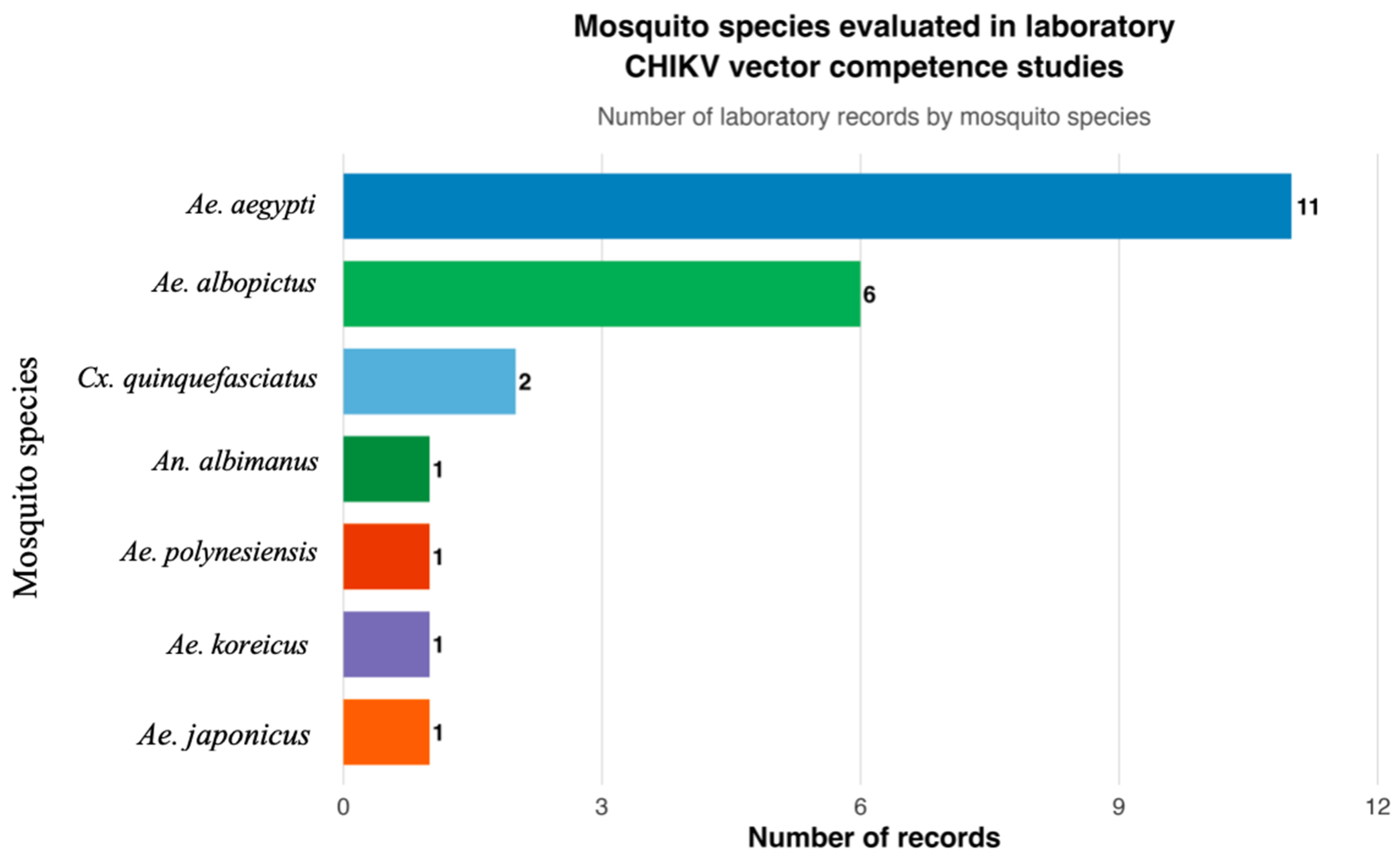
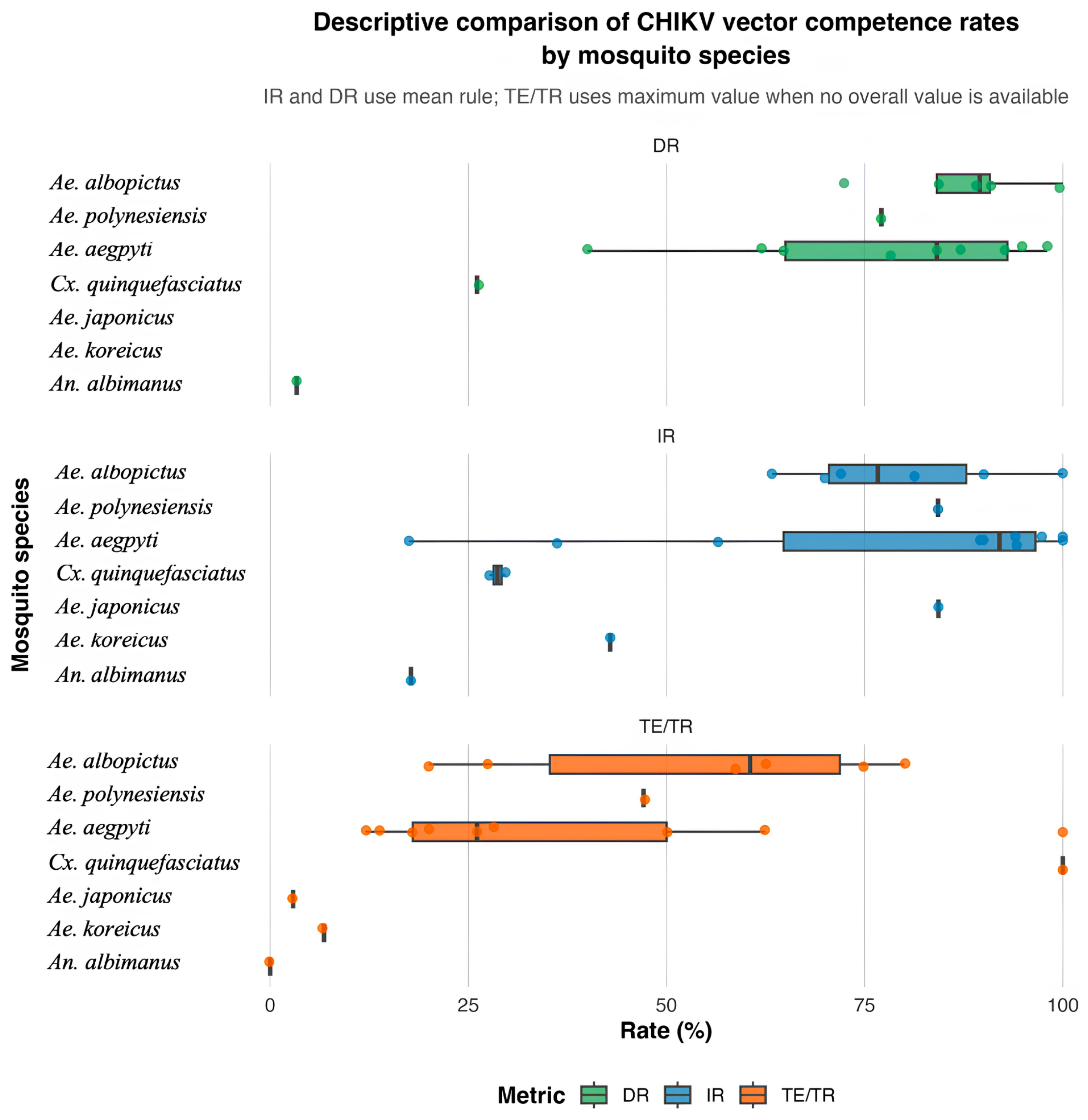
Across the laboratory studies, Ae. aegypti and Ae. albopictus showed the most consistent evidence of CHIKV susceptibility and transmission potential (Table 5). The range of mosquito species evaluated in laboratory vector competence studies is shown in Figure 3, which highlights the predominance of the two principal Aedes vectors among the experimentally tested species. Ae. aegypti was represented in 11 laboratory studies. The calculated mean infection rate was approximately 76%, ranging from 17.5% to 100%. Dissemination data were available in nine studies, with a mean DR of approximately 77% and a range of 25% to 98%. Transmission efficiency (TE), or transmission rate (TR), was reported in nine studies, with a mean of approximately 37%, ranging from 12% to 100%. These findings indicate that Ae. aegypti is generally highly susceptible to CHIKV infection and is frequently capable of supporting viral dissemination under experimental conditions (Table 2 and Figure 4).
Aedes albopictus showed similarly strong vector competence patterns. Among six laboratory studies, all six provided IR data, with a mean infection rate of approximately 79% and a range of 63.3% to 100%. Furthermore, DR was available in five studies, with a mean of approximately 80% and a range of 65% to 90.8%. On the other hand, TE/TR was reported in six studies, with a mean of approximately 47% and a range of 20% to 80%. Compared with Ae. aegypti, Ae. albopictus showed slightly higher mean infection and transmission values in the dataset, whereas the dissemination values were similar. However, this comparison should be interpreted with caution, as the studies differed in terms of virus strain, mosquito origin, incubation period, and experimental design. Nevertheless, the data support Ae. albopictus as a highly competent CHIKV vector, particularly in settings where ECSA or ECSA–Indian Ocean lineage strains are circulating (Table 2, Figure 4).
When the same descriptive values are considered by virus lineage rather than by species alone, a consistent pattern emerges that reflects genotype-by-vector compatibility. ECSA and ECSA-IOL strains, several of which carry the E1-A226V substitution, were associated with the highest transmission efficiencies recorded in the dataset, particularly in Ae. albopictus (for example, ECSA-IOL transmission efficiency peaked around 55% in Tunisian Ae. albopictus with elevated dissemination in European populations), whereas Asian-genotype infections of Ae. aegypti more often produced high infection and dissemination rates but comparatively low infectious saliva transmission (Table 2). West African strains were represented by only a few Ae. aegypti records with modest transmission. A fully quantitative genotype-stratified analysis would be constrained by incomplete genotype reporting and small per-genotype sample sizes (a limitation noted in Section 6); therefore, the values in Table 5 are presented by species, with the genotype indicated per study in Table 2, while the genotype contrasts mentioned above should be read as descriptive rather than statistical. Even so, this pattern reinforces that the compatibility between circulating lineage and locally abundant vectors, rather than by either factor alone, governs outbreak potential in a given region.
Other mosquito species showed more variable or limited evidence. Culex quinquefasciatus was represented in two laboratory studies, with IR values averaging approximately 29%. Dissemination data were limited, with only one study reporting a dissemination rate of 26.1%. Transmission efficiency or transmission rate was extractable from only one study, with maximum values reaching 100%. Although these findings indicate that CHIKV detection or limited transmission-related outcomes may occur under certain experimental conditions, the limited number of studies and inconsistent evidence currently provide insufficient support to establish Cx. quinquefasciatus as an epidemiologically important CHIKV vector.
Among other experimentally evaluated Aedes species, Ae. polynesiensis showed relatively high IR of 84.25%, DR of 77.1%, and TE/TR of 40.7% in the available laboratory records. This suggests that Ae. polynesiensis may be competent for CHIKV under experimental conditions and may have regional importance, particularly in Pacific Island settings. Aedes japonicus also showed a high IR of 84.3%, but the reported TE/TR was very low at 2.9%, indicating that infection alone did not translate into efficient transmission. Aedes koreicus showed a moderate infection rate of 42.9% and low maximum TE/TR of 6.8%, suggesting limited transmission potential based on the available data. Anopheles albimanus showed low IR and DR, with rates of 17.77% and 3.35%, respectively. In addition, there was no transmission detected. These findings support the interpretation that not all mosquito species with detectable infection are epidemiologically important vectors.

5. Detection and Diagnostic Methodologies Used in CHIKV Mosquito Studies

5.1. Molecular Detection

RT-PCR and qPCR were the dominant methods used in field surveillance studies. These methods are suitable for screening mosquito pools and are considered a gold standard for detecting the RNA of arboviruses, showing high efficiency due to its sensitivity and specificity [30,73]. These are especially useful during outbreaks because they can detect CHIKV RNA even when viral loads are low.
However, molecular detection does not confirm virus infection or vector competence. A mosquito may test positive because of a recent blood meal, residual viral RNA, a non-infectious virus, or an abortive infection [74]. Therefore, PCR-positive field results should be interpreted as confirmation of CHIKV detection rather than as direct evidence of transmission.

5.2. Virus Isolation and Infectivity Assays

Virus isolation, plaque assay, focus-forming assay, and cell culture-based methods provide stronger evidence because they indicate viable or infectious viruses. These methods were less common in field surveillance studies but were more frequently used in laboratory vector competence studies [75].
In laboratory studies, infectious virus detection in saliva is particularly important because it directly supports transmission potential. Infection rates and dissemination rates are useful but are less definitive than transmission efficiency [65]. Therefore, laboratory studies that include saliva testing provide stronger evidence for vector competence than studies that assess only body infections (Table 2). Beyond saliva assays, in vivo transmission models offer the most direct confirmation of competence, as experimentally, CHIKV-infected Ae. albopictus has transmitted the virus to naive mice, who subsequently developed viraemia and arthritis [76], while within the present dataset, another study similarly confirmed transmission using an in vivo mouse model [56]. Such animal-based transmission assays remain uncommon but can provide valuable corroboration of saliva-based vector competence metrics.

5.3. Sequencing and Genomic Approaches

Sequencing and next-generation sequencing were used in a smaller number of studies. These approaches are important because they allow for genotype confirmation, outbreak lineage identification, and phylogenetic analysis [77]. The limited use of sequencing contributed to incomplete genotype reporting in field studies (Table 1).
Future field surveillance should incorporate sequencing of CHIKV-positive mosquito pools whenever possible, especially during outbreaks. This would allow for stronger linkage between field detection, virus lineage, and vector species.

6. Limitations

This review has several limitations. Firstly, the literature search was conducted using PubMed and Google Scholar; therefore, some of the regional reports, grey literature, and non-indexed studies may have been missed. In addition, the included studies were heterogeneous in terms of design, mosquito species, sample size, virus strain, diagnostic method, and reporting format.
Many field studies did not report the CHIKV genotype. This limits the analysis of lineage-specific vector associations and regional viral movements. Other than that, most field studies relied on molecular detection without virus isolation. Furthermore, the infection metrics were inconsistently reported across studies. Finally, several non-Aedes and other secondary mosquito species were reported as CHIKV-positive in the field but lacked laboratory confirmation.
These limitations precluded meta-analysis and required a descriptive synthesis. This is important, as mosquito infection estimates can be biassed when pool sizes, sampling intensity, collection timing, and diagnostic sensitivity differ across studies. Even so, the combined evidence provides a useful overview of current CHIKV vector knowledge and identifies priorities for future surveillance and experimental work.

7. Integrated Surveillance and Control of CHIKV Vectors

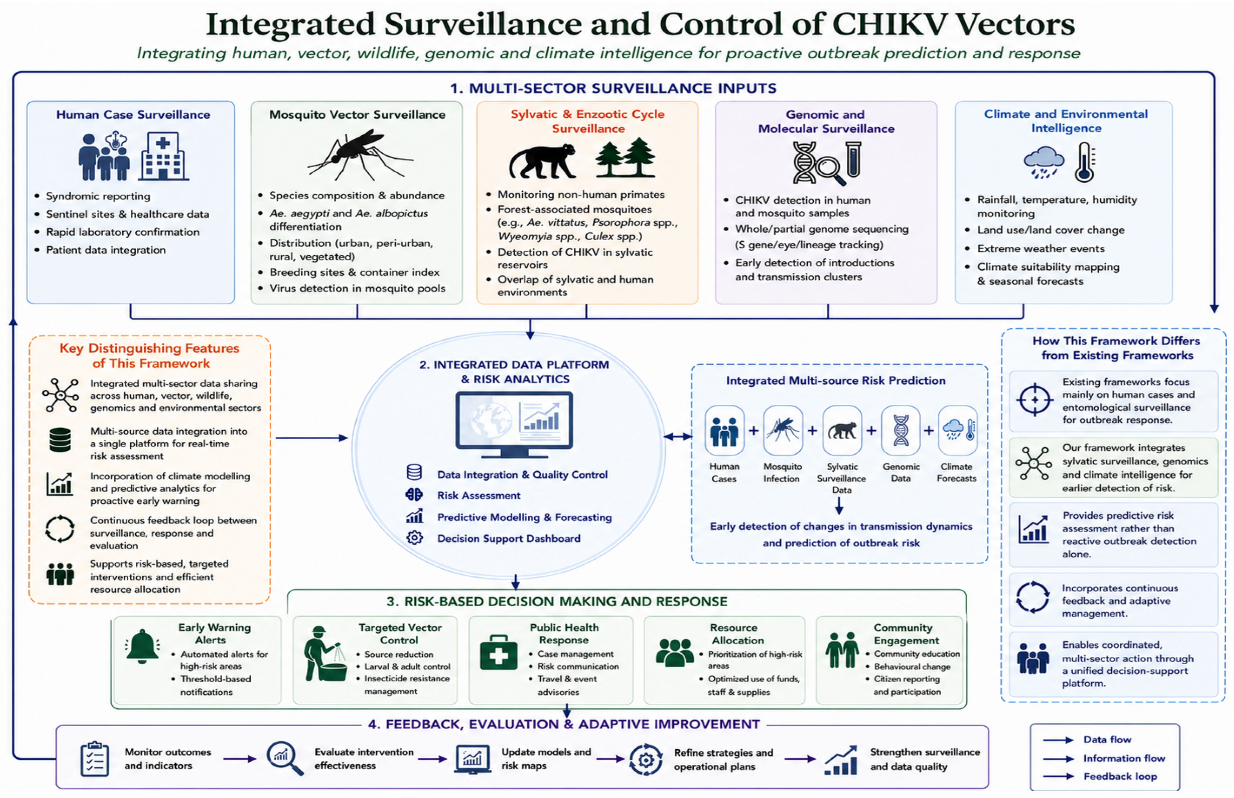
Although CHIKV is primarily recognised as a human arboviral disease, its transmission dynamics are shaped by interactions among humans, mosquitoes, animals, and the environment. Effective preparedness therefore depends on the integration of surveillance and controls to link human-health surveillance, vector surveillance, environmental management, climate monitoring, urban planning, and community-level prevention. A comprehensive integrated framework for CHIKV surveillance and controls is proposed (Figure 5), linking mosquito biology, human infections, environmental conditions, and multi-sectoral activities. The individual components of Figure 5 are not intended to constitute an entirely new surveillance system. Many are already included, to varying degrees, in established arboviral surveillance, integrated vector management, and One Health strategies. For example, existing frameworks commonly emphasise human case detection, laboratory confirmation, entomological surveillance, source reduction, and insecticide-based vector control. The framework proposed here complements these approaches by bringing together the specific evidence synthesised in this review within a single CHIKV-focused structure. In particular, it links the field detection of CHIKV in mosquito populations with the laboratory evidence of vector competence, viral genotype information, environmental and climate indicators, urban infrastructure, and community-level prevention. It also highlights the need for information to move continuously from surveillance to risk assessment, targeted intervention, and subsequent evaluation. This integrated evidence-to-action pathway represents the main distinguishing feature of Figure 5, instead of considering any single surveillance component in isolation. The proposed framework is therefore intended to complement, rather than replace, existing integrated approaches, including the One Health control strategy proposed by Fang et al. [78].
The reviewed evidence highlights the role of mosquito vectors as the central link between environmental conditions and human infection. Aedes aegypti is closely associated with human dwellings, artificial containers, poor water storage practices, and dense urban settlements [68]. These characteristics make CHIKV transmission strongly dependent on human behaviour and the built environment. In contrast, Ae. albopictus can occupy peri-urban, rural, vegetated, and temperate habitats, linking CHIKV risk to broader environmental and ecological changes [68,69]. Therefore, vector control cannot rely solely on insecticide application but must also include environmental sanitation, removal of breeding containers, water management, community engagement, and urban planning to reduce mosquito breeding opportunities.
Climate and environmental changes are also important drivers of CHIKV risk. Temperature, rainfall, humidity, and land-use changes influence mosquito abundance, survival, biting activity, and viral replication [78]. Laboratory vector competence studies show that temperature and extrinsic incubation period can affect infection, dissemination, and transmission outcomes. Warmer conditions may shorten the extrinsic incubation period and increase the chance that mosquitoes become infectious within their lifespan, while changes in rainfall may create more larval habitats [79]. These factors can expand the geographical range and seasonal window of CHIKV transmission, particularly in areas where Ae. albopictus and other invasive mosquitoes are established. Modelling under climate change scenarios projects poleward shifts in Aedes-borne transmission risks, with optimal temperature ranges of 21.3–34.0 °C for Ae. aegypti and 19.9–29.4 °C for Ae. albopictus, as well as the potential exposure of nearly a billion additional people by 2080 (Table 2).
Urban transmission is mainly driven by human–Aedes–human cycles involving Ae. aegypti and Ae. albopictus. However, in some regions, especially in Africa, CHIKV may also be maintained in sylvatic or enzootic cycles involving non-human primates and forest-associated mosquitoes [78,80]. The detection of CHIKV RNA in non-Aedes and other secondary mosquito species, such as Ae. vittatus, Psorophora spp., Wyeomyia spp., Culex spp., and other culicids, suggests that mosquito exposure may occur beyond strictly urban vectors. Although these detections do not confirm vector competence, they indicate that broader ecological surveillance may be necessary in areas where human settlements overlap with forest-associated, peri-domestic, or wildlife-associated habitats. In addition, a full treatment of enzootic maintenance, non-human primate reservoirs, and the vector ecology of forest-associated Aedes (for example, Ae. furcifer and Ae. luteocephalus) would require dedicated sylvatic surveillance, and remains a priority for future work.
Therefore, integrated surveillance is essential to monitor the potential outbreaks of CHIKV. Human case surveillance alone may detect outbreaks only after transmission is already established [81]. Mosquito surveillance can provide earlier warning of viral circulation, particularly when combined with molecular detection, virus isolation, sequencing, and vector abundance monitoring. Environmental surveillance, such as monitoring rainfall patterns, temperature anomalies, land-use changes, and mosquito breeding habitats, can help identify areas at increased risk. Together, these data streams can support more timely and targeted interventions.
Integrated CHIKV control also depends on collaboration across sectors. Public health authorities, entomologists, veterinarians, environmental scientists, urban planners, local governments, and communities all play important roles in CHIKV prevention. Public health teams are needed to detect and report human cases. Entomologists are required to identify vector species, assess vector abundance, and evaluate insecticide resistance. Beyond taxonomic identification, entomologists are also responsible for determining vector hotspots, actively collecting vectors in the field, and instructing and supervising mosquito-control operators in the treatment of breeding sites and resting mosquitoes. Environmental and municipal sectors are needed to manage waste, water storage, drainage, and urban breeding sites. Community participation is also essential, because many Aedes breeding habitats occur in household or peri-household environments.
The findings of this review support the need for integrated CHIKV preparedness. Surveillance should prioritise confirmed vectors, such as Ae. aegypti and Ae. albopictus, but broader mosquito sampling may be useful during outbreaks or in ecologically complex settings. Positive mosquito pools should be subjected to virus isolation and sequencing whenever possible to determine whether infectious viruses are present and which genotype is circulating. Climate and environmental data should be integrated with entomological and epidemiological data to better predict outbreak risk.
Overall, the proposed framework differs from surveillance approaches that treat clinical, entomological, environmental and control activities as largely separate operational streams by explicitly connecting them within a continuous cycle of data integration, risk assessment, response, and evaluation. Its contribution lies in synthesising the evidence reviewed here into a CHIKV-specific, multi-sector structure that can support both routine preparedness and outbreak response. Such coordination is increasingly important as urbanisation, climate change, global travel, and vector expansion alter the geographical landscape of CHIKV transmission. The movement of viraemic travellers, the international trade in used tyres associated with the dispersal of Ae. albopictus, and the continuing establishment of this vector in temperate regions create pathways for cross-border transmission. The introduction of compatible CHIKV variants into areas with established and competent Ae. albopictus populations therefore represents an important risk that internationally coordinated surveillance should address. Recent autochthonous transmission in European settings further demonstrates that surveillance should integrate imported human cases, local vector distributions, viral genotype information, and environmental suitability instead of considering these indicators independently.

8. Conclusions and Future Directions

This review identifies Ae. aegypti and Ae. albopictus as the principal CHIKV vectors, as supported by repeated field detections and laboratory vector competence evidence. Aedes aegypti remains central to urban transmission, whereas Ae. albopictus is important due to its ecological flexibility and continuing expansion into peri-urban and temperate environments.
Other Aedes species, including Ae. vittatus, Ae. polynesiensis, Ae. koreicus, and Ae. japonicus, may have regional or emerging relevance but require further validation. Although Ae. vittatus has demonstrated laboratory competence in East African populations [67], its role in West Africa and other regions remains insufficiently defined. CHIKV RNA detection in Culex, Anopheles, Psorophora, Wyeomyia, and other culicids indicates broader mosquito exposure, but it does not confirm vector competence [42,60,82].
Future CHIKV vector studies should integrate field surveillance with laboratory vector competence assays. Field-positive mosquito pools should be prioritised for virus isolation and sequencing to confirm viral viability, genotypes, and epidemiological relevance. Laboratory studies should use local mosquito populations and locally circulating CHIKV strains whenever possible, and should assess infection, dissemination, and infectious saliva production rather than relying only on infection positivity.
Standardised reporting is also essential. Future studies should clearly report the mosquito species, collection sites, sample sizes, pool sizes, infection rates, minimum infection rates, virus genotypes, detection methods, extrinsic incubation periods, incubation temperatures, and transmission efficiency. Minimum data standards for vector competence experiments, such as those proposed by Wu et al. [83], would improve comparability across CHIKV mosquito studies.
Overall, future CHIKV preparedness requires an integrated framework that brings together human case surveillance, mosquito monitoring, environmental risk assessment, genomic sequencing, community participation, and standardised vector competence assays. This integrated approach would clarify mosquito vector roles, strengthen early-warning systems, and improve targeted prevention of future CHIKV outbreaks.

Author Contributions

Conceptualisation, V.L.L. and W.Y.V.-S.; methodology, W.Y.V.-S., T.K.T., S.S.S., B.T.T. and Y.A.-L.L.; validation, T.K.T., S.S.S., B.T.T. and Y.A.-L.L.; formal analysis, W.Y.V.-S.; investigation, W.Y.V.-S.; resources, V.L.L.; data curation, W.Y.V.-S.; writing—original draft preparation, W.Y.V.-S.; writing—review and editing, W.Y.V.-S., T.K.T., S.S.S., Y.A.-L.L., B.T.T. and V.L.L.; visualisation, W.Y.V.-S.; supervision, V.L.L.; project administration, V.L.L.; funding acquisition, V.L.L. All authors have read and agreed to the published version of the manuscript.

Funding

This study was financially supported by the Kementerian Pendidikan Malaysia, under the Fundamental Research Grant Scheme (Reference code: FRGS/1/2024/SKK13/UM/02/3; Project code: FP066-2024) and Higher Institution Centre of Excellence (HICoE) programme (MO002-2019 and TIDREC-2023).

Data Availability Statement

No new datasets were generated or analysed during the current study. All data discussed in this review were obtained from previously published studies cited in the manuscript.

Acknowledgments

During the preparation of this manuscript, the authors used Gemini (Google) to assist in generating the graphical abstract for Figure 5, which visually summarises the One Health framework for CHIKV surveillance and control. The authors have reviewed and edited the output of this tool and take full responsibility for the content of this publication.

Conflicts of Interest

The authors declare no conflicts of interest. The funders had no role in the design of the study; in the collection, analyses, or interpretation of data; in the writing of the manuscript; or in the decision to publish the results.

Abbreviations

The following abbreviations are used in this manuscript:
AbbreviationDefinition
Ae.Aedes
An.Anopheles
CHIKVChikungunya Virus
Cs.Culiseta
Cx.Culex
DRDissemination Rate
DRCDemocratic Republic of the Congo
ECSAEast/Central/South African Genotype
ECSA-IOLEast/Central/South African–Indian Ocean Lineage
EIPExtrinsic Incubation Period
Hg.Haemagogus
HICoEHigher Institution Centre of Excellence
IFAImmunofluorescence assay
IOLIndian Ocean Lineage
IRInfection Rate
MIRMinimum Infection Rate
NANot Applicable
NGSNext-Generation Sequencing
NRNot Recorded
PCRPolymerase Chain Reaction
Ps.Psorophora
qPCRQuantitative Polymerase Chain Reaction
RT-PCRReverse Transcription Polymerase Chain Reaction
RT-qPCRReverse Transcription Quantitative Polymerase Chain Reaction
TETransmission Efficiency
TIDRECTropical Infectious Diseases Research and Education Centre
TRTransmission Rate
UMUniversiti Malaya
WHOWorld Health Organization
Wy.Wyeomyia

References

  1. World Health Organization. Chikungunya Epidemiology Update, June 2025; WHO: Geneva, Switzerland, 2025; Available online: https://www.who.int/publications/m/item/chikungunya-epidemiology-update-june-2025 (accessed on 15 July 2026).
  2. Wimalasiri-Yapa, B.M.C.R.; Stassen, L.; Huang, X.; Hafner, L.M.; Hu, W.; Devine, G.J.; Yakob, L.; Jansen, C.C.; Faddy, H.M.; Viennet, E.; et al. Chikungunya virus in Asia—Pacific: A systematic review. Emerg. Microbes Infect. 2019, 8, 70–79. [Google Scholar] [CrossRef] [PubMed]
  3. Madewell, Z.J. Arboviruses and Their Vectors. South. Med. J. 2020, 113, 520–523. [Google Scholar] [CrossRef] [PubMed]
  4. Wang, S.; Liu, Y.; Wang, Y.; Zhou, L.; Liu, J. Global and regional burden of chikungunya from 2004 to 2024: A worldwide observational study. J. Glob. Health 2026, 16, 04055. [Google Scholar] [CrossRef] [PubMed]
  5. Kraemer, M.U.G.; Reiner, R.C.; Brady, O.J.; Messina, J.P.; Gilbert, M.; Pigott, D.M.; Yi, D.; Johnson, K.; Earl, L.; Marczak, L.B.; et al. Past and future spread of the arbovirus vectors Aedes aegypti and Aedes albopictus. Nat. Microbiol. 2019, 4, 854–863. [Google Scholar] [CrossRef] [PubMed]
  6. Weaver, S.C.; Chen, R.; Diallo, M. Chikungunya Virus: Role of Vectors in Emergence from Enzootic Cycles. Annu. Rev. Entomol. 2020, 65, 313–332. [Google Scholar] [CrossRef] [PubMed]
  7. Bohers, C.; Mousson, L.; Madec, Y.; Vazeille, M.; Rhim, A.; M’ghirbi, Y.; Bouattour, A.; Failloux, A.B. The recently introduced Aedes albopictus in Tunisia has the potential to transmit chikungunya, dengue and Zika viruses. PLoS Neglected Trop. Dis. 2020, 14, e0008475. [Google Scholar] [CrossRef] [PubMed]
  8. Rodrigues, N.B.; Godoy, R.S.M.; Orfano, A.S.; Chaves, B.A.; Campolina, T.B.; Costa, B.D.A.; Félix, L.D.S.; Silva, B.M.; Norris, D.E.; Pimenta, P.F.P.; et al. Brazilian Aedes aegypti as a Competent Vector for Multiple Complex Arboviral Coinfections. J. Infect. Dis. 2021, 224, 101–108. [Google Scholar] [CrossRef] [PubMed]
  9. Lühken, R.; Rauhöft, L.; Pluskota, B.; Lange, U.; Helms, M.; Becker, N.; Schmidt-Chanasit, J.; Kuhn, C.; Tannich, E.; Jansen, S.; et al. High vector competence for chikungunya virus but heavily reduced locomotor activity of Aedes albopictus from Germany at low temperatures. Parasites Vectors 2024, 17, 502. [Google Scholar] [CrossRef] [PubMed]
  10. Tsetsarkin, K.A.; Vanlandingham, D.L.; McGee, C.E.; Higgs, S. A Single Mutation in Chikungunya Virus Affects Vector Specificity and Epidemic Potential. PLoS Pathog. 2007, 3, e201. [Google Scholar] [CrossRef] [PubMed]
  11. Santos, R.A.; Abreu, F.V.S.; Bello, G.; Fonseca, V.; Lourenco-de-Oliveira, R. Spatial-temporal distribution of chikungunya virus in Brazil: A review on the circulating viral genotypes and Aedes (Stegomyia) albopictus as a potential vector. Front. Public Health 2024, 12, 1496021. [Google Scholar] [CrossRef] [PubMed]
  12. Gu, W.; Novak, R.J. Short Report: Detection Probability of Arbovirus Infection in Mosquito Populations. Am. J. Trop. Med. Hyg. 2004, 71, 636–638. [Google Scholar] [CrossRef]
  13. Gu, W.; Unnasch, T.R.; Katholi, C.R.; Lampman, R.; Novak, R.J. Fundamental issues in mosquito surveillance for arboviral transmission. Trans. R. Soc. Trop. Med. Hyg. 2008, 102, 817–822. [Google Scholar] [CrossRef] [PubMed]
  14. Bustamante, D.M.; Lord, C.C. Sources of Error in the Estimation of Mosquito Infection Rates Used to Assess Risk of Arbovirus Transmission. Am. J. Trop. Med. Hyg. 2010, 82, 1172–1184. [Google Scholar] [CrossRef] [PubMed]
  15. Lutomiah, J.; Mulwa, F.; Mutisya, J.; Koskei, E.; Langat, S.; Nyunja, A.; Koka, H.; Konongoi, S.; Chepkorir, E.; Ofula, V.; et al. Probable contribution of Culex quinquefasciatus mosquitoes to the circulation of chikungunya virus during an outbreak in Mombasa County, Kenya, 2017–2018. Parasites Vectors 2021, 14, 138. [Google Scholar] [CrossRef] [PubMed]
  16. Phumee, A.; Intayot, P.; Sor-Suwan, S.; Jittmittraphap, A.; Siriyasatien, P. Molecular detection of Indian Ocean Lineage Chikungunya virus RNA in field collected Culex quinquefasciatus Say from Bangkok, Thailand but no evidence of virus replication. PLoS ONE 2021, 16, e0246026. [Google Scholar] [CrossRef] [PubMed]
  17. Cruz, R.A.C.; Pinto Nunes Neto, J.; Patroca da Silva, S.; Vieira Pinto da Silva, E.; Juscely Galvão Pereira, G.; Maia Santos, M.; Antônio de Oliveira Monteiro, H.; Barreto Dos Santos, F.; José de Paula Souza E Guimarães, R.; Fortes Aragão, C.; et al. Chikungunya Virus Detection in Aedes aegypti and Culex quinquefasciatus During an Outbreak in the Amazon Region. Viruses 2020, 12, 853. [Google Scholar] [CrossRef] [PubMed]
  18. Teixeira, A.F.; de Brito, B.B.; Correia, T.M.L.; Viana, A.I.S.; Carvalho, J.C.; da Silva, F.A.F.; Santos, M.L.C.; da Silveira, E.A.; Neto, H.P.G.; da Silva, N.M.P.; et al. Simultaneous circulation of Zika, Dengue, and Chikungunya viruses and their vertical co-transmission among Aedes aegypti. Acta Trop. 2021, 215, 105819. [Google Scholar] [CrossRef] [PubMed]
  19. Almeida-Souza, P.A.; Oliveira, C.H.; Brito, L.P.; Teixeira, T.J.; Celestino, I.A.; Penha, G.B.; Dos Santos, R.M.; Mendes, W.M.; Ribeiro, B.M.; Campos, F.S.; et al. High Frequencies of kdr Mutation and Chikungunya Infection in Aedes aegypti Population from Minas Gerais, Brazil. Pathogens 2024, 13, 457. [Google Scholar] [CrossRef] [PubMed]
  20. Banho, C.A.; Parra, M.C.P.; Nascimento, O.B.; Magnani, G.P.; Ferreira, M.V.M.; Lemos, A.P.; de Carvalho Marques, B.; Brancini, M.L.; Sacchetto, L.; Negri, A.F.; et al. Entomological surveillance during a major CHIKV outbreak in northwestern São Paulo: Insights from São José do Rio Preto. medRxiv Prepr. Serv. Health Sci. 2024. [Google Scholar] [CrossRef] [PubMed]
  21. de Melo Ximenes, M.F.F.; de Araújo Galvão, J.M.; Inacio, C.L.S.; Macêdo E Silva, V.P.; Pereira, R.L.D.N.; Pinheiro, M.P.G.; de Medeiros Silva, M.M.; Gomes, C.E.S. Arbovirus expansion: New species of culicids infected by the Chikungunya virus in an urban park of Brazil. Acta Trop. 2020, 209, 105538. [Google Scholar] [CrossRef] [PubMed]
  22. Ortega-López, L.D.; Pondeville, E.; Kohl, A.; León, R.; Betancourth, M.P.; Almire, F.; Torres-Valencia, S.; Saldarriaga, S.; Mirzai, N.; Ferguson, H.M. The mosquito electrocuting trap as an exposure-free method for measuring human-biting rates by Aedes mosquito vectors. Parasites Vectors 2020, 13, 31. [Google Scholar] [CrossRef] [PubMed]
  23. Herrera, J.J.B.; Montalvo, J.P.M.; Tutillo, P.A.Q.; Narváez, F.A.P.; Viteri, D.O.M. Entomological surveillance in active dengue transmission areas of Santo Domingo de los Tsáchilas—Ecuador: Epidemiological role of Aedes aegypti and the recent establishment of Aedes albopictus. Acta Trop. 2026, 274, 107977. [Google Scholar] [CrossRef] [PubMed]
  24. Carrasquilla, M.C.; Ortiz, M.I.; León, C.; Rondón, S.; Kulkarni, M.A.; Talbot, B.; Sander, B.; Vásquez, H.; Cordovez, J.M.; González, C.; et al. Entomological characterization of Aedes mosquitoes and arbovirus detection in Ibagué, a Colombian city with co-circulation of Zika, dengue and chikungunya viruses. Parasites Vectors 2021, 14, 446. [Google Scholar] [CrossRef] [PubMed]
  25. Mantilla-Granados, J.S.; Montilla-López, K.; Sarmiento-Senior, D.; Chapal-Arcos, E.; Velandia-Romero, M.L.; Calvo, E.; Morales, C.A.; Castellanos, J.E. Environmental and anthropic factors influencing Aedes aegypti and Aedes albopictus (Diptera: Culicidae), with emphasis on natural infection and dissemination: Implications for an emerging vector in Colombia. PLoS Neglected Trop. Dis. 2025, 19, e0012605. [Google Scholar] [CrossRef] [PubMed]
  26. Kirstein, O.D.; Ayora-Talavera, G.; Koyoc-Cardeña, E.; Chan Espinoza, D.; Che-Mendoza, A.; Cohuo-Rodriguez, A.; Granja-Pérez, P.; Puerta-Guardo, H.; Pavia-Ruz, N.; Dunbar, M.W.; et al. Natural arbovirus infection rate and detectability of indoor female Aedes aegypti from Mérida, Yucatán, Mexico. PLoS Neglected Trop. Dis. 2021, 15, e0008972. [Google Scholar] [CrossRef] [PubMed]
  27. Hernández-Acosta, E.; Covarrubias, A.M.; Fernández-Salas, I.; Escárcega-Ávila, A.; Olivera, I.P.; Jiménez-Vega, F. Detection of chikungunya virus in Aedes aegypti in Ciudad Juárez, Chihuahua, Mexico. J. Vector Borne Dis. 2025, 62, 226–232. [Google Scholar] [CrossRef] [PubMed]
  28. Musili, F.M.; Mutisya, J.; Langat, S.; Chelangat, B.; Anyango, V.; Chepkorir, E.; Konongoi, S.; Sang, R.; Bastos, A.; Lutomiah, J. Role of invasive Aedes (Aedimorphus) vittatus in chikungunya virus transmission in the newly-invaded coastal island city of Mombasa, Kenya. PLoS Neglected Trop. Dis. 2025, 19, e0013738. [Google Scholar] [CrossRef] [PubMed]
  29. Heath, C.J.; Grossi-Soyster, E.N.; Ndenga, B.A.; Mutuku, F.M.; Sahoo, M.K.; Ngugi, H.N.; Mbakaya, J.O.; Siema, P.; Kitron, U.; Zahiri, N.; et al. Evidence of transovarial transmission of Chikungunya and Dengue viruses in field-caught mosquitoes in Kenya. PLoS Neglected Trop. Dis. 2020, 14, e0008362. [Google Scholar] [CrossRef] [PubMed]
  30. Gomgnimbou, M.K.; Belem, L.R.W.; Bilgo, E.; Amara, M.F.; Laouali, Z.; Ouari, A.; Bayala, T.; Gnada, K.; Yao, R.K.; Namountougou, M.; et al. Potential Performance of Two New RT-PCR and RT-qPCR Methods for Multiplex Detection of Dengue Virus Serotypes 1–4 and Chikungunya Virus in Mosquitoes. Curr. Issues Mol. Biol. 2024, 46, 11048–11056. [Google Scholar] [CrossRef] [PubMed]
  31. Laouali, Z.; Kagoné, H.; Kagoné, T.; Belem, L.R.W.; Konaté, H.; Ouari, A.; Zango, A.; Ouedraogo, S.; Yao, R.K.; Diao, W.R.; et al. Strengthening Arboviral Epidemic Response Through Entomological Surveillance: Insights from Bobo-Dioulasso, Burkina Faso. Curr. Issues Mol. Biol. 2026, 48, 78. [Google Scholar] [CrossRef] [PubMed]
  32. Toé, H.K.; Zongo, S.; Ouédraogo, W.M.; Sana, Z.; Zango, A.; Nana, J.B.; Sombié, A.; Laquera, Z.; Kafando, L.; Ouédraogo, H.S.; et al. Entomological investigation of the 2023 chikungunya outbreak in Pouytenga, Burkina Faso, highlights the virus circulation in Aedes aegypti. Curr. Res. Parasitol. Vector-Borne Dis. 2026, 9, 100358. [Google Scholar] [CrossRef] [PubMed]
  33. Vairo, F.; Aimè Coussoud-Mavoungou, M.P.; Ntoumi, F.; Castilletti, C.; Kitembo, L.; Haider, N.; Carletti, F.; Colavita, F.; Gruber, C.; Iannetta, M.; et al. Chikungunya Outbreak in the Republic of the Congo, 2019—Epidemiological, Virological and entomological Findings of a South-North Multidisciplinary Taskforce Investigation. Viruses 2020, 12, 1020. [Google Scholar] [CrossRef] [PubMed]
  34. De Weggheleire, A.; Nkuba-Ndaye, A.; Mbala-Kingebeni, P.; Mariën, J.; Kindombe-Luzolo, E.; Ilombe, G.; Mangala-Sonzi, D.; Binene-Mbuka, G.; De Smet, B.; Vogt, F.; et al. A Multidisciplinary Investigation of the First Chikungunya Virus Outbreak in Matadi in the Democratic Republic of the Congo. Viruses 2021, 13, 1988. [Google Scholar] [CrossRef] [PubMed]
  35. Joannides, J.; Dzodzomenyo, M.; Azerigyik, F.; Agbosu, E.E.; Pratt, D.; Nyarko Osei, J.H.; Pwalia, R.; Amlalo, G.K.; Appawu, M.; Takashi, H.; et al. Species composition and risk of transmission of some Aedes-borne arboviruses in some sites in Northern Ghana. PLoS ONE 2021, 16, e0234675. [Google Scholar] [CrossRef] [PubMed]
  36. Akyea-Bobi, N.E.; Akorli, J.; Opoku, M.; Akporh, S.S.; Amlalo, G.K.; Osei, J.H.N.; Frempong, K.K.; Pi-Bansa, S.; Boakye, H.A.; Abudu, M.; et al. Entomological risk assessment for transmission of arboviral diseases by Aedes mosquitoes in a domestic and forest site in Accra, Ghana. PLoS ONE 2023, 18, e0295390. [Google Scholar] [CrossRef] [PubMed]
  37. Guarido, M.M.; Fourie, I.; Meno, K.; Mendes, A.; Riddin, M.A.; MacIntyre, C.; Manyana, S.; Johnson, T.; Schrama, M.; Gorsich, E.E.; et al. Alphaviruses Detected in Mosquitoes in the North-Eastern Regions of South Africa, 2014 to 2018. Viruses 2023, 15, 414. [Google Scholar] [CrossRef] [PubMed]
  38. Nwangwu, U.C.; Soumare, H.; Oguzie, J.U.; Nwachukwu, W.E.; Onwude, C.O.; Dogunro, F.A.; Diallo, M.; Ezihe, C.K.; Agashi, N.O.; Eloy, E.I.; et al. Nationwide surveillance detects yellow fever and chikungunya viruses in multiple Aedes mosquito species in Nigeria. Parasites Vectors 2025, 18, 443. [Google Scholar] [CrossRef] [PubMed]
  39. Chand, G.; Godbole, S.; Shivlata, L.; Sahare, L.K.; Ukey, M.; Kaushal, L.S.; Barde, P.V. Molecular xenomonitoring of Dengue, Chikungunya and Zika infections: A year-round study from two Dengue endemic districts of central India. J. Vector Borne Dis. 2021, 58, 135–140. [Google Scholar] [CrossRef] [PubMed]
  40. Munivenkatappa, A.; Nyayanit, D.A.; Yadav, P.D.; Rangappa, M.; Patil, S.; Majumdar, T.; Mohandas, S.; Sinha, D.P.; Jayaswamy, M.M.; OmPrakash, P. Identification Of Phasi Charoen-Like Phasivirus in Field Collected Aedes aegypti from Karnataka State, India. Vector Borne Zoonotic Dis. 2021, 21, 900–909. [Google Scholar] [CrossRef] [PubMed]
  41. Vikram, K.; Nagpal, B.N.; Gupta, S.K.; Tuli, N.R.; Singh, H.; Srivastava, A.; Saxena, R. Co-distribution of dengue and Chikungunya viruses in Aedes mosquitoes of Delhi, India. J. Vector Borne Dis. 2021, 58, 386–390. [Google Scholar] [CrossRef] [PubMed]
  42. Bakhshi, H.; Mousson, L.; Moutailler, S.; Vazeille, M.; Piorkowski, G.; Zakeri, S.; Raz, A.; de Lamballerie, X.; Dinparast-Djadid, N.; Failloux, A.B. Detection of arboviruses in mosquitoes: Evidence of circulation of chikungunya virus in Iran. PLoS Neglected Trop. Dis. 2020, 14, e0008135. [Google Scholar] [CrossRef] [PubMed]
  43. Abbasi, E. Metagenomic surveillance of emerging viruses in mosquito populations from high-risk regions of Iran. J. Virol. Methods 2026, 340, 115301. [Google Scholar] [CrossRef] [PubMed]
  44. Zhou, X.; Xie, X.; Wang, W.; Gao, H.; Wang, K.; Liu, X.; Chen, X.; Jiang, Y.; Yu, H.; Xing, D.; et al. First whole-genome chikungunya virus sequence detected in mosquitoes during the 2025 Foshan outbreak: Evidence of field vector infection and transmission potential in China. Infect. Dis. Poverty 2025, 14, 108. [Google Scholar] [CrossRef] [PubMed]
  45. Haddad, N.; Mousson, L.; Vazeille, M.; Chamat, S.; Tayeh, J.; Osta, M.A.; Failloux, A.B. Aedes albopictus in Lebanon, a potential risk of arboviruses outbreak. BMC Infect. Dis. 2012, 12, 300. [Google Scholar] [CrossRef] [PubMed]
  46. Bohers, C.; Vazeille, M.; Bernaoui, L.; Pascalin, L.; Meignan, K.; Mousson, L.; Jakerian, G.; Karch, A.; de Lamballerie, X.; Failloux, A.B. Aedes albopictus is a competent vector of five arboviruses affecting human health, greater Paris, France, 2023. Euro Surveill. 2024, 29, 2400271. [Google Scholar] [CrossRef] [PubMed]
  47. Visser, T.M.; Wang, H.D.; Abbo, S.R.; Vogels, C.B.F.; Koenraadt, C.J.M.; Pijlman, G.P. Effect of chikungunya, Mayaro and Una virus coinfection on vector competence of Aedes aegypti mosquitoes. One Health 2025, 20, 100991. [Google Scholar] [CrossRef] [PubMed]
  48. da Silva Neves, N.A.; da Silva Ferreira, R.; Morais, D.O.; Pavon, J.A.R.; de Pinho, J.B.; Slhessarenko, R.D. Chikungunya, Zika, Mayaro, and Equine Encephalitis virus detection in adult Culicinae from South Central Mato Grosso, Brazil, during the rainy season of 2018. Braz. J. Microbiol. [Publ. Braz. Soc. Microbiol.] 2022, 53, 63–70. [Google Scholar] [CrossRef] [PubMed]
  49. Leandro, A.S.; Lopes, R.D.; Amaral Martins, C.; Delai, R.M.; Villela, D.A.M.; Maciel-de-Freitas, R. Entomo-virological surveillance followed by serological active survey of symptomatic individuals is helpful to identify hotspots of early arbovirus transmission. Front. Public Health 2022, 10, 1024187. [Google Scholar] [CrossRef] [PubMed]
  50. Silva, D.M.F.D.; Curcio, J.S.; Silva, L.D.C.; Sousa, F.B.; Anunciação, C.E.; Furlaneto, S.M.S.; Silva, V.P.S.M.; Garcia-Zapata, M.T.A.; Silveira-Lacerda, E.P. Detection of arboviruses in Aedes aegypti through transovarian analysis: A study in Goiânia, Goiás. Rev. Da Soc. Bras. De Med. Trop. 2024, 57, e004002023. [Google Scholar] [CrossRef] [PubMed]
  51. Banho, C.A.; Parra, M.C.P.; Nascimento, O.B.; Magnani, G.P.; Ferreira, M.V.M.; Lemos, A.P.; Lourenço, K.L.; de Carvalho Marques, B.; Brancini, M.L.; Sacchetto, L.; et al. Vector surveillance during a major chikungunya outbreak in northwestern São Paulo state, Brazil. npj Viruses 2025, 3, 78. [Google Scholar] [CrossRef] [PubMed]
  52. Robison, A.; Young, M.C.; Byas, A.D.; Rückert, C.; Ebel, G.D. Comparison of Chikungunya Virus and Zika Virus Replication and Transmission Dynamics in Aedes aegypti Mosquitoes. Am. J. Trop. Med. Hyg. 2020, 103, 869–875. [Google Scholar] [CrossRef] [PubMed]
  53. Calvez, E.; Pocquet, N.; Malau, A.; Kilama, S.; Taugamoa, A.; Labrousse, D.; Boussès, P.; Failloux, A.B.; Dupont-Rouzeyrol, M.; Mathieu-Daudé, F. Assessing entomological risk factors for arboviral disease transmission in the French Territory of the Wallis and Futuna Islands. PLoS Neglected Trop. Dis. 2020, 14, e0008250. [Google Scholar] [CrossRef] [PubMed]
  54. Shin, D.; Kang, S.; Smartt, C.T. Profiling Transcripts of Vector Competence between Two Different Aedes aegypti Populations in Florida. Viruses 2020, 12, 823. [Google Scholar] [CrossRef] [PubMed]
  55. Gutiérrez-Bugallo, G.; Boullis, A.; Martinez, Y.; Hery, L.; Rodríguez, M.; Bisset, J.A.; Vega-Rúa, A. Vector competence of Aedes aegypti from Havana, Cuba, for Dengue virus type 1, Chikungunya, and Zika viruses. PLoS Neglected Trop. Dis. 2020, 14, e0008941. [Google Scholar] [CrossRef] [PubMed]
  56. Kaczmarek, M.E.; Herzog, N.L.; Noval, M.G.; Zuzworsky, J.; Shah, Z.; Bajwa, W.I.; Stapleford, K.A. Distinct New York City Aedes albopictus Mosquito Populations Display Differences in Salivary Gland Protein d7 diversity and Chikungunya Virus Replication. Viruses 2020, 12, 698. [Google Scholar] [CrossRef] [PubMed]
  57. Gloria-Soria, A.; Payne, A.F.; Bialosuknia, S.M.; Stout, J.; Mathias, N.; Eastwood, G.; Ciota, A.T.; Kramer, L.D.; Armstrong, P.M. Vector Competence of Aedes albopictus Populations from the Northeastern United States for Chikungunya, Dengue, and Zika Viruses. Am. J. Trop. Med. Hyg. 2020, 104, 1123–1130. [Google Scholar] [CrossRef] [PubMed]
  58. Jansen, S.; Cadar, D.; Lühken, R.; Pfitzner, W.P.; Jöst, H.; Oerther, S.; Helms, M.; Zibrat, B.; Kliemke, K.; Becker, N.; et al. Vector Competence of the Invasive Mosquito Species Aedes koreicus for Arboviruses and Interference with a Novel Insect Specific Virus. Viruses 2021, 13, 2507. [Google Scholar] [CrossRef] [PubMed]
  59. Calvez, E.; Miot, E.F.; Keosenhom, S.; Vungkyly, V.; Viengphouthong, S.; Bounmany, P.; Brey, P.T.; Marcombe, S.; Grandadam, M. Low Transmission of Chikungunya Virus by Aedes aegypti from Vientiane Capital, Lao PDR. Pathogens 2022, 12, 31. [Google Scholar] [CrossRef] [PubMed]
  60. Terradas, G.; Novelo, M.; Metz, H.; Brustolin, M.; Rasgon, J.L. Anopheles albimanus is a Potential Alphavirus Vector in the Americas. Am. J. Trop. Med. Hyg. 2023, 108, 412–423. [Google Scholar] [CrossRef] [PubMed]
  61. Azman, I.K.; Chan, Y.F.; Chua, C.L.; Abd Mutalib, Z.A.; Dass, S.C.; Gill, B.S.; Ismail, N.H.; Jelip, J.; Wan, M.K.; Lee, W.C.; et al. A change in circulating chikungunya virus variant impacts Aedes aegypti vector competence and spatiotemporal distribution of disease in Malaysia. PLoS Neglected Trop. Dis. 2024, 18, e0012632. [Google Scholar] [CrossRef] [PubMed]
  62. Anyango, V.O.; Langat, S.; Mulwa, F.; Mutisya, J.; Koka, H.; Okoyo, C.; Chepkorir, E.; Konongoi, S.; Karanja, A.; Kerubo, G.; et al. Genetic diversity of Aedes aegypti populations from Kisumu and Busia counties, western Kenya, and their vector competence for chikungunya virus. PLoS ONE 2025, 20, e0289191. [Google Scholar] [CrossRef] [PubMed]
  63. Jansen, S.; Cadar, D.; Hey, J.C.; Helms, M.; Lange, U.; Horváth, B.; Jöst, H.; Pfitzner, W.P.; Schmidt-Chanasit, J.; Lühken, R.; et al. The impact of temperature and insect-specific viruses on the transmission of alphaviruses by Aedes japonicus japonicus. Microbiol. Spectr. 2025, 13, e0266824. [Google Scholar] [CrossRef] [PubMed]
  64. Sanon, A.; Wang, L.; Badolo, A.; Delang, L. Aedes aegypti mosquitoes from Burkina Faso can transmit the chikungunya virus and Zika virus of the African lineage, but not the Zika virus of the Asian lineage. J. Gen. Virol. 2025, 106, 002103. [Google Scholar] [CrossRef] [PubMed]
  65. Hafsia, S.; Gomard, Y.; De Graaf, C.; Baudino, F.; Alout, H.; Lebon, C.; Rabarison, P.; Idaroussi, A.B.; Yssouf, A.; Julienne, S.; et al. Vector competence of Aedes albopictus and Aedes aegypti from the islands of the Southwestern Indian Ocean for epidemic Zika, dengue, and chikungunya viruses. Parasites Vectors 2025, 19, 34. [Google Scholar] [CrossRef] [PubMed]
  66. Gutierrez-Bugallo, G.; Calvez, E.; Dollin, C.; Martínez, Y.; Piorkowski, G.; de Lamballerie, X.; Vega-Rúa, A. Vertically infected Aedes aegypti excrete infectious arboviruses in saliva. BMC Biol. 2026, 24, 87. [Google Scholar] [CrossRef] [PubMed]
  67. Mulwa, F.; Lutomiah, J.; Chepkorir, E.; Okello, S.; Eyase, F.; Tigoi, C.; Kahato, M.; Sang, R. Vector competence of Aedes bromeliae and Aedes vittatus mosquito populations from Kenya for chikungunya virus. PLoS Neglected Trop. Dis. 2018, 12, e0006746. [Google Scholar] [CrossRef] [PubMed]
  68. Herath, J.M.M.K.; De Silva, W.A.P.P.; Weeraratne, T.C.; Karunaratne, S.H.P.P. Breeding habitat preference of the Dengue Vector Mosquitoes Aedes aegypti and Aedes albopictus from Urban, Semiurban, and Rural Areas in Kurunegala District, Sri Lanka. J. Trop. Med. 2024, 2024, 4123543. [Google Scholar] [CrossRef] [PubMed]
  69. Waldock, J.; Chandra, N.L.; Lelieveld, J.; Proestos, Y.; Michael, E.; Christophides, G.; Parham, P.E. The role of environmental variables on Aedes albopictus biology and chikungunya epidemiology. Pathog. Glob. Health 2013, 107, 224–241. [Google Scholar] [CrossRef] [PubMed]
  70. Ponlawat, A.; Harrington, L.C. Blood Feeding Patterns of Aedes aegypti and Aedes albopictus in Thailand. J. Med. Entomol. 2005, 42, 844–849. [Google Scholar] [CrossRef] [PubMed]
  71. Delatte, H.; Desvars, A.; Bouétard, A.; Bord, S.; Gimonneau, G.; Vourc’h, G.; Fontenille, D. Blood-Feeding Behavior of Aedes albopictus, a Vector of Chikungunya on La Réunion. Vector-Borne Zoonotic Dis. 2010, 10, 249–258. [Google Scholar] [CrossRef] [PubMed]
  72. Diallo, M.; Thonnon, J.; Traoré-Lamizana, M.; Fontenille, D. Vectors of Chikungunya virus in Senegal: Current data and transmission cycles. Am. J. Trop. Med. Hyg. 1999, 60, 281–286. [Google Scholar] [CrossRef] [PubMed]
  73. Najioullah, F.; Viron, F.; Paturel, L.; Césaire, R. Diagnostic biologique de la dengue. Virologie 2012, 16, 18–31. [Google Scholar] [PubMed]
  74. Wong, H.V.; Vythilingam, I.; Sulaiman, W.Y.; Lulla, A.; Merits, A.; Chan, Y.F.; Sam, I.C. Detection of Persistent Chikungunya Virus RNA but not Infectious Virus in Experimental Vertical Transmission in Aedes aegypti from Malaysia. Am. J. Trop. Med. Hyg. 2016, 94, 182–186. [Google Scholar] [CrossRef] [PubMed]
  75. Heitmann, A.; Jansen, S.; Lühken, R.; Helms, M.; Pluskota, B.; Becker, N.; Kuhn, C.; Schmidt-Chanasit, J.; Tannich, E. Experimental risk assessment for Chikungunya virus transmission based on vector competence, distribution and temperature suitability in Europe, 2018. Euro Surveill. 2018, 23, 1800033. [Google Scholar] [CrossRef] [PubMed]
  76. Hugo, L.E.; Prow, N.A.; Tang, B.; Devine, G.; Suhrbier, A. Chikungunya virus transmission between Aedes albopictus and laboratory mice. Parasites Vectors 2016, 9, 555. [Google Scholar] [CrossRef] [PubMed]
  77. Hill, V.; Dellicour, S.; Giovanetti, M.; Grubaugh, N.D. Phylogenetic insights into the transmission dynamics of arthropod-borne viruses. Nat. Rev. Genet. 2026, 27, 47–61. [Google Scholar] [CrossRef] [PubMed]
  78. Fang, X.; Li, R.; Wu, W.; Zhang, D.; Li, T.; Lian, J.; Lu, J.; Chen, Z. A One Health perspective on chikungunya: From epidemiological insights to integrated control strategies. Anim. Zoonoses 2026, in press. [Google Scholar] [CrossRef]
  79. Christofferson, R.C.; Turner, E.A.; Peña-García, V.H. Identifying Knowledge Gaps through the Systematic Review of Temperature-Driven Variability in the Competence of Aedes aegypti and Ae. albopictus for Chikungunya Virus. Pathogens 2023, 12, 1368. [Google Scholar] [CrossRef] [PubMed]
  80. Yip, S.S.W.; Charm, C.Y.C.; Ng, K.S.; Lam, S.C. Chikungunya fever: A re-emerging mosquito-borne threat demanding proactive prevention strategies in Asia. Front. Public Health 2026, 14, 1745145. [Google Scholar] [CrossRef] [PubMed]
  81. Geffroy, M.; Bogado Machuca, J.V.; Suzán, G.; Esponda, F.; Roche, B. Advances in mosquito-borne disease surveillance using machine learning. New Microbes New Infect. 2026, 71, 101757. [Google Scholar] [CrossRef] [PubMed]
  82. Cottis, S.; Blisnick, A.A.; Failloux, A.B.; Vernick, K.D. Determinants of Chikungunya and O’nyong-Nyong Virus Specificity for Infection of Aedes and Anopheles Mosquito Vectors. Viruses 2023, 15, 589. [Google Scholar] [CrossRef] [PubMed]
  83. Wu, V.Y.; Chen, B.; Christofferson, R.; Ebel, G.; Fagre, A.C.; Gallichotte, E.N.; Sweeny, A.R.; Carlson, C.J.; Ryan, S.J. A minimum data standard for vector competence experiments. Sci. Data 2022, 9, 634. [Google Scholar] [CrossRef] [PubMed]
Figure 1. Country distribution of field CHIKV mosquito surveillance records (n = 34; 28 with confirmed CHIKV detection and six as CHIKV-negative), January 2020–June 2026. Bars are stacked to show CHIKV-positive (blue) and CHIKV-negative (orange) records for each country.
Insects 17 00796 g001
Figure 2. Yearly trend of field surveillance and laboratory vector competence records included in this review, January 2020–June 2026. Field-surveillance bars are stacked to show CHIKV-positive (blue) and CHIKV-negative (grey) records, and laboratory vector competence records (orange) are shown alongside for each year. Bars for 2026 (asterisk, hatched) include only the studies published up to June.
Insects 17 00796 g002
Figure 3. Mosquito species evaluated in laboratory CHIKV vector competence studies (n = 22).
Insects 17 00796 g003
Figure 4. Descriptive comparison of infection rates (IRs), dissemination rates (DRs), and transmission efficiency/rates (TE/TRs) by mosquito species in laboratory vector competence studies. Values are descriptive summaries rather than pooled meta-analytic estimates.
Insects 17 00796 g004
Figure 5. Proposed framework for integrated surveillance and control of CHIKV vectors.
Insects 17 00796 g005
Table 3. Experimental exposure and saliva assay parameters for the laboratory vector competence studies listed in Table 2.
No.StudyMosquito SpeciesInfectious Blood-Meal TitreFeeding/Exposure MethodSaliva Collection/Transmission Assay
1Robison et al. 2020 [52]Ae. aegypti9 × 10^6 PFU/mLWater-jacketed glass feeder; hog-gut membrane; defibrinated calf bloodForced salivation; plaque assay
2Bohers et al. 2020 [7]Ae. albopictus10^7 FFU/mLHemotek feederForced salivation; focus-forming assay
3Calvez et al. 2020 [53]Ae. polynesiensis10^7 PFU/mLHemotek feederForced salivation; plaque assay
4Shin et al. 2020 [54]Ae. aegypti6.82 log10 PFU/mLHemotek feederNot assessed
5Gutiérrez-Bugallo et al. 2020 [55]Ae. aegypti10^7 TCID50/mLHemotek feederForced salivation; FFU/plaque assay
6Kaczmarek et al. 2020 [56]Ae. albopictus1–4 × 10^6 (high)/2–7.5 × 10^5 (low) IVP/mLHemotek feederForced salivation; in vivo mouse transmission
7Gloria-Soria et al. 2020 [57]Ae. albopictus7.4 log10 PFU/mLHemotek feederForced salivation; plaque assay
8Lutomiah et al. 2021 [15]Cx. quinquefasciatus10^8.6 PFU/mLHemotek feederForced salivation; virus isolation/plaque
9Rodrigues et al. 2021 [8]Ae. aegypti1 × 10^5 PFUIntrathoracic inoculation; blood-fed on naive mice post-infectionNot assessed
10Phumee et al. 2021 [16]Cx. quinquefasciatus9.2 × 10^6 PFU/mLArtificial feeding apparatusNot assessed
11Jansen et al. 2021 [58]Ae. koreicus10^6 PFU/mLTwo 50 µL blood droplets in vialSalivation assay; cell culture
12Calvez et al. 2022 [59]Ae. aegypti10^6 TCID50/mLHemotek feederForced salivation; CPE assay
13Terradas et al. 2023 [60]An. albimanus1 × 10^7 FFU/mLGlass feeder; synthetic membrane; 37 °CForced salivation; focus-forming assay
14Azman et al. 2024 [61]Ae. aegypti2–6 log10 TCID50/mLHemotek feederNot assessed
15Bohers et al. 2024 [46]Ae. albopictus10^7 FFU/mLMembrane feederForced salivation; focus-forming assay
16Lühken et al. 2024 [9]Ae. albopictus10^6 PFU/mLTwo 50 µL blood droplets in vialForced salivation; cell-culture assay
17Anyango et al. 2025 [62]Ae. aegypti10^6 PFU/mLHemotek feederForced salivation; plaque/focus assay
18Jansen et al. 2025 [63]Ae. japonicus10^7 PFU/mLTwo 50 µL blood droplets in vialSalivation assay; cell culture
19Sanon et al. 2025 [64]Ae. aegypti1 × 10^7/1 × 10^8/1.4 × 10^9 TCID50/mLOral infection via artificial blood mealForced salivation; TCID50/CPE
20Hafsia et al. 2025 [65]Ae. aegypti;
Ae. albopictus
10^8 PFU/mLHemotek feederForced salivation; plaque assay
21Visser et al. 2025 [47]Ae. aegypti2 × 10^7 TCID50/mL target (mean 4.3 × 10^6)Hemotek feederForced salivation; cell-culture assay
22Gutiérrez-Bugallo et al. 2026 [66]Ae. aegypti10^7 TCID50/mLHemotek feederForced salivation; plaque assay
Table 4. Classification of mosquito species according to the strength of evidence for CHIKV transmission (field detection and laboratory vector competence). Field and laboratory record counts, as well as the studies listed in the authors/reference column, include only the records in which the species was confirmed CHIKV-positive by field detection or was experimentally competent in the laboratory; studies that screened a species without detecting CHIKV are not counted here. The full set of screened studies, including negative results, is compiled in Table 1 and Table 2.
Evidence CategoryMosquito Species/GroupField RecordsLab RecordsInterpretationAuthors/Reference
Principal vectorsAe. aegypti2211Strong field and laboratory evidence; dominant urban vector.Rodrigues et al. [8]; Lutomiah et al. [15]; Cruz et al. [17]; Teixeira et al. [18]; Almeida-Souza et al. [19]; Banho et al. [20]; de Melo Ximenes et al. [21]; Chand et al. [39]; Munivenkatappa et al. [40]; Vikram et al. [41]; Heath et al. [29]; Laouali et al. [31]; Toé et al. [32]; Abbasi [43]; Carrasquilla et al. [24]; Mantilla-Granados J.S. et al. [25]; Kirstein et al. [26]; Hernández-Acosta et al. [27]; Nwangwu et al. [38]; Da Silva-Neves et al. [48]; Leandro et al. [49]; Silva et al. [50]; Banho et al. [51]; Robison et al. [52]; Shin et al. [54]; Gutiérrez-Bugallo et al. [55]; Calvez et al. [59]; Azman et al. [61]; Anyango et al. [62]; Sanon et al. [64]; Hafsia et al. [65]; Visser et al. [47]; Gutierrez-Bugallo et al. [66]
Principal vectorsAe. albopictus96Strong vector competence; important peri-urban/rural/temperate vector.Bohers et al. [7]; Lühken et al. [9]; Banho et al. [20]; de Melo Ximenes et al. [21]; Vairo et al. [33]; Weggheleire et al. [34]; Abbasi [43]; Mantilla-Granados J.S. et al. [25]; Zhou et al. [44]; Da Silva-Neves et al. [48]; Banho et al. [51]; Kaczmarek et al. [56]; Gloria-Soria et al. [57]; Bohers et al. [46]; Hafsia et al. [65]
Potential regional vectorsAe. vittatus10CHIKV-positive in Kenyan field surveillance [28] and laboratory competent in an earlier Kenyan study [67]; continued field validation and regional population studies are warranted.Musili et al. [28]
Emerging/
uncertain vectors
Ae. polynesiensis01Laboratory competence; likely regional island relevance. Calvez et al. [53]
Emerging/
uncertain vectors
Ae. koreicus;
Ae. japonicus
02Experimental susceptibility demonstrated but transmission low or not consistently detected in the laboratory, and no confirmed field vector role: Ae. koreicus [58]; Ae. japonicus [63].Jansen et al. [58]; Jansen et al. [63]
Field-positive but unconfirmedCulex spp.;
Anopheles spp.; Psorophora spp.;
Wyeomyia spp.
Culex: 8;
Anopheles: 1; Psorophora: 1;
Wyeomyia: 1
Cx. quinquefasciatus: 2;
An. albimanus: 1; Psorophora/Wyeomyia: 0
Field RNA detection indicates ecological exposure; laboratory competence not confirmed (Cx. quinquefasciatus [15,16]; An. albimanus [60]) and Psorophora and Wyeomyia were not tested experimentally.Lutomiah et al. [15]; Phumee et al. [16]; Cruz et al. [17]; Almeida-Souza et al. [19]; Banho et al. [20]; de Melo Ximenes et al. [21]; Bakhshi et al. [42]; Da Silva-Neves et al. [48]; Banho et al. [51]; Terradas et al. [60]
Table 5. Descriptive summary of infection rates (IRs), dissemination rates (DRs) and transmission efficiency/rates (TE/TRs) for mosquito species in laboratory CHIKV vector competence studies, 2020–2026. Values are descriptive summaries drawn from the studies in Table 2 (means with observed ranges).
Mosquito SpeciesIR % Mean (Range)DR % Mean (Range)TE/TR % Mean (Range)No. of Lab Studies
Ae. aegypti~76 (17.5–100)~77 (25–98)~37 (12–100)11
Ae. albopictus~79 (63.3–100)~80 (65–90.8)~47 (20–80)6
Cx. quinquefasciatus~29 (25–31.3)26.1 (1 study)TE 6–12.5; TR up to 1002
Ae. polynesiensis84.377.140.71
Ae. japonicus84.3NR2.9 (max)1
Ae. koreicus42.9NR6.8 (max)1
An. albimanus17.83.3501
Disclaimer/Publisher’s Note: The statements, opinions and data contained in all publications are solely those of the individual author(s) and contributor(s) and not of MDPI and/or the editor(s). MDPI and/or the editor(s) disclaim responsibility for any injury to people or property resulting from any ideas, methods, instructions or products referred to in the content.