Article

Identification and RNA Interference-Based Functional Analysis of Chitin Deacetylase Genes in Bemisia tabaci

1
School of Life Sciences, Hebei University, Baoding 071002, China
2
Key Laboratory of Microbial Diversity Research and Application of Hebei Province, Hebei University, Baoding 071002, China
3
Engineering Research Center of Ecological Safety and Conservation in Beijing-Tianjin-Hebei (Xiong’an New Area) of MOE, Hebei University, Baoding 071002, China
*
Author to whom correspondence should be addressed.
Insects 2026, 17(6), 628; https://doi.org/10.3390/insects17060628
Submission received: 7 May 2026 / Revised: 10 June 2026 / Accepted: 12 June 2026 / Published: 15 June 2026

Simple Summary

The whitefly, Bemisia tabaci, is a globally destructive agricultural pest that causes substantial economic losses through direct plant sap feeding and transmission of plant viruses. Current management relies primarily on chemical insecticides. RNA interference-based insect pest control is considered a green and sustainable alternative B. tabaci control strategy. In this study, we identified four key chitin deacetylase genes in B. tabaciBtCDA1, BtCDA2a, BtCDA2b, and BtCDA4—that are involved in the formation of critical structures, such as the insect cuticle and midgut matrix. Delivery of corresponding double-stranded RNAs (dsRNAs) into B. tabaci using the nanomaterial SPc, or allowing whiteflies to feed on transgenic tomato plants producing dsRNAs, effectively silenced the target genes. These findings demonstrate that RNA interference-mediated targeting of these genes is promising for developing novel, environmentally friendly strategies for B. tabaci management.

Abstract

Chitin deacetylases (CDAs) play important roles in the growth and development of insects. In this study, four genes encoding chitin deacetylases (BtCDAs) were identified and characterized in the genome of the whitefly Bemisia tabaci MEAM1 cryptic species through bioinformatic annotation. Phylogenetic analysis showed that insect chitin deacetylases could be divided into five groups, with no Group II, IV, or V CDAs found in B. tabaci. Investigation of the developmental expression patterns of the four BtCDAs revealed that BtCDA1, BtCDA2a, BtCDA2b, and BtCDA4 were expressed at varying levels during the egg and nymph stages, with extremely low expression levels in adults. Delivery of dsRNA targeting BtCDA1, BtCDA2a/b, and BtCDA4 to fourth-instar nymphs of B. tabaci using the nanomaterial SPc resulted in significant gene silencing and mortality. A fusion gene of the three BtCDAs was designed based on the four BtCDA genes and subjected to RNAi experiments, demonstrating that both transgenic tomato and SPc-mediated delivery of fusion gene dsRNA could silence all BtCDA genes. These preliminary results indicate that the RNAi targeting of BtCDAs leads to substantial mortality in B. tabaci, highlighting the potential of BtCDAs as effective targets for RNAi-based pest management strategies.

1. Introduction

Bemisia tabaci (Hemiptera: Aleyrodidae) is a globally important agricultural pest with a host range including various ornamental plants, vegetables, some fiber crops, and grain legumes [1]. B. tabaci possesses piercing-sucking mouthparts for extracting nutrients from plant phloem and transmits numerous plant viruses [2]. Additionally, B. tabaci excretes sticky honeydew, which interferes with plant photosynthesis and thereby reduces crop economic value [3]. Studies have shown that B. tabaci comprises multiple cryptic species, among which Mediterranean (MED, or Q biotype) and Middle East-Asia Minor 1 (MEAM1, or B biotype) are the most invasive and destructive [1,4,5]. Currently, with the growing concern about issues arising from the overuse of chemical insecticides for controlling B. tabaci [6,7,8], RNA interference has been considered as a promising alternative approach for pest control [9]. It is a post-transcriptional gene silencing mechanism whereby the introduction of dsRNA into cells leads to the degradation of specific mRNA, resulting in gene functional defects and even death in insects [10,11,12]. The functionality of RNAi has now been well established in various insects. For example, dsRNA designed against the inhibitor of apoptosis (IAP) gene in the tarnished plant bug (Lygus lineolaris), delivered via microinjection, significantly shortened the lifespan of this pest [13]. Feeding dsRNA targeting the trehalose phosphate synthase (TPS) gene to the brown planthopper (Nilaparvata lugens) significantly reduced TPS gene expression levels in this insect [14]. As we have reported previously, nanoparticle-mediated dsRNA delivery achieved efficient RNA interference of chitin metabolism genes in B. tabaci, causing increased nymph mortality and adult emergence failure [15].
Chitin is a linear polymer composed of numerous N-acetylglucosamine units linked by β-1,4-glycosidic bonds and represents one of the most important biopolymers in nature [16]. In insects, chitin serves as a structural component of extracellular matrices, including the exoskeletal cuticle and the peritrophic matrix (PM) of the midgut [17,18,19]. Insect growth and development are strictly dependent on the ability to remodel chitin structures [20]. Consequently, insects continuously synthesize and degrade chitin in a highly regulated manner to facilitate molting and peritrophic matrix renewal [21]. Aberrations in chitin metabolism can result in molting defects and even mortality [16,22].
Chitin deacetylases (CDAs) are key enzymes involved in chitin modification, belonging to carbohydrate esterase family 4 (CE4). Their catalytic function involves hydrolyzing acetyl groups from N-acetylglucosamine residues in chitin molecules to produce chitosan [23]. CDAs are widely distributed in bacteria, fungi, and insects. Notably, this enzyme is highly conserved in insects [24]. The first insect CDA protein was identified by Guo et al. from the midgut of cabbage looper (Trichoplusia ni) [25]. In recent years, with the increasing availability of genomic and transcriptomic data, CDA gene families in various insects have been systematically identified and functionally characterized [26,27,28,29,30,31]. Studies have shown that insect CDA genes are typically divided into five groups (I–V), with distinct differences in domain composition, expression patterns, and biological functions among members of different groups [31]. Group I CDA members include CDA1 and CDA2, while Group II consists of CDA3. These two groups of CDAs possess a chitin-binding peritrophin-A domain (CBD), a polysaccharide deacetylase-like catalytic domain (CE4), and a low-density lipoprotein receptor class A domain (LDLa). Group III and IV CDA members are CDA4 and CDA5, respectively. These CDAs retain the CBD and CE4 domains but lack the LDLa domain. Group V contains only a CE4 domain, with members including CDA6, CDA7, CDA8, and CDA9 [23]. In the red flour beetle (Tribolium castaneum), RNAi of Group I CDAs resulted in larval molting failure, whereas interference of Group III CDAs affected peritrophic matrix integrity and increased susceptibility to pathogens [32]. In hemipteran pests such as the brown planthopper (N. lugens) and the white-backed planthopper (Sogatella furcifera), CDA genes have also been demonstrated to affect nymph molting and survival, showing potential as RNA interference targets [28,29].
Among the diverse nanocarriers developed for dsRNA delivery, star polycation (SPc) emerges as a particularly promising candidate. It combines high structural stability, effective protection of dsRNA from nuclease degradation, efficient cellular uptake, and negligible toxicity to host plants and the environment. Recent studies, including our previous work [33], have demonstrated the utility of SPc as an efficient RNAi delivery system for gene function analysis in tiny insects like B. tabaci [34].
In this study, we used bioinformatic tools to make a whole-genome identification of chitin deacetylases (BtCDAs) in the B. tabaci genome. The molecular phylogenic relationship of BtCDAs with CDAs from other insect species was analyzed. Based on the temporal expression patterns of these genes, we performed RNA interference of BtCDAs on B. tabaci nymphs to evaluate their roles in B. tabaci growth and development, with the aim of providing novel targets and theoretical reference for RNAi-based B. tabaci control strategies.

2. Materials and Methods

2.1. Insect Rearing

Adults of the MEAM1 cryptic species of B. tabaci were collected and identified from the campus of Hebei University (Wusi East Road Campus, Baoding, Hebei Province, China) in October 2022. Subsequently, the B. tabaci strain has been maintained continuously on cotton (Gossypium hirsutum) seedlings in insect-proof cages. The rearing conditions were as follows: temperature 26 ± 1 °C, relative humidity (RH) 60 ± 10%, and a photoperiod of 16 h light and 8 h dark. Every 3 to 5 generations, the mitochondrial cytochrome oxidase I (mtDNA COI) gene sequence was amplified using polymerase chain reaction (PCR) and sequenced to monitor the strain purity [35].

2.2. Identification and Molecular Cloning of B.tabaci CDA Genes

The genome data of B. tabaci MEAM1 cryptic species were retrieved from the NCBI database (https://www.ncbi.nlm.nih.gov/datasets/genome/GCF_001854935.1/, accessed on 1 September 2024). Insect CDA protein sequences were downloaded from NCBI Entrez (https://www.ncbi.nlm.nih.gov/, accessed on 1 September 2025). The domains of these sequences were annotated using InterProScan software (Version 5.76-107.0) with CDD, Pfam, and SignalP_EUK programs [36]. For curation of the insect CDA protein sequences, proteins containing domains other than LDLa (cd00112 in CDD database and PF00057 in Pfam database), chitin deacetylase (cd10974 and cd10975 in CDD database and PF01522 in Pfam database), chitin binding (PF01607 in Pfam database), and signal peptides (SignalP-noTM in SignalP_EUK database) were excluded from the dataset. A total of 1000 randomly selected insect CDA sequences were aligned using the MAFFT7 multiple sequence alignment tool [37]. The hmmbuild program in the HMMER3.4 software package was used to generate a profile hidden Markov model (HMM) from the alignment file, which was then employed to search the B. tabaci proteome data using hmmsearch program in the same software package to obtain B. tabaci CDA candidate sequences [38].

2.3. Bioinformatic Analysis of BtCDAs

The molecular weight, isoelectric point, and hydrophilicity/hydrophobicity of the candidates BtCDAs were predicted using the ExPASy Proteomics Server (http://expasy.org/, accessed on 1 November 2025). Subcellular localization was predicted using WoLF PSORT II (https://www.genscript.com/wolf-psort.html, accessed on 15 October 2025). Signal peptides were predicted using SignalP 6.0 (https://services.healthtech.dtu.dk/services/SignalP-6.0/, accessed on 15 October 2025). Protein domains of candidate BtCDA sequences were annotated using InterProScan software (Version 5.76-107.0) with CDD, Pfam, and SignalP_EUK programs [36]. Phylogenetic trees were constructed using MEGA 11 software with the neighbor-joining (NJ) method and 1000 bootstrap replicates [39]. Protein domain architectures and phylogenetic trees were visualized using TBtools (Version 2.146) [40].

2.4. Gene Cloning Using RT-PCR

B. tabaci total RNA was extracted using a TRIzol Reagent (Invitrogen, Carlsbad, CA, USA) according to the manufacturer’s instructions. The concentration and integrity of RNA were checked using a spectrophotometer (BIO-DL, Shanghai, China) and 1% agarose gel electrophoresis. To synthesize the first-strand cDNA and amplify the cDNA sequences of BtCDAs, a HiFiScript gDNA Removal RT MasterMix kit (CWBIO, Taizhou, China) was used, and a PrimeSTAR Max DNA Polymerase (Takara, Dalian, China) was employed. Primers used are listed in Supplementary Table S1. The PCR products were ligated with a pTOPO-TA/Blunt vector (Aidlab, Beijing, China) for dideoxy sequencing (Sangon, Shanghai, China). After sequence alignment, the cDNA sequences were submitted and deposited in NCBI under accession numbers PZ357124–PZ357127.

2.5. Developmental Expression Analysis of BtCDAs

To analyze the expression patterns of BtCDAs, approximately 200 eggs, 100 first- and second-instar nymphs, 50 third-instar nymphs, 40 fourth-instar nymphs, and 20 newly emerged (within 24 h post-eclosion) mixed-sex adults were collected. RNA extraction and cDNA synthesis were performed following the procedures described in Section 2.4. Reverse-transcription quantitative PCR (RT-qPCR) analysis was performed on an FQD-96A real-time PCR system (Bioer, Hangzhou, China) using a ChamQ Blue Universal SYBR qPCR Master Mix (Vazyme, Nanjing, China). All primers used for RT-qPCR are listed in Supplementary Table S1. The elongation factor 1-alpha (EF1α) gene (NCBI accession: EE600682) served as the reference gene for analyzing BtCDA expression levels across developmental stages. Each sample was analyzed in triplicate, and target gene expression levels were calculated using the 2−ΔΔCt method [41].

2.6. dsRNA Synthesis and RNAi Experiment

Two 400-base pair (bp) DNA fragments of btCDA1 and btCDA4 genes were selected as templates for in vitro dsRNA synthesis. For BtCDA2a and BtCDA2b, the two spliced mRNA isoforms of BtCDA2, a shared 400 bp sequence was used as dsRNA transcription template. A chemically synthesized (Sangon, Shanghai, China) 300 bp fusion construct, designated as 3CDA, was used to target a 100 bp region from the three B. tabaci CDA paralogs, BtCDA1, BtCDA2a/b, and BtCDA4. For cloning of the 3CDA fragment and screening of recombinants, the primers T7CDA4-F and T7CDA2a/2b-R were used. Specific primers with 5’ flanking T7 promoter sequences are listed in Supplementary Table S1. dsRNA targeting the enhanced green fluorescent protein (EGFP) gene served as a negative control in the RNAi experiment. dsRNA was prepared using a T7 RNAi Transcription Kit (Vazyme, Nanjing, China). The quality of the dsRNA was verified by 1% agarose gel electrophoresis and assessed using a microvolume spectrophotometer.
In vitro transcription of dsRNA and subsequent RNAi experiments were conducted using 30 ng of dsRNA. To facilitate dsRNA delivery, a nanomaterial SPc, or star polycation, was used to package dsRNA [33,34]. The preparation of the SPc/dsRNA complex and application of dsRNA on B. tabaci have been described previously [15]. Briefly, 50 B. tabaci nymphs of similar size at the late third or early fourth instar stage were selected and placed on the abaxial surface of cotton leaves. A 10 nL dsRNA/SPc mixture (containing 3 μg/μL dsRNA and 3 μg/μL SPc) was dropped onto the notum of the nymphs using a Nanoliter2020 nanoliter injector (WPI, Sarasota, FL, USA). At 24, 48, and 72 h post-treatment, expression levels of BtCDAs were assessed by RT-qPCR. Additionally, 75 nymphs were selected per biological replicate to record the mortality rate and adult eclosion rate at 12 h intervals post-treatment. Three biological replicates were established for all dsRNA treatment groups.
To minimize potential off-target effects, all dsRNA sequences were searched against the B. tabaci transcriptome to exclude sequences with significant homology (at least 19 bp continuous matches) to non-target genes using the NCBI blastn tool (Version 2.12.0).

2.7. Construction of dsRNA Expression Vectors and Plant Transformation

Plant dsRNA expression vectors were constructed following the method described by Yan et al. [42]. Primers with 5′- flanking universal sequences (Supplementary Table S1) were designed to amplify target fragments. To construct a plant expression plasmid, 50 ng of purified PCR product, 200 ng of pRNAi-GG plasmid, 0.5 μL of Bsa I, 0.5 μL of T4 DNA ligase, 1 μL of T4 DNA ligase buffer (New England Biolabs, Ipswich, MA, USA), and sterile distilled water were mixed to make a final volume of 10 μL. The reaction mixture was incubated at 37 °C for 2 h, followed by 80 °C for 5 min. The resulting product was transformed into Escherichia coli DH5α competent cells. After verification by dideoxy-sequencing, the recombinant plasmids were introduced into Agrobacterium tumefaciens LBA4404 competent cells. Recombinant plasmids were introduced into tomato (Solanum lycopersicum cv. Micro-Tom) following the method of Park et al. [43].

2.8. Plant-Mediated RNAi

Adults were used for the feeding assay because they are easy to transfer and can actively feed on transgenic plants. For the feeding assay, approximately 100 newly emerged (1-day-old) adult whiteflies were released into each insect-proof cage (20 cm × 20 cm × 45 cm) containing one transgenic tomato plant. The whiteflies were allowed to feed on the plants for 3 days. After 3 days, surviving adults were collected for RNA extraction and RT-qPCR analysis (primers are listed in Supplementary Table S1). The dsEGFP-expressing tomato plants were used as the control. Three biological replicates were performed for each treatment.

2.9. Statistical Analysis

All experimental data were analyzed using GraphPad Prism 10.0 software (GraphPad Software Inc., San Diego, CA, USA). For developmental expression data, one-way ANOVA with Tukey’s HSD test was used for multiple comparisons. Gene expression levels following dsRNA treatment were analyzed by two-way ANOVA with Sidak’s multiple comparisons test. Mortality data after dsRNA treatment were analyzed by two-way ANOVA with Dunnett’s post hoc test. Eclosion rates were analyzed by Welch’s ANOVA with the Games–Howell post hoc test for multiple comparisons.

3. Results

3.1. Identification and Screening of BtCDAs

Using the HMMER3.4 package suite, seven candidate chitin deacetylases (CDAs) from the B. tabaci genome were identified, designated BtCDA1, BtCDA2a, BtCDA2b, BtCDA4, BtCDA-like1 (NCBI accession: XP_018903165), BtCDA-like2 (NCBI accession: XP_018903166), and BtCDA-like3 (NCBI accession: XP_018903167). Based on phylogenetic analysis and domain prediction (Figure 1A), BtCDA-like1, BtCDA-like2, and BtCDA-like3 were clustered into Group IV but lacked the chitin-binding domain (ChBD); consequently, these three genes were excluded from the B. tabaci CDA family. Finally, four proteins possessing the essential CDA domains were confirmed as CDA members in the B. tabaci genome (Table 1).

3.2. Molecular Characterization and Phylogenetic Analysis of BtCDAs

cDNA sequences of the four BtCDA genes were cloned and sequenced. The open reading frames (ORFs) were determined to be 1638, 1671, 1653, and 1488 bp in length, encoding proteins of 545, 556, 550, and 495 amino acid residues, respectively (Table 1). Subcellular localization prediction indicated that all four proteins are secreted into the extracellular matrix, consistent with previous studies on chitin deacetylases [44,45].
Phylogenetic analysis revealed that the red flour beetle, T. castaneum, possesses CDAs belonging to all five groups (Groups I–V) [31,32], whereas only Group I and Group III CDAs were identified in B. tabaci (Figure 1A). Specifically, BtCDA1, BtCDA2a, and BtCDA2b were classified into Group I, and BtCDA4 into Group III (Figure 1A). This distribution pattern differs from the discovery of Group I, III, and IV CDAs in other Hemipteran insects, like N. lugens, Acyrthosiphon pisum, and S. furcifera [28,29]. Notably, BtCDA2a and BtCDA2b are two alternatively spliced isoforms of BtCDA2, exhibiting 96.18% nucleotide sequence identity in their coding DNA sequence (CDS) regions (Figure 1B).

3.3. Developmental Expression Patterns of BtCDAs

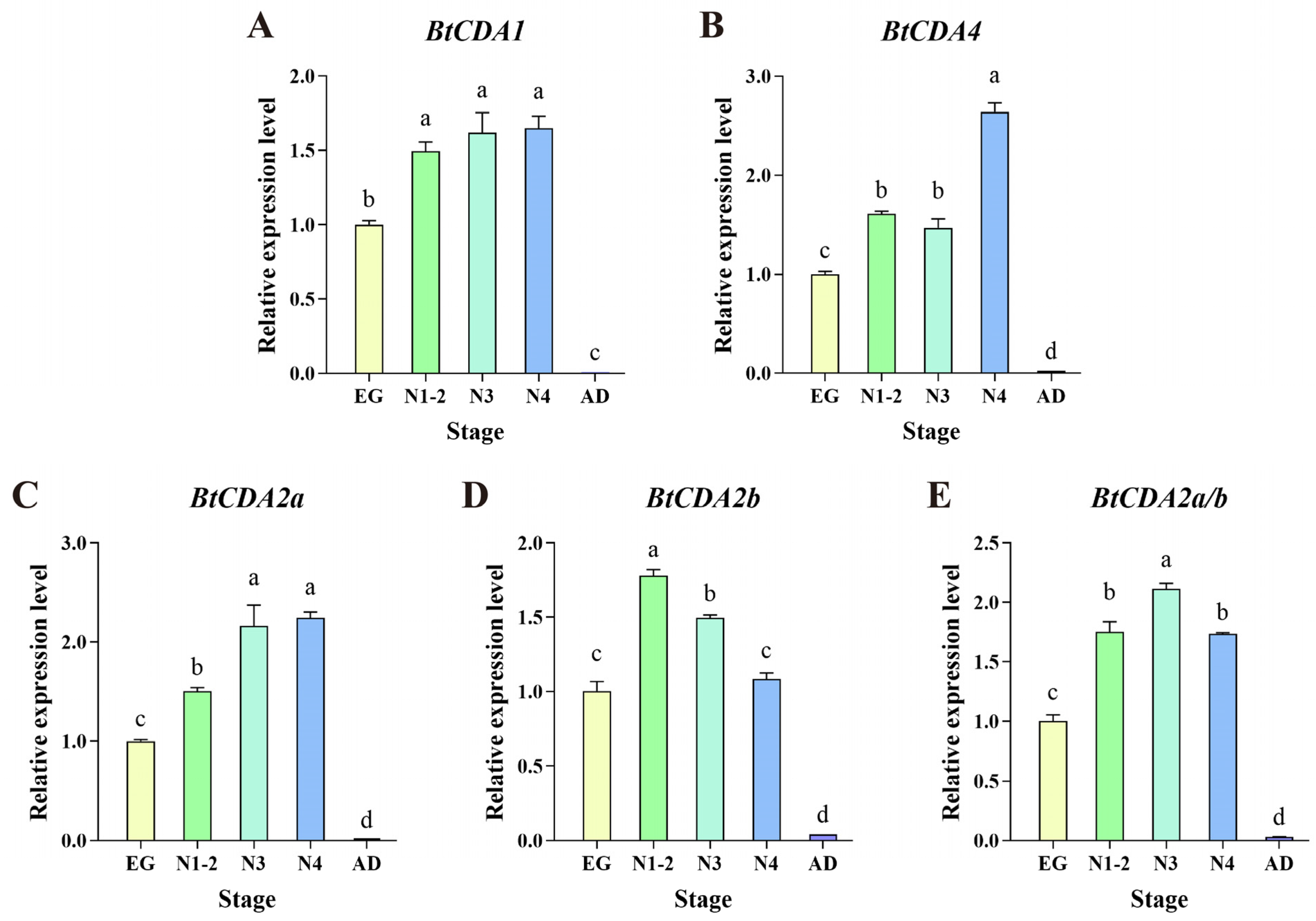
To investigate the expression profiles of BtCDA genes, RT-qPCR was performed to determine their transcript levels across different developmental stages of B. tabaci (Figure 2). Transcript levels of BtCDA1, BtCDA2a, and BtCDA4 increased progressively during egg and nymphal development. In contrast, BtCDA2b expression declined from the first through fourth instar, suggesting the difference in gene function between BtCDA2a and BtCDA2b. Notably, all four genes showed very low expression levels in adults.

3.4. Functional Analysis of BtCDAs by RNAi

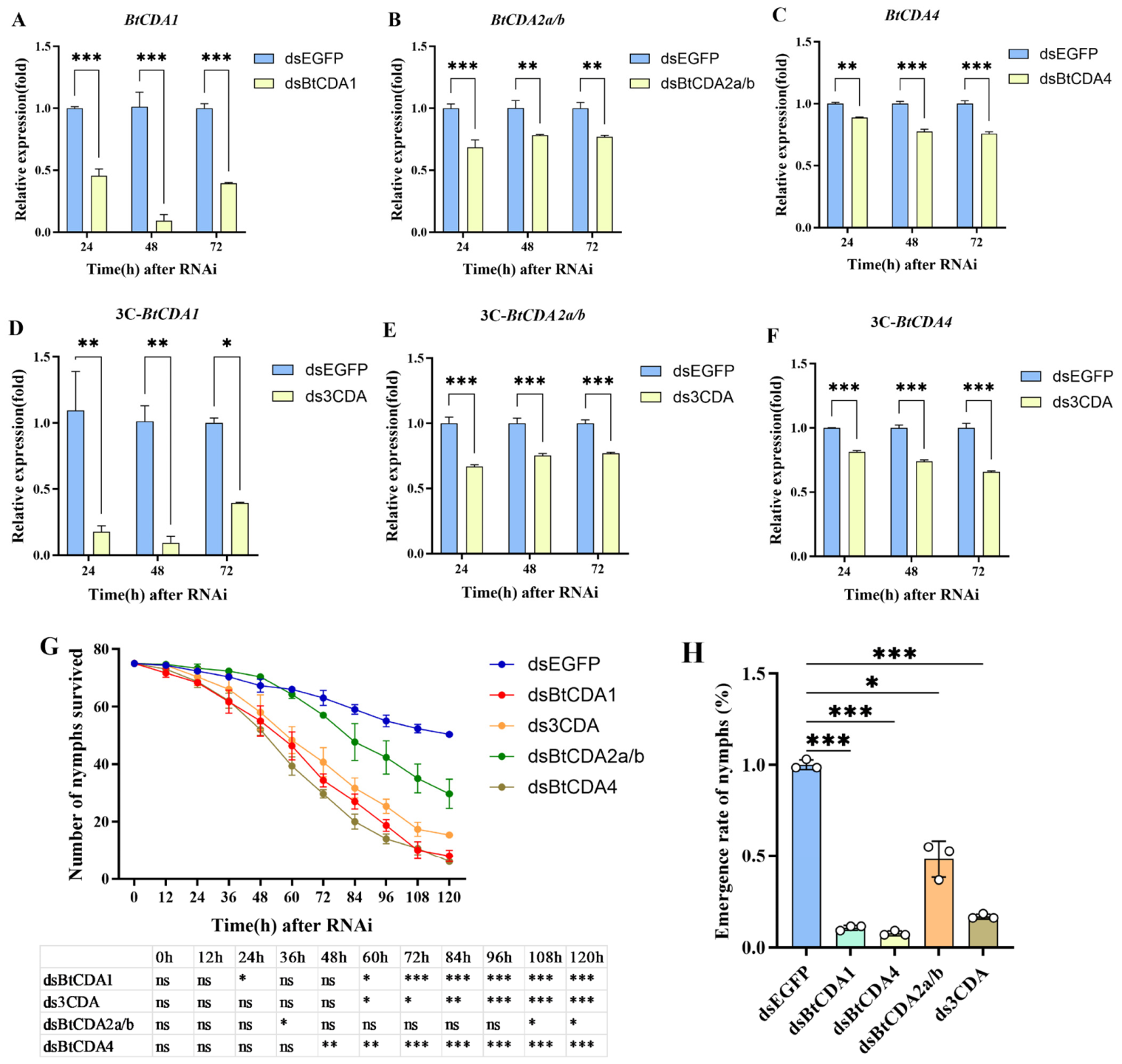
To evaluate the biological functions of chitin deacetylase genes in B. tabaci, we performed functional analysis of CDAs through SPc-mediated RNAi. At 72 h post-dsRNA treatment, the expression levels of all CDA genes were significantly downregulated compared with the control group treated with dsEGFP (Figure 3A–C). The expression levels of BtCDA1, BtCDA2a/b, and BtCDA4 were reduced by approximately 60.7%, 23.2%, and 24.4%, respectively. Notably, for BtCDA1 and BtCDA4, transcript levels at 72 h were slightly higher than at 48 h, showing a rebound of expression after the initial silencing. A similar temporal pattern has been observed in S. furcifera after dsRNA injection, where target gene expression at 72 h was higher than at 48 h [29]. Furthermore, dsBtCDA4 treatment induced continuous and significant mortality in B. tabaci nymphs starting from 48 h post-treatment. dsBtCDA1 treatment caused continuous and significant mortality in B. tabaci nymphs starting from 60 h post-treatment. In contrast, continuous significant differences were only observed for dsBtCDA2a/b treatment starting from 108 h post-treatment. The corrected mortality rates of B. tabaci nymphs at 120 h were 84.1%, 88.1%, and 40.8% for dsBtCDA1, dsBtCDA4, and dsBtCDA2a/b treatments, respectively (Figure 3G). For dsRNA treatments targeting different CDA genes, the final relative eclosion rates were 11.5%, 7.5%, and 48.1% (dsBtCDA1, dsBtCDA4, and dsBtCDA2a/b, respectively) (Figure 3H).
For RNAi of the fusion gene 3CDA, all three CDA genes exhibited downregulated expression, with BtCDA1 showing the most prominent reduction. At 72 h post-ds3CDA treatment, the expression levels of BtCDA1, BtCDA2a/b, and BtCDA4 decreased by approximately 60.7%, 23.1%, and 34.3%, respectively (Figure 3D–F). Compared with interference of single CDA genes, the fusion gene interference demonstrated more significant downregulation of gene expression. Furthermore, ds3CDA treatment caused continuous and significant mortality in B. tabaci nymphs starting from 60 h post-treatment. At 120 h, the corrected mortality rate of B. tabaci nymphs reached 69.5%, with a relative eclosion rate of 16.8% (Figure 3G,H).

3.5. Expression Levels of BtCDAs in B. tabaci Adults Feeding on Transgenic Tomato Plants

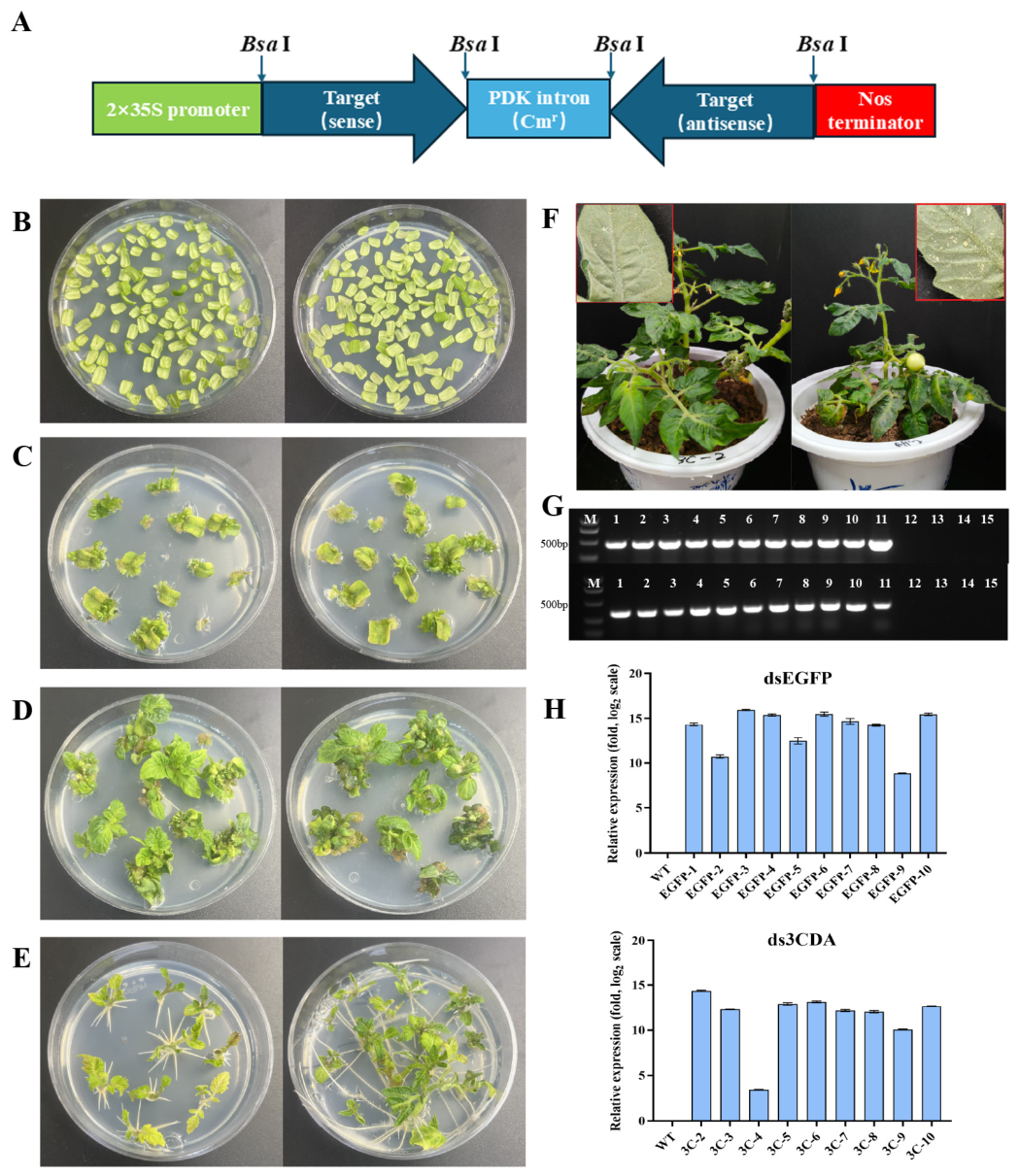
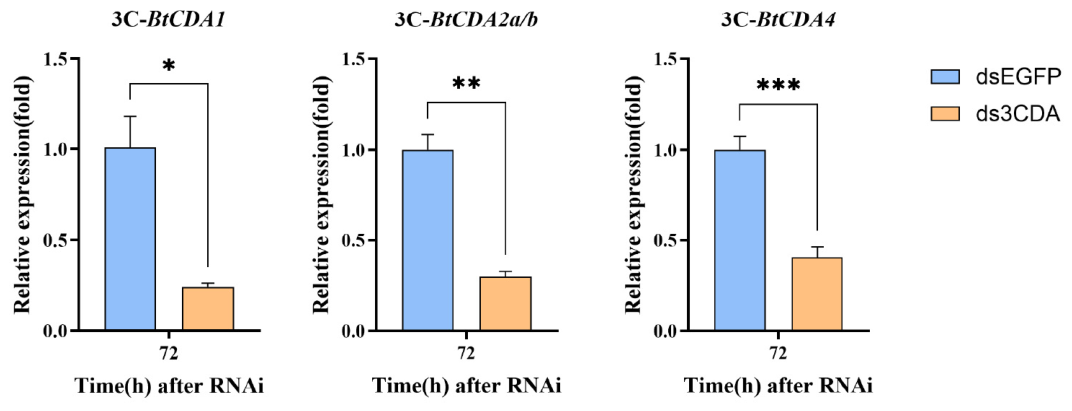
Transgenic tomato plants expressing dsRNAs of 3CDA and EGFP were constructed via Agrobacterium-mediated transformation using the pRNAi-GG plasmid (Figure 4A) (Figure 4B–F). Based on PCR genotyping and RT-qPCR expression level analyses, 10 dsEGFP transgenic plants and 9 ds3CDA transgenic plants were obtained (Figure 4G,H). We selected high-expressing transgenic plants (EGFP-3, 4, 6 and 3CDA-2, 5, 6) for subsequent feeding experiments. Whiteflies were released on each of these lines separately, and the results were pooled as mean ± SEM. The results showed that after B. tabaci fed on transgenic plant leaves for 3 days, the expression levels of all BtCDA genes were significantly downregulated compared with those in whiteflies fed on dsEGFP tomato plants. The average expression levels of BtCDA1, BtCDA2a/b, and BtCDA4 were 23.9%, 30.0%, and 40.5% of control levels, respectively (Figure 5). Three weeks after releasing adult B. tabaci, observation of the abaxial side of transgenic tomato leaves revealed fewer eggs, nymphs, and exuviae of B. tabaci on plants expressing ds3CDA compared to those expressing dsEGFP, indicating an inhibitory effect of dsCDA expression on the colonization of B. tabaci.

4. Discussion

Chitin deacetylases (CDAs) are key enzymes in chitin modification, playing indispensable roles in insect growth, molting, and metamorphosis [16,23]. In this study, we identified and cloned four CDA genes (BtCDA1, BtCDA2a, BtCDA2b, and BtCDA4) from the sweetpotato whitefly, B. tabaci. Phylogenetic analysis revealed that insect CDAs can be classified into five groups (Groups I–V) [31]. Among these, BtCDA1 and BtCDA2 belong to Group I and BtCDA4 to Group III, while Groups II, IV, and V were not detected in B. tabaci. This is inconsistent with the findings in other hemipteran insects such as the brown planthopper (N. lugens), pea aphid (A. pisum), and the white-backed planthopper (S. furcifera). These hemipteran insects were reported to contain Group I, III, and IV CDAs [28,29], lacking Group II and Group V CDAs, which are commonly present in holometabolous insects such as the red flour beetle, T. castaneum [32]. Despite the absence of Group IV CDAs, three CDA-like proteins, which cluster with insect Group IV CDAs but lack the chitin-binding peritrophin-A domain, were present in B. tabaci (Figure 1A), suggesting an evolutionary divergence between B. tabaci and other hemipteran insects. The conserved reduction in the CDA families likely reflects an evolutionary adaptation to hemimetabolous development and piercing-sucking feeding habits in Hemiptera, characterized by the absence of a pupal stage and relatively simple peritrophic matrix structure in the midgut, thereby reducing functional requirements for certain CDA subtypes [28]. Notably, two highly homologous paralogs, BtCDA2a and BtCDA2b, exist in Group I CDA of B. tabaci, sharing 96.18% nucleotide sequence identity in their coding sequences. A similar scenario has been reported in N. lugens, where CDA2 exhibits alternative splicing variants with differential expression across developmental stages [28]. This suggests that Group I CDAs may have undergone functional divergence in hemipteran insects, with different isoforms potentially assuming distinct physiological roles—some primarily responsible for cuticular chitin modification and others participating in the formation of specific structures, such as wings or joints, a phenomenon well-documented in T. castaneum and Drosophila [32,46]. The observed developmental expression patterns of BtCDAs can be well explained by their proposed physiological roles. The high transcription levels during the egg and nymph stages correlate with the active periods of chitin synthesis and remodeling required for embryonic development, hatching, and successive molts. Conversely, the extremely low expression in adults is expected, as the adult cuticle is largely sclerotized and no longer undergoes expansion, and the peritrophic matrix, once formed, may require less active chitin modification. This pattern further supports the notion that targeting BtCDAs with RNAi during the nymph stage, when these genes are most active, is a highly effective strategy for pest control [47].
Through SPc nanoparticle-mediated dsRNA delivery, this study systematically evaluated the biological functions of the four BtCDA genes. Topical application of dsRNA or nanocarrier-dsRNA complexes via droplets on the insect cuticle has been successfully demonstrated in several species, including B. tabaci [15], Apolygus lucorum [48], S. frugiperda [49], and Dendroctonus valens [50]. This simple, non-invasive strategy offers a practical means to partially mimic field-spraying conditions [15]. RNAi results showed that silencing BtCDA4 and BtCDA1 caused 84.1% and 88.1% corrected mortality in fourth instar nymphs of B. tabaci, respectively, whereas silencing BtCDA2a/b resulted in only 40.8% mortality. These findings are highly consistent with observations in N. lugens and S. furcifera: in N. lugens, RNAi of NlCDA4 (Group III) caused over 95% mortality, with a stronger lethal effect than Group I CDAs [28]; in S. furcifera, silencing of SfCDA4 similarly induced severe molting defects and high mortality, while the effect of SfCDA2 was relatively mild [29]. Collectively, these results indicate that Group III CDA (CDA4) may play a more critical role than Group I CDA during the nymph-to-adult molting transition in hemipteran insects. This functional distinction may be attributed to the unique domain architecture of CDA4, which contains only a chitin-binding domain (CBD) and a CE4 catalytic domain but lacks the LDLa domain—a simplified structure that may confer distinct catalytic activity or substrate specificity [23,24]. Furthermore, the relatively low mortality observed upon BtCDA2a/b silencing in this study, combined with their high sequence similarity, suggests that these two genes may be functionally redundant, making it difficult to completely abolish their overall function with single dsRNA treatment. This partially explains why the fusion gene 3CDA (simultaneously targeting BtCDA1, BtCDA2a/b, and BtCDA4) achieved more efficient gene silencing and higher mortality (69.5% corrected mortality at 120 h, with only 16.8% relative eclosion rate). The fusion gene strategy, which concatenates multiple target fragments into a single dsRNA molecule, enables simultaneous interference of multiple key genes, enhancing RNAi efficiency while potentially reducing the risk of pest resistance to single-target dsRNAs [51]. This multi-target synergistic interference strategy has demonstrated significant advantages in RNAi-based pest control research on hemipteran insects [51,52].
The SPc nanoparticle used in this study significantly enhanced dsRNA stability and delivery efficiency, achieving sustained gene silencing for up to 72 h. SPc is a specially designed amphiphilic cationic polymer that can self-assemble with dsRNA into nanocomplexes of approximately 100–200 nm through electrostatic interactions [34]. Nanocarriers effectively protect dsRNA from degradation by ribonucleases in insect hemolymph and gut [15,53]. In the green mirid bug (A. lucorum), SPc-mediated delivery of dsRNA targeting ecdysone receptor and trehalase genes effectively inhibited insect growth and development [48]. In the present study and our previous studies [15], SPc/dsRNA complexes were directly applied to the dorsal epidermis of fourth instar nymphs of B. tabaci, a simple practice causing minimal damage to the insects, overcoming the limitations of microinjection methods, which are tedious and unsuitable for large-scale application.
The SPc-mediated RNAi silencing efficiencies observed in this study range from 60.7% to 23.2% at 72 h. This is only a moderate level of gene silencing in comparison to reported SPc-mediated RNAi. For example, in our previous work, SPc-mediated RNAi of five other chitin metabolism-related genes led to target gene expression being decreased by over 70% at 72 h post RNAi [15]. Significant variation in gene silencing efficiency is a common feature of nanomaterial-mediated RNAi. Specifically, RNAi mediated by various nanomaterials targeting distinct genes has yielded gene silencing efficiencies ranging from 23% to 98% [54]. The relatively moderate knockdown levels in this study might be attributed to the specific target genes, the dsRNA delivery strategy, and the length of the dsRNA constructs [55,56], highlighting the critical need to optimize both target gene selection and dsRNA design.
In the present work, we further constructed transgenic tomato plants expressing ds3CDA and validated the feasibility of plant-mediated RNAi through feeding assays. Results showed that after adult whiteflies fed on transgenic tomato leaves for 3 days, the expression levels of BtCDA1, BtCDA2a/b, and BtCDA4 decreased by 4.18-fold, 3.33-fold, and 2.47-fold compared to the control, respectively. Plant-mediated RNAi technology has achieved multiple successes in hemipteran pest control. Transgenic soybean plants expressing dsRNA targeting Leguminivora glycinivorella 18S ribosomal RNA showed enhanced resistance to this pest, achieving 35% mortality [57]. In B. tabaci, transgenic tobacco expressing dsRNA of BtTPS1 and BtTPS2 caused over 90% adult mortality and reduced fecundity [58]. Although the present study does not report data on developmental and lethal effects of the plant-expressed dsRNA against B. tabaci, our work nonetheless provides valuable evidence in establishing CDA genes as effective gene silencing targets for B. tabaci by plant-mediated RNAi. Further investigation will be conducted to analyze the abundance of B. tabaci eggs, nymphs, and exuviae on plants expressing ds3CDA or dsEGFP. Additionally, we will perform a long-term investigation to evaluate the gene silencing effects in B. tabaci nymphs rather than adults feeding on dsRNA-expressing transgenic plants.
In this study, both SPc nanoparticle-mediated delivery and transgenic plant-mediated expression of dsRNA were effective in silencing BtCDA genes. The SPc-mediated method resulted in a more potent gene silencing effect (e.g., decrease by 1.67-fold of BtCDA1 at 72 h), likely due to the high concentration of dsRNA delivered directly to the target nymphs. However, this method is labor-intensive, cost-inefficient, and limited to laboratory applications. In contrast, plant-mediated RNAi, while achieving a moderate level of silencing (decrease by 2.47- to 4.18-fold compared to the control) in our short-term observation, offers a practical and scalable approach for field application. The continuous production of dsRNA by the plant provides sustained exposure, which could suppress pest populations over multiple generations. In this work, we used the pRNAi-GG vector, which produces hairpin RNA structures that are generally more stable than linear dsRNA. Additionally, the transgenic tomato plants showed consistent silencing effects over the experimental period, suggesting that dsRNA levels were sufficient to trigger RNAi in B. tabaci. Future optimization could include the design of artificial microRNA [59], co-expression of dsRNA-binding proteins [60], or the use of tissue-specific promoters to enhance dsRNA accumulation and stability in phloem, the primary feeding site of whiteflies [61].
In the future, further work will be undertaken to develop sprayable dsRNA formulations, explore synergistic effects of multiple RNAi targets, and systematically evaluate the environmental safety and resistance risks of RNAi technology to facilitate its transition from laboratory to field applications.

5. Conclusions

In conclusion, in this study, we identified four CDA members from the genome of the MEAM1 cryptic species of B. tabaci. Significant expression of the four btCDAs was detected in the egg and various nymphal stages. Both nanoparticle-mediated dsRNA delivery and transgenic plant-mediated RNAi strategies were proven to be feasible in B. tabaci control. These findings provide novel target genes and technical insights for RNAi-based green management of B. tabaci.

Supplementary Materials

The following supporting information can be downloaded at: https://www.mdpi.com/article/10.3390/insects17060628/s1, Table S1: Primers used in this study.

Author Contributions

Conceptualization, J.L.; methodology, J.L., D.K., Y.H., Y.X. and Y.W.; validation, D.K., J.L., Y.H., Y.X. and Y.W.; formal analysis, D.K. and J.L.; investigation, D.K., J.L. and Y.H.; resources, J.L. and D.K.; data curation, D.K.; writing—original draft preparation, D.K. and J.L.; writing—review and editing, J.L.; visualization, D.K. and J.L.; supervision, J.L.; and funding acquisition, J.L. All authors have read and agreed to the published version of the manuscript.

Funding

This work was funded by the S&T Program of Hebei (22322910D), which is sponsored by the Hebei Provincial Department of Science and Technology.

Data Availability Statement

All data in this study, such as the gene entry number, are available on the NCBI website (National Center for Biotechnology Information).

Acknowledgments

The SPc nanoparticles utilized in this paper were generously provided by Jie Shen from China Agricultural University.

Conflicts of Interest

The authors declare no conflicts of interest.

References

  1. De Barro, P.J.; Liu, S.S.; Boykin, L.M.; Dinsdale, A.B. Bemisia tabaci: A statement of species status. Annu. Rev. Entomol. 2011, 56, 1–19. [Google Scholar] [CrossRef] [Scilit] [PubMed]
  2. Ning, W.; Shi, X.; Liu, B.; Pan, H.; Wei, W.; Zeng, Y.; Sun, X.; Xie, W.; Wang, S.; Wu, Q.; et al. Transmission of tomato yellow leaf curl virus by Bemisia tabaci as affected by whitefly sex and biotype. Sci. Rep. 2015, 5, 10744. [Google Scholar] [CrossRef] [Scilit] [PubMed]
  3. Li, Y.; Mbata, G.N.; Punnuri, S.; Simmons, A.M.; Shapiro-Ilan, D.I. Bemisia tabaci on vegetables in the southern United States: Incidence, impact, and management. Insects 2021, 12, 198. [Google Scholar] [CrossRef] [Scilit]
  4. Dinsdale, A.; Cook, L.; Riginos, C.; Buckley, Y.M.; De Barro, P. Refined global analysis of Bemisia tabaci (Hemiptera: Sternorrhyncha: Aleyrodoidea: Aleyrodidae) mitochondrial Cytochrome Oxidase 1 to identify species level genetic boundaries. Ann. Entomol. Soc. Am. 2010, 103, 196–208. [Google Scholar] [CrossRef] [Scilit]
  5. Boykin, L.M.; De Barro, P.J. A practical guide to identifying members of the Bemisia tabaci species complex: And other morphologically identical species. Front. Ecol. Evol. 2014, 2, 45. [Google Scholar] [CrossRef] [Scilit]
  6. Houndété, T.A.; Kétoh, G.K.; Hema, O.S.; Brévault, T.; Glitho, I.A.; Martin, T. Insecticide resistance in field populations of Bemisia tabaci (Hemiptera: Aleyrodidae) in West Africa. Pest Manag. Sci. 2010, 66, 1181–1185. [Google Scholar] [CrossRef] [Scilit]
  7. Pan, H.; Preisser, E.L.; Chu, D.; Wang, S.; Wu, Q.; Carriére, Y.; Zhou, X.; Zhang, Y. Insecticides promote viral outbreaks by altering herbivore competition. Ecol. Appl. 2015, 25, 1585–1595. [Google Scholar] [CrossRef] [Scilit]
  8. Abdullah, N.M.M.; Singh, J.; Sohal, B.S. Behavioral hormoligosis in oviposition preference of Bemisia tabaci on cotton. Pestic. Biochem. Physiol. 2006, 84, 10–16. [Google Scholar] [CrossRef] [Scilit]
  9. Zhang, J.; Khan, S.A.; Heckel, D.G.; Bock, R. Next-Generation insect-resistant plants: RNAi-mediated crop protection. Trends Biotechnol. 2017, 35, 871–882. [Google Scholar] [CrossRef] [Scilit]
  10. Baulcombe, D. RNA silencing in plants. Nature 2004, 431, 356–363. [Google Scholar] [CrossRef] [Scilit]
  11. Novina, C.D.; Sharp, P.A. The RNAi revolution. Nature 2004, 430, 161–164. [Google Scholar] [CrossRef] [Scilit]
  12. Geley, S.; Müller, C. RNAi: Ancient mechanism with a promising future. Exp. Gerontol. 2004, 39, 985–998. [Google Scholar] [CrossRef] [Scilit]
  13. Walker, W.B.; Allen, M.L. RNA interference-mediated knockdown of IAP in Lygus lineolaris induces mortality in adult and pre-adult life stages. Entomol. Exp. Appl. 2011, 138, 83–92. [Google Scholar] [CrossRef] [Scilit]
  14. Chen, J.; Zhang, D.; Yao, Q.; Zhang, J.; Dong, X.; Tian, H.; Chen, J.; Zhang, W. Feeding-based RNA interference of a trehalose phosphate synthase gene in the brown planthopper, Nilaparvata lugens. Insect Mol. Biol. 2010, 19, 777–786. [Google Scholar] [CrossRef] [Scilit] [PubMed]
  15. Kong, D.; Gu, H.; Gao, Y.; Hou, Y.; Li, J. Nanomaterial-mediated RNAi targeting chitin metabolism genes in MEAM1 cryptic species of Bemisia tabaci (Hemiptera: Aleyrodidae). Insects 2026, 17, 2. [Google Scholar] [CrossRef] [Scilit]
  16. Zhu, K.Y.; Merzendorfer, H.; Zhang, W.; Zhang, J.; Muthukrishnan, S. Biosynthesis, turnover, and functions of chitin in insects. Annu. Rev. Entomol. 2016, 61, 177–196. [Google Scholar] [CrossRef] [Scilit] [PubMed]
  17. Merzendorfer, H. The cellular basis of chitin synthesis in fungi and insects: Common principles and differences. Eur. J. Cell Biol. 2011, 90, 759–769. [Google Scholar] [CrossRef] [Scilit]
  18. Moussian, B. Recent advances in understanding mechanisms of insect cuticle differentiation. Insect Biochem. Mol. Biol. 2010, 40, 363–375. [Google Scholar] [CrossRef] [Scilit]
  19. Merzendorfer, H.; Zimoch, L. Chitin metabolism in insects: Structure, function and regulation of chitin synthases and chitinases. J. Exp. Biol. 2003, 206, 4393–4412. [Google Scholar] [CrossRef] [Scilit]
  20. Arakane, Y.; Muthukrishnan, S. Insect chitinase and chitinase-like proteins. Cell. Mol. Life Sci. 2010, 67, 201–216. [Google Scholar] [CrossRef] [Scilit]
  21. Zhu, Q.; Arakane, Y.; Banerjee, D.; Beeman, R.W.; Kramer, K.J.; Muthukrishnan, S. Domain organization and phylogenetic analysis of the chitinase-like family of proteins in three species of insects. Insect Biochem. Mol. Biol. 2008, 38, 452–466. [Google Scholar] [CrossRef] [Scilit]
  22. Zhang, M.; Du, M.-Y.; Wang, G.-X.; Wang, Z.-Y.; Lu, Y.-J. Identification, mRNA expression, and functional analysis of chitin synthase 2 gene in the rusty grain beetle, Cryptolestes ferrugineus. J. Stored Prod. Res. 2020, 87, 101622. [Google Scholar] [CrossRef] [Scilit]
  23. Liu, X.; Zhang, J.; Zhu, K.Y. Chitin in arthropods: Biosynthesis, modification, and metabolism. Adv. Exp. Med. Biol. 2019, 1142, 169–207. [Google Scholar] [CrossRef] [Scilit]
  24. Liang, B.; Song, W.; Xing, R.; Liu, S.; Yu, H.; Li, P. The source, activity influencing factors and biological activities for future development of chitin deacetylase. Carbohydr. Polym. 2023, 321, 121335. [Google Scholar] [CrossRef] [Scilit]
  25. Guo, W.; Li, G.; Pang, Y.; Wang, P. A novel chitin-binding protein identified from the peritrophic membrane of the cabbage looper, Trichoplusia ni. Insect Biochem. Mol. Biol. 2005, 35, 1224–1234. [Google Scholar] [CrossRef] [Scilit]
  26. Zhong, X.W.; Wang, X.H.; Tan, X.; Xia, Q.Y.; Xiang, Z.H.; Zhao, P. Identification and molecular characterization of a chitin deacetylase from Bombyx mori peritrophic membrane. Int. J. Mol. Sci. 2014, 15, 1946–1961. [Google Scholar] [CrossRef] [Scilit]
  27. Tetreau, G.; Cao, X.; Chen, Y.R.; Muthukrishnan, S.; Jiang, H.; Blissard, G.W.; Kanost, M.R.; Wang, P. Overview of chitin metabolism enzymes in Manduca sexta: Identification, domain organization, phylogenetic analysis and gene expression. Insect Biochem. Mol. Biol. 2015, 62, 114–126. [Google Scholar] [CrossRef] [Scilit]
  28. Xi, Y.; Pan, P.L.; Ye, Y.X.; Yu, B.; Zhang, C.X. Chitin deacetylase family genes in the brown planthopper, Nilaparvata lugens (Hemiptera: Delphacidae). Insect Mol. Biol. 2014, 23, 695–705. [Google Scholar] [CrossRef] [Scilit]
  29. Yang, X.B.; Zhou, C.; Gong, M.F.; Yang, H.; Long, G.Y.; Jin, D.C. Identification and RNAi-based functional analysis of four chitin deacetylase genes in Sogatella furcifera (Hemiptera: Delphacidae). J. Insect Sci. 2021, 21, 9. [Google Scholar] [CrossRef] [Scilit]
  30. Yu, H.-Z.; Liu, M.-H.; Wang, X.-Y.; Yang, X.; Wang, W.-L.; Geng, L.; Yu, D.; Liu, X.-L.; Liu, G.-Y.; Xu, J.-P. Identification and expression profiles of chitin deacetylase genes in the rice leaf folder, Cnaphalocrocis medinalis. J. Asia Pac. Entomol. 2016, 19, 691–696. [Google Scholar] [CrossRef] [Scilit]
  31. Dixit, R.; Arakane, Y.; Specht, C.A.; Richard, C.; Kramer, K.J.; Beeman, R.W.; Muthukrishnan, S. Domain organization and phylogenetic analysis of proteins from the chitin deacetylase gene family of Tribolium castaneum and three other species of insects. Insect Biochem. Mol. Biol. 2008, 38, 440–451. [Google Scholar] [CrossRef] [Scilit]
  32. Arakane, Y.; Dixit, R.; Begum, K.; Park, Y.; Specht, C.A.; Merzendorfer, H.; Kramer, K.J.; Muthukrishnan, S.; Beeman, R.W. Analysis of functions of the chitin deacetylase gene family in Tribolium castaneum. Insect Biochem. Mol. Biol. 2009, 39, 355–365. [Google Scholar] [CrossRef] [Scilit]
  33. Li, J.H.; Qian, J.; Xu, Y.Y.; Yan, S.; Shen, J.; Yin, M.Z. A facile-synthesized star polycation constructed as a highly efficient gene vector in pest management. ACS Sustain. Chem. Eng. 2019, 7, 6316–6322. [Google Scholar] [CrossRef] [Scilit]
  34. Ma, Z.; Zheng, Y.; Chao, Z.; Chen, H.; Zhang, Y.; Yin, M.; Shen, J.; Yan, S. Visualization of the process of a nanocarrier-mediated gene delivery: Stabilization, endocytosis and endosomal escape of genes for intracellular spreading. J. Nanobiotechnol. 2022, 20, 124. [Google Scholar] [CrossRef] [Scilit]
  35. Zhang, L.P.; Zhang, Y.J.; Zhang, W.J.; Wu, Q.J.; Xu, B.Y.; Chu, D. Analysis of genetic diversity among different geographical populations and determination of biotypes of Bemisia tabaci in China. J. Appl. Entomol. 2005, 129, 121–128. [Google Scholar] [CrossRef] [Scilit]
  36. Jones, P.; Binns, D.; Chang, H.-Y.; Fraser, M.; Li, W.; McAnulla, C.; McWilliam, H.; Maslen, J.; Mitchell, A.; Nuka, G.; et al. InterProScan 5: Genome-scale protein function classification. Bioinformatics 2014, 30, 1236–1240. [Google Scholar] [CrossRef] [Scilit]
  37. Katoh, K.; Standley, D.M. MAFFT multiple sequence alignment software version 7: Improvements in performance and usability. Mol. Biol. Evol. 2013, 30, 772–780. [Google Scholar] [CrossRef] [Scilit]
  38. Eddy, S.R. Accelerated profile hmm searches. PLoS Comput. Biol. 2011, 7, e1002195. [Google Scholar] [CrossRef] [Scilit]
  39. Tamura, K.; Stecher, G.; Kumar, S. MEGA11: Molecular evolutionary genetics analysis version 11. Mol. Biol. Evol. 2021, 38, 3022–3027. [Google Scholar] [CrossRef] [Scilit]
  40. Chen, C.; Chen, H.; Zhang, Y.; Thomas, H.R.; Frank, M.H.; He, Y.; Xia, R. TBtools: An integrative toolkit developed for interactive analyses of big biological data. Mol. Plant 2020, 13, 1194–1202. [Google Scholar] [CrossRef] [Scilit]
  41. Livak, K.J.; Schmittgen, T.D. Analysis of relative gene expression data using real-time quantitative PCR and the 2−ΔΔCt method. Methods 2001, 25, 402–408. [Google Scholar] [CrossRef] [Scilit]
  42. Yan, P.; Shen, W.; Gao, X.; Li, X.; Zhou, P.; Duan, J. High-throughput construction of intron-containing hairpin RNA vectors for RNAi in plants. PLoS ONE 2012, 7, e38186. [Google Scholar] [CrossRef] [Scilit]
  43. Park, S.H.; Morris, J.L.; Park, J.E.; Hirschi, K.D.; Smith, R.H. Efficient and genotype-independent Agrobacterium-mediated tomato transformation. J. Plant Physiol. 2003, 160, 1253–1257. [Google Scholar] [CrossRef] [Scilit]
  44. Tsigos, I.; Martinou, A.; Kafetzopoulos, D.; Bouriotis, V. Chitin deacetylases: New, versatile tools in biotechnology. Trends Biotechnol. 2000, 18, 305–312. [Google Scholar] [CrossRef] [Scilit]
  45. Grifoll-Romero, L.; Pascual, S.; Aragunde, H.; Biarnés, X.; Planas, A. Chitin deacetylases: Structures, specificities, and biotech applications. Polymers 2018, 10, 352. [Google Scholar] [CrossRef] [Scilit] [PubMed]
  46. Luschnig, S.; Bätz, T.; Armbruster, K.; Krasnow, M.A. Serpentine and vermiform encode matrix proteins with chitin binding and deacetylation domains that limit tracheal tube length in Drosophila. Curr. Biol. 2006, 16, 186–194. [Google Scholar] [CrossRef] [Scilit]
  47. Doucet, D.; Retnakaran, A. Insect Chitin: Metabolism, Genomics and Pest Management. In Advances in Insect Physiology, Vol 43: Insect Growth Disruptors; Dhadialla, T.S., Ed.; Elsevier: Amsterdam, The Netherlands, 2012; Volume 43, pp. 437–511. [Google Scholar]
  48. Qiao, H.; Jiang, Q.; Zhao, J.; Xiao, L.; Zhu-Salzman, K.; Xu, D.; Xu, G.; Shen, J.; Gu, A.; Hao, D.; et al. Nano-delivery platform with strong protection and efficient delivery: Preparation of self-assembled RNA pesticide with dual RNAi targets against Apolygus lucorum. J. Nanobiotechnol. 2025, 23, 93. [Google Scholar] [CrossRef] [Scilit] [PubMed]
  49. Guo, S.; Li, Z.; Zhao, X.; Zhang, D.; Ayra-Pardo, C.; Kan, Y.; Li, D. Additive insecticidal effects of chitosan/dsRNA nanoparticles targeting V-ATPaseD and emamectin benzoate–lufenuron formulations against Spodoptera frugiperda (J.E. Smith) (Lepidoptera: Noctuidae). Insects 2025, 16, 348. [Google Scholar] [CrossRef] [Scilit]
  50. Cao, Q.; Sun, Y.; Kong, D.; Han, J.; Wei, J.; Li, J. Potential of RNAi targeting Juvenile Hormone Acid Methyltransferase (JHAMT) for controlling Dendroctonus valens LeConte (Coleoptera: Scolytidae). Forests 2026, 17, 628. [Google Scholar] [CrossRef] [Scilit]
  51. Xu, Q.Q.; Shang, F.; Feng, S.Y.; Xie, Q.P.; Zhang, W.; Wang, Z.G.; Wang, J.J. Design the fusion double-strand RNAs to control two global sap-sucking pests. Pestic. Biochem. Physiol. 2024, 205, 106114. [Google Scholar] [CrossRef] [Scilit]
  52. Wang, Z.G.; Qin, C.Y.; Chen, Y.; Yu, X.Y.; Chen, R.Y.; Niu, J.; Wang, J.J. Fusion dsRNA designs incorporating multiple target sequences can enhance the aphid control capacity of an RNAi-based strategy. Pest Manag. Sci. 2024, 80, 2689–2697. [Google Scholar] [CrossRef] [Scilit] [PubMed]
  53. Christiaens, O.; Whyard, S.; Vélez, A.M.; Smagghe, G. Double-stranded RNA technology to control insect pests: Current status and challenges. Front. Plant Sci. 2020, 11, 451. [Google Scholar] [CrossRef] [Scilit]
  54. Liu, J.S.; He, Q.Y.; Lin, X.F.; Smagghe, G. Recent progress in nanoparticle-mediated RNA interference in insects: Unveiling new frontiers in pest control. J. Insect Physiol. 2025, 167, 104884. [Google Scholar] [CrossRef] [Scilit]
  55. Liu, S.S.; Jaouannet, M.; Dempsey, D.M.A.; Imani, J.; Coustau, C.; Kogel, K.H. RNA-based technologies for insect control in plant production. Biotechnol. Adv. 2020, 39, 107463. [Google Scholar] [CrossRef] [Scilit]
  56. San Miguel, K.; Scott, J.G. The next generation of insecticides: dsRNA is stable as a foliar-applied insecticide. Pest Manag. Sci. 2016, 72, 801–809. [Google Scholar] [CrossRef] [Scilit]
  57. Wang, Z.; Li, T.; Ni, H.; Wang, G.; Liu, X.; Cao, Y.; Li, W.; Meng, F. Transgenic soybean plants expressing Spb18S dsRNA exhibit enhanced resistance to the soybean pod borer Leguminivora glycinivorella (Lepidoptera: Olethreutidae). Arch. Insect Biochem. Physiol. 2018, 98, e21461. [Google Scholar] [CrossRef] [Scilit]
  58. Gong, C.; Yang, Z.; Hu, Y.; Wu, Q.; Wang, S.; Guo, Z.; Zhang, Y. Silencing of the BtTPS genes by transgenic plant-mediated RNAi to control Bemisia tabaci MED. Pest Manag. Sci. 2022, 78, 1128–1137. [Google Scholar] [CrossRef] [Scilit] [PubMed]
  59. Bao, W.; Li, A.; Zhang, Y.; Diao, P.; Zhao, Q.; Yan, T.; Zhou, Z.; Duan, H.; Li, X.; Wuriyanghan, H. Improvement of host-induced gene silencing efficiency via polycistronic-tRNA-amiR expression for multiple target genes and characterization of RNAi mechanism in Mythimna separata. Plant Biotechnol. J. 2021, 19, 1370–1385. [Google Scholar] [CrossRef] [Scilit]
  60. Gillet, F.X.; Garcia, R.A.; Macedo, L.L.P.; Albuquerque, E.V.S.; Silva, M.C.M.; Grossi-de-Sa, M.F. Investigating engineered ribonucleoprotein particles to improve oral RNAi delivery in crop insect pests. Front. Physiol. 2017, 8, 256. [Google Scholar] [CrossRef] [Scilit] [PubMed]
  61. Javaid, S.; Amin, I.; Jander, G.; Mukhtar, Z.; Saeed, N.A.; Mansoor, S. A transgenic approach to control hemipteran insects by expressing insecticidal genes under phloem-specific promoters. Sci. Rep. 2016, 6, 34706. [Google Scholar] [CrossRef] [Scilit]
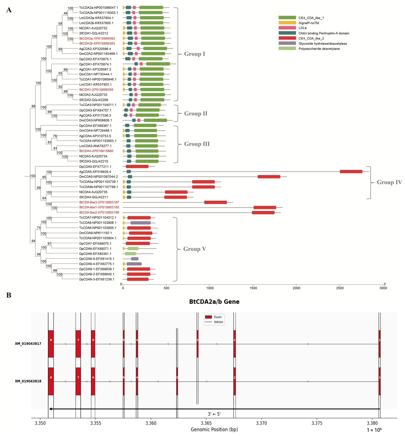
Figure 1. Phylogeny and sequence similarity of candidate BtCDAs. (A) Phylogenetic analyses and domain organization of insect chitin deacetylases (CDAs) from Anopheles gambiae (Ag), Drosophila melanogaster (Dm), T. castaneum (Tc), Daphnia pulex (Dp), Locusta migratoria (Lm), N. lugens (Nl), S. furcifera (Sf), and B. tabaci (Bt). The amino acid sequences of CDAs were classified into five groups (I–V). Protein names in red font indicate sequences derived from Bemisia tabaci. (B) Genomic sequence structure of two alternatively spliced BtCDA2 genes. BtCDA2a and BtCDA2b use different exon 3, resulting in approximately 100 bp sequence divergence. Arrows indicate that BtCDA2a/b are transcribed in the right-to-left direction in the figure.
Insects 17 00628 g001
Figure 2. Expression of four BtCDAs at various developmental stages in B. tabaci. (AE) Relative expression levels of BtCDA1 (A), BtCDA2a (B), BtCDA2b (C), BtCDA4 (D), and BtCDA2a/b (E) at Egg (EG), N1–N4 (first–fourth instar nymph), and adult (AD) stages. Different lowercase letters above the bars indicate significant differences in expression levels among developmental stages for a given gene (one-way ANOVA followed by Tukey’s HSD test, p < 0.05). Bars: ±standard error of the mean (SEM).
Insects 17 00628 g002
Figure 3. Silencing of BtCDA genes by SPc-delivered dsRNA in B. tabaci. (AC) Relative expression levels of BtCDA1, BtCDA2a/b, and BtCDA4 at 24, 48, and 72 h after treatment with dsRNA targeting the corresponding single gene. (DF) Relative expression levels of BtCDA1, BtCDA2a/b, and BtCDA4 at 24, 48, and 72 h after treatment with dsRNA targeting the fused 3CDA gene. (G) Corrected mortality of fourth-instar nymphs after treatment with ds3CDA, dsBtCDA1, dsBtCDA2a/b, dsBtCDA4, or dsEGFP (control). Mortality was recorded every 12 h up to 120 h. (H) Relative emergence rate of adults after each dsRNA treatment. All values are shown as mean ± SEM. Asterisks indicate significant differences compared with the dsEGFP control (ns, not significant; *, p < 0.05; **, p < 0.01; ***, p < 0.001).
Insects 17 00628 g003
Figure 4. Construction and molecular characterization of transgenic tomato lines expressing dsRNA targeting 3CDA or EGFP. (A) Schematic representation of the pRNAi-GG expression cassette used for tomato transformation. (BF) Key steps of Agrobacterium-mediated tomato transformation: (B) co-cultivation of explants with Agrobacterium, (C) screening of transformed cells on medium containing antibiotics, (D) shoot differentiation, and (E) rooting of regenerated shoots. (F) Potted transgenic plants expressing ds3CDA and dsEFGP. Pests colonizing on the surface of the back of the leaf blades are shown in the red-boxed photos. (G) Genomic identification of transgenic lines by PCR: detection of dsEGFP (upper panel) and ds3CDA (lower panel) fragments. M, DNA marker; lanes 1–10 represent independent lines; lane 11 indicates positive plasmid control; and lanes 12–15 are wild-type control. (H) Relative expression levels of the dsRNA in transgenic lines detected by RT-qPCR. RT-qPCR data of Plant 3C-1 are missing due to the death of the plant prior to RT-qPCR analysis, potentially due to unanticipated experimental factors. For the same reason, during the release of whitefly adults to evaluate developmental and lethal effects, the majority of the transgenic plants withered and died prematurely prior to the completion of observation and statistical analyses.
Insects 17 00628 g004
Figure 5. Expression levels of BtCDA genes in B. tabaci adults after feeding on transgenic tomato plants expressing ds3CDA. Whitefly adults were allowed to feed on transgenic tomato leaves for 3 days. Relative expression of BtCDA1, BtCDA2a/b, and BtCDA4 was determined by RT-qPCR. The dsEGFP transgenic tomato line was used as the control. All values are shown as mean ± SEM. Asterisks indicate significant differences compared with the control (Welch’s t-test; *, p < 0.05; **, p < 0.01; ***, p < 0.001).
Insects 17 00628 g005
Table 1. Characteristics of chitin deacetylase genes in B. tabaci.
NameProteinmRNAAaSubcellular
Localization
Hydrophilic/HydrophobicSP
BtCDA1XP_018899356XM_019043811545ECMHydrophilic+
BtCDA2aXP_018899362XM_019043817556ECMHydrophilic+
BtCDA2bXP_018899363XM_019043818550ECMHydrophilic+
BtCDA4XP_018915980XM_019060435495ECMHydrophilic+
Note: ECM indicates an extracellular matrix; SP indicates a signal peptide; + indicate presence, respectively.
Disclaimer/Publisher’s Note: The statements, opinions and data contained in all publications are solely those of the individual author(s) and contributor(s) and not of MDPI and/or the editor(s). MDPI and/or the editor(s) disclaim responsibility for any injury to people or property resulting from any ideas, methods, instructions or products referred to in the content.