Review

In Vitro Analyses of the Toxicity, Immunological, and Gene Expression Effects of Cobalt-Chromium Alloy Wear Debris and Co Ions Derived from Metal-on-Metal Hip Implants

1
Biomedical Engineering Department, University of Strathclyde, Wolfson Centre, Glasgow G4 0NW, UK
2
Strathclyde Institute for Pharmacy & Biomedical Sciences, University of Strathclyde, Glasgow G4 0RE, UK
3
Department of Orthopaedic Surgery, Southern General Hospital, Glasgow G11 6NT, UK
4
Current affiliation, Leeds Institute of Cardiovascular and Metabolic Medicine (LICAMM), University of Leeds, Leeds LS2 9JT, UK
*
Author to whom correspondence should be addressed.
Lubricants 2015, 3(3), 539-568; https://doi.org/10.3390/lubricants3030539
Submission received: 30 April 2015 / Revised: 12 June 2015 / Accepted: 8 July 2015 / Published: 14 July 2015
(This article belongs to the Special Issue Tribological Performance of Artificial Joints)

Abstract

:
Joint replacement has proven to be an extremely successful and cost-effective means of relieving arthritic pain and improving quality of life for recipients. Wear debris-induced osteolysis is, however, a major limitation and causes orthopaedic implant aseptic loosening, and various cell types including macrophages, monocytes, osteoblasts, and osteoclasts, are involved. During the last few years, there has been increasing concern about metal-on-metal (MoM) hip replacements regarding adverse reactions to metal debris associated with the MoM articulation. Even though MoM-bearing technology was initially aimed to extend the durability of hip replacements and to reduce the requirement for revision, they have been reported to release at least three times more cobalt and chromium ions than metal-on-polyethylene (MoP) hip replacements. As a result, the toxicity of metal particles and ions produced by bearing surfaces, both locally in the periprosthetic space and systemically, became a concern. Several investigations have been carried out to understand the mechanisms responsible for the adverse response to metal wear debris. This review aims at summarising in vitro analyses of the toxicity, immunological, and gene expression effects of cobalt ions and wear debris derived from MoM hip implants.

1. Introduction

Joint replacement has proven to be an extremely successful and cost-effective means of relieving arthritic pain and improving quality of life for recipients [1]. A primary replacement is an initial replacement procedure undertaken on a joint and involves replacing either part (partial) or all (total) of the articular surface. Revision hip replacements are repeat-operations of previous hip replacements where one or more of the prosthetic components are replaced, removed, or one or more components are added [2].
The total number of joint replacement procedures recorded by the National Joint Registry of England, Wales, and Northern Ireland (NJR) exceeded 1.6 million records between 1 April 2003 and 31 March 2013, with 2012/13 having the highest ever annual number of submissions at 205,686. The total number of primary hip procedures was 620,400. Of these, 76,274 were entered into the NJR during 2013, as reported in the NJR 11th Annual Report [3]. Similarly, there have been 410,767 hip replacements reported to the Australian Registry up to 31 December 2013. Of these, 40,180 were entered during 2013 [2].
Total joint replacement surgery has traditionally been reserved for elderly patients with advanced arthrosis who postoperatively would be less active. However, this scenario has now substantially changed, and many patients now receive total hip arthroplasty at a younger age. For example, Furnes et al. [4] compared implant survival of metal-on-metal (MoM) with that of metal-on-highly-cross-linked-polyethylene in patients between 45 and 64 years old. Kanda et al. [5] reported the case of a 42-year-old patient presenting with femoral head migration after an arthroplasty performed 22 years earlier. Moreover, it has been estimated that 10,000 to 30,000 patients less than 25 years of age have undergone joint replacement procedures in the last five years, and it is likely that many of those are paediatric patients [6]. It can also be anticipated that the rate of joint replacement in paediatric patients will increase, particularly given the popularity of this surgery and the incidence of diagnoses that may result in joint replacement surgery. Currently, over 294,000 individuals younger than 21 years of age are estimated to have juvenile arthritis [7]. The clinical outcome is generally excellent, but many young patients still need implant replacement within 10–15 years [8], and some may experience complications, including implant failure. Out of the 620,400 procedures recorded by the NJR, 14,903 had an associated first revision. The most commonly cited indications were aseptic loosening (cited in 3659 procedures), pain (3489), dislocation/subluxation (2545), and infection (2072) [3]. In accordance with this, the Australian registry reported the most common reasons for revision of primary total conventional hip replacement were loosening/lysis (2550 procedures), followed by prosthesis dislocation (2251), fracture (1576), and infection (1534) [2].
Wear debris-induced osteolysis is a major cause of orthopaedic implant aseptic loosening, and various cell types including macrophages, monocytes, osteoblasts, and osteoclasts are involved [9]. During the last few years, there has been increasing concern about MoM hip replacements regarding adverse reactions to metal debris associated with the MoM articulation [10]. As a consequence, on 22 July 2008, there was a voluntary recall of the Zimmer Durom® Acetabular Component (“Durom Cup”) because the instructions for use/surgical technique were inadequate, which led to a higher than expected revision rate. Following this, on 24 August 2010, there was a voluntary recall of the DePuy ASR™ total hip system because of new, unpublished data from the UK joint registry indicating the revision rates within five years were approximately 13%. Two years later, on 1 June 2012, Smith & Nephew Orthopaedics initiated a market withdrawal for metal liners of the R3 acetabular system due to a higher than expected number of revision surgeries associated with the use of the device in total hip replacements outside the US [11]. Since a patient with an adverse reaction to metal debris can be asymptomatic [12], may have low metal ion levels [13], and may have normal cross-sectional imaging [14], diagnosing an adverse reaction is challenging. Even though MoM-bearing technology was initially aimed to extend the durability of hip replacements and to reduce the requirement for revision, they have been reported to release at least three times more cobalt and chromium ions than metal-on-polyethylene (MoP) hip replacements [15,16]. As a result, the toxicity of metal particles and ions produced by bearing surfaces, both locally in the periprosthetic space and systemically, became a concern [17]. Several investigations have been carried out to understand the mechanisms responsible for the adverse response to metal wear debris. This review aims at summarising in vitro analyses of the toxicity, immunological, and gene expression effects of cobalt ions and wear debris derived from MoM hip implants.

2. Wear Particle Generation and Metal Ion Release
The degradation products of any orthopaedic implant include two basic types of debris: particles and soluble (or ionic) debris [18]. MoM joints are exposed to a complex in vivo environment with mechanical and electrochemical degradation mechanisms that influence the longevity of the device [19]. The wear of MoM joints is of particular concern because particulate debris and release of Co/Cr ions can lead to adverse tissue reactions including necrosis, hypersensitivity, and pseudotumors [20,21].
Wear characteristics of MoM total hip replacements have two distinctive stages. Initially, the femoral and acetabular components show a relatively rapid but decreasing wear rate over the first 1–2 × 10⁶ cycles in a hip joint simulator, or for one or two years in vivo, generally referred to as the “bedding-in” or “running-in” stage. Once this process has been completed, the rate of wear becomes reasonably steady and hence is referred to as “steady-state” [22,23]. Most wear debris particles are created in the running-in phase [23]. Wear debris is released from the articular surfaces after joint arthroplasty as a result of friction between articulating implant components or between cement and implant [24]. There are three types of mechanical wear mechanisms: fatigue, abrasive, and adhesive [9]. The first is caused by cyclic stresses inducing micro-fractures to occur within materials due to fatigue; once these micro-fractures reach the surface, wear particles are generated through delamination [25]. Abrasive wear can be split into two sub-categories; two-body and three-body. Two-body abrasive wear involves the roughness of a hard surface in contact with a softer material; particulates are released from the softer material due to ploughing. Three-body abrasive wear involves three materials instead; for example, they can include bone cement or fragments of bone between two articulating surfaces. Finally, adhesive wear involves intermolecular bonds of the weaker material bonding to the stronger material, resulting in shearing [9].
Metal corrosion is the degradation process affecting the surface of metallic materials due to their reaction with the surrounding environment. Most metallic materials are susceptible to corrosion attack if a tenacious surface oxide layer does not exist. Where the surface layer is permeable to oxygen and moisture, the corrosion process will continue and lead to eventual failure. Among the variety of corrosion mechanisms, metal corrosion is driven mainly by electrochemical potential. During exposure to aqueous environments, atoms of the metal surface experience an anodic process; electrons are released from the atoms forming metallic ions (oxidation). The localized electrical potential accelerates the oxidation process until the electrochemical potential is balanced [26]. Fretting corrosion is a type of mechanically assisted chemical degradation. Fretting corrosion damage is determined by a combination of metal atom dissolution through the fractured passive layer and metal oxide reformation. The oxide film is fractured by the contact and friction of rough articulating surfaces and exposure of the pure metal surface to a corrosive medium [27,28]. The physiological environment is considered corrosive. This makes the corrosion of metallic materials a slow and continuous process, which leads to the release of metal ions [29,30]. Corrosion damage is a very important issue for metallic implants that can affect their biocompatibility and mechanical integrity [31]. Chloride ions, amino acids, and proteins in the body can accelerate corrosion. Metallic biomaterials in aqueous solutions comprise active and passive surfaces that are simultaneously in contact with electrolytes. In this environment, the surface oxide film repeats a process of partial dissolution and re-precipitation. Metal ions are gradually released when dissolution is faster than re-precipitation [32]. Under physiological conditions, corrosion occurs as an electrochemical process in which electron exchange occurs at the metal surface [33]. The rate of this phenomenon is determined, in part, by the surface area. Since wear debris released from metal components is, for the most part, in the nanometre size range, it has a high surface area that increases the rate of corrosion [28]. When CoCr alloy is in contact with body fluids, cobalt is completely dissolved, and the surface oxide changes into chromium oxide containing a small amount of molybdenum oxide [34]. Elevated levels of Co and Cr ions occur in the peripheral blood and in the hip synovial fluid after CoCr alloy MoM hip replacement, and there is concern about the toxicity and biological effects of such ions both locally and systemically [35,36] (Table 1).
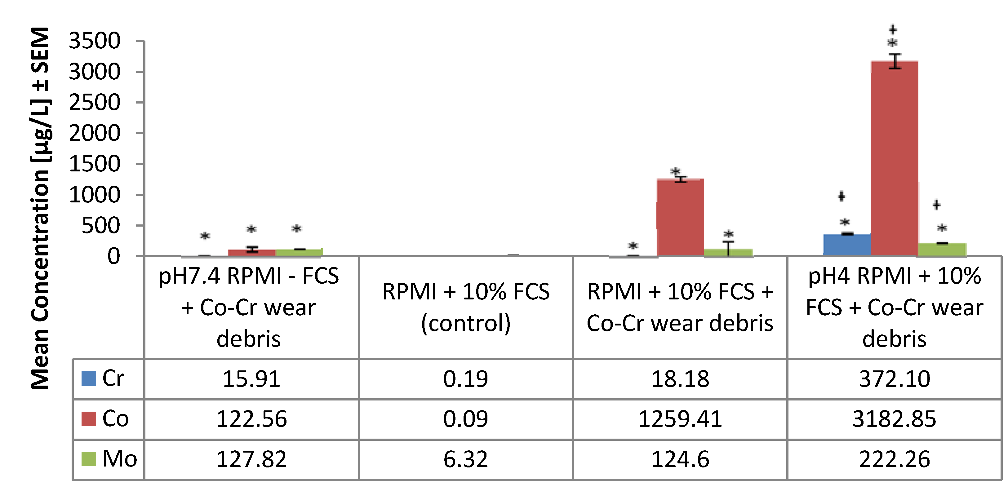
It has been reported that 20%–30% material loss can be attributed to corrosion-related damage, and that not only includes the pure corrosion process but also the wear induced/enhanced corrosion process that is defined as tribocorrosion [23]. Mechanically assisted corrosion, also referred to as tribocorrosion, is an irreversible process that occurs on the surface causing a deterioration of the material due to the combined wear and corrosion actions that simultaneously take place [31]. Tribocorrosion, present at bearing surfaces and within modular taper connections between components of the arthroplasty device, has been proposed to be the primary process by which ions and particles are generated [37]. The released ions can activate the immune system by forming complexes with native proteins [38]. Chromium and cobalt have similar protein binding affinity, and bind to proteins in proportion to the concentration ratio [39]. Once a metal is bound to a protein, it can be systemically transported and either stored or excreted [28]. Additionally, the presence of wear debris in the peri-implant area leads to macrophage phagocytosis of particulate debris and activation to stimulation of the release of a variety of mediators, such as free radicals and nitric oxide, and a myriad of proinflammatory cytokines and chemokines [40]. It has been reported that local acidification may develop during acute and chronic inflammation [41] and high hydrogen ion concentrations down to pH 5.4 have been found in inflamed tissue [42]. In turn, such an acidic environment, created by actively metabolizing immune cells, may enhance the corrosion process, increasing the metal ions being released. To illustrate this, Posada et al. [43] showed that significantly higher concentrations of Co and Cr were released when CoCr metal wear debris were incubated at low pH (Figure 1). Their findings suggest that the osteolysis process generated by wear debris may be exacerbated by the lowering of pH at an inflammation site. This is in line with reports of synovial fluid acidosis correlating with radiological joint destruction in rheumatoid-arthritis knee joints [44,45].
Figure 1. Metal ions released in vitro from CoCr alloy into RPMI-1640 medium at pH 7.4 and 4.0. Results are expressed as mean values (±standard error of the mean (SEM), n = 3). FCS = foetal calf serum. * Significantly different from control values (p < 0.05) by one-way Analysis Of Variance (ANOVA) followed by Dunnett’s multiple comparison test. Significant difference between pH 7.4 and pH 4.0.
Lubricants 03 00539 g001

2.1. Chromium

After entering the body from an exogenous source, Cr3+ binds to plasma proteins such as transferrin, an iron-transporting protein. Regardless of the source, Cr3+ is widely distributed in the body and accounts for most of the chromium in plasma or tissues. The Cr3+ is taken up as a protein complex into bone marrow, lungs, lymph nodes, spleen, kidney, and liver, with the highest uptake being in the lungs [46]. It has been shown that cell membranes are relatively impermeable to Cr3+. When varying amounts of radioactive Cr3+ were added to whole blood in vitro, almost all of the radioactivity (94%–99%) remained in the plasma with an insignificant amount retained in the red blood cells (RBC). Similar results were obtained in vivo [47]. Similarly, low permeability of Cr3+ was found in Chinese hamster lung V79 cells exposed to Cr3+ complexes [48]. Additionally, it has been shown that the cellular uptake of Cr6+ is several-fold greater than that of Cr3+ ion, because trivalent chromium is predominantly octahedral and diffuses slowly [49]. In contrast to Cr3+, Cr6+ is rapidly taken up by RBCs and reduced to Cr3+ inside the cell. Cr6+ enters the cell through non-specific anionic channels, such as the phosphate and sulphate anion exchange pathway [50,51]. Once within the cell, Cr6+ is reduced metabolically by the redox system to short-lived intermediates Cr5+, Cr4+, and ultimately to the most stable species Cr3+ [52,53,54]. Cr3+ interacts and forms complexes with DNA, protein, and lipids resulting in increased chromium intracellular levels [51,52,53].
Table 1. Metal ion levels measured in whole blood and synovial fluid from patients with metal-on-metal (MoM) hip replacements.
AuthorImplantBody fluidFollow upMean Concentration (µg/L)
CoCr
Daniel, et al. [55]MoM resurfacingWhole bloodUp to 4 yearsPre-op0.20.3
1 year1.32.4
4 years1.21.1
Ziaee, et al. [56]MoM resurfacingWhole bloodMean of 53 monthsControl0.30.2
Patients1.41.9
Antoniou, et al. [57]MoM (THA and resurfacing), MoP (THA)Whole blood1 yearControl1.80.1
MoM THA2.60.6
MoM resurfacing2.40.5
MoP THA1.70.1
non-steep (component abduction <55°)2.43.6
Wretenberg [58]MoM THA (Case report)Whole blood37 years-22.919.4
Hart, et al. [59]Painful MoM resurfacingsWhole bloodmedian of 27 monthsUnilateral4.5 *3.0 *
Bilateral10.6 *7.9 *
Hart, et al. [60]Failed MoM resurfacingWhole bloodMean 51 months-112.661.7
Langton, et al. [61]MoM resurfacing (ASR, BHR)Whole bloodminimum of 12 monthsASR2.7 *4.2 *
BHR1.8 *4.2 *
Adverse reactions69.029.3
Davda, et al. [62]Symptomatic MoM, THA and resurfacingSynovial fluidMean of 36 monthsUnexplained pain1127.0 *1337.0 *
Defined cause of failure1014.0 *1512.0 *
Hart, et al. [63]MoM, THA and resurfacingWhole blood39–42 monthsFailed6.9 *5.0 *
Well-functioning1.7 *2.3 *
Non-pseudotumor1.9 *2.1 *
Pseudotumor9.2 *12.0 *
Malviya, et al. [64]MoM, MoP, THAWhole blood2 yearsMoM5.22.8
MoP1.60.8
Fritzsche, et al. [65]bilateral MoM resurfacing followed by unilateral MoM THA (Case report)Whole blood, aspirate of pseudotumor3 months after revision surgeryBlood138.039.0
Aspirate of pseudotumor258.01011.0
Well-functioning2.31.6
Matthies, et al. [66]MoM, THA and resurfacingWhole bloodMedian of 39 monthsNo pseudotumor2.93.2
Pseudotumor11.06.7
Lass, et al. [67]MoM, THASynovial fluidminimum of 18 years-113.4 *54.0 *
* Ion concentrations expressed as median values. Pre-op = previous to operation, THA = total hip arthroplasty, MoM = metal-on-metal, MoP = metal-on-polyethylene, BHR = Birmingham Hip Resurfacing, ASR = Acetabular System Resurfacing, DePuy.
Excretion of Cr occurs primarily via urine, with no major retention in organs. Approximately 10% of an absorbed dose is eliminated by biliary excretion, with smaller amounts excreted in hair, nails, milk, and sweat. Clearance from plasma is generally rapid (within hours), whereas elimination from tissues is slower (with a half-life of several days) [68].
The toxicity, mutagenicity, and carcinogenicity of chromium compounds are well-established phenomena [46,50,69,70]. Long-term occupational inhalational exposure to Cr levels 100–1000 times higher than those found in the natural environment have been associated with squamous cell carcinoma and adenocarcinoma in exposed workers [71]. Epidemiological studies carried out in the UK, Europe, Japan, and the United States have consistently shown that workers in occupations where particulate chromates are generated or used have an elevated risk of respiratory disease, fibrosis, perforation of the nasal septum, development of nasal polyps, and lung cancer [72]. Additionally, during the intracellular reduction of Cr6+ to the stable Cr3+, reactive intermediates (for example, reactive oxygen species (ROS), pentavalent and tetravalent chromium species) are generated, which causes a wide variety of DNA lesions including Cr-DNA adducts, DNA-protein crosslinks, DNA-DNA crosslinks, and oxidative damage [68,73].
Cr toxicity is associated with its oxidation state. However, it is still controversial whether Cr is released as Cr6+ in patients with MoM devices, with some reports supporting this idea and others disproving it [60,70,74]. In the authors’ experience, analysis of the speciation of Cr is fraught with difficulty due to the instability of Cr6+, which tends to oxidise to Cr3+ very rapidly. There is a general consensus, however, that Cr(III) is elevated in the biological fluids of all patients with MoM-type implants [63,75].

2.2. Cobalt

For the general population, diet is the main source of exposure to Co and it is readily absorbed from the small intestine [68,76]. Most of the consumed Co is excreted in the urine and the little that is retained is mainly in the liver and kidneys [68]. Under physiological conditions, this element is mostly accumulated in the liver, kidneys, heart, and spleen, while minimum concentrations are found in the blood serum and tissues of the brain and pancreas [77]. Molecular details of the mode of Co uptake into cells are not well known [76,78]. However, it is likely that it is transported into the cells by broad-specificity divalent metal transporters [78]. It has been shown that P2X7, a transmembrane ionotropic receptor, is involved in the uptake of divalent cations and Co [79]. In the same way, a protein named divalent metal transporter 1 (DMT1) has been shown to have a broad substrate specificity favouring divalent metals including Co2+ [80,81,82]. Additionally, the cellular uptake of Co may be mediated both by active transport ion pumps (i.e., Ca2+/Mg2+ ATPase and Na+/K+ ATPase) and endocytosis [84].
The only biological known function of Co is as an integral part of vitamin B12, which is incorporated into enzymes that participate in reactions essential to DNA synthesis, fatty acid synthesis, and energy production [76,78]. Even though Co has a role in biological systems, overexposure results in toxicity [78], which involves development of hypoxia, increases in the level of ROS, suppression of Adenosine triphosphate (ATP) synthesis, and initiation of apoptotic and necrotic cell death [77]. Co ions can directly induce DNA damage, interfere with DNA repair, and cause DNA-protein crosslinking and sister chromatid exchange [68]. The exact mechanism for Co carcinogenicity remains to be elucidated, but it has been established that free radical generation contributes to its toxicity and carcinogenicity [83].

3. Biological Effects

3.1. Toxicity

Particulate wear debris generated by MoM has an average particle size range of 30 to 100 nm [84]. The reduced size of the particles allows their entry into tissues and organs and diffusion throughout the body, and interaction with different types of cells [85]. Concern about the toxicity of such particles has led to a number of studies assessing the effects of CoCr metal wear debris in vitro on a variety of cells [86,87,88]. It has been established that both Co ions and Co nanoparticles are cytotoxic and induce apoptosis and, at high concentrations, they induce necrosis with an inflammatory response [89]. Papageorgiou et al. [86] compared the cytotoxic and genotoxic effects of nanoparticles and micron-sized particles of CoCr alloy using human fibroblasts. Their results showed that exposure to both nano- and micron-sized particles of CoCr alloy, at the same particle mass per cell, causes different types and amounts of cellular damage. Posada et al. [88] investigated the effects of the combined exposure to CoCr nanoparticles and cobalt ions released from a resurfacing implant on monocytes (U937 cells), and used much lower concentrations of nanoparticles than the previous study [85]. They showed that metal debris in combination with Co ions had a direct effect on cell viability. Interestingly, they showed that previous exposure to Co ions seems to sensitise U937 cells to the toxic effects of both Co ions themselves and to nanoparticles, pointing to the potential for interaction in vivo. Their results indicate that even low doses of CoCr nanoparticles can exert cytotoxic effects. Dalal et al. [87] compared the responses of human osteoblasts, fibroblasts, and macrophages exposed to different metal-based particles (i.e., cobalt-chromium (CoCr) alloy, titanium (Ti) alloy, zirconium (Zr) oxide, and Zr alloy). They found that CoCr-alloy particles were by far the most toxic and decreased viability and proliferation of human osteoblasts, fibroblasts, and macrophages. VanOs et al. [90] used commercially available 60 nm and 700 nm round chromium oxide (Cr2O3) particles to analyse the cytotoxic effects of chromium oxide particles on macrophage responses in vitro. With both particle sizes, cell mortality increased, resulting in a significant decrease in total cell numbers, as well as a significant increase in late apoptosis and necrosis. Tsaousi et al. [91] investigated the in vitro cytotoxicity and genotoxic effects of alumina ceramic (Al2O3) particles in comparison with CoCr alloy particles. They found no significant differences in cell viability between control and ceramic-treated cells, at all doses and time-points studied. However, and in agreement with the studies mentioned above, cells exposed to CoCr alloy particles showed both dose- and time-dependent cytotoxicity including damage to, and loss of, chromosomes.
The apoptotic effects of Co ions have mainly been reported at concentrations starting from 100 μM, where Co induced cell death and apoptosis in a dose- and time-dependent manner [92,93,94]. Catelas et al. [95] demonstrated that macrophage mortality induced by metal ions depends on the type and concentration of metal ions as well as the duration of their exposure. Overall, apoptosis was predominant after 24 h with both Co2+ (0–10 ppm) and Cr3+ (0–500 ppm) ions, but high concentrations induced mainly necrosis at 48 h. This same group also showed that Co2+ and Cr3+ induced mortality and apoptosis in J774 macrophages [96,97]. In a similar way, Akbar et al. [94] reported that exposure to high concentrations of metal ions (10 and 100 μM Cr6+, 100 μM Co2+) initiated apoptosis that resulted in decreased lymphocyte proliferation. A variety of soluble metals, including Co2+ and Cr3+, at a range of concentrations between 0.05 and 5 mM, were found to induce Jurkat T-lymphocyte DNA damage, apoptosis, and/or direct necrosis in a metal- and concentration-dependent manner [98].
From all these reports, it seems evident that CoCr nanoparticles and metal ions released from MoM implants have toxic effects in vitro and may pose a health risk to patients, regardless of whether their implant is well-functioning or failing. This toxicity helps explain the higher prevalence of adverse reactions to metal debris when compared to ceramic or polyethylene particles. The long-term effects of the exposure to these particles and ions remain a concern. Adverse health effects caused by accumulated metal particles in the periprosthetic tissues include osteolysis [99], inflammation, pain, and pseudotumours [100]. Case et al. [101] reported that the accumulation of metal particles in lymph nodes causes structural changes such as necrosis and fibrosis. Multiple reports [102,103,104,105,106,107,108,109] have described patients with MoM implants who presented systemic adverse effects including neurological symptoms such as auditory impairment/deafness, visual impairment/blindness, peripheral neuropathy/dysesthesia of the extremities, poor concentration/cognitive decline, cardiomyopathy, and hypothyroidism. Several authors have associated these adverse effects with grossly elevated systemic Co blood levels. For example, Devlin et al. [110] reviewed 10 cases of suspected prosthetic hip-associated cobalt toxicity and reported that these patients had findings consistent with cobalt toxicity, including thyroid, cardiac, and neurologic dysfunction. Similarly, Bradberry et al. [111] reviewed some cases in which patients exposed to high circulating concentrations of cobalt from failed hip replacements developed neurological damage, hypothyroidism, and/or cardiomyopathy. Finally, Clark et al. [112] reported that chronic exposure to MoM hip resurfacing is associated with subtle structural changes in the visual pathways and the basal ganglia in asymptomatic patients. Consistent with this notion, revision surgery to remove the defective metal hip prostheses resulted in lowered blood concentrations of metal ions and improved symptoms. Evidence is accumulating that systemic elevated concentrations of Co ions, due to the presence of wear debris, pose a serious health risk for some patients bearing CoCr MoM implants. There has also been speculation of a potential carcinogenic effect, however, recent reports [113,114] suggest that CoCr-containing hip implants are unlikely to be associated with an increased risk of cancer.

3.2. Immunological

Wear debris products generated at the articulation site may lead to a chronic inflammatory reaction in the periprosthetic region, resulting in implant failure caused by macrophage-stimulated osteolysis and aseptic loosening [115,116], which is the principal biological mechanism underlying prosthesis failure according to the National Joint Registry of England, Wales, and Northern Ireland [117].
It has been established and accepted that the presence of implant devices and wear debris incites a foreign body inflammatory reaction [118]. Metallic debris derived from alloy implants induces macrophage activation and triggers immune responses resulting in the release of an array of proinflammatory mediators including Tumor necrosis factor alpha (TNF)-α, IL-1β, IL-6, and IL-8 [119]. TNF-α, IL-1, and IL-6 induce osteoclast differentiation and maturation, which lead to bone resorption and, ultimately, aseptic loosening [120] (Figure 2). Dalal et al. [87] reported an increase in TNF-α and IL-8 production by human osteoblasts, fibroblasts, and macrophages in response to different metal-based particles. Interestingly, they observed that the greatest cytokine responses of macrophages were to CoCr alloy particles. Posada et al. [88] also reported higher levels of secretion of IL-6, TNF-α, and interferon (IFN)-γ by resting monocyte-like cells (U937) after exposure to high concentrations of metal debris and the combination of metal debris and Co ions. Devitt et al. [121] investigated the in vitro effect of Co ions on a variety of cell lines by measuring production of IL-8 and Monocyte chemoattractant protein-1 (MCP-1) and found that Co ions enhanced the secretion of both cytokines in renal epithelial cells, gastric and colon epithelium, monocytes and neutrophils, and small airway epithelial cells. These investigations suggest a key role of Co ions in the immune response to wear debris.
Figure 2. Schematic diagram of macrophage-stimulated osteolysis.
Lubricants 03 00539 g002
Despite the understanding of implant-related cytokine/chemokine networks that are released by different peri-implant cell types, knowledge about the mechanisms mediating cellular interaction with debris particles and the subsequent activation of macrophages to produce and release the inflammatory mediators remain incomplete [122]. This has led to a number of studies seeking to define such mechanisms. Toll-like receptors (TLRs) are mainly found on monocytes and macrophages, and have been previously shown to activate the inflammatory cascade by triggering the expression of various cytokines (IL-1, IL-6, TNF-a), growth factors (macrophage colony stimulating factor-1), and chemokines (MIP-1 a, MCP-1), and activating various downstream signalling pathways (nuclear factor-κB (NF-κB), protein kinase B (AKT/PKB), and mitogen-activated protein kinase (MAPK) [123]. Tyson-Capper et al. [124] demonstrated that Co ions released from MoM joint replacement implants stimulate innate immune responses via direct activation of TLR4. Similarly, Potnis et al. [125] also demonstrated that Co-alloy particles trigger immune responses via the Toll-like receptor 4 (TLR4) myeloid differentiation primary response protein 88 (MyD88)-dependent signalling pathway. Since a key element in the initiation of the innate immune response against pathogens is the recognition of components commonly found on the pathogen, referred to as pathogen-associated molecular patterns (PAMP) [126], these studies suggest that wear particles could have a PAMP-like behaviour and bind to TLRs being expressed by macrophages, which then initiates signalling pathways leading to the stimulation of the immune response. Additionally, endogenous molecules generated upon tissue injury, termed damage-associated molecular patterns (DAMPs), directly activate TLRs [127]. Proteins released by the damaged periprosthetic tissue include several heat shock proteins, biglycan, and fragments of extracellular molecules, such as oligosaccharides of hyaluronic acid and heparan sulphate, all of which are known activators of TLR2 and TLR4 [1].
Metal debris and ions can activate the immune system by inducing a delayed-type hypersensitivity reaction [38]. The most common sensitizing orthopaedic metals are nickel, cobalt, and chromium [38,128,129,130]. It is thought that stimulated T-cells generate pro-osteoclastogenetic factors that can alter bone homeostasis [115] and therefore contribute to osteolysis. The prevalence of metal sensitivity among the general population is approximately 10% to 15% and the prevalence of metal sensitivity among patients with well-functioning and poorly functioning implants has been reported to be ~25% and 60%, respectively, as measured by dermal patch testing [38]. The response of metal-specific lymphocytes to metals as debris or in the form of metal ions has been linked to poor implant performance. Cell-mediated type-IV hypersensitivity reaction characterized by vasculitis with perivascular and intramural lymphocytic infiltration of the postcapillary venules, swelling of the vascular endothelium, recurrent localized bleeding, and necrosis has been reported following MoM hip replacements [131]. Lymphocyte infiltrates have also been reported in the soft-tissue masses, described as pseudo-tumours, following MoM resurfacing arthroplasty [21,132]. Additionally, metal-specific T-cells have been isolated from patients with contact dermatitis, indicating a T-cell-led inflammatory reaction against a metal-derived antigen [133]. For example, Cr exposure has been shown to upset the immunoregulatory balance between Th1 and Th2 cells that controls different immune effects or functions through the production of distinct cytokines [134]. In the work of these authors, the effects of cadmium, chromium, inorganic mercury, and inorganic lead exposure on the immune system were determined by measuring cytokine production of human peripheral blood mononuclear cells. Their results showed that the cytokines assayed were differentially affected by heavy metal exposure. Of particular interest, Cr significantly increased the production of IL-1β while decreasing the production of IFN-γ, IL-6, IL-8, and IL-10.
It has been suggested that activation of T-cells following exposure to biomaterial particles is driven by macrophages and requires synergistic signals primed by both antigen presentation and costimulation [135,136,137]. Bainbridge et al. [138] examined the expression of CD80 and CD86 costimulatory molecules in U937 cells that had been exposed to titanium aluminium vanadium alloy (TiAlV) implant wear debris. This was compared to the expression of these costimulatory molecules in tissues taken from patients with aseptic loosening. They demonstrated the increased expression of costimulatory molecules in response to wear particles both at the bone implant interface and in vitro. These findings reinforce the hypothesis that macrophages have the potential to aid T-cell activation in response to metal or metal ions from orthopaedic implants, as well as to augment any T-cell mediated response.
Several studies have described perivascular lymphocytes in tissue membranes around failed MoM implants apparently not associated with infection, and the authors have interpreted this inflammation as an immunologic reaction against metal ions or metal particles associated with those articulations [131,139,140,141,142]. Polyzois et al. [143] reviewed the evidence of local and systemic toxicity of wear debris from total hip arthroplasty and found extensive evidence and experimental data supporting the fact that orthopaedic metals induce local immunological effects characterized by an unusual lymphocytic infiltration and cell-mediated hypersensitivity. Thomas et al. [144] reported the case of a patient who developed eczema and impaired wound-healing following the fixation of an ankle fracture with titanium-based implants. Histological analysis of the tissue around the implant demonstrated inflammation primarily with lymphocytes, and a contact allergy to nickel and cobalt was found in the absence of titanium hyper-reactivity, raising the question of a prior unknown nickel exposure as the source of the complications. Similarly, Gao et al. [145] reported a case of systemic dermatitis caused by Cr (serum Cr level of 61.9 µg/L) after total knee arthroplasty. Another feature reported as a metal-induced systemic T-lymphocyte-mediated hypersensitivity reaction is the formation of periprosthetic soft-tissue masses in patients with MoM devices [132,146]. A delayed T-lymphocyte-mediated self-perpetuating response can also create extensive tissue damage [122]. To date, the only means to predict/diagnose those individuals that will have an excessive immune response to metal exposure that may lead to premature implant failure are lymphocyte transformation test and patch testing (for skin reactions). However, they are not so useful in the evaluation of deep tissue metal allergy [122,145]. Although complications in metal-allergic patients appear to be generally rare [115], metal nanoparticles and high metal ion concentrations remain a concern as they could trigger early events leading to implant failure or a shorter implant lifespan in sensitized patients. Moreover, it remains unclear whether sensitization is a direct cause of implant loosening and failure, or if it is a consequence of particle loading due to device loosening.

3.3. Gene Expression

Over the past few years numerous investigations have been carried out to study the effects of different ions and particulate wear debris on the expression of an array of cytokine and toxicology related genes in vitro. TNF-α, IL-1, and IL-6 are cytokines that have been reported to play a central role in the induction of implant osteolysis [147,148]. Extensive work has been carried out on cytokine production by macrophages in response to wear debris. Sethi et al. [40] studied the macrophage response to cross-linked ultra-high molecular weight polyethylene (UHMWPE) and compared it to conventional UHMWPE as well as TiAlV and cobalt-chrome alloy (CoCr). At 24 and 48 h, macrophages cultured on TiAlV and CoCr alloy expressed higher levels of IL-1α, IL-1β, IL-6, and TNF-α than when grown on UHMWPE materials according to real time reverse transcription polymerase chain reaction (qRT-PCR) analysis. Jakobsen et al. [149] compared surfaces of as-cast and wrought CoCrMo alloy and TiAlV alloy when incubated with mouse macrophage J774A.1 cells and reported a significant increase in the levels of expression of TNF-α, IL-6, IL-1α, and β from cells incubated with alloys compared to non-stimulated cells. Garrigues et al. [150] used microarrays to investigate alterations in the phenotype of macrophages as they interact with UHMWPE and TiAlV alloy particulate wear debris. Their findings further validate the important roles of TNF-α, IL-1β, IL-1α, IL-6, MIP1α, and MIP1β in osteolysis. In a recent study, Posada et al. [43] examined the ability of the metal debris and Co ions to induce general toxicology-related gene expression of human monocyte-like U937 cells. In some experiments, they pre-treated the cells with Co ions prior to exposure to CoCr particles, in order to simulate the in vivo situation where a patient may receive a second MoM implant in either a bilateral or a revision procedure. Analysis of qRT-PCR results found significant up-regulation of inducible nitric oxide synthase (NOS2) and Bcl2-associated athanogene (BAG1) in Co pre-treated cells which were subsequently exposed to Co ions and debris. They showed that metal debris was more effective as an inducer of gene expression when cells had been pre-treated with Co ions. Overexpression of NOS2, which leads to an over production of NO, could have a predominant role in the inflammation and acidification of the peri-implant microenvironment, which in turn could exacerbate the corrosion of the nanoparticles. Co-expression of BAG1 and Bcl-2 has been shown to increase protection from cell death [151,152]. Consequently, up-regulation of BAG1 could be interpreted as part of a defence mechanism for delaying cell death in response to metal toxicity, particularly Co toxicity, in this case. Since the main gene expression fold changes were observed in cells pre-treated with Co ions, patients with a MoM implant undergoing revision surgery or receiving a second MoM device may potentially be at higher risk of implant failure.
As well as macrophages, other cell types have been reported as being involved in the biological response to implant wear debris. As a result, there are similar studies on monocytes, lymphocytes, osteocytes, and osteoblasts. For example, the effects of CoCr particles on osteocytes were tested by Kanaji et al. [148]. CoCr treatment of murine long bone osteocyte Y4 (MLO-Y4) osteocytes significantly up-regulated TNFα gene expression after 3 and 6 h and TNF-α protein production after 24 h, but down-regulated IL-6 gene expression after 6 h. MG-63 osteoblasts were treated by Vermes et al. [153] with titanium, titanium alloy, chromium orthophosphate, polyethylene, and polystyrene particles and they reported that each type of particle significantly suppressed procollagen alpha1[I] gene expression (p < 0.05), whereas other osteoblast-specific genes (osteonectin, osteocalcin, and alkaline phosphatase) did not show significant changes. The effect of particulate derivatives of nickel and cobalt alloys on the mRNA levels of chemokine receptors CCR1 and CCR2 on monocytes/macrophages from whole blood were analysed by Fujiyoshi and Hunt [154]. Although there were no significant differences in the level of CCR1 mRNA in monocytes/macrophages incubated with NiCr particulates, there was a down-regulation in the level of CCR2 in cells incubated with NiCr and CoCr particles. All these investigations indicate that wear debris and metal ions derived from MoM implants can cause an adverse tissue response by modulating gene expression on several types of cells, which suggests that osteolysis and subsequent aseptic loosening is the result of the concerted action of the different cell types.
Previous studies have stated that ions released from the wear debris could also affect gene expression. It has been reported that Cr+3 and Co+2 ions could induce damage to proteins in macrophage-like cells in vitro, probably through the formation of reactive oxygen species (ROS) [155,156]. U937 cells were exposed to Cr+6 and Co+2 ions by Tkaczyk et al. [157]. Cr+6 induced the protein expression of Mn-superoxide dismutase, Cu/Zn superoxide dismutase, catalase, glutathione peroxidase, and heme oxygenase-1 (HO-1) but had no effect on the expression of their mRNA, whereas Co+2 induced the expression of both protein and mRNA of HO-1 only. Co+2 had no effect on the expression of the other proteins. The overexpression of HO-1 has been suggested to play an important role in cellular protection against oxidant-mediated cell damage [158]. This suggests that the results from Tkaczyk et al. [157] show that Cr and Co ions cause oxidant-mediated cell damage. Type-I collagen gene expression was evaluated by Hallab et al. [159] after treating MG-63 cells with increasing concentrations (from 0.001 to 10 mM) of a variety of metal ions including Co and Cr. At toxic concentrations (1 mM), Co depressed osteoblast function by significantly decreasing the levels of type I collagen gene expression to 40% of control values. Queally et al. [160] showed that 10 ppm Co ions induce chemokine secretion in primary human osteoblasts by measuring the up-regulation of IL-8 and MCP-1 gene expression in osteoblasts stimulated with 0–10 ppm Co2+. The level of expression of one of the principal proteinases capable of degrading native fibrillar collagens in the extracellular matrix, MMP-1, and its tissue inhibitors (TIMP-1) in U937 cells exposed to Co2+ and Cr3+ ions for 24 h, was determined by Luo et al. [161] who showed that these ions induce up-regulation in a dose-dependent manner. Their expression was studied to gain insight into the regulation of extracellular matrix degradation and tissue remodelling around hip prostheses. Altered expression of MMP-1 and TIMP-1 in the periprosthetic tissues has led to the hypothesis that their imbalance could contribute to the loosening of total hip prosthesis [162]. The findings from Luo et al. [161] suggest that Co and Cr ions can up-regulate the MMP-1 expression in vivo. These studies provide more evidence of potential gene expression modulation by wear debris and ions derived from MoM implants.
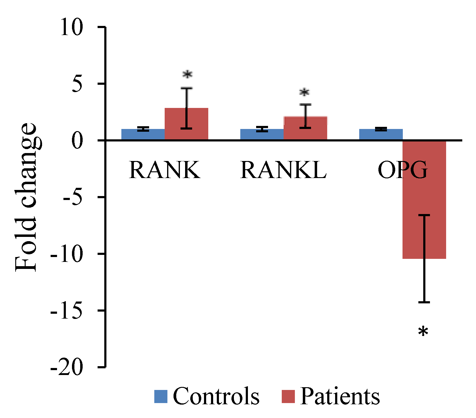
Receptor activator of nuclear factor-κB ligand (RANKL), its receptor, receptor activator of nuclear factor-κB (RANK), and its soluble inhibitor osteoprotegerin (OPG) are recognized as key regulators of osteoclast formation that regulate bone resorption in both health and disease [163]. Several studies have demonstrated the expression of mRNA encoding RANKL, OPG, and RANK in peri-implant tissues associated with osteolytic zones [164,165,166,167]. Jiang et al. [168] demonstrated a significantly elevated gene expression of RANKL in CoCr particle-challenged osteoblasts. Similarly, Pioletti and Kottelat [169] showed an increase of osteoblast gene expression for RANKL after exposure to Ti particles. Zijlstra et al. [170] determined the effects of Co and Cr ions on the expression of bone turnover regulatory proteins RANKL and OPG on human osteoblast-like cells. They found that the RANKL/OPG ratio increased after 72 h of exposure to 10 μg/L Co, 1 μg/L Cr, and higher, and at 1 μg/L Co + Cr and higher, indicating net bone loss. These findings are interesting since they seem to suggest that even in well-functioning MoM implants with systemic Co and Cr levels around 1 μg/L, local periprosthetic osteolytic reactions may take place. In a pilot study in our laboratory, gene expression of RANK, RANKL, and OPG in peripheral blood from six patients that had MoM hip implants for at least one year was investigated and correlated with the whole blood metal ion levels at the time of the analysis. There was a significant up-regulation of RANK and RANKL and significant down-regulation of OPG when compared to controls (no implant) (Figure 3). It has been suggested that the RANKL/OPG ratio is raised significantly in patients with severe osteolysis and that this imbalance is involved in bone resorption mechanisms [171]. Since OPG was down-regulated, patients had higher ratios (27.69 ± 10.53, mean ± SEM) when compared to controls (1 ± 0.15, mean ± SEM), suggesting an imbalance in the bone turnover system favouring bone resorption. However, a clear relationship between RANKL/OPG ratios and ion levels could not be established. Although changes in gene expression were identified, the lack of pre-surgery data made it impossible to determine whether the presence of the MoM implant was the cause of such changes.
All the studies mentioned in this section have been carried out in order to understand how wear debris affects the levels of expression of genes involved in osteolysis in tissues surrounding the joint implant. They suggest different mechanisms for transcriptional activation of the genes investigated, which could indicate that gene expression is modulated in a dose- and particle-dependent manner and as the result of several signals coming together.
Thus, the biological responses to metal wear debris are complex, involving regulation at different cellular and molecular levels to try and maintain intra- and extra-cellular homeostasis. When cells fail, they tip the balance towards inflammation and acidification. This acidification of the peri-implant microenvironment in turn enhances the corrosion of the nanoparticles and release of metal ions, which exacerbates the adverse reaction ultimately resulting in osteolysis and subsequent aseptic loosening.
Figure 3. Fold variations of target genes in blood samples from controls and from patients with MoM hip implants. Results are mean values ± standard deviation (SD) (six biological samples with three technical replicates per gene assayed) expressed as the negative reciprocal. * Significantly different from control values by Analysis of variance (ANOVA) followed by Dunnet’s comparison test (p < 0.05) (Posada et al., unpublished data).
Lubricants 03 00539 g003

4. Discussion

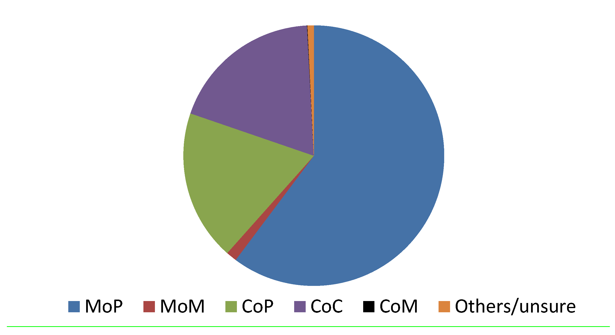
Since the recall of MoM devices in 2010, the trends in bearing surface materials have changed, showing ceramic-on-polyethylene becoming more popular (Figure 4). The use of MoM has declined dramatically and the proportion of MoM resurfacing implants has decreased from a peak in 2006 to account for only 1.1% of implants in 2013 [117]. Although the recalls have taken most of the defective implant designs off the market, there are still tens of thousands of patients in the UK alone with these implants still in situ.
Figure 4. Number of primary hip replacements registered in 2013 by bearing surface. Metal-on-polyethylene (MoP): 45,944. Metal-on-metal (MoM, includes both total and resurfacing): 1014. Ceramic-on-polyethylene (CoP): 14,258. Ceramic-on-ceramic (CoC): 14,433. Ceramic-on-metal (CoM): 32. Others/unsure: 593 [3].
Lubricants 03 00539 g004
The Medicines and Healthcare Products Regulatory Agency (MHRA) issued information and advice about the follow-up of both symptomatic and asymptomatic patients implanted with MoM hip replacements, which include appropriate imaging (Metal Artifact Reduction Sequence (MARS) MRI/ultrasound), whole blood metal ion levels, and situations where revision may need to be considered. The MHRA has suggested that MARS MRI scans (or ultrasound scans) should carry more weight in decision-making than blood ion levels alone, and combined whole blood cobalt and chromium levels of greater than 7 ppb (7 μg/L), indicates potential for soft tissue reaction [172]. This decision is becoming more established among orthopaedic surgeons, but it does leave the quandary of the patients with circulating metal ion levels in several hundreds of µg/L and normal cross-sectional imaging. For these patients, many of whom have a well-functioning implant, it is difficult to understand whether or not the effects of particles and ions will ultimately impact their health locally or distally to their implant. It remains unclear whether these patients should be advised on a revision to avoid long-term systemic adverse effects on organs such as the heart and brain. It is worth noting that the 7 ppb threshold is a means of understanding how well the hip is performing in vivo [173], and revision is only considered if imaging is abnormal and/or blood metal ion levels rise [174]. Additionally, although this threshold is not based on soft tissue damage, levels of greater than 7 ppb are associated with significant soft-tissue reactions and failed MoM hips [63].
Although there has been widespread research on the various functions of different cytokines, questions concerning how inflammatory responses are triggered by wear particles remain largely unanswered. Specific organelles could play an important role in the cellular response triggered by wear particles. A growing body of evidence has suggested a role for endoplasmic reticulum (ER) stress in initiating inflammation, which is now thought to be fundamental to the pathogenesis of inflammatory diseases [175,176]. There is evidence suggesting that metal particles can cause increasing ER stress in various types of cells [177,178]. This presents the possibility that wear particles, produced around the prosthesis, have the potential to stimulate ER stress and thus may play a role in particle-induced osteolysis.
Chronic environmental exposure to some metal compounds, including arsenic, nickel, chromium, and cadmium, has been known to induce cancers and other diseases in exposed individuals [179]. While it has been shown that these metals disturb a vast array of cellular processes, such as redox state and various intracellular stress-signalling pathways, their ability to induce acute and/or chronic pathologies remains poorly understood. Sources of potential environmental exposure to these metals include occupational exposure and environmental contamination from industrial production [180]. Additionally, with all the evidence on particles and ions derived from metal orthopaedic implants, such replacements should be considered as an additional source of exposure. Emerging epidemiological studies show that the carcinogenic potential of some toxic metals may involve epigenetic changes, including silencing of DNA repair and tumor-suppressor genes [179]. The combinations of mechanisms, which confer long-term programming to genes and could bring about a change in gene function without changing gene sequence, are termed epigenetic [181]. As artificial articulations are being implanted in younger people, epigenetic studies could help assess how long-term exposure to metal debris and ions could bring about epigenetic changes altering gene expression which may have significant health-related consequences for these patients.

5. Conclusions

Despite the clinical success of hip replacements, this review has summarised some of the work addressing the still-present concern regarding the toxicity of metal particles and ions produced at the articulation site of a MoM implant. A vast literature on the effects of different kinds of debris as well as ions exists with many groups engaged in the discussion of mechanisms involved.
The potential dangers to patient health from CoCr alloy wear debris generation and the release of Co and Cr ions into circulation have been recognized. However, until the regulations for thoroughly testing the safety of medical devices such as hip replacements and other orthopaedic implants is more stringent, there is a very real possibility that 20 years from now we will be reading articles on the award of compensation for the next generation of articulations.

Acknowledgments

OMP is grateful for the financial support of an Overseas Research Scholarship and funds from the University of Strathclyde.

Author Contributions

The main author is Olga M. Posada, who wrote the first draft of the manuscript, and the revisions of this text to form the final draft were contributed by Rothwelle J. Tate, R. M. Dominic Meek, and M. Helen Grant. Dominic Meek provided clinical advice on the text.

Conflicts of Interest

The authors declare no conflict of interest.

References

  1. Cobelli, N.; Scharf, B.; Crisi, G.M.; Hardin, J.; Santambrogio, L. Mediators of the inflammatory response to joint replacement devices. Nat. Rev. Rheumatol. 2011, 7, 600–608. [Google Scholar]
  2. Australian Orthopaedic Association National Joint Replacement Registry. Annual Report 2014. Available online: https://aoanjrr.dmac.adelaide.edu.au/annual-reports-2014 (accessed on 20 January 2015).
  3. National Joint Registry. 11th Annual Report. Available online: http://www.njrcentre.org.uk/njrcentre/Reports,PublicationsandMinutes/Annualreports/tabid/86/Default.aspx. (accessed on 20 January 2015).
  4. Furnes, O.; Paxton, E.; Cafri, G.; Graves, S.; Bordini, B.; Comfort, T.; Rivas, M.C.; Banerjee, S.; Sedrakyan, A. Distributed analysis of hip implants using six national and regional registries: Comparing metal-on-metal with metal-on-highly cross-linked polyethylene bearings in cementless total hip arthroplasty in young patients. J. Bone Joint Surg. Am. 2014, 96A, 25–33. [Google Scholar]
  5. Kanda, A.; Kaneko, K.; Obayashi, O.; Mogami, A. A 42-year-old patient presenting with femoral head migration after hemiarthroplasty performed 22 years earlier: A case report. J. Med. Case Rep. 2015, 9, 17. [Google Scholar]
  6. Sedrakyan, A.; Romero, L.; Graves, S.; Davidson, D.; de Steiger, R.; Lewis, P.; Solomon, M.; Vial, R.; Lorimer, M. Survivorship of hip and knee implants in pediatric and young adult populations. J. Bone Joint Surg. Am. 2014, 96A, 73–78. [Google Scholar]
  7. Eikmans, M.; Rekers, N.V.; Anholts, J.D.H.; Heidt, S.; Claas, F.H.J. Blood cell mRNAs and microRNAs: Optimized protocols for extraction and preservation. Blood 2013, 121, E81–E89. [Google Scholar]
  8. Malchau, H.; Herberts, P.; Eisler, T.; Garellick, G.; Soderman, P. The swedish total hip replacement register. J. Bone Joint Surg. Am. 2002, 84A, 2–20. [Google Scholar]
  9. Prokopovich, P. Interactions between mammalian cells and nano- or micro-sized wear particles: Physico-chemical views against biological approaches. Adv. Colloid Interface Sci. 2014, 213, 36–47. [Google Scholar]
  10. Reito, A.; Puolakka, T.; Elo, P.; Pajamaki, J.; Eskelinen, A. High prevalence of adverse reactions to metal debris in small-headed ASR™ hips. Clin. Orthop. Relat. Res. 2013, 471, 2954–2961. [Google Scholar]
  11. U.S. Food and Drug Administration (FDA). Medical devices. Available online: http://www.fda.gov/MedicalDevices/ProductsandMedicalProcedures/ImplantsandProsthetics/MetalonMetalHipImplants/ucm241770.htm (accessed on 20 January 2015).
  12. Wynn-Jones, H.; Macnair, R.; Wimhurst, J.; Chirodian, N.; Derbyshire, B.; Toms, A.; Cahir, J. Silent soft tissue pathology is common with a modern metal-on-metal hip arthroplasty. Acta Orthop. 2011, 82, 301–307. [Google Scholar]
  13. Langton, D.J.; Jameson, S.S.; Joyce, T.J.; Gandhi, J.N.; Sidaginamale, R.; Mereddy, P.; Lord, J.; Nargol, A.V.F. Accelerating failure rate of the asr total hip replacement. J. Bone Joint Surg. Br. 2011, 93B, 1011–1016. [Google Scholar]
  14. Hayter, C.L.; Gold, S.L.; Koff, M.F.; Perino, G.; Nawabi, D.H.; Miller, T.T.; Potter, H.G. MRI findings in painful metal-on-metal hip arthroplasty. Am. J. Roentgenol. 2012, 199, 884–893. [Google Scholar]
  15. Hart, A.J.; Hester, T.; Sinclair, K.; Powell, J.J.; Goodship, A.E.; Pele, L.; Fersht, N.L.; Skinner, J. The association between metal ions from hip resurfacing and reduced T-cell counts. J. Bone Joint Surg. Br. 2006, 88, 449–454. [Google Scholar]
  16. Keegan, G.M.; Learmonth, I.D.; Case, C.P. Orthopaedic metals and their potential toxicity in the arthroplasty patient—Review of current knowledge and future strategies. J. Bone Joint Surg. Br. 2007, 89B, 567–573. [Google Scholar]
  17. Moroni, A.; Savarino, L.; Hoque, M.; Cadossi, M.; Baldini, N. Do ion levels in hip resurfacing differ from metal-on-metal tha at midterm? Clin. Orthop. Relat. Res. 2011, 469, 180–187. [Google Scholar]
  18. Hallab, N.J.; Jacobs, J.J. Biologic effects of implant debris. Bull. NYU Hospital Joint Dis. 2009, 67, 182–188. [Google Scholar]
  19. Mathew, M.T.; Nagelli, C.; Pourzal, R.; Fischer, A.; Laurent, M.P.; Jacobs, J.J.; Wimmer, M.A. Tribolayer formation in a metal-on-metal (MoM) hip joint: An electrochemical investigation. J. Mech. Behav. Biomed. Mater. 2014, 29, 199–212. [Google Scholar]
  20. Clayton, R.A.E.; Beggs, I.; Salter, D.M.; Grant, M.H.; Patton, J.T.; Porter, D.E. Inflammatory pseudotumor associated with femoral nerve palsy following metal-on-metal resurfacing of the hip. J. Bone Joint Surg. Am. 2008, 90A, 1988–1993. [Google Scholar]
  21. Pandit, H.; Glyn-Jones, S.; McLardy-Smith, P.; Gundle, R.; Whitwell, D.; Gibbons, C.L.M.; Ostlere, S.; Athanasou, N.; Gill, H.S.; Murray, D.W. Pseudotumours associated with metal-on-metal hip resurfacings. J. Bone Joint Surg. Br. 2008, 90B, 847–851. [Google Scholar]
  22. Dowson, D. Tribological principles in metal-on-metal hip joint design. Proc. Inst. Mech. Eng. H J. Eng. Med. 2006, 220, 161–171. [Google Scholar]
  23. Yan, Y.; Neville, A.; Dowson, D. Biotribocorrosion—An appraisal of the time dependence of wear and corrosion interactions: I. The role of corrosion. J. Phys. D Appl. Phys. 2006, 39, 3200–3205. [Google Scholar]
  24. Konttinen, Y.T.; Pajarinen, J. Surgery adverse reactions to metal-on-metal implants. Nat. Rev. Rheumatol. 2013, 9, 5–6. [Google Scholar]
  25. Teoh, S.H. Fatigue of biomaterials: A review. Int. J. Fatigue 2000, 22, 825–837. [Google Scholar]
  26. Ryu, J.J.; Shrotriya, P. Synergistic Mechanisms of Bio-Tribocorrosion in Medical Implants. In Bio-Tribocorrosion in Biomaterials and Medical Implants; Yan, Y., Ed.; Elsevier: Sawston, Cambridge, UK, 2013; pp. 25–44. [Google Scholar]
  27. Hallab, N.J.; Jacobs, J.J. Orthopedic implant fretting corrosion. Corros. Rev. 2003, 21, 183–213. [Google Scholar]
  28. Sargeant, A.; Goswami, T. Hip implants—Paper VI—Ion concentrations. Mater. Des. 2007, 28, 155–171. [Google Scholar]
  29. Singh, R.; Dahotre, N.B. Corrosion degradation and prevention by surface modification of biometallic materials. J. Mater. Sci. Mater. Med. 2007, 18, 725–751. [Google Scholar]
  30. Cadosch, D.; Chan, E.; Gautschi, O.P.; Filgueira, L. Metal is not inert: Role of metal ions released by biocorrosion in aseptic loosening-current concepts. J. Biomed. Mater. Res. A 2009, 91A, 1252–1262. [Google Scholar]
  31. Doni, Z.; Alves, A.C.; Toptan, F.; Gomes, J.R.; Ramalho, A.; Buciumeanu, M.; Palaghian, L.; Silva, F.S. Dry sliding and tribocorrosion behaviour of hot pressed cocrmo biomedical alloy as compared with the cast CoCrMo and Ti6Al4V alloys. Mater. Des. 2013, 52, 47–57. [Google Scholar]
  32. Zeng, Y.; Feng, W. Metal allergy in patients with total hip replacement: A review. J. Int. Med. Res. 2013, 41, 247–252. [Google Scholar]
  33. Steinemann, S.G. Metal implants and surface reactions. Inj. Int. J. Care Inj. 1996, 27, 16–22. [Google Scholar]
  34. Hanawa, T. Metal ion release from metal implants. Mater. Sci. Eng. C Biomim. Supramol. Syst. 2004, 24, 745–752. [Google Scholar]
  35. Friesenbichler, J.; Maurer-Ertl, W.; Sadoghi, P.; Lovse, T.; Windhager, R.; Leithner, A. Serum metal ion levels after rotating-hinge knee arthroplasty: Comparison between a standard device and a megaprosthesis. Int. Orthop. 2012, 36, 539–544. [Google Scholar]
  36. Penny, J.O.; Varmarken, J.E.; Ovesen, O.; Nielsen, C.; Overgaard, S. Metal ion levels and lymphocyte counts: ASR hip resurfacing prosthesis vs. standard THA 2-year results from a randomized study. Acta Orthop. 2013, 84, 130–137. [Google Scholar]
  37. Gilbert, J.L.; Sivan, S.; Liu, Y.; Kocagoez, S.B.; Arnholt, C.M.; Kurtz, S.M. Direct in vivo inflammatory cell-induced corrosion of cocrmo alloy orthopedic implant surfaces. J. Biomed. Mater. Res. A 2015, 103, 211–223. [Google Scholar] [CrossRef] [PubMed]
  38. Hallab, N.; Merritt, K.; Jacobs, J.J. Metal sensitivity in patients with orthopaedic implants. J. Bone Joint Surg. Am. 2001, 83A, 428–436. [Google Scholar]
  39. Yang, J.; Black, J. Competitive-binding of chromium, cobalt and nickel to serum-proteins. Biomaterials 1994, 15, 262–268. [Google Scholar]
  40. Sethi, R.K.; Neavyn, M.J.; Rubash, H.E.; Shanbhag, A.S. Macrophage response to cross-linked and conventional UHMWPE. Biomaterials 2003, 24, 2561–2573. [Google Scholar]
  41. Rajamaki, K.; Nordstrom, T.; Nurmi, K.; Akerman, K.E.O.; Kovanen, P.T.; Oorni, K.; Eklund, K.K. Extracellular acidosis is a novel danger signal alerting innate immunity via the NLRP3 inflammasome. J. Biol. Chem. 2013, 288, 13410–13419. [Google Scholar]
  42. Steen, K.H.; Steen, A.E.; Reeh, P.W. A dominant role of acid ph in inflammatory excitation and sensitization of nociceptors in rat skin, in-vitro. J. Neurosci. 1995, 15, 3982–3989. [Google Scholar]
  43. Posada, O.M.; Gilmour, D.; Tate, R.J.; Grant, M.H. CoCr wear particles generated from cocr alloy metal-on-metal hip replacements, and cobalt ions stimulate apoptosis and expression of general toxicology-related genes in monocyte-like U937 cells. Toxicol. Appl. Pharmacol. 2014, 281, 125–135. [Google Scholar]
  44. Geborek, P.; Saxne, T.; Pettersson, H.; Wollheim, F.A. Synovial-fluid acidosis correlates with radiological joint destruction in rheumatoid-arthritis knee joints. J. Rheumatol. 1989, 16, 468–472. [Google Scholar]
  45. Mansson, B.; Geborek, P.; Saxne, T.; Bjornsson, S. Cytidine deaminase activity in synovial-fluid of patients with rheumatoid-arthritis—Relation to lactoferrin, acidosis, and cartilage proteoglycan release. Ann. Rheum. Dis. 1990, 49, 594–597. [Google Scholar]
  46. Bagchi, D.; Stohs, S.J.; Downs, B.W.; Bagchi, M.; Preuss, H.G. Cytotoxicity and oxidative mechanisms of different forms of chromium. Toxicology 2002, 180, 5–22. [Google Scholar]
  47. Gray, S.J.; Sterling, K. The tagging of red cells and plasma proteins with radioactive chromium. J. Clin. Invest. 1950, 29, 1604–1613. [Google Scholar]
  48. Dillon, C.T.; Lay, P.A.; Bonin, A.M.; Cholewa, M.; Legge, G.J.F. Permeability, cytotoxicity, and genotoxicity of Cr(III) complexes and some Cr(V) analogues in V79 chinese hamster lung cells. Chem. Res. Toxicol. 2000, 13, 742–748. [Google Scholar]
  49. Biedermann, K.A.; Landolph, J.R. Role of valence state and solubility of chromium compounds on induction of cytotoxicity, mutagenesis, and anchorage independence in diploid human fibroblasts. Cancer Res. 1990, 50, 7835–7842. [Google Scholar]
  50. Tkaczyk, C.; Huk, O.L.; Mwale, F.; Antoniou, J.; Zukor, D.J.; Petit, A.; Tabrizian, M. The molecular structure of complexes formed by chromium or cobalt ions in simulated physiological fluids. Biomaterials 2009, 30, 460–467. [Google Scholar]
  51. Raja, N.S.; Sankaranarayanan, K.; Dhathathreyan, A.; Nair, B.U. Interaction of chromium(III) complexes with model lipid bilayers: Implications on cellular uptake. Biochim. Biophys. Acta 2011, 1808, 332–340. [Google Scholar]
  52. Fornsaglio, J.L.; O’Brien, T.J.; Patierno, S.R. Differential impact of ionic and coordinate covalent chromium (Cr)-DNA binding on DNA replication. Mol. Cell Biochem. 2005, 279, 149–155. [Google Scholar]
  53. Shrivastava, H.Y.; Ravikumar, T.; Shanmugasundaram, N.; Babu, M.; Nair, B.U. Cytotoxicity studies of chromium(III) complexes on human dermal fibroblasts. Free Radic. Biol. Med. 2005, 38, 58–69. [Google Scholar]
  54. Afolaranmi, G.A.; Tettey, J.; Meek, R.M.D.; Grant, M.H. Release of chromium from orthopaedic arthroplasties. Open Orthop. J. 2008, 2, 10–18. [Google Scholar]
  55. Daniel, J.; Ziaee, H.; Pradhan, C.; Pynsent, P.B.; McMinn, D.J.W. Blood and urine metal ion levels in young and active patients after Birmingham hip resurfacing arthroplasty—Four-year results of a prospective longitudinal study. J. Bone Joint Surg. Br. 2007, 89B, 169–173. [Google Scholar]
  56. Ziaee, H.; Daniel, J.; Datta, A.K.; Blunt, S.; McMinn, D.J.W. Transplacental transfer of cobalt and chromium in patients with metal-on-metal hip arthroplasty—A controlled study. J. Bone Joint Surg. Br. 2007, 89B, 301–305. [Google Scholar]
  57. Antoniou, J.; Zukor, D.J.; Mwale, F.; Minarik, W.; Petit, A.; Huk, O.L. Metal ion levels in the blood of patients after hip resurfacing: A comparison between twenty-eight and thirty-six-millimeter-head metal-on-metal prostheses. J. Bone Joint Surg. Am. 2008, 90A, 142–148. [Google Scholar]
  58. Wretenberg, P. Good function but very high concentrations of cobalt and chromium ions in blood 37 years after metal-on-metal total hip arthroplasy. Med. Devices (Auckland, N.Z.) 2008, 1, 31–32. [Google Scholar]
  59. Hart, A.J.; Sabah, S.; Henckel, J.; Lewis, A.; Cobb, J.; Sampson, B.; Mitchell, A.; Skinner, J.A. The painful metal-on-metal hip resurfacing. J. Bone Joint Surg. Br. 2009, 91B, 738–744. [Google Scholar]
  60. Hart, A.J.; Quinn, P.D.; Sampson, B.; Sandison, A.; Atkinson, K.D.; Skinner, J.A.; Powell, J.J.; Mosselmans, J.F.W. The chemical form of metallic debris in tissues surrounding metal-on-metal hips with unexplained failure. Acta Biomater. 2010, 6, 4439–4446. [Google Scholar]
  61. Langton, D.J.; Jameson, S.S.; Joyce, T.J.; Hallab, N.J.; Natu, S.; Nargol, A.V.F. Early failure of metal-on-metal bearings in hip resurfacing and large-diameter total hip replacement a consequence of excess wear. J. Bone Joint Surg. Br. 2010, 92B, 38–46. [Google Scholar]
  62. Davda, K.; Lali, F.V.; Sampson, B.; Skinner, J.A.; Hart, A.J. An analysis of metal ion levels in the joint fluid of symptomatic patients with metal-onmetal hip replacements. J. Bone Joint Surg. Br. 2011, 93B, 738–745. [Google Scholar]
  63. Hart, A.J.; Sabah, S.A.; Bandi, A.S.; Maggiore, P.; Tarassoli, P.; Sampson, B.; Skinner, J.A. Sensitivity and specificity of blood cobalt and chromium metal ions for predicting failure of metal-on-metal hip replacement. J. Bone Joint Surg. Br. 2011, 93B, 1308–1313. [Google Scholar]
  64. Malviya, A.; Ramaskandhan, J.R.; Bowman, R.; Kometa, S.; Hashmi, M.; Lingard, E.; Holland, J.P. What advantage is there to be gained using large modular metal-on-metal bearings in routine primary hip replacement? A preliminary report of a prospective randomised controlled trial. J. Bone Joint Surg. Br. 2011, 93B, 1602–1609. [Google Scholar]
  65. Fritzsche, J.; Borisch, C.; Schaefer, C. Case report: High chromium and cobalt levels in a pregnant patient with bilateral metal-on-metal hip arthroplasties. Clin. Orthop. Relat. Res. 2012, 470, 2325–2331. [Google Scholar]
  66. Matthies, A.K.; Skinner, J.A.; Osmani, H.; Henckel, J.; Hart, A.J. Pseudotumors are common in well-positioned low-wearing metal-on-metal hips. Clin. Orthop. Relat. Res. 2012, 470, 1895–1906. [Google Scholar]
  67. Lass, R.; Grubl, A.; Kolb, A.; Stelzeneder, D.; Pilger, A.; Kubista, B.; Giurea, A.; Windhager, R. Comparison of synovial fluid, urine, and serum ion levels in metal-on-metal total hip arthroplasty at a minimum follow-up of 18 years. J. Orthop. Res. 2014, 32, 1234–1240. [Google Scholar]
  68. Valko, M.; Morris, H.; Cronin, M.T.D. Metals, toxicity and oxidative stress. Curr. Med. Chem. 2005, 12, 1161–1208. [Google Scholar]
  69. De Flora, S. Threshold mechanisms and site specificity in chromium(VI) carcinogenesis. Carcinogenesis 2000, 21, 533–541. [Google Scholar]
  70. Merritt, K.; Brown, S.A. Release of hexavalent chromium from corrosion of stainless-steel and cobalt-chromium alloys. J. Biomed. Mater. Res. 1995, 29, 627–633. [Google Scholar]
  71. MacDonald, S.J. Can a safe level for metal ions in patients with metal-on-metal total hip arthroplasties be determined? J. Arthroplast. 2004, 19, 71–77. [Google Scholar]
  72. Nickens, K.P.; Patierno, S.R.; Ceryak, S. Chromium genotoxicity: A double-edged sword. Chem. Biol. Interact. 2010, 188, 276–288. [Google Scholar]
  73. Codd, R.; Dillon, C.T.; Levina, A.; Lay, P.A. Studies on the genotoxicity of chromium: From the test tube to the cell. Coord. Chem. Rev. 2001, 216, 537–582. [Google Scholar]
  74. Shettlemore, M.G.; Bundy, K.J. Examination of in vivo influences on bioluminescent microbial assessment of corrosion product toxicity. Biomaterials 2001, 22, 2215–2228. [Google Scholar]
  75. De Smet, K.; de Haan, R.; Calistri, A.; Campbell, P.A.; Ebramzadeh, E.; Pattyn, C.; Gill, H.S. Metal ion measurement as a diagnostic tool to identify problems with metal-on-metal hip resurfacing. J. Bone Joint Surg. Am. 2008, 90A, 202–208. [Google Scholar]
  76. Catalani, S.; Rizzetti, M.C.; Padovani, A.; Apostoli, P. Neurotoxicity of cobalt. Hum. Exp. Toxicol. 2012, 31, 421–437. [Google Scholar]
  77. Kravenskaya, Y.V.; Fedirko, N.V. Mechanisms underlying interaction of zinc, lead, and cobalt with nonspecific permeability pores in the mitochondrial membranes. Neurophysiology 2011, 43, 163–172. [Google Scholar]
  78. Bleackley, M.R.; MacGillivray, R.T.A. Transition metal homeostasis: From yeast to human disease. Biometals 2011, 24, 785–809. [Google Scholar]
  79. Virginio, C.; Church, D.; North, R.A.; Surprenant, A. Effects of divalent cations, protons and calmidazolium at the rat P2X7 receptor. Neuropharmacology 1997, 36, 1285–1294. [Google Scholar]
  80. Park, J.D.; Cherrington, N.J.; Klaassen, C.D. Intestinal absorption of cadmium is associated with divalent metal transporter 1 in rats. Toxicol. Sci. 2002, 68, 288–294. [Google Scholar]
  81. Griffin, K.P.; Ward, D.T.; Liu, W.; Stewart, G.; Morris, I.D.; Smith, C.P. Differential expression of divalent metal transporter DMT1 (Slc11a2) in the spermatogenic epithelium of the developing and adult rat testis. Am. J. Physiol. Cell Physiol. 2005, 288, C176–C184. [Google Scholar]
  82. Forbes, J.R.; Gros, P. Iron, manganese, and cobalt transport by Nramp1 (Slc11a1) and Nramp2 (Slc11a2) expressed at the plasma membrane. Blood 2003, 102, 1884–1892. [Google Scholar]
  83. De Boeck, M.; Kirsch-Volders, M.; Lison, D. Cobalt and antimony: Genotoxicity and carcinogenicity. Mutat. Res. 2003, 533, 135–152. [Google Scholar]
  84. Catelas, I.; Wimmer, M.A. New insights into wear and biological effects of metal-on-metal bearings. J. Bone Joint Surg. Am. 2011, 93A, 76–83. [Google Scholar]
  85. Lucarelli, M.; Gatti, A.M.; Savarino, G.; Quattroni, P.; Martinelli, L.; Monari, E.; Boraschi, D. Innate defence functions of macrophages can be biased by nano-sized ceramic and metallic particles. Eur. Cytokine Netw. 2004, 15, 339–346. [Google Scholar]
  86. Papageorgiou, I.; Brown, C.; Schins, R.; Singh, S.; Newson, R.; Davis, S.; Fisher, J.; Ingham, E.; Case, C.P. The effect of nano- and micron-sized particles of cobalt-chromium alloy on human fibroblasts in vitro. Biomaterials 2007, 28, 2946–2958. [Google Scholar]
  87. Dalal, A.; Pawar, V.; McAllister, K.; Weaver, C.; Hallab, N.J. Orthopedic implant cobalt-alloy particles produce greater toxicity and inflammatory cytokines than titanium alloy and zirconium alloy-based particles in vitro, in human osteoblasts, fibroblasts, and macrophages. J. Biomed. Mater. Res. A 2012, 100A, 2147–2158. [Google Scholar]
  88. Posada, O.M.; Tate, R.J.; Grant, M.H. Effects of CoCr metal wear debris generated from metal-on-metal hip implants and co ions on human monocyte-like U937 cells. Toxicol. Vitro 2014, 29, 271–280. [Google Scholar]
  89. Simonsen, L.O.; Harbak, H.; Bennekou, P. Cobalt metabolism and toxicology-A brief update. Sci. Total Environ. 2012, 432, 210–215. [Google Scholar]
  90. VanOs, R.; Lildhar, L.L.; Lehoux, E.A.; Beaule, P.E.; Catelas, I. In vitro macrophage response to nanometer-size chromium oxide particles. J. Biomed. Mater. Res. B Appl. Biomater. 2014, 102, 149–159. [Google Scholar]
  91. Tsaousi, A.; Jones, E.; Case, C.P. The in vitro genotoxicity of orthopaedic ceramic (Al2O3) and metal (CoCr alloy) particles. Mutat. Res. 2010, 697, 1–9. [Google Scholar]
  92. Araya, J.; Maruyama, M.; Inoue, A.; Fujita, T.; Kawahara, J.; Sassa, K.; Hayashi, R.; Kawagishi, Y.; Yamashita, N.; Sugiyama, E.; et al. Inhibition of proteasome activity is involved in cobalt-induced apoptosis of human alveolar macrophages. Am. J. Physiol. Lung Cell. Mol. Physiol. 2002, 283, L849–L858. [Google Scholar]
  93. Zou, W.G.; Yan, M.D.; Xu, W.J.; Huo, H.R.; Sun, L.Y.; Zheng, Z.C.; Liu, X.Y. Cobalt chloride induces PC12 cells apoptosis through reactive oxygen species and accompanied by AP-1 activation. J. Neurosci. Res. 2001, 64, 646–653. [Google Scholar]
  94. Akbar, M.; Brewer, J.M.; Grant, M.H. Effect of chromium and cobalt ions on primary human lymphocytes in vitro. J. Immunotoxicol. 2011, 8, 140–149. [Google Scholar]
  95. Catelas, I.; Petit, A.; Vali, H.; Fragiskatos, C.; Meilleur, R.; Zukor, D.J.; Antoniou, J.; Huk, O.L. Quantitative analysis of macrophage apoptosis vs. Necrosis induced by cobalt and chromium ions in vitro. Biomaterials 2005, 26, 2441–2453. [Google Scholar]
  96. Catelas, I.; Petit, A.; Zukor, D.J.; Huk, O.L. Cytotoxic and apoptotic effects of cobalt and chromium ions on J774 macrophages—Implication of caspase-3 in the apoptotic pathway. J. Mater. Sci. Mater. Med. 2001, 12, 949–953. [Google Scholar]
  97. Catelas, I.; Petit, A.; Zukor, D.J.; Antoniou, J.; Huk, O.L. TNF-alpha secretion and macrophage mortality induced by cobalt and chromium ions in vitro—Qualitative analysis of apoptosis. Biomaterials 2003, 24, 383–391. [Google Scholar]
  98. Caicedo, M.; Jacobs, J.J.; Reddy, A.; Hallab, N.J. Analysis of metal ion-induced DNA damage, apoptosis, and necrosis in human (Jurkat) T-cells demonstrates Ni2+, and V3+ are more toxic than other metals: Al3+, Be2+, Co2+, Cr3+, Cu2+, Fe3+, Mo5+, Nb5+, Zr2+. J. Biomed. Mater. Res. A 2008, 86A, 905–913. [Google Scholar]
  99. Huber, M.; Reinisch, G.; Zenz, P.; Zweymueller, K.; Lintner, F. Postmortem study of femoral osteolysis associated with metal-on-metal articulation in total hip replacement an analysis of nine cases. J. Bone Joint Surg. Am. 2010, 92A, 1720–1731. [Google Scholar]
  100. Langton, D.J.; Joyce, T.J.; Jameson, S.S.; Lord, J.; Van Orsouw, M.; Holland, J.P.; Nargol, A.V.F.; de Smet, K.A. Adverse reaction to metal debris following hip resurfacing the influence of component type, orientation and volumetric wear. J. Bone Joint Surg. Br. 2011, 93B, 164–171. [Google Scholar]
  101. Case, C.P.; Langkamer, V.G.; James, C.; Palmer, M.R.; Kemp, A.J.; Heap, P.F.; Solomon, L. Widespread dissemination of metal debris from implants. J. Bone Joint Surg. Br. 1994, 76, 701–712. [Google Scholar]
  102. Tower, S.S. Arthroprosthetic cobaltism: Neurological and cardiac manifestations in two patients with metal-on-metal arthroplasty a case report. J. Bone Joint Surg. Am. 2010, 92, 2847–2851. [Google Scholar]
  103. Tower, S.S. Metal on metal hip implants arthroprosthetic cobaltism associated with metal on metal hip implants. Br. Med. J. 2012, 344, e430. [Google Scholar]
  104. Oldenburg, M.; Wegner, R.; Baur, X. Severe cobalt intoxication due to prosthesis wear in repeated total hip arthroplasty. J. Arthroplast. 2009, 24, 825.e815–825.e820. [Google Scholar]
  105. Steens, W.; von Foerster, G.; Katzer, A. Severe cobalt poisoning with loss of sight after ceramic-metal pairing in a hip—A case report. Acta Orthop. 2006, 77, 830–832. [Google Scholar]
  106. Ikeda, T.; Takahashi, K.; Kabata, T.; Sakagoshi, D.; Tomita, K.; Yamada, M. Polyneuropathy caused by cobalt-chromium metallosis after total hip replacement. Muscle Nerve 2010, 42, 140–143. [Google Scholar]
  107. Machado, C.; Appelbe, A.; Wood, R. Arthroprosthetic cobaltism and cardiomyopathy. Heart Lung Circ. 2012, 21, 759–760. [Google Scholar]
  108. Pelclova, D.; Sklensky, M.; Janicek, P.; Lach, K. Severe cobalt intoxication following hip replacement revision: Clinical features and outcome. Clin. Toxicol. 2012, 50, 262–265. [Google Scholar]
  109. Rizzetti, M.C.; Liberini, P.; Zarattini, G.; Catalani, S.; Pazzaglia, U.; Apostoli, P.; Padovani, A. Loss of sight and sound. Could it be the hip? Lancet 2009, 373, 1052. [Google Scholar]
  110. Devlin, J.J.; Pomerleau, A.C.; Brent, J.; Morgan, B.W.; Deitchman, S.; Schwartz, M. Clinical features, testing, and management of patients with suspected prosthetic hip-associated cobalt toxicity: A systematic review of cases. J. Med. Toxicol. 2013, 9, 405–415. [Google Scholar]
  111. Bradberry, S.M.; Wilkinson, J.M.; Ferner, R.E. Systemic toxicity related to metal hip prostheses. Clin. Toxicol. 2014, 52, 837–847. [Google Scholar]
  112. Clark, M.J.; Prentice, J.R.; Hoggard, N.; Paley, M.N.; Hadjivassiliou, M.; Wilkinson, J.M. Brain structure and function in patients after metal-on-metal hip resurfacing. Am. J. Neuroradiol. 2014, 35, 1753–1758. [Google Scholar]
  113. Makela, K.T.; Visuri, T.; Pulkkinen, P.; Eskelinen, A.; Remes, V.; Virolainen, P.; Junnila, M.; Pukkala, E. Risk of cancer with metal-on-metal hip replacements: Population based study. Br. Med. J. 2012, 345, e4646. [Google Scholar]
  114. Christian, W.V.; Oliver, L.D.; Paustenbach, D.J.; Kreider, M.L.; Finley, B.L. Toxicology-based cancer causation analysis of cocr-containing hip implants: A quantitative assessment of genotoxicity and tumorigenicity studies. J. Appl. Toxicol. 2014, 34, 939–967. [Google Scholar]
  115. Frigerio, E.; Pigatto, P.D.; Guzzi, G.; Altomare, G. Metal sensitivity in patients with orthopaedic implants: A prospective study. Contact Dermat. 2011, 64, 273–279. [Google Scholar]
  116. Romesburg, J.W.; Wasserman, P.L.; Schoppe, C.H. Metallosis and metal-induced synovitis following total knee arthroplasty: Review of radiographic and CT findings. J. Radiol. Case Rep. 2010, 4, 7–17. [Google Scholar]
  117. Desrochers, J.; Amrein, M.W.; Matyas, J.R. Microscale surface friction of articular cartilage in early osteoarthritis. J. Mech. Behav. Biomed. Mater. 2013, 25, 11–22. [Google Scholar]
  118. Anderson, J.M.; Rodriguez, A.; Chang, D.T. Foreign body reaction to biomaterials. Semin. Immunol. 2008, 20, 86–100. [Google Scholar]
  119. Kaufman, A.M.; Alabre, C.I.; Rubash, H.E.; Shanbhag, A.S. Human macrophage response to uhmwpe, tialv, cocr, and alumina particles: Analysis of multiple cytokines using protein arrays. J. Biomed. Mater. Res. A 2008, 84A, 464–474. [Google Scholar]
  120. Cachinho, S.C.P.; Pu, F.R.; Hunt, J.A. Cytokine secretion from human peripheral blood mononuclear cells cultured in vitro with metal particles. J. Biomed. Mater. Res. A 2013, 101A, 1201–1209. [Google Scholar]
  121. Devitt, B.M.; Queally, J.M.; Vioreanu, M.; Butler, J.S.; Murray, D.; Doran, P.P.; OʼByrne, J.M. Cobalt ions induce chemokine secretion in a variety of systemic cell lines. Acta Orthop. 2010, 81, 756–764. [Google Scholar]
  122. Landgraeber, S.; Jaeger, M.; Jacobs, J.J.; Hallab, N.J. The pathology of orthopedic implant failure is mediated by innate immune system cytokines. Mediat. Inflamm. 2014, 2014, 185150. [Google Scholar]
  123. Valladares, R.D.; Nich, C.; Zwingenberger, S.; Li, C.; Swank, K.R.; Gibon, E.; Rao, A.J.; Yao, Z.; Goodman, S.B. Toll-like receptors-2 and 4 are overexpressed in an experimental model of particle-induced osteolysis. J. Biomed. Mater. Res. A 2014, 102, 3004–3011. [Google Scholar]
  124. Tyson-Capper, A.J.; Lawrence, H.; Holland, J.P.; Deehan, D.J.; Kirby, J.A. Metal-on-metal hips: Cobalt can induce an endotoxin-like response. Ann. Rheum. Dis. 2013, 72, 460–461. [Google Scholar]
  125. Potnis, P.A.; Dutta, D.K.; Wood, S.C. Toll-like receptor 4 signaling pathway mediates proinflammatory immune response to cobalt-alloy particles. Cell. Immunol. 2013, 282, 53–65. [Google Scholar]
  126. Werling, D.; Jungi, T.W. Toll-like receptors linking innate and adaptive immune response. Vet. Immunol. Immunopathol. 2003, 91, 1–12. [Google Scholar]
  127. Piccinini, A.M.; Midwood, K.S. Dampening inflammation by modulating TLR signalling. Mediat. Inflamm. 2010, 2010, 1. [Google Scholar]
  128. Minang, J.T.; Arestrom, I.; Troye-Blomberg, M.; Lundeberg, L.; Ahlborg, N. Nickel, cobalt, chromium, palladium and gold induce a mixed Th1- and Th2-type cytokine response in vitro in subjects with contact allergy to the respective metals. Clin. Exp. Immunol. 2006, 146, 417–426. [Google Scholar]
  129. Hegewald, J.; Uter, W.; Pfahlberg, A.; Geier, J.; Schnuch, A.; IVDK. A multifactorial analysis of concurrent patch-test reactions to nickel, cobalt, and chromate. Allergy 2005, 60, 372–378. [Google Scholar]
  130. Fors, R.; Stenberg, B.; Stenlund, H.; Persson, M. Nickel allergy in relation to piercing and orthodontic appliances—A population study. Contact Dermatitis 2012, 67, 342–350. [Google Scholar]
  131. Willert, H.G.; Buchhorn, G.H.; Fayyazi, A.; Flury, R.; Windler, M.; Koster, G.; Lohmann, C.H. Metal-on-metal bearings and hypersensitivity in patients with artificial hip joints—A clinical and histomorphological study. J. Bone Joint Surg. Am. 2005, 87A, 28–36. [Google Scholar]
  132. Boardman, D.R.; Middleton, F.R.; Kavanagh, T.G. A benign psoas mass following metal-on-metal resurfacing of the hip. J. Bone Joint Surg. Br. 2006, 88B, 402–404. [Google Scholar]
  133. Moulon, C.; Vollmer, J.; Weltzien, H.U. Characterization of processing requirements acid metal cross-reactivities in T cell clones from patients with allergic contact dermatitis to nickel. Eur. J. Immunol. 1995, 25, 3308–3315. [Google Scholar]
  134. Villanueva, M.B.G.; Koizumi, S.; Jonai, H. Cytokine production by human peripheral blood mononuclear cells after exposure to heavy metals. J. Health Sci. 2000, 46, 358–362. [Google Scholar]
  135. Jiranek, W.A.; Machado, M.; Jasty, M.; Jevsevar, D.; Wolfe, H.J.; Goldring, S.R.; Goldberg, M.J.; Harris, W.H. Production of cytokines around loosened cemented acetabular components—Analysis with immunohistochemical techniques and in-situ hybridization. J. Bone Joint Surg. Am. 1993, 75A, 863–879. [Google Scholar]
  136. Goodman, S.B.; Huie, P.; Song, Y.; Schurman, D.; Maloney, W.; Woolson, S.; Sibley, R. Cellular profile and cytokine production at prosthetic interfaces—Study of tissues retrieved from revised hip and knee replacements. J. Bone Joint Surg. Br. 1998, 80B, 531–539. [Google Scholar]
  137. Voronov, I.; Santerre, J.P.; Hinek, A.; Callahan, J.W.; Sandhu, J.; Boynton, E.L. Macrophage phagocytosis of polyethylene particulate in vitro. J. Biomed. Mater. Res. 1998, 39, 40–51. [Google Scholar]
  138. Bainbridge, J.A.; Revell, P.A.; Al-Saffar, N. Costimulatory molecule expression following exposure to orthopaedic implants wear debris. J. Biomed. Mater. Res. 2001, 54, 328–334. [Google Scholar]
  139. Bohler, M.; Kanz, F.; Schwarz, B.; Steffan, I.; Walter, A.; Plenk, H.; Knahr, K. Adverse tissue reactions to wear particles form co-alloy articulations, increased by alumina-blasting particle contamination from cementless Ti-based total hip implants—A report of seven revisions with early failure. J. Bone Joint Surg. Br. 2002, 84B, 128–136. [Google Scholar]
  140. Campbell, P.; Ebramzadeh, E.; Nelson, S.; Takamura, K.; de Smet, K.; Amstutz, H.C. Histological features of pseudotumor-like tissues from metal-on-metal hips. Clin. Orthop. Relat. Res. 2010, 468, 2321–2327. [Google Scholar]
  141. Davies, A.P.; Willert, H.G.; Campbell, P.A.; Learmonth, I.D.; Case, C.P. An unusual lymphocytic perivascular infiltration in tissues around contemporary metal-on-metal joint replacements. J. Bone Joint Surg. Am. 2005, 87A, 18–27. [Google Scholar]
  142. Korovessis, P.; Petsinis, G.; Repanti, M.; Repantis, T. Metallosis after contemporary metal-on-metal total hip arthroplasty—Five to nine-year follow-up. J. Bone Joint Surg. Am. 2006, 88A, 1183–1191. [Google Scholar]
  143. Polyzois, I.; Nikolopoulos, D.; Michos, I.; Patsouris, E.; Theocharis, S. Local and systemic toxicity of nanoscale debris particles in total hip arthroplasty. J. Appl. Toxicol. 2012, 32, 255–269. [Google Scholar]
  144. Thomas, P.; Thomas, M.; Summer, B.; Dietrich, K.; Zauzig, M.; Steinhauser, E.; Krenn, V.; Arnholdt, H.; Flaig, M.J. Impaired wound-healing, local eczema, and chronic inflammation following titanium osteosynthesis in a nickel and cobalt-allergic patient: A case report and review of the literature. J. Bone Joint Surg. Am. 2011, 93, e61. [Google Scholar]
  145. Gao, X.; He, R.-X.; Yan, S.-G.; Wu, L.-D. Dermatitis associated with chromium following total knee arthroplasty. J. Arthroplast. 2011, 26. [Google Scholar] [CrossRef]
  146. Kwon, Y.M.; Thomas, P.; Summer, B.; Pandit, H.; Taylor, A.; Beard, D.; Murray, D.W.; Gill, H.S. Lymphocyte proliferation responses in patients with pseudotumors following metal-on-metal hip resurfacing arthroplasty. J. Orthop. Res. 2010, 28, 444–450. [Google Scholar]
  147. Masui, T.; Sakano, S.; Hasegawa, Y.; Warashina, H.; Ishiguro, N. Expression of inflammatory cytokines, rankl and opg induced by titanium, cobalt-chromium and polyethylene particles. Biomaterials 2005, 26, 1695–1702. [Google Scholar]
  148. Kanaji, A.; Caicedo, M.S.; Virdi, A.S.; Sumner, D.R.; Hallab, N.J.; Sena, K. Co-Cr-Mo alloy particles induce tumor necrosis factor alpha production in MLO-Y4 osteocytes: A role for osteocytes in particle-induced inflammation. Bone 2009, 45, 528–533. [Google Scholar]
  149. Jakobsen, S.S.; Larsen, A.; Stoltenberg, M.; Bruun, J.M.; Soballe, K. Effects of as-cast and wrought cobalt-chrome-molybdenum and titanium-aluminium-vanadium alloys on cytokine gene expression and protein secretion in J774a.1 macrophages. Eur. Cells Mater. 2007, 14, 45–54. [Google Scholar]
  150. Garrigues, G.E.; Cho, D.R.; Rubash, H.E.; Goldring, S.R.; Herndon, J.H.; Shanbhag, A.S. Gene expression clustering using self-organizing maps: Analysis of the macrophage response to particulate biomaterials. Biomaterials 2005, 26, 2933–2945. [Google Scholar]
  151. Takayama, S.; Sato, T.; Krajewski, S.; Kochel, K.; Irie, S.; Millan, J.A.; Reed, J.C. Cloning and functional-analysis of BAG-1—A novel Bcl-2-binding protein with anti-cell death activity. Cell 1995, 80, 279–284. [Google Scholar]
  152. Terada, S.; Komatsu, T.; Fujita, T.; Terakawa, A.; Nagamune, T.; Takayama, S.; Reed, J.C.; Suzuki, E. Co-expression of Bcl-2 and BAG-1, apoptosis suppressing genes, prolonged viable culture period of hybridoma and enhanced antibody production. Cytotechnology 1999, 31, 143–151. [Google Scholar]
  153. Vermes, C.; Chandrasekaran, R.; Jacobs, J.J.; Galante, J.O.; Roebuck, K.A.; Glant, T.T. The effects of particulate wear debris, cytokines, and growth factors on the functions of MG-63 osteoblasts. J. Bone Joint Surg. Am. 2001, 83A, 201–211. [Google Scholar]
  154. Fujiyoshi, K.; Hunt, J.A. The effect of particulate material on the regulation of chemokine receptor expression in leukocytes. Biomaterials 2006, 27, 3888–3896. [Google Scholar]
  155. Petit, A.; Mwale, F.; Tkaczyk, C.; Antoniou, J.; Zukor, D.J.; Huk, O.L. Induction of protein oxidation by cobalt and chromium ions in human U937 macrophages. Biomaterials 2005, 26, 4416–4422. [Google Scholar]
  156. Petit, A.; Mwale, F.; Tkaczyk, C.; Antoniou, J.; Zukor, D.J.; Huk, O.L. Cobalt and chromium ions induce nitration of proteins in human U937 macrophages in vitro. J. Biomed. Mater. Res. A 2006, 79A, 599–605. [Google Scholar]
  157. Tkaczyk, C.; Huk, O.L.; Mwale, F.; Antoniou, J.; Zukor, D.J.; Petit, A.; Tabrizian, M. Effect of chromium and cobalt ions on the expression of antioxidant enzymes in human U937 macrophage-like cells. J. Biomed. Mater. Res. A 2010, 94A, 419–425. [Google Scholar]
  158. Rothfuss, A.; Speit, G. Overexpression of heme oxygenase-1 (HO-1) in V79 cells results in increased resistance to hyperbaric oxygen (HBO)-induced DNA damage. Environ. Mol. Mutagen. 2002, 40, 258–265. [Google Scholar]
  159. Hallab, N.J.; Vermes, C.; Messina, C.; Roebuck, K.A.; Glant, T.T.; Jacobs, J.J. Concentration- and composition-dependent effects of metal ions on human MG-63 osteoblasts. J. Biomed. Mater. Res. 2002, 60, 420–433. [Google Scholar]
  160. Queally, J.M.; Devitt, B.M.; Butler, J.S.; Malizia, A.P.; Murray, D.; Doran, P.P.; OʼByrne, J.M. Cobalt ions induce chemokine secretion in primary human osteoblasts. J. Orthop. Res. 2009, 27, 855–864. [Google Scholar]
  161. Luo, L.; Petit, A.; Antoniou, J.; Zukor, D.J.; Huk, O.L.; Liu, R.C.W.; Winnik, F.M.; Mwale, F. Effect of cobalt and chromium ions on MMP-1 TIMP-1, and TNF-alpha gene expression in human U937 macrophages: A role for tyrosine kinases. Biomaterials 2005, 26, 5587–5593. [Google Scholar]
  162. Takagi, M. Neutral proteinases and their inhibitors in the loosening of total hip prostheses. Acta Orthop. Scand. 1996, 67, 1–29. [Google Scholar]
  163. Crotti, T.N.; Smith, M.D.; Findlay, D.M.; Zreiqat, H.; Ahern, M.J.; Weedon, H.; Hatzinikolous, G.; Capone, M.; Holding, C.; Haynes, D.R. Factors regulating osteoclast formation in human tissues adjacent to peri-implant bone loss: Expression of receptor activator NF kappaB, RANK ligand and osteoprotegerin. Biomaterials 2004, 25, 565–573. [Google Scholar]
  164. Haynes, D.R.; Crotti, T.N.; Loric, M.; Bain, G.I.; Atkins, G.J.; Findlay, D.M. Osteoprotegerin and receptor activator of nuclear factor kappaB ligand (RANKL) regulate osteoclast formation by cells in the human rheumatoid arthritic joint. Rheumatology 2001, 40, 623–630. [Google Scholar]
  165. Holding, C.A.; Findlay, D.M.; Stamenkov, R.; Neale, S.D.; Lucas, H.; Dharmapatni, A.; Callary, S.A.; Shrestha, K.R.; Atkins, G.J.; Howie, D.W.; et al. The correlation of RANK, RANKL and TNF alpha expression with bone loss volume and polyethylene wear debris around hip implants. Biomaterials 2006, 27, 5212–5219. [Google Scholar]
  166. Mao, X.; Pan, X.Y.; Zhao, S.; Peng, X.C.; Cheng, T.; Zhang, X.L. Protection against titanium particle-induced inflammatory osteolysis by the proteasome inhibitor bortezomib in vivo. Inflammation 2012, 35, 1378–1391. [Google Scholar]
  167. Chen, D.S.; Zhang, X.L.; Guo, Y.Y.; Shi, S.F.; Mao, X.; Pan, X.Y.; Cheng, T. MMP-9 inhibition suppresses wear debris-induced inflammatory osteolysis through downregulation of RANK/RANKL in a murine osteolysis model. Int. J. Mol. Med. 2012, 30, 1417–1423. [Google Scholar]
  168. Jiang, Y.; Jia, T.; Gong, W.; Wooley, P.H.; Yang, S.-Y. Effects of Ti, PMMA, UHMWPE, and Co-Cr wear particles on differentiation and functions of bone marrow stromal cells. J. Biomed. Mater. Res. A 2013, 101, 2817–2825. [Google Scholar]
  169. Pioletti, D.P.; Kottelat, A. The influence of wear particles in the expression of osteoclastogenesis factors by osteoblasts. Biomaterials 2004, 25, 5803–5808. [Google Scholar]
  170. Zijlstra, W.P.; Bulstra, S.K.; van Raay, J.; van Leeuwen, B.M.; Kuijer, R. Cobalt and chromium ions reduce human osteoblast-like cell activity in vitro, reduce the OPG to RANKL ratio, and induce oxidative stress. J. Orthop. Res. 2012, 30, 740–747. [Google Scholar]
  171. Perez-Sayans, M.; Manuel Somoza-Marin, J.; Barros-Angueira, F.; Gandara Rey, J.M.; Garcia-Garcia, A. RANK/RANKL/OPG role in distraction osteogenesis. Oral Surg. Oral Med. Oral Pathol. Oral Radiol. Endodontol. 2010, 109, 679–686. [Google Scholar]
  172. Medicines and Healthcare Products Regulatory agency (MHRA). Medical device alert: All metal-on-metal (MoM) hip replacements (MDA/2012/036). Available online: http://www.mhra.gov.uk/Publications/Safetywarnings/MedicalDeviceAlerts/CON155761. (accessed on 20 January 2015).
  173. Sampson, B.; Hart, A. Clinical usefulness of blood metal measurements to assess the failure of metal-on-metal hip implants. Ann. Clin. Biochem. 2012, 49, 118–131. [Google Scholar]
  174. Anderson, J.M. In vitro and in vivo monocyte, macrophage, foreign body giant cell, and lymphocyte interactions with biomaterials. In Biological Interactions on Material Surfaces; Springer US: New York, NY, USA, 2009; pp. 225–244. [Google Scholar]
  175. Zhang, K.; Kaufman, R.J. From endoplasmic-reticulum stress to the inflammatory response. Nature 2008, 454, 455–462. [Google Scholar]
  176. Hansson, G.K.; Libby, P. The immune response in atherosclerosis: A double-edged sword. Nat. Rev. Immunol. 2006, 6, 508–519. [Google Scholar]
  177. Tsai, Y.-Y.; Huang, Y.-H.; Chao, Y.-L.; Hu, K.-Y.; Chin, L.-T.; Chou, S.-H.; Hour, A.-L.; Yao, Y.-D.; Tu, C.-S.; Liang, Y.-J.; et al. Identification of the nanogold particle-induced endoplasmic reticulum stress by omic techniques and systems biology analysis. ACS Nano 2011, 5, 9354–9369. [Google Scholar]
  178. Zhang, R.; Piao, M.J.; Kim, K.C.; Kim, A.D.; Choi, J.-Y.; Choi, J.; Hyun, J.W. Endoplasmic reticulum stress signaling is involved in silver nanoparticles-induced apoptosis. Int. J. Biochem. Cell Biol. 2012, 44, 224–232. [Google Scholar]
  179. Martinez-Zamudio, R.; Ha, H.C. Environmental epigenetics in metal exposure. Epigenetics 2011, 6, 820–827. [Google Scholar]
  180. Salnikow, K.; Zhitkovich, A. Genetic and epigenetic mechanisms in metal carcinogenesis and cocarcinogenesis: Nickel, arsenic, and chromium. Chem. Res. Toxicol. 2008, 21, 28–44. [Google Scholar]
  181. Stoccoro, A.; Karlsson, H.L.; Coppede, F.; Migliore, L. Epigenetic effects of nano-sized materials. Toxicology 2013, 313, 3–14. [Google Scholar] [CrossRef] [PubMed]