
Breast Density Evaluation According to BI-RADS 5th Edition on Digital Breast Tomosynthesis: AI Automated Assessment Versus Human Visual Assessment

 ,
,  , ,
, , 
Abstract
1. Introduction
2. Materials and Methods
2.1. Study Design
2.2. Visual Mammographic Density Assessment by Radiologists
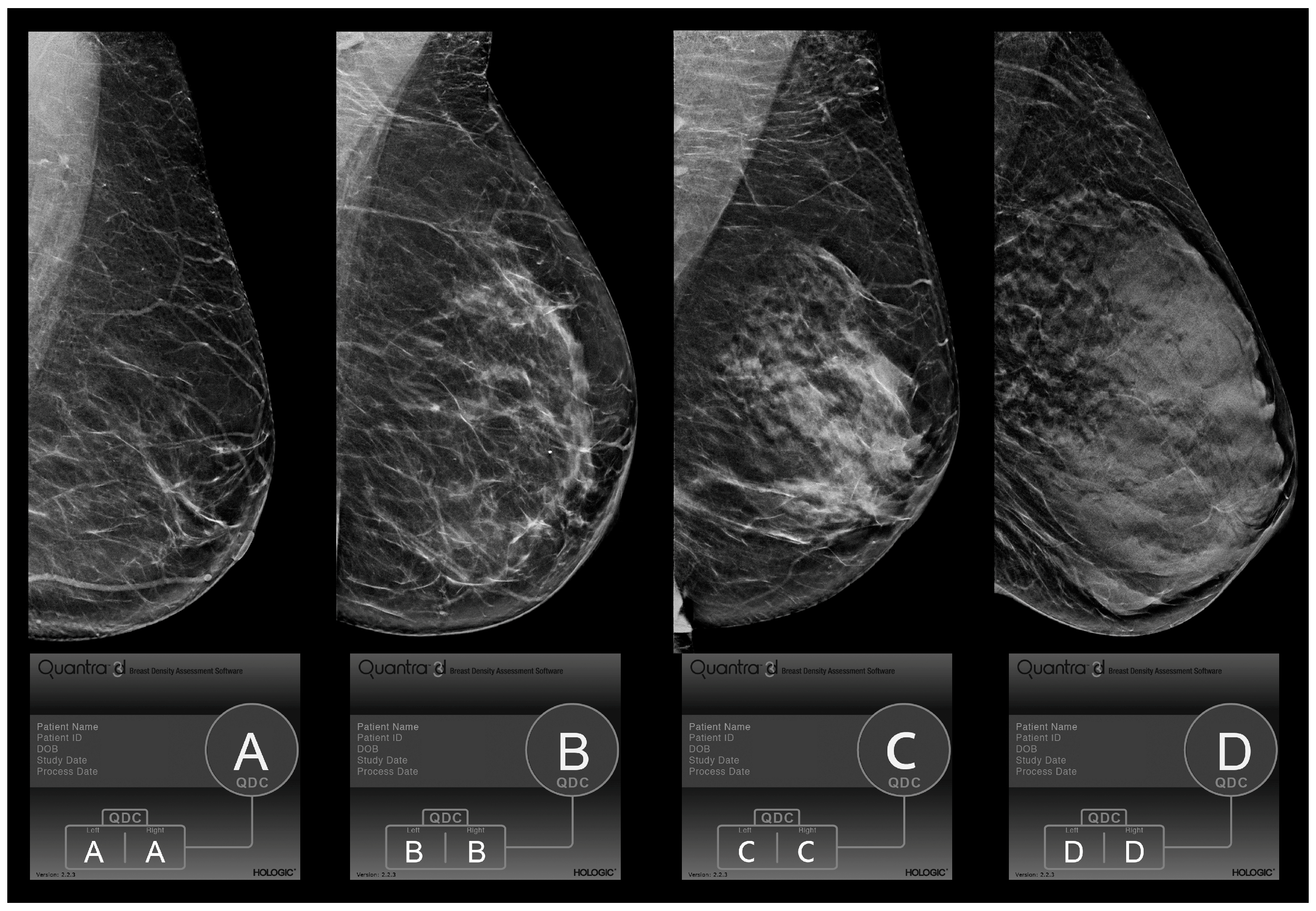
2.3. Automated Breast Density Assessment
2.4. Ethical Considerations and Data Availability
2.5. Statistical Analysis
3. Results
4. Discussion
5. Conclusions
Author Contributions
Funding
Institutional Review Board Statement
Informed Consent Statement
Data Availability Statement
Conflicts of Interest
References
- Carney, P.A.; Miglioretti, D.L.; Yankaskas, B.C.; Kerlikowske, K.; Rosenberg, R.; Rutter, C.M.; Geller, B.M.; Abraham, L.A.; Taplin, S.H.; Dignan, M.; et al. Individual and combined effects of age, breast density, and hormone re-placement therapy use on the accuracy of screening mammography. Ann. Intern. Med. 2003, 138, 168–175. [Google Scholar] [CrossRef] [PubMed]
- McCormack, V.A.; dos Santos Silva, I. Breast Density and Parenchymal Patterns as Markers of Breast Cancer Risk: A Meta-analysis. Cancer Epidemiol. Biomark. Prev. 2006, 15, 1159–1169. [Google Scholar] [CrossRef] [PubMed]
- Leborgne, R. The Breast in Roentgen Diagnosis; Impresora Uruguaya: Montevideo, Uruguay, 1953. [Google Scholar]
- Wolfe, J.N. Risk for breast cancer development determined by mammographic parenchymal pattern. Cancer 1976, 37, 2486–2492. [Google Scholar] [CrossRef] [PubMed]
- Johns, P.C.; Yaffe, M.J. X-ray characterisation of normal and neoplastic breast tissues. Phys. Med. Biol. 1987, 32, 675–695. [Google Scholar] [CrossRef] [PubMed]
- Freer, P. Mammographic Breast Density: Impact on Breast Cancer Risk and Implications for Screening. Radio Graph. 2015, 35, 302–315. [Google Scholar] [CrossRef]
- Nguyen, T.L.; Li, S.; Dite, G.S.; Aung, Y.K.; Evans, C.F.; Trinh, H.N.; Baglietto, L.; Stone, J.; Song, Y.; Sung, J.; et al. Interval breast cancer risk associations with breast density, family history and breast tissue aging. Int. J. Cancer 2019, 147, 375–382. [Google Scholar] [CrossRef] [PubMed]
- Boyd, N.F.; Guo, H.; Martin, L.J.; Sun, L.; Stone, J.; Fishell, E.; Jong, R.A.; Hislop, G.; Chiarelli, A.; Minkin, S.; et al. Mammographic Density and the Risk and Detection of Breast Cancer. N. Engl. J. Med. 2007, 356, 227–236. [Google Scholar] [CrossRef]
- Lee, C.I.; Chen, L.E.; Elmore, J.G. Risk-based Breast Cancer Screening: Implications of Breast Density. Med. Clin. N. Am. 2017, 101, 725–741. [Google Scholar] [CrossRef]
- Are You Dense Advocacy. D.E.N.S.E._State Efforts. 2014. Available online: http://areyoudenseadvocacy.org/dense/ (accessed on 5 August 2022).
- Boyd, N.F.; Martin, L.J.; Yaffe, M.J.; Minkin, S. Mammographic density and breast cancer risk: Current understanding and future prospects. Breast Cancer Res. 2011, 13, 223. [Google Scholar] [CrossRef]
- Ekpo, E.U.; Hogg, P.; Highnam, R.; McEntee, M.F. Breast composition: Measurement and clinical use. Radiography 2015, 21, 324–333. [Google Scholar] [CrossRef]
- Ang, T.; Harkness, E.F.; Maxwell, A.J.; Lim, Y.Y.; Emsley, R.; Howell, A.; Evans, D.G.; Astley, S.; Gadde, S. Visual assessment of breast density using visual analogue scales: Observer variability, reader attributes and reading time. Proc. SPIE 2017, 10136, 1013608. [Google Scholar]
- Garrido-Estepa, M.; Ruiz-Perales, F.; Miranda, J.; Ascunce, N.; González-Román, I.; Sánchez-Contador, C.; Santamariña, C.; Moreo, P.; Vidal, C.; Pollán, M.; et al. Evaluation of mammographic density patterns: Reproducibility and concordance among scale. BMC Cancer 2010, 10, 485. [Google Scholar] [CrossRef] [PubMed]
- Gastounioti, A.; McCarthy, A.M.; Pantalone, L.; Synnestvedt, M.; Kontos, D.; Conant, E.F. Effect of Mammographic Screening Modality on Breast Density Assessment: Digital Mammography versus Digital Breast Tomosynthesis. Radiology 2019, 291, 320–327. [Google Scholar] [CrossRef] [PubMed]
- D’Orsi, C.J.; Mendelson, E.B.; Ikeda, D.M.; et al. Breast Imaging Reporting and Data System: ACR BI-RADS—Breast Imaging Atlas; American College of Radiology: Reston, VA, USA, 2003. [Google Scholar]
- Sickles, E.A.; D’Orsi, C.J.; Bassett, L.W.; et al. ACR BI-RADS Mammography. In ACR BI-RADS Atlas, Breast Imaging Reporting and Data System, 5th ed.; D’Orsi, C.J., Sickles, E.A., Mendelson, E.B., Morris, E.A., et al., Eds.; American College of Radiology: Reston, VA, USA, 2013. [Google Scholar]
- Gard, C.C.; Bowles, E.J.A.; Miglioretti, D.L.; Taplin, S.H.; Rutter, C.M. Misclassification of Breast Imaging Reporting and Data System (BI-RADS) Mammographic Density and Implications for Breast Density Reporting Legislation. Breast J. 2015, 21, 481–489. [Google Scholar] [CrossRef] [PubMed]
- Ciatto, S.; Houssami, N.; Apruzzese, A.; Bassetti, E.; Brancato, B.; Carozzi, F.; Catarzi, S.; Lamberini, M.; Marcelli, G.; Pellizzoni, R.; et al. Categorizing breast mammographic density: Intra- and interobserver reproducibility of BI-RADS density categories. Breast 2005, 14, 269–275. [Google Scholar] [CrossRef] [PubMed]
- Gemici, A.A.; Bayram, E.; Hocaoglu, E.; Inci, E. Comparison of breast density assessments according to BI-RADS 4th and 5th editions and experience level. Acta Radiol. Open 2020, 9, 2058460120937381. [Google Scholar] [CrossRef]
- Ekpo, E.U.; Mello-Thoms, C.; Rickard, M.; Brennan, P.C.; McEntee, M.F. Breast density (BD) assessment with digital breast tomo-synthesis (DBT): Agreement between Quantra™ and 5th edition BI-RADS®. Breast 2016, 30, 185–190. [Google Scholar] [CrossRef]
- Kshirsagar, A.; Quantra, T.M. Quantra™ 2.2 Software Design Intent and Clinical Performance; Clinical Solutions, Research and Development, Hologic, Inc.: Marlborough, MA, USA, 2020. [Google Scholar]
- Landis, J.R.; Koch, G.G. The measurement of observer agreement for categorical data. Biometrics 1977, 33, 159–174. [Google Scholar] [CrossRef]
- Alonzo-Proulx, O.; Mawdsley, G.E.; Patrie, J.T.; Yaffe, M.J.; Harvey, J.A. Reliability of automated breast density measurements. Radiology 2015, 275, 366–376. [Google Scholar] [CrossRef]
- Kallenberg, M.G.; Lokate, M.; van Gils, C.H.; Karssemeijer, N. Automatic breast density segmentation: An integration of different approaches. Phys. Med. Biol. 2011, 56, 2715–2729. [Google Scholar] [CrossRef]
- Brandt, K.R.; Scott, C.G.; Ma, L.; Mahmoudzadeh, A.P.; Jensen, M.R.; Whaley, D.H.; Wu, F.F.; Malkov, S.; Hruska, C.B.; Norman, A.D.; et al. Comparison of Clinical and Automated Breast Density Measurements: Implications for Risk Prediction and Supplemental Screening. Radiology 2016, 279, 710–719. [Google Scholar] [CrossRef] [PubMed]
- Youk, J.H.; Gweon, H.M.; Son, E.J.; Eun, N.L.; Kim, J.-A. Fully automated measurements of volumetric breast density adapted for BIRADS 5th edition: A comparison with visual assessment. Acta Radiol. 2020, 62, 1148–1154. [Google Scholar] [CrossRef] [PubMed]
- Eng, A.; Gallant, Z.; Shepherd, J.; McCormack, V.; Li, J.; Dowsett, M.; Vinnicombe, S.; Allen, S.; Dos-Santos-Silva, I. Digital mammographic density and breast cancer risk: A case–control study of six alternative density assessment methods. Breast Cancer Res. 2014, 16, 439. [Google Scholar] [CrossRef]
- Hologic Inc. Understanding Quantra 2.0 User Manual—MAN-02004 Rev 002; Hologic: Bedford, MA, USA, 2012. [Google Scholar]
- Pahwa, S.; Hari, S.; Thulkar, S.; Angraal, S. Evaluation of breast parenchymal density with QUANTRA software. Indian J. Radiol. Imaging 2015, 25, 391–396. [Google Scholar] [CrossRef] [PubMed]
- Ekpo, E.U.; McEntee, M.F.; Rickard, M.; Brennan, P.C.; Kunduri, J.; Demchig, D.; Mello-Thoms, C. Quantra™ should be considered a tool for two-grade scale mammographic breast density classification. Br. J. Radiol. 2016, 89, 20151057. [Google Scholar] [CrossRef]
- van der Waal, D.; den Heeten, G.J.; Pijnappel, R.M.; Schuur, K.H.; Timmers, J.M.H.; Verbeek, A.L.M.; Broeders, M.J.M. Comparing visually assessed BI-RADS breast density and automated volumetric breast density software: A cross-sectional study in a breast cancer screening setting. PLoS ONE 2015, 10, e0136667. [Google Scholar] [CrossRef] [PubMed]
- Østerås, B.H.; Martinsen, A.C.; Brandal, S.H.; Chaudhry, K.N.; Eben, E.; Haakenaasen, U.; Falk, R.S.; Skaane, P. Classification of fatty and dense breast parenchyma: Comparison of automatic volumetric density measurement and radiologists’ classification and their inter-observer variation. Acta Radiol. 2016, 57, 1178–1185. [Google Scholar] [CrossRef] [PubMed]
- Youk, J.H.; Kim, S.J.; Son, E.J.; Gweon, H.M.; Kim, J.-A. Comparison of Visual Assessment of Breast Density in BI-RADS 4th and 5th Editions With Automated Volumetric Measurement. Am. J. Roentgenol. 2017, 209, 703–708. [Google Scholar] [CrossRef]
- Winkler, N.S.; Raza, S.; Mackesy, M.; Birdwell, R.L. Breast Density: Clinical Implications and Assessment Methods. Radiographics 2015, 35, 316–324. [Google Scholar] [CrossRef]
- Machida, Y.; Tozaki, M.; Shimauchi, A.; Yoshida, T. Breast density: The trend in breast cancer screening. Breast Cancer 2015, 22, 253–261. [Google Scholar] [CrossRef]
- Heindl, F.; Fasching, P.A.; Hein, A.; Hack, C.C.; Heusinger, K.; Gass, P.; Pöschke, P.; Stübs, F.A.; Schulz-Wendtland, R.; Hartmann, A.; et al. Mammographic density and prognosis in primary breast cancer patients. Breast 2021, 59, 51–57. [Google Scholar] [CrossRef] [PubMed]
- van der Waal, D.; Verbeek, A.L.M.; Broeders, M.J.M. Breast density and breast cancer-specific survival by detection mode. BMC Cancer 2018, 18, 386. [Google Scholar] [CrossRef] [PubMed]
- Tari, D.U.; Santonastaso, R.; Pinto, F. Consequences of the impact of COVID-19 pandemic on breast cancer at a single Italian institution. Explor. Target. Anti-Tumor Ther. 2022, 3, 414–422. [Google Scholar] [CrossRef] [PubMed]


| Kappa Values | Type of Agreement |
|---|---|
| 0.00–0.20 | Slight |
| 0.21–0.40 | Fair |
| 0.41–0.60 | Moderate |
| 0.61–0.80 | Substantial |
| 0.81–1.00 | Almost perfect |
| Density Category | Radiologists | Consensus | QDC | ||
|---|---|---|---|---|---|
| 1 | 2 | 3 | |||
| Total (1075) | |||||
| A | 166 (15.4%) | 163 (15.1%) | 218 (20.3%) | 179 (16.6%) | 123 (11.5%) |
| B | 492 (45.8%) | 462 (43%) | 367 (34.1%) | 447 (41.6%) | 425 (39.5%) |
| Non-dense | 658 (61.2%) | 625 (58.1%) | 585 (54.4%) | 626 (58.2%) | 548 (51%) |
| C | 354 (32.9%) | 392 (36.5%) | 409 (38.1%) | 385 (35.8%) | 445 (41.4%) |
| D | 63 (5.9%) | 58 (5.4%) | 81 (7.5%) | 64 (6%) | 82 (7.6%) |
| Dense | 417 (38.8%) | 450 (41.9%) | 490 (45.6%) | 449 (41.8%) | 527 (49%) |
| 40–49 (87) | |||||
| A | 4 (4.6%) | 4 (4.6%) | 5 (5.8%) | 3 (3.5%) | 3 (3.4%) |
| B | 27 (31%) | 18 (20.7%) | 15 (17.2%) | 21 (24.1%) | 14 (16.1%) |
| Non-dense | 31 (35.6%) | 22 (25.3%) | 20 (23%) | 24 (27.6%) | 17 (19.5%) |
| C | 41 (47.1%) | 54 (62.1%) | 53 (60.9%) | 50 (57.5%) | 54 (62.1%) |
| D | 15 (17.3%) | 11 (12.6%) | 14 (16.1%) | 13 (14.9%) | 16 (18.4%) |
| Dense | 56 (64.4%) | 65 (74.7%) | 67 (77%) | 63 (72.4%) | 70 (80.5%) |
| 50–69 (997) | |||||
| A | 150 (16.2%) | 148 (16%) | 196 (21.1%) | 164 (17.7%) | 114 (12.3%) |
| B | 433 (46.7%) | 410 (44.2%) | 328 (35.4%) | 395 (42.6%) | 377 (40.7%) |
| Non-dense | 583 (62.9%) | 558 (60.2%) | 524 (56.5%) | 559 (60.3%) | 491 (53%) |
| C | 297 (32%) | 323 (34.8%) | 338 (36.5%) | 318 (34.3%) | 372 (40.1%) |
| D | 47 (5.1%) | 46 (5%) | 65 (7.0%) | 50 (5.4%) | 64 (6.9%) |
| Dense | 344 (37.1%) | 369 (39.8%) | 403 (43.5%) | 368 (39.7%) | 436 (47%) |
| >70 (61) | |||||
| A | 12 (19.7%) | 11 (18%) | 17 (27.9%) | 12 (19.7%) | 6 (9.8%) |
| B | 32 (52.5%) | 34 (55.8%) | 24 (39.3%) | 31 (50.8%) | 34 (55.8%) |
| Non-dense | 44 (72.2%) | 45 (73.8%) | 41 (67.2%) | 43 (70.5%) | 40 (65.6%) |
| C | 16 (26.2%) | 15 (24.6%) | 18 (29.5%) | 17 (27.9%) | 19 (31.1%) |
| D | 1 (1.6%) | 1 (1.6%) | 2 (3.3%) | 1 (1.6%) | 2 (3.3%) |
| Dense | 17 (27.8%) | 16 (26.2%) | 20 (32.8%) | 18 (29.5%) | 21 (34.4%) |
| Observers | OVERALL | 40–49 | 50–69 | >70 | ||||
|---|---|---|---|---|---|---|---|---|
| K | CI | K | CI | K | CI | K | CI | |
| Rad1/Rad2 | 0.82 | (0.77–0.86) | 0.78 | (0.64–0.92) | 0.82 | (0.78–0.87) | 0.69 | (0.51–0.87) |
| Rad1/Rad3 | 0.77 | (0.73–0.82) | 0.70 | (0.56–0.84) | 0.78 | (0.74–0.83) | 0.63 | (0.45–0.81) |
| Rad2/Rad3 | 0.83 | (0.79–0.87) | 0.77 | (0.63–0.91) | 0.83 | (0.78–0.87) | 0.78 | (0.61–0.96) |
| Rad1/QDC | 0.69 | (0.65–0.73) | 0.65 | (0.51–0.78) | 0.70 | (0.66–0.75) | 0.44 | (0.27–0.62) |
| Rad2/QDC | 0.77 | (0.73–0.81) | 0.72 | (0.58–0.86) | 0.78 | (0.73–0.82) | 0.57 | (0.39–0.74) |
| Rad3/QDC | 0.78 | (0.74–0.82) | 0.76 | (0.62–0.90) | 0.78 | (0.73–0.82) | 0.65 | (0.48–0.81) |
| CON/QDC | 0.77 | (0.73–0.81) | 0.74 | (0.60–0.88) | 0.77 | (0.73–0.82) | 0.60 | (0.43–0.78) |
| Observers | OVERALL | 40–49 | 50–69 | >70 | ||||||||
|---|---|---|---|---|---|---|---|---|---|---|---|---|
| K | CI | p | K | CI | p | K | CI | p | K | CI | p | |
| Rad1/Rad2 | 0.87 | (0.81–0.93) | <0.05 | 0.76 | (0.56–0.96) | <0.05 | 0.88 | (0.82–0.94) | <0.05 | 0.79 | (0.54–1.04) | 0.65 |
| Rad1/Rad3 | 0.81 | (0.75–0.87) | <0.05 | 0.70 | (0.50–0.90) | <0.05 | 0.81 | (0.75–0.88) | <0.05 | 0.81 | (0.56–1.06) | <0.5 |
| Rad2/Rad3 | 0.88 | (0.82–0.94) | <0.05 | 0.81 | (0.60–1.02) | <0.5 | 0.88 | (0.82–0.94) | <0.05 | 0.84 | (0.60–1.09) | <0.05 |
| Rad1/QDC | 0.76 | (0.70–0.82) | <0.05 | 0.61 | (0.42–0.80) | <0.05 | 0.77 | (0.71–0.84) | <0.05 | 0.62 | (0.37–0.87) | <0.5 |
| Rad2/QDC | 0.82 | (0.76–0.88) | <0.05 | 0.84 | (0.63–1.04) | <0.05 | 0.82 | (0.76–0.88) | <0.05 | 0.65 | (0.41–0.90) | <0.5 |
| Rad3/QDC | 0.86 | (0.80–0.92) | <0.05 | 0.83 | (0.62–1.04) | <0.5 | 0.86 | (0.80–0.93) | <0.05 | 0.74 | (0.49–0.99) | 0.7 |
| CON/QDC | 0.83 | (0.77–0.89) | <0.05 | 0.78 | (0.57–0.98) | <0.05 | 0.83 | (0.77–0.89) | <0.05 | 0.74 | (0.49–0.99) | <0.5 |
| Results | BI-RADS Categories | Dense—Non Dense Categories | ||
|---|---|---|---|---|
| N (%) | Average age ± SD | N (%) | Average age ± SD | |
| Concordant | 861 (80.1%) | 58.1 ± 7.1 | 983 | 58.3 ± 7.1 |
| Discordant | 214 (19.1%) | 59.0 ± 7.3 | 92 | 58.5 ± 7.3 |
| p value | 0.09 | 0.82 | ||
Disclaimer/Publisher’s Note: The statements, opinions and data contained in all publications are solely those of the individual author(s) and contributor(s) and not of MDPI and/or the editor(s). MDPI and/or the editor(s) disclaim responsibility for any injury to people or property resulting from any ideas, methods, instructions or products referred to in the content. |
© 2023 by the authors. Licensee MDPI, Basel, Switzerland. This article is an open access article distributed under the terms and conditions of the Creative Commons Attribution (CC BY) license (https://creativecommons.org/licenses/by/4.0/).
Share and Cite
Tari, D.U.; Santonastaso, R.; De Lucia, D.R.; Santarsiere, M.; Pinto, F. Breast Density Evaluation According to BI-RADS 5th Edition on Digital Breast Tomosynthesis: AI Automated Assessment Versus Human Visual Assessment. J. Pers. Med. 2023, 13, 609. https://doi.org/10.3390/jpm13040609
Tari DU, Santonastaso R, De Lucia DR, Santarsiere M, Pinto F. Breast Density Evaluation According to BI-RADS 5th Edition on Digital Breast Tomosynthesis: AI Automated Assessment Versus Human Visual Assessment. Journal of Personalized Medicine. 2023; 13(4):609. https://doi.org/10.3390/jpm13040609
Chicago/Turabian StyleTari, Daniele Ugo, Rosalinda Santonastaso, Davide Raffaele De Lucia, Marika Santarsiere, and Fabio Pinto. 2023. "Breast Density Evaluation According to BI-RADS 5th Edition on Digital Breast Tomosynthesis: AI Automated Assessment Versus Human Visual Assessment" Journal of Personalized Medicine 13, no. 4: 609. https://doi.org/10.3390/jpm13040609
APA StyleTari, D. U., Santonastaso, R., De Lucia, D. R., Santarsiere, M., & Pinto, F. (2023). Breast Density Evaluation According to BI-RADS 5th Edition on Digital Breast Tomosynthesis: AI Automated Assessment Versus Human Visual Assessment. Journal of Personalized Medicine, 13(4), 609. https://doi.org/10.3390/jpm13040609