Pretreatment Tumor Volume and Tumor Sphericity as Prognostic Factors in Patients with Oral Cavity Squamous Cell Carcinoma: A Prospective Clinical Study in 95 Patients


 , ,
, , 
Abstract
1. Introduction
2. Materials and Methods
2.1. CT-Scan Acquisition Protocol
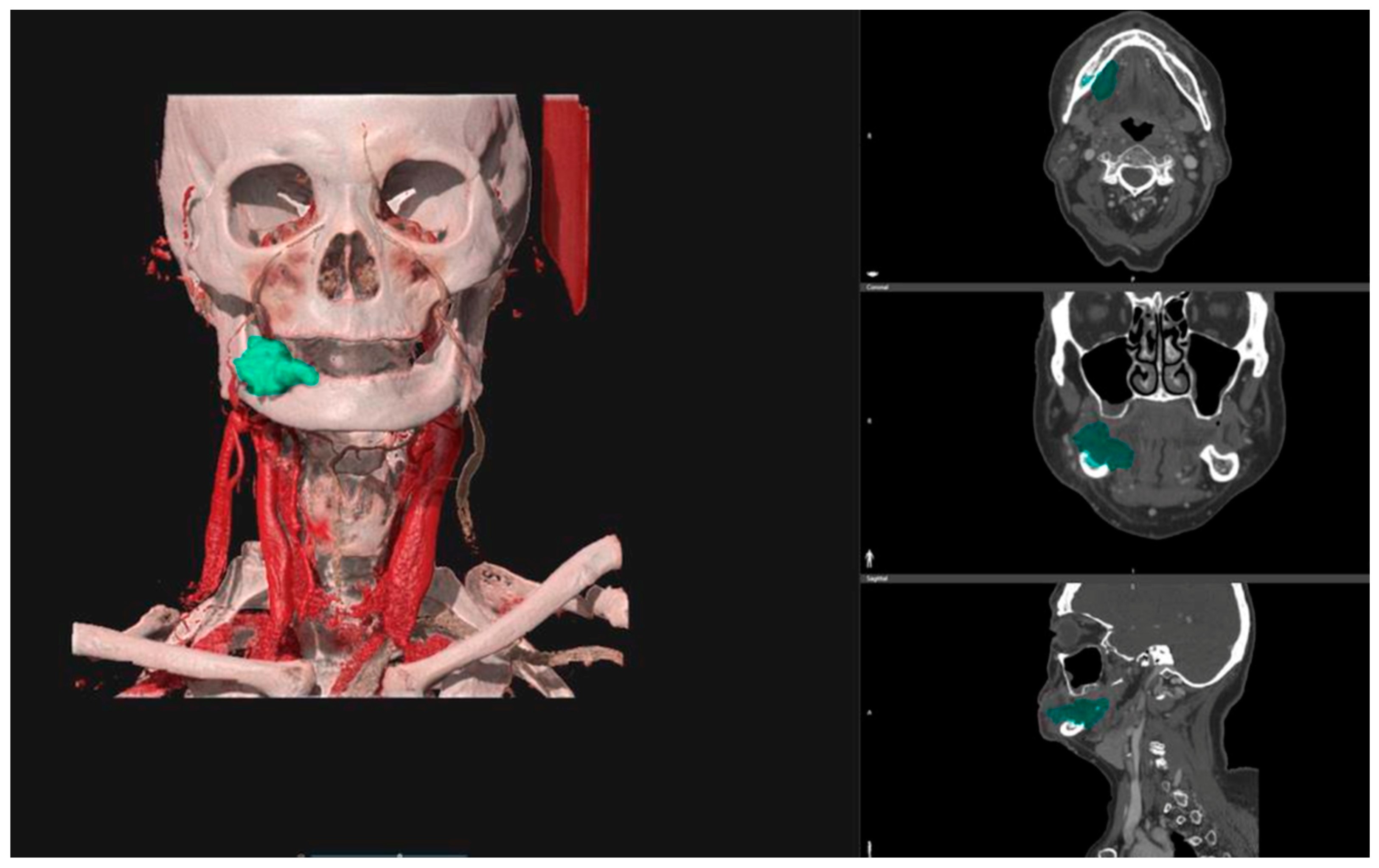
2.2. Tumor Volume (Vt) Calculation
2.3. Tumor Sphericity (St) Calculation
2.4. Statistical Analysis
3. Results
3.1. Descriptive Analysis
3.2. Continuous Variables: Tumor Volume (Vt) and Tumor Sphericity (St)
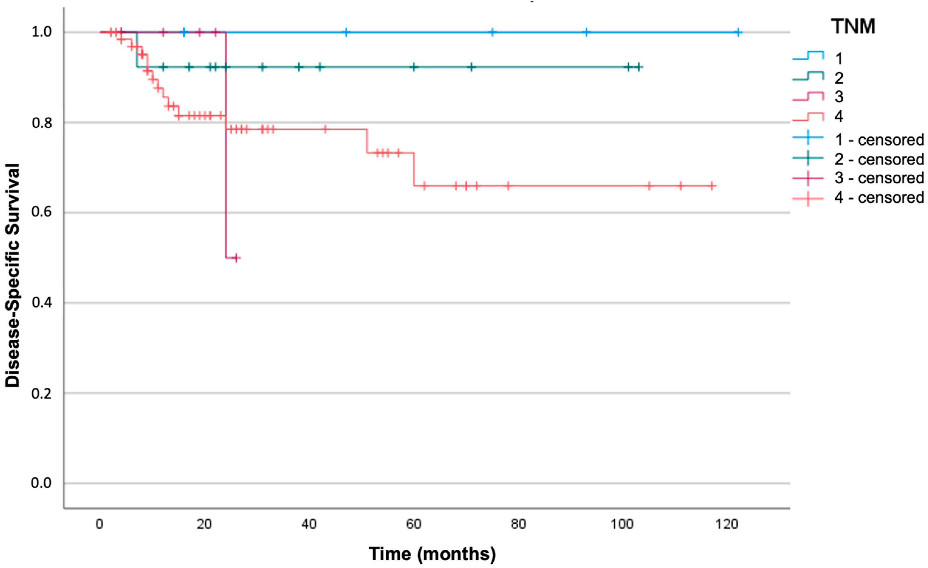
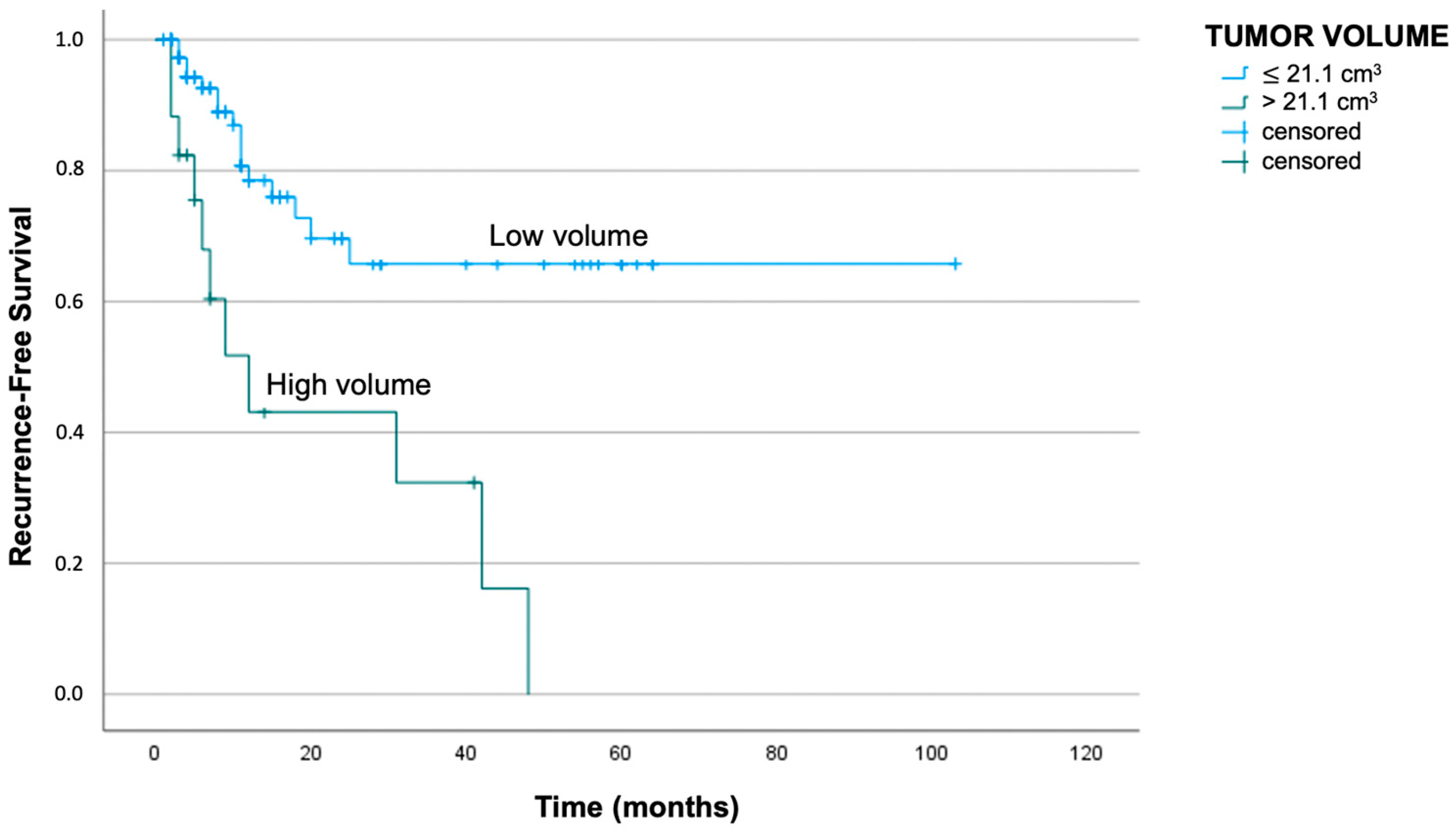
3.3. Survival Analysis
3.4. Univariate and Multivariate Analyses
4. Discussion
5. Conclusions
Author Contributions
Funding
Institutional Review Board Statement
Informed Consent Statement
Data Availability Statement
Acknowledgments
Conflicts of Interest
References
- World Health Organization Classification of Tumour. Tumours of the oral cavity and oropharynx. In Pathology and Genetics of Head and Neck Tumours, 3rd ed.; Barbes, L., Eveson, J.W., Reichar, P., Sidransky, D., Eds.; IARC Press: Lyon, France, 2005; Volume 9, pp. 163–175. [Google Scholar]
- Shah, J.P.; Gil, Z. Current concepts in management of oral cancer—Surgery. Oral Oncol. 2009, 45, 394–401. [Google Scholar] [CrossRef] [Scilit]
- Ridge, J.A.; Lydiatt, W.M.; Patel, S.G.; Glastonbury, C.M.; Brandwein-Weber, M.; Ghossein, R.A.; Shah, J.P. Oral cavity. In AJCC Cancer Staging Manual, 8th ed.; Amin, M.B., Edge, S.B., Greene, F.L., Byrd, D.R., Brookland, R.K., Washington, M.K., Gershenwald, J.E., Compton, C.C., Hess, K.R., Sullivan, D.C., et al., Eds.; Springer: New York, NY, USA, 2017; pp. 79–94. [Google Scholar]
- Pollaers, K.; Hinton-Bayre, A.; Friedland, P.L.; Farah, C.S. AJCC 8th Edition oral cavity squamous cell carcinoma staging-Is it an improvement on the AJCC 7th Edition? Oral Oncol. 2018, 82, 23–28. [Google Scholar] [CrossRef] [Scilit]
- Mukherji, S.K.; Toledano, A.Y.; Beldon, C.; Schmalfuss, I.M.; Cooper, J.S.; Sicks, J.D.; Amdur, R.; Sailer, S.; Loevner, L.A.; Kousouboris, P.; et al. Interobserver reliability of computed tomography-derived primary tumor volume measurement in patients with supraglottic carcinoma. Cancer 2005, 103, 2616–2622. [Google Scholar] [CrossRef] [Scilit]
- Shiao, J.C.; Mohamed, A.S.R.; Messer, J.A.; Hutcheson, K.A.; Johnson, J.M.; Enderling, H.; Kamal, M.; Warren, B.W.; Pham, B.; Morrison, W.H.; et al. Quantitative pretreatment CT volumetry: Association with oncologic outcomes in patients with T4a squamous carcinoma of the larynx. Head Neck 2017, 39, 1609–1620. [Google Scholar] [CrossRef] [Scilit]
- Kurek, R.; Kalogera-Fountzila, A.; Muskalla, K.; Dafni, U.; Schnabel, T.; Kober, B.; Röddiger, S.; Martin, T.; Fountzilas, G.; Zamboglou, N. Usefulness of tumor volumetry as a prognostic factor of survival in head and neck cancer. Strahlenther. Onkol. 2003, 179, 292–297. [Google Scholar] [CrossRef] [Scilit]
- Nixon, I.J.; Palmer, F.L.; Lakin, P.; Kattan, M.M.; Lee, N.Y.; Ganly, I. Pathologically determined tumor volume vs pathologic T stage in the prediction of outcome after surgical treatment of oropharyngeal squamous cell carcinoma. JAMA Otolaryngol. Head Neck Surg. 2013, 139, 1151–1155. [Google Scholar] [CrossRef] [Scilit]
- Yuan, Y.; Ren, J.; Shi, Y.; Tao, X. MRI-based radiomic signature as predictive marker for patients with head and neck squamous cell carcinoma. Eur. J. Radiol. 2019, 117, 193–198. [Google Scholar] [CrossRef] [Scilit]
- Mes, S.W.; van Velden, F.H.P.; Peltenburg, B.; Peeters, C.F.W.; Te Beest, D.E.; van de Wiel, M.A.; Mekke, J.; Mulder, D.C.; Martens, R.M.; Castelijns, J.A.; et al. Outcome prediction of head and neck squamous cell carcinoma by MRI radiomic signatures. Eur. Radiol. 2020, 30, 6311–6321. [Google Scholar] [CrossRef] [Scilit]
- Alfieri, S.; Romanò, R.; Bologna, M.; Calareso, G.; Corino, V.; Mirabile, A.; Ferri, A.; Bellanti, L.; Poli, T.; Marcantoni, A.; et al. Prognostic role of pre-treatment magnetic resonance imaging (MRI)-based radiomic analysis in effectively cured head and neck squamous cell carcinoma (HNSCC) patients. Acta Oncol. 2021, 60, 1192–1200. [Google Scholar] [CrossRef] [Scilit]
- Been, M.J.; Watkins, J.; Manz, R.M.; Gentry, L.R.; Leverson, G.E.; Harari, P.M.; Hartig, G.K. Tumor volume as a prognostic factor in oropharyngeal squamous cell carcinoma treated with primary radiotherapy. Laryngoscope 2008, 118, 1377–1382. [Google Scholar] [CrossRef] [Scilit]
- Chen, S.W.; Yang, S.N.; Liang, J.A.; Lin, F.J.; Tsai, M.H. Prognostic impact of tumor volume in patients with stage III-IVA hypopharyngeal cancer without bulky lymph nodes treated with definitive concurrent chemoradiotherapy. Head Neck 2009, 31, 709–716. [Google Scholar] [CrossRef] [Scilit]
- Mukoyama, N.; Suzuki, H.; Hanai, N.; Sone, M.; Hasegawa, Y. Pathological tumor volume predicts survival outcomes in oral squamous cell carcinoma. Oncol. Lett. 2018, 16, 2471–2477. [Google Scholar] [CrossRef] [Scilit]
- Lin, C.S.; de Oliveira Santos, A.B.; Silva, E.L.; de Matos, L.L.; Moyses, R.A.; Kulcsar, M.A.; Pinto, F.R.; Brandão, L.G.; Cernea, C.R. Tumor volume as an independent predictive factor of worse survival in patients with oral cavity squamous cell carcinoma. Head Neck 2017, 39, 960–964. [Google Scholar] [CrossRef] [Scilit]
- Tarsitano, A.; Ricotta, F.; Cercenelli, L.; Bortolani, B.; Battaglia, S.; Lucchi, E.; Marchetti, C.; Marcelli, E. Pretreatment tumor volume and tumor sphericity as prognostic factors in patients with oral cavity squamous cell carcinoma. J. Craniomaxillofac. Surg. 2019, 47, 510–515. [Google Scholar] [CrossRef] [Scilit]
- Sutton, D.N.; Brown, J.S.; Rogers, S.N.; Vaughan, E.D.; Woolgar, J.A. The prognostic implications of the surgical margin in oral squamous cell carcinoma. Int. J. Oral Maxillofac. Surg. 2003, 32, 30–34. [Google Scholar] [CrossRef] [Scilit]
- Larsen, S.R.; Johansen, J.; Sørensen, J.A.; Krogdahl, A. The prognostic significance of histological features in oral squamous cell carcinoma. J. Oral Pathol. Med. 2009, 38, 657–662. [Google Scholar] [CrossRef] [Scilit]
- van der Schroeff, M.P.; Baatenburg de Jong, R.J. Staging and prognosis in head and neck cancer. Oral Oncol. 2009, 45, 356–360. [Google Scholar] [CrossRef] [Scilit]
- Mücke, T.; Mitchell, D.A.; Ritschl, L.M.; Tannapfel, A.; Wolff, K.D.; Kesting, M.R.; Loeffelbein, D.J.; Kanatas, A. Influence of tumor volume on survival in patients with oral squamous cell carcinoma. J. Cancer Res. Clin. Oncol. 2015, 141, 1007–1011. [Google Scholar] [CrossRef] [Scilit]
- Cercenelli, L.; Zoli, M.; Bortolani, B.; Curti, N.; Gori, D.; Rustici, A.; Mazzatenta, D.; Marcelli, E. 3D Virtual Modeling for Morphological Characterization of Pituitary Tumors: Preliminary Results on Its Predictive Role in Tumor Resection Rate. Appl. Sci. 2022, 12, 4275. [Google Scholar] [CrossRef] [Scilit]
- Dhara, A.K.; Mukhopadhyay, S.; Saha, P.; Garg, M.; Khandelwal, N. Differential geometry-based techniques for characterization of boundary roughness of pulmonary nodules in CT images. Int. J. Comput. Assist. Radiol. Surg. 2016, 11, 337–349. [Google Scholar] [CrossRef] [Scilit]
- Choi, T.W.; Kim, J.H.; Yu, M.H.; Park, S.J.; Han, J.K. Pancreatic neuroendocrine tumor: Prediction of the tumor grade using CT findings and computerized texture analysis. Acta Radiol. 2018, 59, 383–392. [Google Scholar] [CrossRef] [Scilit] [PubMed]
- Apostolova, I.; Steffen, I.G.; Wedel, F.; Lougovski, A.; Marnitz, S.; Derlin, T.; Amthauer, H.; Buchert, R.; Hofheinz, F.; Brenner, W. Asphericity of pretherapeutic tumour FDG uptake provides independent prognostic value in head-and-neck cancer. Eur. Radiol. 2014, 24, 2077–2087. [Google Scholar] [CrossRef] [Scilit] [PubMed]
- Hofheinz, F.; Lougovski, A.; Zöphel, K.; Hentschel, M.; Steffen, I.G.; Apostolova, I.; Wedel, F.; Buchert, R.; Baumann, M.; Brenner, W.; et al. Increased evidence for the prognostic value of primary tumor asphericity in pretherapeutic FDG PET for risk stratification in patients with head and neck cancer. Eur. J. Nucl. Med. Mol. Imaging 2015, 42, 429–437. [Google Scholar] [CrossRef] [Scilit] [PubMed]




| Variables | Frequencies (%) | |
|---|---|---|
| Sex | Male | 47/95 (49%) |
| Female | 48/95 (51%) | |
| T stage | T1–T2 | 30/95 (32%) |
| T3–T4 | 65/95 (68%) | |
| N stage | N– | 53/95 (56%) |
| N+ | 42/95 (44%) | |
| Grading | Low | 16/95 (16%) |
| Moderate | 67/95 (71%) | |
| High | 12/95 (13%) | |
| PNI 1 | Negative | 75/95 (79%) |
| Positive | 20/95 (21%) | |
| Lymphovascular infiltration | Negative | 90/95 (95%) |
| Positive | 5/95 (5%) | |
| ECS 2 | Negative | 70/95 (74%) |
| Positive | 25/95 (26%) | |
| Margins | Negative | 84/95 (88%) |
| Positive | 11/95 (12%) |
| Total (95 Cases) | Disease Free and Alive (68 Cases) | Recurrence (27 Cases) | Tumor-Related Death (15 Cases) | |
|---|---|---|---|---|
| mean Vt * | 13.6 cm3 | 11.1 cm3 | 19.8 cm3 | 23.3 cm3 |
| mean St ** | 0.63 | 0.65 | 0.59 | 0.59 |
| Variables | p-Value | Hazard Ratio | Confidence Interval (95%) |
|---|---|---|---|
| T stage | 0.06 | 2.57 | 0.98–6.71 |
| N stage | 0.04 | 2.20 | 1.06–4.60 |
| PNI 1 | <0.01 | 4.68 | 2.21–9.93 |
| Tumor grading | 0.27 | 1.98 | 0.59–6.65 |
| Lymphovascular infiltration | 0.31 | 1.86 | 0.56–6.24 |
| Surgical margins | 0.19 | 1.91 | 0.73–5.01 |
| Variables | p-Value | Hazard Ratio | Confidence Interval (95%) |
|---|---|---|---|
| T stage | 0.29 | 1.98 | 0.56–7.03 |
| N stage | <0.01 | 5.75 | 1.77–18.60 |
| PNI 1 | <0.01 | 6.72 | 2.39–18.91 |
| Tumor grading | 0.26 | 1.72 | 0.67–4.42 |
| Lymphovascular infiltration | 0.22 | 2.54 | 0.57–11.38 |
| Surgical margins | 0.01 | 4.64 | 1.57–13.68 |
| Variables | p-Value | Hazard Ratio | Confidence Interval (95%) |
|---|---|---|---|
| N stage | 0.05 | 2.54 | 1.93–3.04 |
| PNI 1 | 0.03 | 2.32 | 1.34–3.96 |
| Vt 2 | 0.04 | 1.96 | 1.65–2.43 |
| St 3 | 0.02 | 2.87 | 1.68–3.30 |
| Variables | p-Value | Hazard Ratio | Confidence Interval (95%) |
|---|---|---|---|
| N stage | 0.01 | 5.98 | 1.67–21.41 |
| PNI 1 | <0.01 | 4.84 | 1.63–14.37 |
| Surgical margins | 0.04 | 3.37 | 1.04–10.92 |
| Vt 2 | 0.61 | 1.00 | 1.00–1.00 |
| St 3 | 0.65 | 0.19 | 0.00–261.23 |
Disclaimer/Publisher’s Note: The statements, opinions and data contained in all publications are solely those of the individual author(s) and contributor(s) and not of MDPI and/or the editor(s). MDPI and/or the editor(s) disclaim responsibility for any injury to people or property resulting from any ideas, methods, instructions or products referred to in the content. |
© 2023 by the authors. Licensee MDPI, Basel, Switzerland. This article is an open access article distributed under the terms and conditions of the Creative Commons Attribution (CC BY) license (https://creativecommons.org/licenses/by/4.0/).
Share and Cite
Lucchi, E.; Cercenelli, L.; Maiolo, V.; Bortolani, B.; Marcelli, E.; Tarsitano, A. Pretreatment Tumor Volume and Tumor Sphericity as Prognostic Factors in Patients with Oral Cavity Squamous Cell Carcinoma: A Prospective Clinical Study in 95 Patients. J. Pers. Med. 2023, 13, 1601. https://doi.org/10.3390/jpm13111601
Lucchi E, Cercenelli L, Maiolo V, Bortolani B, Marcelli E, Tarsitano A. Pretreatment Tumor Volume and Tumor Sphericity as Prognostic Factors in Patients with Oral Cavity Squamous Cell Carcinoma: A Prospective Clinical Study in 95 Patients. Journal of Personalized Medicine. 2023; 13(11):1601. https://doi.org/10.3390/jpm13111601
Chicago/Turabian StyleLucchi, Elisabetta, Laura Cercenelli, Vincenzo Maiolo, Barbara Bortolani, Emanuela Marcelli, and Achille Tarsitano. 2023. "Pretreatment Tumor Volume and Tumor Sphericity as Prognostic Factors in Patients with Oral Cavity Squamous Cell Carcinoma: A Prospective Clinical Study in 95 Patients" Journal of Personalized Medicine 13, no. 11: 1601. https://doi.org/10.3390/jpm13111601
APA StyleLucchi, E., Cercenelli, L., Maiolo, V., Bortolani, B., Marcelli, E., & Tarsitano, A. (2023). Pretreatment Tumor Volume and Tumor Sphericity as Prognostic Factors in Patients with Oral Cavity Squamous Cell Carcinoma: A Prospective Clinical Study in 95 Patients. Journal of Personalized Medicine, 13(11), 1601. https://doi.org/10.3390/jpm13111601