

Metabolic Disorders, the Microbiome as an Endocrine Organ, and Their Relations with Obesity: A Literature Review

 ,
,  ,
,
Abstract
:1. Introduction
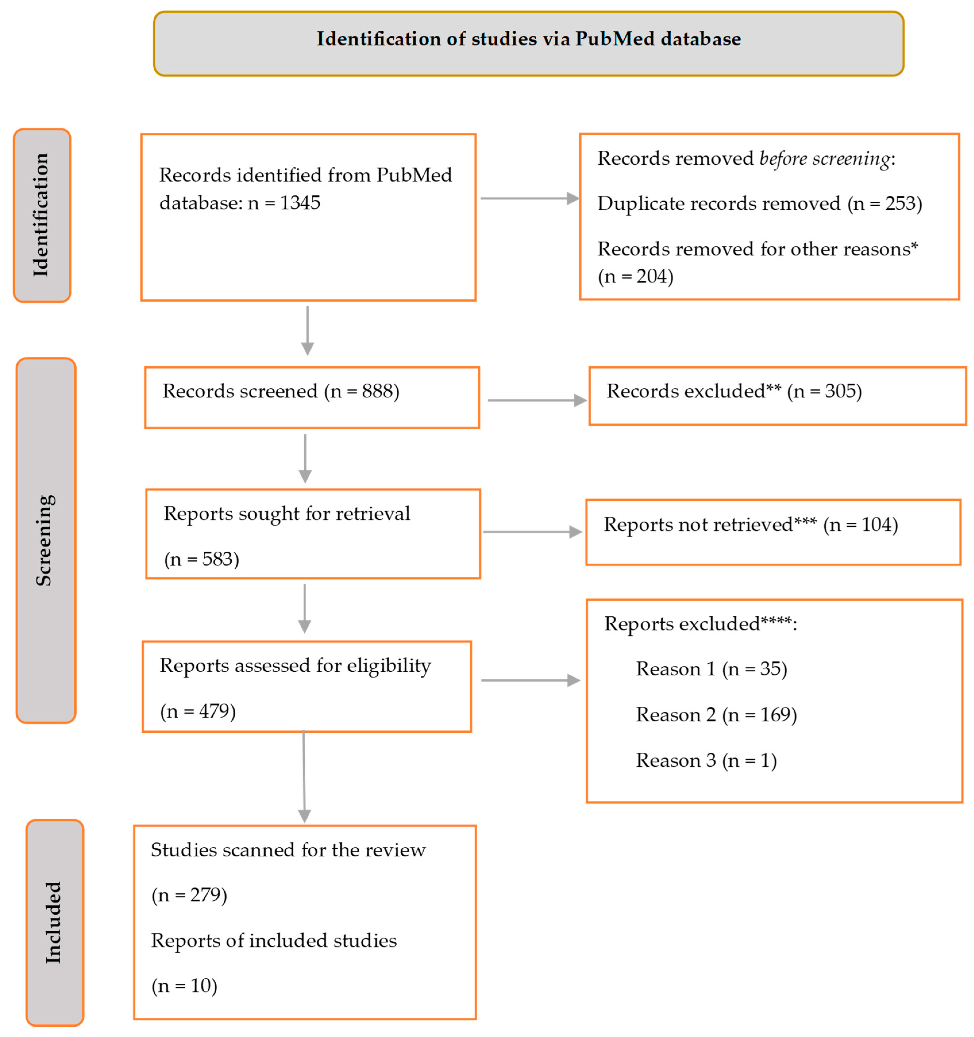
2. Materials and Methods
3. Results
4. Discussion
4.1. Hormones Involved in Visceral Obesity and Microbiota
4.2. Neurotransmitters and Neuropeptides Ivolved in Obesity
4.3. Microbiota
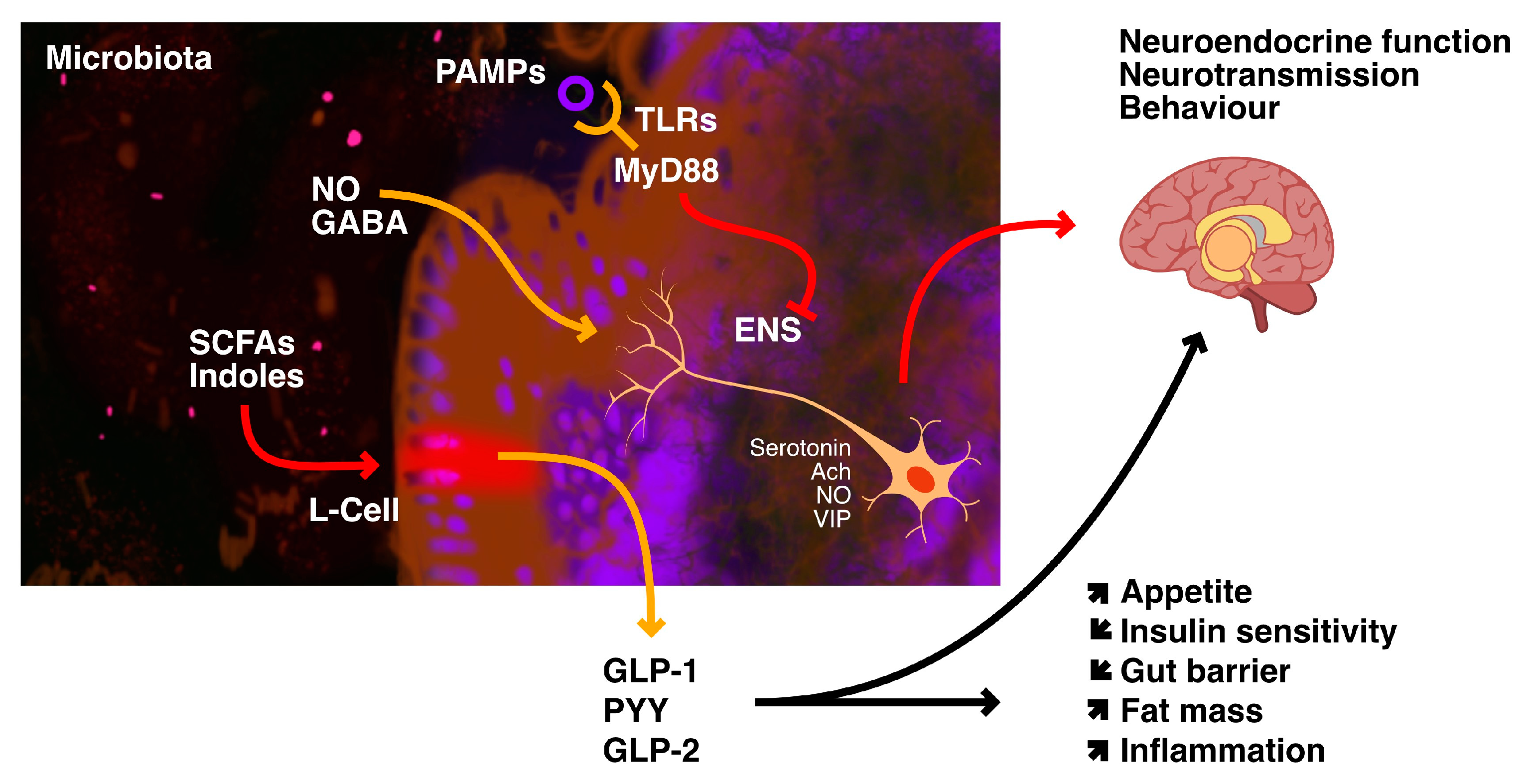
4.4. Gut–Brain Axis
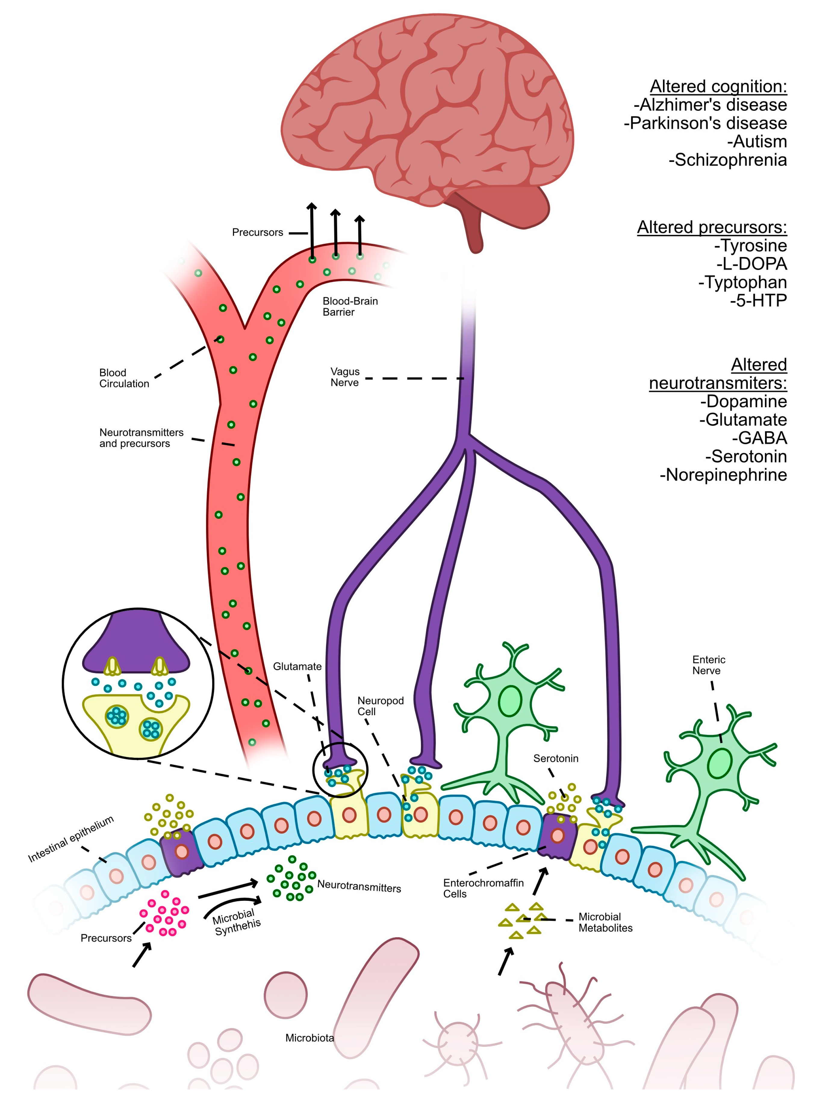
4.5. Gut Microbiota and Neurotransmitters
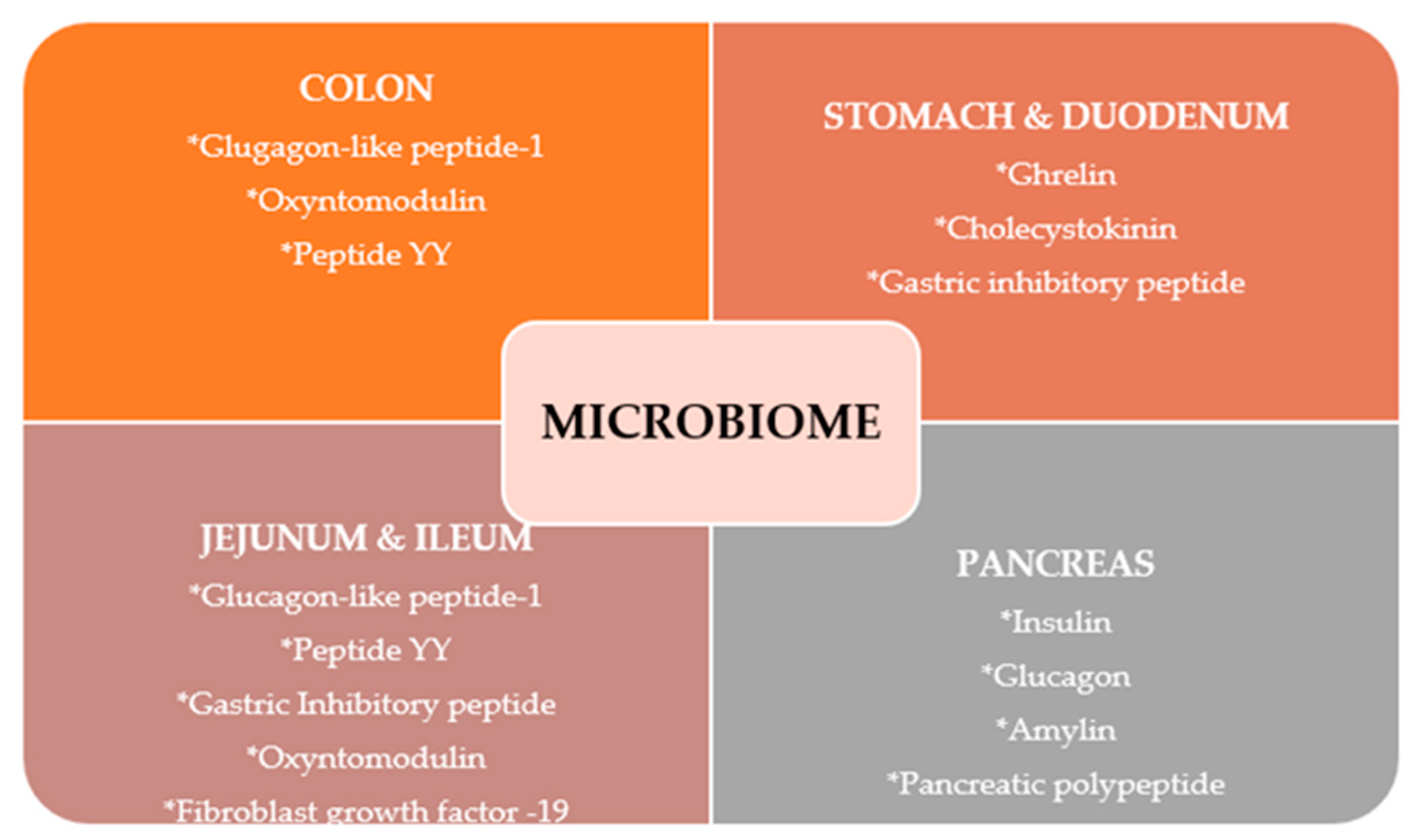
4.6. Incretin Effect
4.7. GIP
5. Conclusions
Author Contributions
Funding
Institutional Review Board Statement
Informed Consent Statement
Data Availability Statement
Conflicts of Interest
References
- World Health Organisation. (n.d.). Retrieved 5 July 2023. Available online: https://www.who.int/news-room/fact-sheets/detail/obesity-and-overweight (accessed on 6 July 2023).
- Heymsfield, S.B.; Scherzer, R.; Pietrobelli, A.; Lewis, C.E.; Grunfeld, C. Body mass index as a phenotypic expression of adiposity: Quantitative contribution of muscularity in a population-based sample. Int. J. Obes. 2009, 33, 1363–1373. [Google Scholar] [CrossRef]
- Field, A.E.; Cook, N.R.; Gillman, M.W. Weight status in childhood as a predictor of becoming overweight or hypertensive in early adulthood. Obes. Res. 2005, 13, 163–169. [Google Scholar] [CrossRef] [PubMed]
- Lin, X.; Li, H. Obesity: Epidemiology, Pathophysiology, and Therapeutics. Front. Endocrinol. 2021, 12, 706978. [Google Scholar] [CrossRef] [PubMed]
- World Health Organisation. (n.d.). Retrieved 5 July 2023. Available online: https://www.who.int/tools/growth-reference-data-for-5to19-years/indicators/bmi-for-age (accessed on 10 July 2023).
- World Obesity Atlas 2023. (n.d.). Retrieved 5 July 2023. Available online: https://www.worldobesity.org/resources/resource-library/world-obesity-atlas-2023 (accessed on 5 July 2023).
- Thursby, E.; Juge, N. Introduction to the human gut microbiota. Biochem. J. 2017, 474, 1823–1836. [Google Scholar] [CrossRef] [PubMed]
- Krajmalnik-Brown, R.; Ilhan, Z.E.; Kang, D.W.; DiBaise, J.K. Effects of gut microbes on nutrient absorption and energy regulation. Nutr. Clin. Pract. 2012, 27, 201–214. [Google Scholar] [CrossRef]
- Ciobârcă, D.; Cătoi, A.F.; Copăescu, C.; Miere, D.; Crișan, G. Bariatric Surgery in Obesity: Effects on Gut Microbiota and Micronutrient Status. Nutrients 2020, 12, 235. [Google Scholar] [CrossRef]
- Hemarajata, P.; Versalovic, J. Effects of probiotics on gut microbiota: Mechanisms of intestinal immunomodulation and neuromodulation. Ther. Adv. Gastroenterol. 2013, 6, 39–51. [Google Scholar] [CrossRef]
- Castaner, O.; Goday, A.; Park, Y.M.; Lee, S.H.; Magkos, F.; Shiow, S.T.E.; Schröder, H. The Gut Microbiome Profile in Obesity: A Systematic Review. Int. J. Endocrinol. 2018, 2018, 4095789. [Google Scholar] [CrossRef]
- Attaye, I.; van Oppenraaij, S.; Warmbrunn, M.V.; Nieuwdorp, M. The Role of the Gut Microbiota on the Beneficial Effects of Ketogenic Diets. Nutrients 2021, 14, 191. [Google Scholar] [CrossRef] [PubMed]
- Dziewiecka, H.; Buttar, H.S.; Kasperska, A.; Ostapiuk-Karolczuk, J.; Domagalska, M.; Cichoń, J.; Skarpańska-Stejnborn, A. Physical activity induced alterations of gut microbiota in humans: A systematic review. BMC Sports Sci. Med. Rehabil. 2022, 14, 122. [Google Scholar] [CrossRef]
- Liu, Y.; Wang, Y.; Ni, Y.; Cheung, C.K.Y.; Lam, K.S.L.; Wang, Y.; Xia, Z.; Ye, D.; Guo, J.; Tse, M.A.; et al. Gut Microbiome Fermentation Determines the Efficacy of Exercise for Diabetes Prevention. Cell Metab. 2020, 31, 77–91.e5. [Google Scholar] [CrossRef] [PubMed]
- Motiani, K.K.; Collado, M.C.; Eskelinen, J.J.; Virtanen, K.A.; Löyttyniemi, E.; Salminen, S.; Nuutila, P.; Kalliokoski, K.K.; Hannukainen, J.C. Exercise Training Modulates Gut Microbiota Profile and Improves Endotoxemia. Med. Sci. Sports Exerc. 2020, 52, 94–104. [Google Scholar] [CrossRef]
- Kahleova, H.; Rembert, E.; Alwarith, J.; Yonas, W.N.; Tura, A.; Holubkov, R.; Agnello, M.; Chutkan, R.; Barnard, N.D. Effects of a Low-Fat Vegan Diet on Gut Microbiota in Overweight Individuals and Relationships with Body Weight, Body Composition, and Insulin Sensitivity. A Randomized Clinical Trial. Nutrients 2020, 12, 2917. [Google Scholar] [CrossRef]
- Dong, T.S.; Luu, K.; Lagishetty, V.; Sedighian, F.; Woo, S.L.; Dreskin, B.W.; Katzka, W.; Chang, C.; Zhou, Y.; Arias-Jayo, N.; et al. A High Protein Calorie Restriction Diet Alters the Gut Microbiome in Obesity. Nutrients 2020, 12, 3221. [Google Scholar] [CrossRef] [PubMed]
- Dicks, L.M.T. Gut Bacteria and Neurotransmitters. Microorganisms 2022, 10, 1838. [Google Scholar] [CrossRef] [PubMed]
- Neish, A.S. Microbes in gastrointestinal health and disease. Gastroenterology 2009, 136, 65–80. [Google Scholar] [CrossRef]
- Leong, K.S.W.; Jayasinghe, T.N.; Wilson, B.C.; Derraik, J.G.B.; Albert, B.B.; Chiavaroli, V.; Svirskis, D.M.; Beck, K.L.; Conlon, C.A.; Jiang, Y.; et al. Effects of Fecal Microbiome Transfer in Adolescents With Obesity: The Gut Bugs Randomized Controlled Trial. JAMA Netw. Open 2020, 3, e2030415. [Google Scholar] [CrossRef]
- Clemente-Suárez, V.J.; Redondo-Flórez, L.; Beltrán-Velasco, A.I.; Martín-Rodríguez, A.; Martínez-Guardado, I.; Navarro-Jiménez, E.; Laborde-Cárdenas, C.C.; Tornero-Aguilera, J.F. The Role of Adipokines in Health and Disease. Biomedicines 2023, 11, 1290. [Google Scholar] [CrossRef]
- Ilyas, Z.; Perna, S.; Al-Thawadi, S.; Alalwan, T.A.; Riva, A.; Petrangolini, G.; Gasparri, C.; Infantino, V.; Peroni, G.; Rondanelli, M. The effect of Berberine on weight loss in order to prevent obesity: A systematic review. Biomed. Pharmacother. 2020, 127, 110137. [Google Scholar] [CrossRef]
- Liu, L.F.; Craig, C.M.; Tolentino, L.L.; Choi, O.; Morton, J.; Rivas, H.; Cushman, S.W.; Engleman, E.G.; McLaughlin, T. Adipose tissue macrophages impair preadipocyte differentiation in humans. PLoS ONE 2017, 12, e0170728. [Google Scholar] [CrossRef]
- Chait, A.; den Hartigh, L.J. Adipose Tissue Distribution, Inflammation and Its Metabolic Consequences, Including Diabetes and Cardiovascular Disease. Front. Cardiovasc. Med. 2020, 7, 22. [Google Scholar] [CrossRef] [PubMed]
- Yoshida, Y.; Shimizu, I.; Shimada, A.; Nakahara, K.; Yanagisawa, S.; Kubo, M.; Fukuda, S.; Ishii, C.; Yamamoto, H.; Ishikawa, T.; et al. Brown adipose tissue dysfunction promotes heart failure via a trimethylamine N-oxide-dependent mechanism. Sci. Rep. 2022, 12, 14883. [Google Scholar] [CrossRef] [PubMed]
- Mai, K.; Li, L.; Wiegand, S.; Brachs, M.; Leupelt, V.; Ernert, A.; Kühnen, P.; Hübner, N.; Robinson, P.; Chen, W.; et al. An Integrated Understanding of the Molecular Mechanisms of How Adipose Tissue Metabolism Affects Long-term Body Weight Maintenance. Diabetes 2019, 68, 57–65. [Google Scholar] [CrossRef] [PubMed]
- Choe, S.S.; Huh, J.Y.; Hwang, I.J.; Kim, J.I.; Kim, J.B. Adipose Tissue Remodeling: Its Role in Energy Metabolism and Metabolic Disorders. Front. Endocrinol. 2016, 7, 30. [Google Scholar] [CrossRef] [PubMed]
- Tupone, D.; Madden, C.J.; Morrison, S.F. Autonomic regulation of brown adipose tissue thermogenesis in health and disease: Potential clinical applications for altering BAT thermogenesis. Front. Neurosci. 2014, 8, 14. [Google Scholar] [CrossRef]
- Zorena, K.; Jachimowicz-Duda, O.; Ślęzak, D.; Robakowska, M.; Mrugacz, M. Adipokines and Obesity. Potential Link to Metabolic Disorders and Chronic Complications. Int. J. Mol. Sci. 2020, 21, 3570. [Google Scholar] [CrossRef]
- Le Roy, C.I.; Bowyer, R.C.E.; Castillo-Fernandez, J.E.; Pallister, T.; Menni, C.; Steves, C.J.; Berry, S.E.; Spector, T.D.; Bell, J.T. Dissecting the role of the gut microbiota and diet on visceral fat mass accumulation. Sci. Rep. 2019, 9, 9758. [Google Scholar] [CrossRef]
- Zulian, A.; Cancello, R.; Ruocco, C.; Gentilini, D.; Di Blasio, A.M.; Danelli, P.; Micheletto, G.; Cesana, E.; Invitti, C. Differences in visceral fat and fat bacterial colonization between ulcerative colitis and Crohn’s disease. An in vivo and in vitro study. PLoS ONE 2013, 8, e78495. [Google Scholar] [CrossRef]
- Longo, M.; Zatterale, F.; Naderi, J.; Parrillo, L.; Formisano, P.; Raciti, G.A.; Beguinot, F.; Miele, C. Adipose Tissue Dysfunction as Determinant of Obesity-Associated Metabolic Complications. Int. J. Mol. Sci. 2019, 20, 2358. [Google Scholar] [CrossRef]
- Al-Mansoori, L.; Al-Jaber, H.; Prince, M.S.; Elrayess, M.A. Role of Inflammatory Cytokines, Growth Factors and Adipokines in Adipogenesis and Insulin Resistance. Inflammation 2022, 45, 31–44. [Google Scholar] [CrossRef]
- Horwitz, A.; Birk, R. Adipose Tissue Hyperplasia and Hypertrophy in Common and Syndromic Obesity—The Case of BBS Obesity. Nutrients 2023, 15, 3445. [Google Scholar] [CrossRef] [PubMed]
- Jung, U.J.; Choi, M.S. Obesity and its metabolic complications: The role of adipokines and the relationship between obesity, inflammation, insulin resistance, dyslipidemia and nonalcoholic fatty liver disease. Int. J. Mol. Sci. 2014, 15, 6184–6223. [Google Scholar] [CrossRef] [PubMed]
- Heianza, Y.; Sun, D.; Ma, W.; Zheng, Y.; Champagne, C.M.; Bray, G.A.; Sacks, F.M.; Qi, L. Gut-microbiome-related LCT genotype and 2-year changes in body composition and fat distribution: The POUNDS Lost Trial. Int. J. Obes. 2018, 42, 1565–1573. [Google Scholar] [CrossRef] [PubMed]
- Cuevas-Sierra, A.; Milagro, F.I.; Guruceaga, E.; Cuervo, M.; Goni, L.; García-Granero, M.; Martinez, J.A.; Riezu-Boj, J.I. A weight-loss model based on baseline microbiota and genetic scores for selection of dietary treatments in overweight and obese population. Clin. Nutr. 2022, 41, 1712–1723. [Google Scholar] [CrossRef]
- Leyrolle, Q.; Cserjesi, R.; DG HMulders, M.; Zamariola, G.; Hiel, S.; Gianfrancesco, M.A.; Portheault, D.; Amadieu, C.; Bindels, L.B.; Leclercq, S.; et al. Prebiotic effect on mood in obese patients is determined by the initial gut microbiota composition: A randomized, controlled trial. Brain Behav. Immun. 2021, 94, 289–298. [Google Scholar] [CrossRef] [PubMed]
- Zeng, Q.; Li, D.; He, Y.; Li, Y.; Yang, Z.; Zhao, X.; Liu, Y.; Wang, Y.; Sun, J.; Feng, X.; et al. Discrepant gut microbiota markers for the classification of obesity-related metabolic abnormalities. Sci. Rep. 2019, 9, 13424. [Google Scholar] [CrossRef] [PubMed]
- Ghusn, W.; De la Rosa, A.; Sacoto, D.; Cifuentes, L.; Campos, A.; Feris, F.; Hurtado, M.D.; Acosta, A. Weight Loss Outcomes Associated With Semaglutide Treatment for Patients With Overweight or Obesity. JAMA Netw. Open 2022, 5, e2231982. [Google Scholar] [CrossRef]
- Zhou, T.; Heianza, Y.; Chen, Y.; Li, X.; Sun, D.; DiDonato, J.A.; Pei, X.; LeBoff, M.S.; Bray, G.A.; Sacks, F.M.; et al. Circulating Gut Microbiota Metabolite Trimethylamine N-Oxide (TMAO) and Changes in Bone Density in Response to Weight Loss Diets: The POUNDS Lost Trial. Diabetes Care 2019, 42, 1365–1371. [Google Scholar] [CrossRef]
- Christensen, P.; Meinert Larsen, T.; Westerterp-Plantenga, M.; Macdonald, I.; Martinez, J.A.; Handjiev, S.; Poppitt, S.; Hansen, S.; Ritz, C.; Astrup, A.; et al. Men and women respond differently to rapid weight loss: Metabolic outcomes of a multi-centre intervention study after a low-energy diet in 2500 overweight, individuals with pre-diabetes (PREVIEW). Diabetes Obes. Metab. 2018, 20, 2840–2851. [Google Scholar] [CrossRef]
- Shank, L.M.; Tanofsky-Kraff, M.; Radin, R.M.; Shomaker, L.B.; Wilfley, D.E.; Young, J.F.; Brady, S.; Olsen, C.H.; Reynolds, J.C.; Yanovski, J.A. Remission of loss of control eating and changes in components of the metabolic syndrome. Int. J. Eat Disord. 2018, 51, 565–573. [Google Scholar] [CrossRef]
- Kwee, L.C.; Ilkayeva, O.; Muehlbauer, M.J.; Bihlmeyer, N.; Wolfe, B.; Purnell, J.Q.; Xavier Pi-Sunyer, F.; Chen, H.; Bahnson, J.; Newgard, C.B.; et al. Metabolites and diabetes remission after weight loss. Nutr. Diabetes 2021, 11, 10. [Google Scholar] [CrossRef] [PubMed]
- Pearl, R.L.; Wadden, T.A.; Hopkins, C.M.; Shaw, J.A.; Hayes, M.R.; Bakizada, Z.M.; Alfaris, N.; Chao, A.M.; Pinkasavage, E.; Berkowitz, R.I.; et al. Association between weight bias internalization and metabolic syndrome among treatment-seeking individuals with obesity. Obesity 2017, 25, 317–322. [Google Scholar] [CrossRef] [PubMed]
- Sharma, A.M. Implementing Obesity Care in Health Systems. Helsinki, Finland. Retrieved 12 August 2023. Available online: https://www.allianzadipositasschweiz.ch/fileadmin/user_upload/2_Adipositas/News/1_Setting_up_an_obesity_program_Bern_2022.pdf (accessed on 26 October 2022).
- Kim, J.Y. Optimal Diet Strategies for Weight Loss and Weight Loss Maintenance. J. Obes. Metab. Syndr. 2021, 30, 20–31. [Google Scholar] [CrossRef] [PubMed]
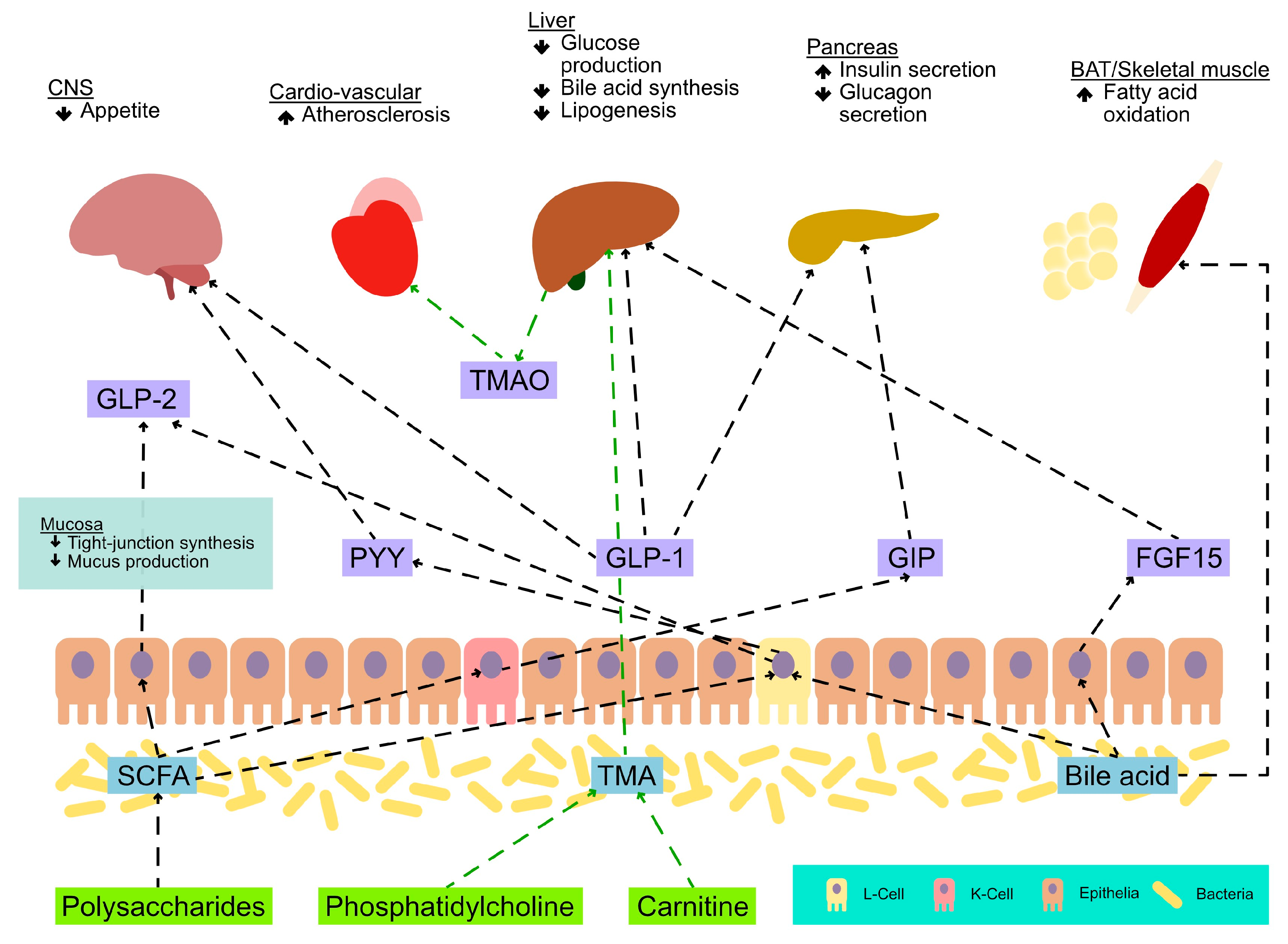
- Brown, J.M.; Hazen, S.L. The gut microbial endocrine organ: Bacterially derived signals driving cardiometabolic diseases. Annu. Rev. Med. 2015, 66, 343–359. [Google Scholar] [CrossRef] [PubMed]
- Camilleri, M. Gastrointestinal hormones and regulation of gastric emptying. Curr. Opin. Endocrinol. Diabetes Obes. 2019, 26, 3–10. [Google Scholar] [CrossRef]
- Côté, C.D.; Zadeh-Tahmasebi, M.; Rasmussen, B.A.; Duca, F.A.; Lam, T.K.T. Hormonal signaling in the gut. J. Biol. Chem. 2014, 289, 11642–11649. [Google Scholar] [CrossRef]
- Fusco, W.; Lorenzo, M.B.; Cintoni, M.; Porcari, S.; Rinninella, E.; Kaitsas, F.; Lener, E.; Mele, M.C.; Gasbarrini, A.; Collado, M.C.; et al. Short-Chain Fatty-Acid-Producing Bacteria: Key Components of the Human Gut Microbiota. Nutrients 2023, 15, 2211. [Google Scholar] [CrossRef]
- Khan, I.; Ullah, N.; Zha, L.; Bai, Y.; Khan, A.; Zhao, T.; Che, T.; Zhang, C. Alteration of Gut Microbiota in Inflammatory Bowel Disease (IBD): Cause or Consequence? IBD Treatment Targeting the Gut Microbiome. Pathogens 2019, 8, 126. [Google Scholar] [CrossRef]
- Salvi, P.S.; Cowles, R.A. Butyrate and the Intestinal Epithelium: Modulation of Proliferation and Inflammation in Homeostasis and Disease. Cells 2021, 10, 1775. [Google Scholar] [CrossRef]
- Ney, L.M.; Wipplinger, M.; Grossmann, M.; Engert, N.; Wegner, V.D.; Mosig, A.S. Short chain fatty acids: Key regulators of the local and systemic immune response in inflammatory diseases and infections. Open Biol. 2023, 13, 230014. [Google Scholar] [CrossRef]
- Martin, C.R.; Osadchiy, V.; Kalani, A.; Mayer, E.A. The Brain-Gut-Microbiome Axis. Cell. Mol. Gastroenterol. Hepatol. 2018, 6, 133–148. [Google Scholar] [CrossRef] [PubMed]
- Cani, P.D.; Knauf, C. How gut microbes talk to organs: The role of endocrine and nervous routes. Mol. Metab. 2016, 5, 743–752. [Google Scholar] [CrossRef]
- The Human Microbiome Project Consortium. Structure. function and diversity of the healthy human microbiome. Nature 2012, 486, 207–214. [Google Scholar] [CrossRef] [PubMed]
- Turnbaugh, P.J.; Ley, R.E.; Hamady, M.; Fraser-Liggett, C.M.; Knight, R.; Gordon, J.I. The Human Microbiome Project. Nature 2007, 449, 804–810. [Google Scholar] [CrossRef] [PubMed]
- Hansen, T.H.; Gøbel, R.J.; Hansen, T.; Pedersen, O. The gut microbiome in cardio-metabolic health. Genome Med. 2015, 7, 33. [Google Scholar] [CrossRef]
- Carabotti, M.; Scirocco, A.; Maselli, M.A.; Severi, C. The gut-brain axis: Interactions between enteric microbiota, central and enteric nervous systems. Ann. Gastroenterol. 2015, 28, 203–209. [Google Scholar]
- Breit, S.; Kupferberg, A.; Rogler, G.; Hasler, G. Vagus Nerve as Modulator of the Brain-Gut Axis in Psychiatric and Inflammatory Disorders. Front. Psychiatry 2018, 9, 44. [Google Scholar] [CrossRef]
- Silva, Y.P.; Bernardi, A.; Frozza, R.L. The Role of Short-Chain Fatty Acids From Gut Microbiota in Gut-Brain Communication. Front. Endocrinol. 2020, 11, 25. [Google Scholar] [CrossRef]
- Chen, Y.; Xu, J.; Chen, Y. Regulation of Neurotransmitters by the Gut Microbiota and Effects on Cognition in Neurological Disorders. Nutrients 2021, 13, 2099. [Google Scholar] [CrossRef]
- Sittipo, P.; Choi, J.; Lee, S.; Lee, Y.K. The function of gut microbiota in immune-related neurological disorders: A review. J. Neuroinflammation 2022, 19, 154. [Google Scholar] [CrossRef]
- Wei, L.; Singh, R.; Ghoshal, U.C. Enterochromaffin Cells-Gut Microbiota Crosstalk: Underpinning the Symptoms, Pathogenesis, and Pharmacotherapy in Disorders of Gut-Brain Interaction. J. Neurogastroenterol. Motil. 2022, 28, 357–375. [Google Scholar] [CrossRef] [PubMed]
- Rowland, I.; Gibson, G.; Heinken, A.; Scott, K.; Swann, J.; Thiele, I.; Tuohy, K. Gut microbiota functions: Metabolism of nutrients and other food components. Eur. J. Nutr. 2018, 57, 1–24. [Google Scholar] [CrossRef]
- Wolf, G. Insulin resistance and obesity: Resistin, a hormone secreted by adipose tissue. Nutr. Rev. 2004, 62, 389–394. [Google Scholar] [CrossRef] [PubMed]
- Li, Y.; Yang, Q.; Cai, D.; Guo, H.; Fang, J.; Cui, H.; Gou, L.; Deng, J.; Wang, Z.; Zuo, Z. Resistin, a Novel Host Defense Peptide of Innate Immunity. Front. Immunol. 2021, 12, 699807. [Google Scholar] [CrossRef] [PubMed]
- Laterra, J.; Keep, R.; Betz, L.A. Blood—Brain Barrier. In Basic Neurochemistry: Molecular, Cellular and Medical Aspects, 6th ed.; Siegel, G.J., Agranoff, B.W., Albers, R.W., Eds.; Lippincott-Raven: Pennsylvania Furnace, PA, USA, 1999. Available online: https://www.ncbi.nlm.nih.gov/books/NBK28180/ (accessed on 12 July 2023).
- Hamamah, S.; Aghazarian, A.; Nazaryan, A.; Hajnal, A.; Covasa, M. Role of Microbiota-Gut-Brain Axis in Regulating Dopaminergic Signaling. Biomedicines 2022, 10, 436. [Google Scholar] [CrossRef] [PubMed]
- Gupta, A.; Osadchiy, V.; Mayer, E.A. Brain-gut-microbiome interactions in obesity and food addiction. Nat. Rev. Gastroenterol. Hepatol. 2020, 17, 655–672. [Google Scholar] [CrossRef]
- Yu, Y.; Yang, W.; Li, Y.; Cong, Y. Enteroendocrine Cells: Sensing Gut Microbiota and Regulating Inflammatory Bowel Diseases. Inflamm. Bowel. Dis. 2020, 26, 11–20. [Google Scholar] [CrossRef]
- Pais, R.; Gribble, F.M.; Reimann, F. Stimulation of incretin secreting cells. Ther. Adv. Endocrinol. Metab. 2016, 7, 24–42. [Google Scholar] [CrossRef]
- Rehfeld, J.F. The Origin and Understanding of the Incretin Concept. Front. Endocrinol. 2018, 9, 387. [Google Scholar] [CrossRef]
- Gilbert, M.P.; Pratley, R.E. GLP-1 Analogs and DPP-4 Inhibitors in Type 2 Diabetes Therapy: Review of Head-to-Head Clinical Trials. Front. Endocrinol. 2020, 11, 178. [Google Scholar] [CrossRef]
- Zhao, X.; Wang, M.; Wen, Z.; Lu, Z.; Cui, L.; Fu, C.; Xue, H.; Liu, Y.; Zhang, Y. GLP-1 Receptor Agonists: Beyond Their Pancreatic Effects. Front. Endocrinol. 2021, 12, 721135. [Google Scholar] [CrossRef] [PubMed]
- Priyadarshini, M.; Kotlo, K.U.; Dudeja, P.K.; Layden, B.T. Role of Short Chain Fatty Acid Receptors in Intestinal Physiology and Pathophysiology. Compr. Physiol. 2018, 8, 1091–1115. [Google Scholar] [PubMed]
- Leeming, E.R.; Johnson, A.J.; Spector, T.D.; Le Roy, C.I. Effect of Diet on the Gut Microbiota: Rethinking Intervention Duration. Nutrients 2019, 11, 2862. [Google Scholar] [CrossRef] [PubMed]
- Sun, L.J.; Li, J.N.; Nie, Y.Z. Gut hormones in microbiota-gut-brain cross-talk. Chin. Med. J. 2020, 133, 826–833. [Google Scholar] [CrossRef]
- Müller, T.D.; Blüher, M.; Tschöp, M.H.; DiMarchi, R.D. Anti-obesity drug discovery: Advances and challenges. Nat. Rev. Drug Discov. 2022, 21, 201–223. [Google Scholar] [CrossRef] [PubMed]

| 2020 | 2025 | 2030 | 2035 | |
|---|---|---|---|---|
| Number with overweight or obesity (BMI ≥ 25 kg/m2) (millions) | 2603 | 3041 | 3507 | 4005 |
| Number with obesity (BMI ≥ 30 kg/m2) (millions) | 988 | 1249 | 1556 | 1914 |
| Proportion of the population with overweight or obesity (BMI ≥ 25 kg/m2) | 38% | 42% | 46% | 51% |
| Proportion of the population with obesity (BMI ≥ 30 kg/m2) | 14% | 17% | 20% | 24% |
| Study | Study Design | Pico Framework | Results of the Study | Conclusions | Links—MD *, MB **, and Obesity |
|---|---|---|---|---|---|
| Heianza et al. [36] | RCT | Population: 583 patients with G allele as Bifidobacterium-abundance-increasing allele. Intervention: The subjects were assigned at random to 1 of 4 diets for weight loss that varied in their macronutrient composition. Comparison: The study assessed adiposity measures over a span of two years, examining the correlation between the LCT genotype and weight-loss interventions. Outcomes: To see if there is a connection between the gut microbiota and obesity. | The researchers observed that alterations in overall body fat percentage, abdominal fat percentage, superficial adipose tissue mass, visceral adipose tissue mass, and total adipose tissue mass were markedly impacted by the LCT genotype and dietary protein consumption. The study found that those who had the G allele of the LCT variation rs4988235 saw a more significant decrease in many measures of body fat, including whole-body total percentage of fat, abdominal fat, superficial adipose tissue, visceral adipose tissue, and total adipose tissue, in response to a high-protein diet. In contrast, the G allele is often linked to less mitigation of these consequences when exposed to a low-protein dietary regimen. | The influence of the Bifidobacterium-related LCT genotype and dietary protein intake on the long-term enhancement of body fat composition and distribution was shown to be significant. The implementation of a dietary regimen that is both low in calories and rich in protein has the potential to assist individuals who are classified as obese or overweight, particularly those who possess the G allele of the LCT variant rs4988235, in decreasing their adiposity. | YES |
| Cuevas-Sierra et al. [37] | RCT | Population: Two hypocaloric diets were given a random assignment to 190 overweight and obese Spanish individuals for a period of four months. Intervention: A diet with moderately high protein was followed by 61 women and 29 men, and a diet with low fat was followed by 72 women and 28 men. Comparison: Four microbiota subscores related to the proportion of BMI loss for each diet were created using baseline fecal DNA, which was sequenced. Outcomes: To see if there is a connection between the gut microbiota and obesity. | The groups who used the MHP diet showed a large rise in protein consumption and a moderately significant drop in fat consumption, whereas the LF-diet group showed an increase in carbohydrate consumption and a considerable decrease in fat consumption. Women showed significantly reduced values for hip circumference after the MHP diet and lower values for leptin after the LF diet, but significant increases in HDL cholesterol. Following the LF diet, men showed considerably bigger reductions in weight, waist circumference, LDL cholesterol, and triglycerides, but a greater reduction in adiponectin levels with the MHP diet. | Despite having lower baseline values than women, men experienced a greater decline in adiponectin. According to the diet suggested for each group, the proportion of macronutrient intake showed substantial alterations as expected. As a result, a most effective weight loss plan based on this model was significantly assigned to a total of 72% of women and 84% of men who took part in this study. | YES |
| Leyrolle et al. [38] | RCT | Population: 106 obese patients. Intervention: Patients were assigned to two groups: prebiotic vs. placebo. Comparison: In addition to dietary guidance to consume inulin-rich or -poor vegetables for three months and to limit calorie consumption, patients received 16 g per day of native inulin or maltodextrin. Outcomes: To assess if there is a link between microbiota and obesity. | Except for inhibition, which was better in the prebiotic group, baseline mood and cognitive metrics did not change between the prebiotic and placebo groups. The placebo group had considerably more alcohol consumption at baseline. The therapy differently altered emotional competence. Even though within-group comparisons were not significant, emotional competence does, in fact, tend to rise in the prebiotic group while falling in the placebo group. Only in the prebiotic group did within-group comparisons show a significant reduction in negative feeling as judged by the Scale of Positive and Negative Experience and better flexibility. | Overall, the conclusion is that gut microbiota could be used to forecast how a prebiotic strategy will affect obese participants’ mood. It will be easier to tailor these methods if key gut bacteria in the body’s reaction to food-based therapy are identified. According to this research, Coprococcus may have neuroactive qualities and can be used as a gut microbiota biomarker for reaction to prebiotics. | YES |
| Zeng et al. [39] | RCT | Population: 1914 individuals average 41 years old, representing four typical lifestyles and living conditions in China. Intervention: Males made up 58%, and 11% of the total were healthy adults with normal BMIs and body weights. Comparison: Depending on the results of their physical examination and body mass index, the participants were divided into three groups: a healthy group, an obesity group without metabolic abnormalities, and an obesity group with abnormal clinical indications. Outcomes: To assess if there is a link between microbiota and obesity. | Patients with metabolic disorders showed changed GM components in comparison to obese patients without abnormalities, and Clostridium XIVa helped distinguish between obese patients with high serum cholesterol or blood pressure. These indicators revealed common GM changes in obese patients with various metabolic disorders, suggesting that other variables (such as genetic variation) may play a role in the development of several metabolic diseases. Based on these findings, the authors hypothesized that MDs were first brought on by obesity-related GM changes, and that further specific pathogenic aspects beyond GM dysbiosis needed to be investigated. | As a result, the study provided markers for obese individuals with diverse MDs, identified GM characteristics, and demonstrated the relationships between bacterial commensals and other clinical indications. These findings provided prospective GM targets for adjuvant therapies in the treatment of obesity with metabolic abnormalities and revealed the roles of GM in the etiology of metabolic disorders. | YES |
| Ghusn et al. [40] | RCT | Population: A total of 175 patients with BMI of 27 or more. Intervention: The patients were given weekly subcutaneous injections of semaglutide for a duration of at least three months. Comparison: A total of 132 female individuals were included in the study at the 3-month mark, whereas the number of patients decreased to 102 at the 6-month mark. Outcomes: To assess any connections of microbiota with obesity. | Following a period of three months, the mean reduction in weight was seen to be 6.7 kg, equivalent to 5.9% of an individual’s initial body weight. Subsequently, following a span of six months, the average weight loss increased to 12.3 kg, corresponding to 10.9% of one’s initial body weight. Among the cohort of 102 individuals who were subjected to monitoring over a period of 6 months, it was seen that a substantial proportion, namely 87.3%, had achieved a reduction in their body weight of no less than 5%. Furthermore, a significant percentage of 54.9% had managed to lose at least 10% of their initial body weight. Additionally, a noteworthy subset of 23.5% had successfully achieved a weight loss of at least 15%, while a smaller fraction of 7.8% had accomplished a reduction of no less than 20% of their initial body weight. At the 3-month and 6-month marks, those diagnosed with type 2 diabetes had comparatively lower average weight loss in comparison to those without the illness. Specifically, the weight loss percentages were 3.9% and 6.3% at 3 months and 7.2% and 11.8% at 6 months, respectively. | The results of this cohort study suggest that the weight reduction achieved with weekly dosages of 1.7 mg and 2.4 mg of semaglutide is similar to that found in randomized clinical trials. | YES |
| Zhou et al. [41] | RCT | Population: 264 overweight and obese patients. Intervention: From the beginning of the dietary intervention to six months later, blood levels of TMAO, choline, and l-carnitine were measured. Comparison: There were four different diets: two low fat, two high fat, two intermediate protein, and two high proteins. Outcomes: To find out if variations in BMD after two years were related to variations in plasma TMAO, choline, and l-carnitine levels from baseline to six months. | The researchers discovered that a higher loss in bone mineral density (BMD) at 6 months and 2 years was connected to a greater decline in plasma levels of TMAO from baseline to 6 months. Independent of changes in body weight, the larger decline in TMAO was also linked to a bigger loss in spine BMD at 2 years. The correlations were unaffected by the glycemic and diabetic status at baseline. In relation to changes in spine BMD and hip BMD after 6 months, L-carnitine alterations showed interactions with dietary fat intake. In the low-fat-diet group, those who saw the least drop in L-carnitine experienced less bone loss than in the high-fat-diet group. | Independent of diet treatments with different macronutrient contents and baseline diabetes risk factors, TMAO may protect against BMD decline during weight loss. The relationship between changes in plasma L-carnitine levels and changes in BMD may be altered by dietary fat. The results emphasize the significance of researching the link between TMAO and bone health in diabetic patients. | YES |
| Christensen et al. [42] | RCT | Population: 2224 individuals (1504 women and 720 men). Intervention: participants followed a low energy diet (LED) for 2 months. Comparison: Phase 1 consisted of an eight-week weight-loss phase using the LED. Phase 2 was a 148-week ongoing randomized lifestyle intervention that emphasized nutrition and exercise. Outcomes: To evaluate behavior modification for weight loss maintenance. | Men lost more weight than women (11.8% vs. 10.3%, respectively), but improvements in insulin resistance were comparable in both sexes. Men experienced greater declines in heart rate, fibromyalgia, the Z-score for the metabolic syndrome, and the C-peptide, whereas women experienced greater declines in HDL cholesterol, free fat mass, hip circumference, and pulse pressure. A total of 35% of participants returned to normoglycemia after the LED. | Women and men experienced distinct outcomes from the 8-week low-energy diet. These findings, which point to gender-specific alterations following weight loss, are therapeutically significant. It is crucial to investigate whether rapid weight reduction in women causes higher declines in free fat mass, hip circumference, and HDL cholesterol, which could jeopardize long-term weight maintenance and cardiovascular health. | YES |
| Shank et al. [43] | RCT | Population: 103 adolescent girls reported losing control of their eating (LOC), thus gaining weight. Intervention: The girls underwent assessments for the metabolic syndrome at baseline and again six months later. Comparison: Participants were randomly assigned to either a 12-week interpersonal group psychotherapy program or a group health education control program. Outcomes: Considering baseline age, depressive symptoms, fat mass, and height, the primary impacts of LOC status at treatment’s conclusion on metabolic syndrome components at a 6-month follow-up were studied. | Adolescents who had loss of control (LOC) remission at the conclusion of their therapy exhibited decreased levels of triglycerides, increased levels of high-density lipoprotein (HDL), and decreased levels of low-density lipoprotein (LDL) at the 6-month follow-up, in contrast to adolescents who continued to have persistent LOC. Notably, there were no discernible variations in these lipid components at baseline between the two groups. There were no significant differences seen in any other component based on the eating status of individuals with limited or no control (LOC) over their eating behavior. | Improvements in various metabolic syndrome components are linked to LOC eating remission. Future studies should continue to clarify the connection between LOC eating and physical health to conclude whether metabolic health may be improved in the long run by abstinence from LOC eating. The consumption of low-quality, energy-dense foods is a potentially modifiable lifestyle factor that might be strategically addressed in order to mitigate the risk of developing total or partial metabolic syndrome, provided that it leads to sustained improvements in metabolic well-being. | YES |
| Kwee et al. [44] | RCT | Population: 2458 participants were studied from 2006 and 2009 to 31 January 2015. Intervention: Using mass-spectrometry-based techniques, the quantitative levels of 135 metabolites were assessed at baseline. Comparison: The results were compared to see which group managed to have a diabetes remission status. Outcomes: To assess the change in diabetes-related clinical variables from pre-intervention to two years after-intervention. | Two metabolite factors, one with betaine and choline and the other with branched chain amino acids and tyrosine, were linked to the remission of diabetes. | The circulating baseline biomarkers for diabetes remission that the authors identified have independent associations as well as incremental predictive powers when included in a clinical model. | YES |
| Pearl et al. [45] | RCT | Population: 178 obese adults signed up for a weight-loss trial. Intervention: The participants filled in the Weight Bias Internalization Scale (WBI) and Patient Health Questionnaire. Comparison: The adults were investigated to determine whether WBI and metabolic syndrome (MS) are related. Outcomes: If participants lost less than 5% of their starting weight during a 14-week diet run-in period, they were randomly allocated to a 1-year weight reduction maintenance program to examine its effects. | Participants with higher WBI had an increased chance of fulfilling the criteria for MS. Greater likelihood of having high triglycerides were indicated by higher WBI. When categorically analyzed, high (vs. low) WBI indicated a higher likelihood of metabolic syndrome and high triglycerides. | Self-stigmatizing obese people may be at higher risk for cardiovascular and metabolic problems. Further exploring the biological and behavioral processes that connect WBI and metabolic syndrome is important. | YES |
| Neuro- Transmitters | Precursor | Gut Microbiota | Cells of Intestine | Gut–Brain Axis |
|---|---|---|---|---|
| Glutamate (GLU) | Acetate | Lactobacillus plantarum Bacteroides vulgatus Campylobacter jejuni | Enteroendocrine cells | The transmission of sensory information originating from the intestines to the brain occurs through the vagus nerve. |
| GABA | Acetate | Bifidobacterium Bacteroides fragilis Parabacteroides Eubacterium | Myenteric neurons Mucosal endocrine-like cells | This neurotransmitter modulates the neuro-synaptic transmission in the GI nervous system and has an impact on intestinal motility and inflammation. |
| Acetylcholine | Choline | Lactobacillus plantarum Bacillus acetylcholini Bacillus subtilis Escherichia coli Staphylococcus aureus | Myenteric neurons | The myenteric neurons in the human colon are responsible for the production of 33% of the total output. The regulation of intestinal motility, secretion, and enteric neurotransmission is of paramount importance in maintaining proper gastrointestinal function. |
| Dopamine | Tyrosine l-DOPA | Staphylococcus | Affects gastric secretion, motility, and mucosal blood flow. Affects gastric tone and motility through nigro–vagal pathway in a Parkinson’s disease rat model. | |
| Serotonin | 5-HTP Tryptophan | Staphylococcus Clostridial species | Enterochromaffin cells | Enhance gastrointestinal peristalsis. |
| Norepinephrine | Tyrosine | Modulates energy intake and thermal homeostasis. | ||
| Tyramine | Tyrosine | Staphylococcus Providencia | The substance that precedes or serves as a precursor to octopamine. | |
| Phenyle- thylamine | Phenyl- alanine | Staphylococcus | ||
| Tryptamine | Tryptophan | Staphylococcus Ruminococcus gnavus Clostridium sporogenes | The stimulation of serotonin secretion in enterochromaffin cells. Enhances gastrointestinal function. |
Disclaimer/Publisher’s Note: The statements, opinions and data contained in all publications are solely those of the individual author(s) and contributor(s) and not of MDPI and/or the editor(s). MDPI and/or the editor(s) disclaim responsibility for any injury to people or property resulting from any ideas, methods, instructions or products referred to in the content. |
© 2023 by the authors. Licensee MDPI, Basel, Switzerland. This article is an open access article distributed under the terms and conditions of the Creative Commons Attribution (CC BY) license (https://creativecommons.org/licenses/by/4.0/).
Share and Cite
Ispas, S.; Tuta, L.A.; Botnarciuc, M.; Ispas, V.; Staicovici, S.; Ali, S.; Nelson-Twakor, A.; Cojocaru, C.; Herlo, A.; Petcu, A. Metabolic Disorders, the Microbiome as an Endocrine Organ, and Their Relations with Obesity: A Literature Review. J. Pers. Med. 2023, 13, 1602. https://doi.org/10.3390/jpm13111602
Ispas S, Tuta LA, Botnarciuc M, Ispas V, Staicovici S, Ali S, Nelson-Twakor A, Cojocaru C, Herlo A, Petcu A. Metabolic Disorders, the Microbiome as an Endocrine Organ, and Their Relations with Obesity: A Literature Review. Journal of Personalized Medicine. 2023; 13(11):1602. https://doi.org/10.3390/jpm13111602
Chicago/Turabian StyleIspas, Sorina, Liliana Ana Tuta, Mihaela Botnarciuc, Viorel Ispas, Sorana Staicovici, Sevigean Ali, Andreea Nelson-Twakor, Cristina Cojocaru, Alexandra Herlo, and Adina Petcu. 2023. "Metabolic Disorders, the Microbiome as an Endocrine Organ, and Their Relations with Obesity: A Literature Review" Journal of Personalized Medicine 13, no. 11: 1602. https://doi.org/10.3390/jpm13111602
APA StyleIspas, S., Tuta, L. A., Botnarciuc, M., Ispas, V., Staicovici, S., Ali, S., Nelson-Twakor, A., Cojocaru, C., Herlo, A., & Petcu, A. (2023). Metabolic Disorders, the Microbiome as an Endocrine Organ, and Their Relations with Obesity: A Literature Review. Journal of Personalized Medicine, 13(11), 1602. https://doi.org/10.3390/jpm13111602