Article

Influence of ApoE Genotype and Clock T3111C Interaction with Cardiovascular Risk Factors on the Progression to Alzheimer’s Disease in Subjective Cognitive Decline and Mild Cognitive Impairment Patients

1
Department of Neuroscience, Psychology, Drug Research and Child Health-University of Florence–Viale Pieraccini 6, 50139 Florence, Italy
2
IRCCS Fondazione Don Carlo Gnocchi, via di Scandicci 269, 50143 Florence, Italy
*
Author to whom correspondence should be addressed.
J. Pers. Med. 2020, 10(2), 45; https://doi.org/10.3390/jpm10020045
Submission received: 11 May 2020 / Revised: 26 May 2020 / Accepted: 27 May 2020 / Published: 29 May 2020
(This article belongs to the Special Issue Novel Biomarkers in Alzheimer’s Disease)

Abstract

:
Background: Some genes could interact with cardiovascular risk factors in the development of Alzheimer’s disease. We aimed to evaluate the interaction between ApoE ε4 status, Clock T3111C and Per2 C111G polymorphisms with cardiovascular profile in Subjective Cognitive Decline (SCD) and Mild Cognitive Impairment (MCI). Methods: We included 68 patients who underwent clinical evaluation; neuropsychological assessment; ApoE, Clock and Per2 genotyping at baseline; and neuropsychological follow-up every 12–24 months for a mean of 13 years. We considered subjects who developed AD and non-converters. Results: Clock T3111C was detected in 47% of cases, Per2 C111G in 19% of cases. ApoE ε4 carriers presented higher risk of heart disease; Clock C-carriers were more frequently smokers than non C-carriers. During the follow-up, 17 patients progressed to AD. Age at baseline, ApoE ε 4 and dyslipidemia increased the risk of conversion to AD. ApoE ε4 carriers with history of dyslipidemia showed higher risk to convert to AD compared to ApoE ε4− groups and ApoE ε4+ without dyslipidemia patients. Clock C-carriers with history of blood hypertension had a higher risk of conversion to AD. Conclusions: ApoE and Clock T3111C seem to interact with cardiovascular risk factors in SCD and MCI patients influencing the progression to AD.

1. Introduction

Alzheimer’s disease (AD) is characterized by a slow but progressive trend, with a presymptomatic phase that can last from years to decades [1]. Subjective Cognitive Decline (SCD) is defined as a self-experienced persistent decline in cognitive capacity in comparison with the subject’s previously status, during which the subject has normal performance on standardized cognitive tests [2]. Mild cognitive impairment (MCI) concerns an objective cognitive impairment with minimal impact on instrumental activity of daily living [3], and it is considered an intermediate phase between normal cognition and dementia. MCI is associated with an increased risk of positive AD biomarkers and with an annual conversion rate of 5%–17% to AD [4,5]. For SCD, the annual conversion rate (ACR) to MCI is 3.6%–6.6%, while it is 1.5%–2.3% to dementia [4,6].
A growing amount of evidence has underlined the importance of cardiovascular health on the risk of developing AD [7,8,9]. It has been reported that approximately one-third of AD cases worldwide may be attributable to cardiovascular risk factors, including hypertension, obesity, diabetes, smoking, and physical inactivity [10].
In addition, it is well known that genetic aspects play a central role in development of AD [11]. Apolipoprotein E e4 carrier status (ApoE e4) is a well-defined genetic risk factor, and recently, current research has been focused on clock genes. Clock (Circadian Locomotor Output Cycle Kaput, chromosome 4q12) and Per2 (Period2, chromosome 2q37.3) are part of the transcriptional-translational feedback loops regulating circadian rhythm [12]. Several polymorphisms of these genes have been recently studied to elucidate their role in sleep-wake cycle alterations, aging, psychiatric disturbance and neurodegeneration [13,14]. Moreover, Clock and Per2 polymorphisms have been associated with overweight and glucose and lipid metabolism impairments [15,16]. Some studies have investigated the possible role of the Clock T3111C polymorphism on the quality of aging in very elderly patients [17], while other studies focused on influence of Per2 C111G polymorphism on lipid metabolism in adults with metabolic syndrome [16]. On the basis of the above-mentioned initial findings about of the influence of these polymorphisms on cardiovascular profile, the aim of the present study was to define the interaction between Clock T3111C and Per2 C111G, ApoE ε4, and cognitive function, in relation to cardiovascular risk factors, in SCD and MCI patients and in the progression to AD.

2. Materials and Methods

2.1. Participants and Clinical Assessment

As part of a longitudinal, clinical-neuropsychological-genetic survey on SCD and MCI, we included 74 consecutive spontaneous patients who self-referred to the Centre for Alzheimer’s Disease and Adult Cognitive Disorders of Careggi Hospital in Florence between April 1996 and May 2014. All participants underwent a comprehensive family and clinical history, general and neurological examination, extensive neuropsychological investigation, estimation of premorbid intelligence, as well as assessment of depression. A positive family history was defined as one or more first-degree relatives with documented cognitive decline. Sleep quality was assessed according to anamnestic data: we considered as “poor sleepers” patients who had difficulties in falling asleep or woke up early or have frequent sleep interruptions; patients that did not report sleep disturbances were classified as “good sleepers”. For this study, inclusion criteria were: (1) complaining of cognitive decline with a duration of ≥6 months; (2) normal functioning on the Activities of Daily Living and the Instrumental Activities of Daily Living scales; (3) unsatisfied criteria for dementia at baseline [18]; (4) attainment of the clinical endpoint, i.e., conversion to AD according to the NIA-AA [18] criteria during follow up, regardless of follow-up duration; (5) a follow-up time of more than 2 years from the baseline visit for those patients who did not develop AD. Exclusion criteria were: (1) history of head injury, current neurological and/or systemic disease, symptoms of psychosis, major depression, alcoholism or other substance abuse; (2) the complete data loss of patients’ follow-up; (3) progression to dementia other than AD.
From the initial sample, we excluded six patients: two patients had a follow-up shorter than 2 years; two diagnosed with psychiatric disturbance, and one with Fronto-Temporal Dementia, according to Neary criteria [19]; one patient received a diagnosis of Vascular Dementia [20]. Therefore, in the end 68 patients were included.
We divided this sample into two groups: 41 patients classified as SCD, according to the terminology proposed by the Subjective Cognitive Decline Initiative (SCD-I) Working Group [2] (i.e., presence of a self-experienced persistent decline in cognitive capacities with normal performance on standardized cognitive tests); 27 patients classified as MCI according to (NIA-AA) criteria for the diagnosis of MCI [3] (i.e., evidence of lower performance in one or more cognitive domains with preserved independence of function in daily life).
All patients underwent clinical and neuropsychological follow-up every 12 or 24 months. All of them were genotyped for ApoE (Apolipoprotein E), Clock and Per2.
On the basis of progression to AD during the follow-up, patients were classified respectively into converters and non-converters. All subjects gave their informed consent for inclusion before they participated in the study. The study was conducted in accordance with the Declaration of Helsinki, and the protocol was approved by the Ethics Committee (DSM study).

2.2. Neuropsychological Assessment

All patients were evaluated by means of an extensive neuropsychological battery [21]. The battery consisted of global measurements [Mini-Mental State Examination (MMSE)], tasks exploring verbal and spatial short- term memory (Digit Span; Corsi Tapping Test) and verbal long-term memory [Five Words and Paired Words Acquisition (FWA, PWA); recall after 10min (FWR10, PWR10); recall after 24-h (FWR24, PWR24); Babcock Short Story Immediate and Delayed Recall (BS, BSR)]; language (Token Test; Category Fluency Task); and visuo-motor functions (Copying Drawings) [21]. Visuospatial abilities were also evaluated by Rey–Osterrieth Complex Figure copy, and visuospatial long-term memory was assessed by means of recall of Rey–Osterrieth Complex Figure test [22]; attention/executive function was explored by means of Dual Task [23], Phonemic Fluency Test [24], and Trail Making Test [25]. Everyday memory was assessed by means of Rivermead Behavioral Memory Test (RBMT) [26]. All raw test scores were adjusted for age, education and gender according to the correction factor reported in validation studies for the Italian population [21,22,23,24,25,26]. In order to estimate pre-morbid intelligence, all patients were given the TIB (“Test di Intelligenza Breve”) [27], an Italian version of the National Adult Reading Test [28]. The presence and severity of depressive symptoms was evaluated by means of the 22-item Hamilton Depression Rating Scale (HRSD) [29].

2.3. Apolipoprotein E ε4, Clock T3111C and Per2 C111G Genotyping

A standard automated method (QIAcube, QIAGEN) was used to isolate DNA from peripheral blood samples. ApoE genotypes were investigated by high resolution melting analysis (HRMA). Two sets of PCR primers were designed to amplify the regions encompassing rs7412 [NC_000019.9:g.45412079C>T] and rs429358 (NC_000019.9:g.45411941T>C). The samples with known ApoE genotypes, which had been validated by DNA sequencing, were used as standard references. The ApoE genotype was coded as ApoE ε4− (no ApoE ε4 alleles) and ApoE ε4+ (presence of one or two ApoE ε4 alleles).
The analyses of Clock and Per2 were performed using HRMA in order to detect the 3111T/C Clock polymorphism using primers as reported [30] and the Per2 C111G polymorphism with the following primers Forward 5′-ACAGAAAGAGTCAAATGGGTGC-3′, Reverse 5′-TGTCCACATCTTCCTGCAGT-3′ with Annealing temperature 60 °C.

2.4. Statistical Analysis

Patient groups were characterized using means and standard deviations (SD). We tested for the normality distribution of the data using the Kolmogorov–Smirnov test. Depending on the distribution of our data, we used t-test or non-parametric Mann–Whitney U Tests for between-groups’ comparisons. We used chi-square test to compare categorical data. We analyzed survival curves using the Kaplan–Meier estimator. Finally, we used logistic regression to analyze the role of some cardiovascular risk factors in worsening cognition. All statistical analyses were performed with SPSS software v.25 (SPSS Inc., Chicago, IL, USA). The significance level was set at p < 0.05.

3. Results

3.1. Participants and Clinical Assessment

In the whole cohort, 32 of 68 patients (47%) were Clock C carriers (29 TC, 3 CC), while 13 of 68 (19%) were Per2 G carriers (13 CG, 0 GG); 7 of 68 (10%) carried both Clock C and Per2 G alleles. The genotypic distributions of the Clock and Per2 genes in this sample were in Hardy–Weinberg equilibrium (Clock T3111C χ2 = 0.91, p > 0.05; Per2 C111G χ2 = 0.77, p > 0.05). The prevalence of Clock T3111C and Per2 C111G polymorphisms did not significantly differ between SCD and MCI (Clock T3111C: 51.2% in SCD and 40.7% in MCI; Per2 C111G: 19.5% in SCD and 18.5% in MCI); moreover, there were not any differences in the prevalence of both Clock C and Per2 G carriers in SCD and MCI groups. (Table 1).
There were no differences between Clock C carriers and non C carriers with regards to age at onset of symptoms, age at baseline visit, disease duration, sex, family history of AD, years of education, TIB, MMSE, and ApoE ε4 allele status. With respect to CV risk factors, there was a higher proportion of smokers in Clock C carriers than in non C carriers (19.40% vs. 2.80%, χ2 = 4,892, p = 0.027) (Table 2).
Comparing Per2 G carriers and non G carriers, there were no differences in age at onset of symptoms, age at baseline visit, disease duration, sex, family, MMSE, and ApoE ε4 allele status. Per2 G carriers had lower premorbid intelligence score on TIB (104.29 ± 10.74 vs. 109.98 ± 8.06, p = 0.049), less years of education (7 ± 3.05 vs 10.73 ± 4.51, p = 0.007), and lower frequency of family history of AD (15.38% vs. 60%, χ2 = 8.37, p = 0.004) (Table 2). There were no differences in CV risk factors proportion between G and non G carriers.
We did not find any differences in sleep quality between Clock C carriers and Clock non C carriers (χ2 = 0.136, p = 0.454), neither between Per2 G carriers and Per2 non G carriers (χ2 = 0.879, p = 0.273) (Table 2).
ApoE ε4+ patients have a higher proportion of history of heart disease than ApoE ε4− (8.70% vs. 0.00%, χ2 = 3.944, p = 0.047). We did not find any statistically significant difference between ApoE ε4+ and ApoE ε4− as far age at onset of symptoms, age at baseline evaluation, disease duration (time from onset of symptoms and baseline evaluation), follow-up time, familiarity, sex, education, and MMSE (Table 2).

3.2. Description of Sample at Follow-Up

During the follow-up, 17 patients (25%, 2 SCD and 15 MCI) converted to AD (converters) while 51 patients did not progress to AD (non-converters). Mean conversion time was 4.73 ± 3.91 years (range: 1.41–14.01, IQR = 3.71 years). Mean follow-up time of non-converters was 13.03 ± 4.48 years (range: 4.06–23.74, IQR = 6.73 years). There were no differences between converters and non-converters with respect to disease duration, sex, family history of AD, years of education, and MMSE at baseline. Converters had higher age at the onset of symptoms (68.20 ± 7.49 vs. 61.25 ± 6.21, p = 0.016), age at baseline (71.60 ± 6.19 vs. 63.42 ± 6.96, p = 0.003) and greater proportion of ApoE ε4 (56.25% vs. 25.49%, χ2 = 5.22, p = 0.022). (Table 3).
There were no significant differences between converters and non-converters in the prevalence of Clock T3111C (35.3% vs. 51.0%, χ2 = 1.26, p = 0.262) and Per2 C111G (23.5% vs. 17.6%, χ2 = 0.285 p = 0.593) polymorphism (Table 3).
In order to ascertain the effects of cardiovascular risk factors on the conversion to AD, we performed a proportional hazards regression analysis considering conversion time as time and “conversion to AD” as dependent variable. We considered as covariates age at onset, age at baseline, ApoE, Clock and Per2 genotype, hypertension, diabetes, dyslipidemia, heart disease, and smoking habit. Dyslipidemia (p = 0.041, HR = 3.08, 95% I.C. = 1.05: 9.09), age at baseline (p = 0.001, HR = 1.16, 95% I.C. = 1.07:1.27) and ApoE ε4 (p = 0.001, HR = 6.21, 95% I.C. = 2.04:18.9) were statistically significantly associated with an increased likelihood of conversion to AD (Table 4).

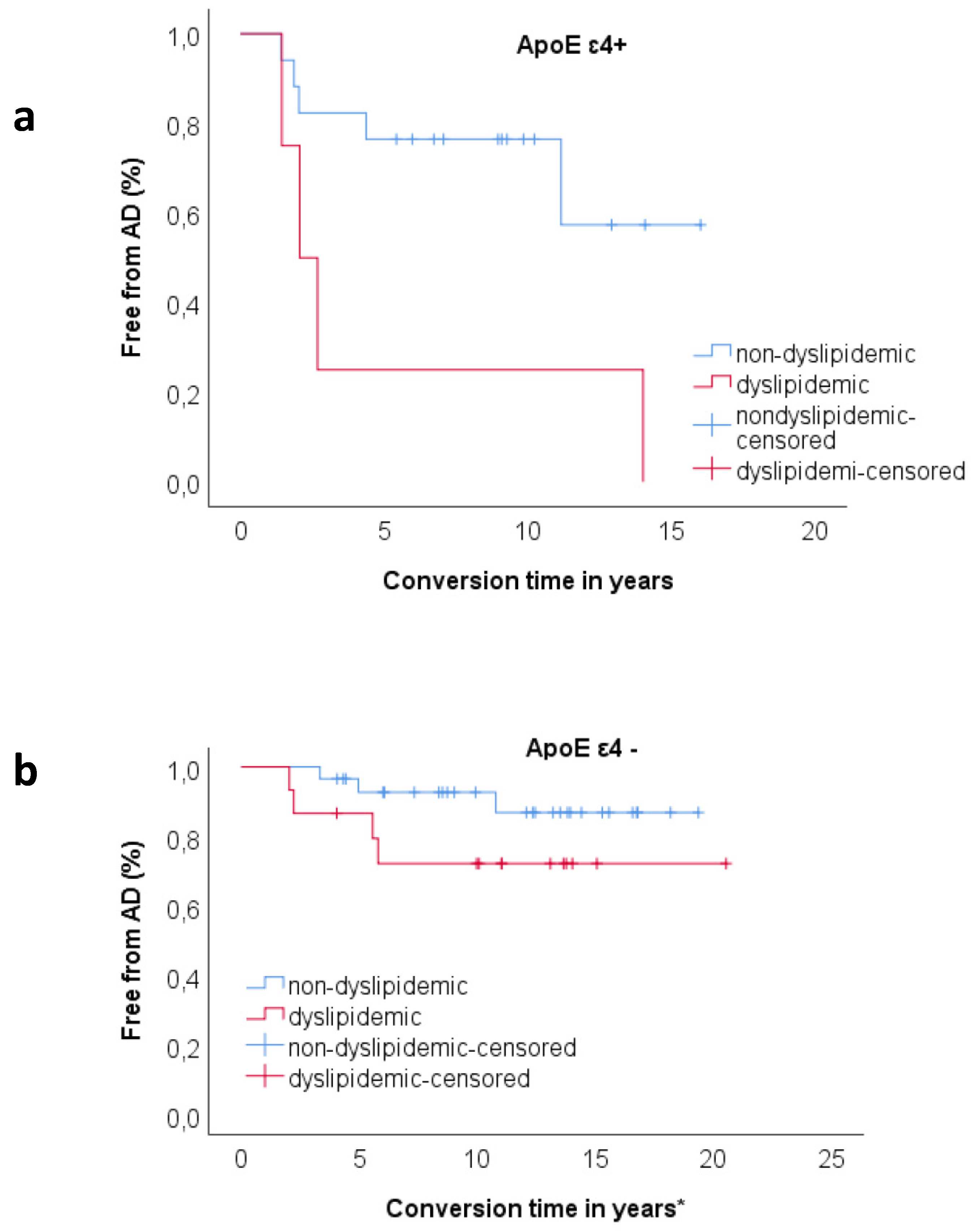
3.3. Relationship between ApoE and Dyslipidemia

In order to explore the relationship between dyslipidemia and ApoE genotype, we divided the sample according to ApoE ε4 status (ε4+ and ε4−). In the ε4+ sample, a Kaplan–Meier survival analysis showed significant difference in survival distributions between patients with history of dyslipidemia and patients without history of dyslipidemia (χ2 = 4.42, p = 0.036), as 100% of dyslipidemic patients and 29.4% of non-dyslipidemic patients converted to AD (Figure 1a). When we performed the same analysis on the ε4− sample, we found no statistically significant effect of dyslipidemia on rate of progression to AD (χ2 = 1.92, p = 0.166) (Figure 1b).
Finally, we ranked the whole sample according to history of dyslipidemia and ApoE genotype (non-dyslipidemic/ε4−, n = 29; non-dyslipidemic/ε4+, n = 17; dyslipidemic/ε4−, n = 15; dyslipidemic/ε4, n = 4) and Kaplan–Meier survival analysis was conducted to compare the proportions of conversions in the four different groups. Patients in dyslipidemic/ε4+ group had a higher rate of conversion to AD compared to non-dyslipidemic/ ε4− (χ2 = 25.47, p < 0.001), non-dyslipidemic/ε4+ (χ2 = 4.42, p = 0.036) and dyslipidemic/ε4− (χ2 = 7.64, p = 0.006). Proportion of conversion in non-dyslipidemic/ε4+ was higher as compared to non-dyslipidemic/ε4− (χ2 = 3.73, p = 0.05). There was no significant difference between non-dyslipidemic/ε4−, dyslipidemic/ε4− and dyslipidemic/ε4− (Figure 2).

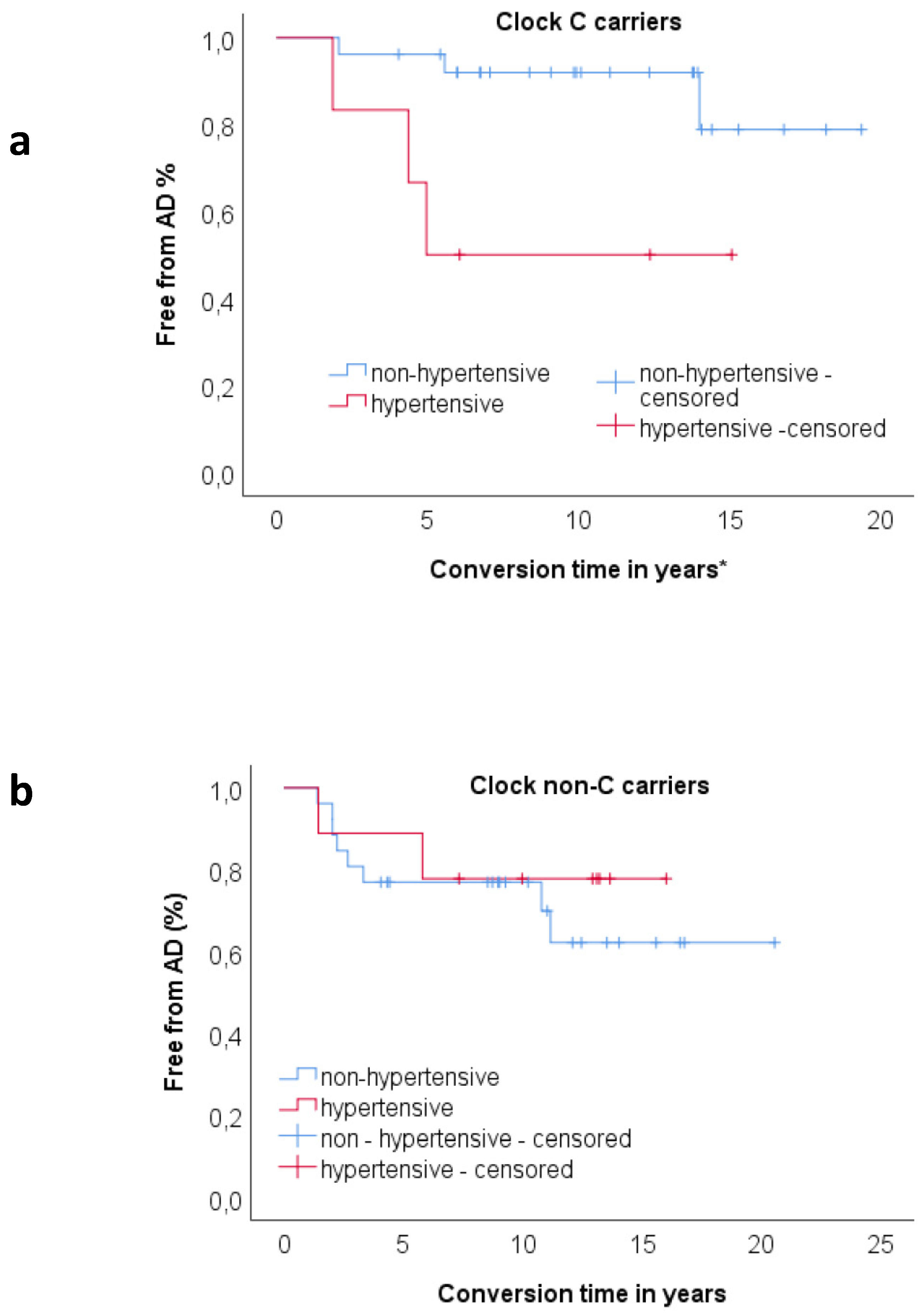
3.4. Relationship between Clock and Per2 and Risk Factors on Progression to AD

In order to explore the relationship between CV risk factors and Clock polymorphism, we divided the sample according to Clock genotype status (C carriers and non C carriers). For each sample, we performed a proportional hazards regression analysis considering conversion time as time; “conversion to AD” as dependent variable; and age at onset, age at baseline, ApoE genotype, hypertension, diabetes, dyslipidemia, heart disease, and smoking habit as covariates. In the C carriers sample, hypertension at baseline (p = 0.025, HR = 3.625) was statistically significantly associated with an increased likelihood of conversion to AD (Table 4). In the Clock non C carriers, none of the CV factors were included in the model as only age at baseline was statistically significantly associated with high risk of progression to AD.
A Kaplan–Meier survival analysis showed significant difference in survival distributions between patients with history of hypertension and patients without history of hypertension (χ2 = 4.42, p = 0.036) only in the Clock C carriers sample (Figure 3a). In this group, 50% of hypertensive patients and 11.5% of non-hypertensive patients converted to AD. When we performed the same analysis on the Clock non C carriers sample, we found no statistically significant effect of hypertension on rate of progression to AD; (Figure 3b).
We performed the proportional hazard regression analysis ranking patients according to Per2 polymorphism, including as covariates age at onset, age at baseline, ApoE genotype, hypertension, diabetes, dyslipidemia, heart disease, and smoking. In the G carriers sample, none of the covariates showed a statistically significant effect on risk of conversion to AD. In the Per2 non G carriers, none of the CV factors were included in the model as only age at baseline was statistically significantly associated with high risk of progression to AD (p = 0.010, HR = 1.185).

4. Discussion

This study investigated the interaction between genetic features (Clock T3111C, Per2 C111G polymorphisms, and ApoE genotype) and cardiovascular risk factors in a sample of SCD and MCI patients. We are not aware of any previous studies exploring this topic on these groups of patients.
The frequency of these polymorphisms in our cohort was similar to prevalence data reported in a previous study on healthy Italian population [31].
We found that Clock T3111C carriers were more frequent smokers compared to non-carriers of the polymorphism. Our result may suggest an implication also of Clock T3111C on nicotine dependence. Other researches have shown that clock genes are associated with substance abuse, including alcohol, cocaine and cannabis [32]. In fact, circadian genes have a direct role in in the regulation of dopaminergic transmission, especially in reward circuitry. This evidence could represent the biological substrate for the role of Clock genes in the development of addicted behaviours [33,34].
We found a correlation between Per2 C111G polymorphism with years of education and family history of AD. This result confirms previous evidence by our group [35], but it is difficult to interpret this finding due to the absence of previous works on this issue.
For future researches on wider sample, we will aim to clarify our current findings. With regard to cardiovascular risk factors, we did not find any association with Per2 C111G.
We found that ApoE ε4 carriers had more frequent history of heart disease than ε4 non carriers. Our result is supported by previous studies. In particular, two different meta-analysis [36,37] showed a different distribution of coronary disease risk according to ApoE genotype. Furthermore, this association could be independent from other cardiovascular risk factors, as we did not find any correlation with diabetes, dyslipidemia, smoking, or hypertension.
A multivariate analysis showed that age at baseline, ApoE e4 and dyslipidemia increase the risk of progression to AD.
With regard to age, this is not surprising as it is recognized to be the major risk factor for AD [38,39].
The effect of ApoE ε4 allele on risk of progression to AD has been widely demonstrated by previous studies [40,41,42,43].
Last, lipid disorders have been said to have a role in cognitive impairment and their treatment has been studied as a prevention tool, but evidence about this topic is not yet conclusive [44].
The interaction between ApoE ε4 and dyslipidemia on cognition is not yet completely understood [36]. In order to explore this point, we ranked the patients according to ApoE genotype. The effect of dyslipidemia on progression to AD was confirmed only in ApoE ε4 carriers. Furthermore, we showed that patients who were ApoE ε4 carriers and had history of dyslipidemia showed higher risk to convert to AD both compared to ApoEε4− groups and ApoE ε4+ without dyslipidemia patients. According to this analysis, dyslipidemia could be synergistic with E4 carrier status in contributing to AD pathogenesis as reported by other authors [44].
Previous studies suggested an association between Clock gene polymorphisms with different cardiovascular risk factors, as well as with the cognitive state [15,16,17]. In order to investigate a possible interaction between Clock T3111C and cardiovascular risk factors, we ranked patients according to Clock genotype. We found that Clock C carriers with history of blood hypertension had a higher risk of conversion to AD than Clock C carriers without hypertension. This difference was not detected in the Clock non C carrier group. No other work, to the best of our knowledge, has previously studied the possible influence of Clock gene polymorphisms on the effect of hypertension in conversion to AD in this particular group of patients.
A limitation of this study is the small size of our cohort. In future we aim to expand our sample, also including a healthy control group, to support our present results.
Secondly, the lack of quantitative data about dyslipidemia and hypertension did not allow us to understand if our results might be different according to the level of blood lipids and blood pressure. Another limitation of this study is that AD diagnosis was not supported by AD biomarkers. Future researches including cerebrospinal fluid amyloid beta, tau and p-tau levels or neuroimaging data, as amyloid PET, could provide interesting and additional information. Finally, as it is a single-center study, there may be biases with regard to assessment and diagnosis procedures. On the other hand, this study has some remarkable strengths. First of all, to the best of our knowledge, this is the first prospective study that assessed the interaction of these clock genes polymorphisms with cardiovascular factors on the risk or progression to AD in cohort of well-defined SCD and MCI patients. The second strength is the very long mean follow-up time of 13 years. Moreover, non-converter patients had a longer follow-up time than patients who converted to AD. This is crucial information as follow-up time could influence rate of conversion to AD. The long follow-up time in our sample allows us to minimize the risk of classifying as stable subjects carrying an Alzheimer pathology who will convert later. Indeed, the present study suggests an association between ApoE genotype and Clock T3111C with different cardiovascular risk factors in a cohort of SCD and MCI patients. This interaction could influence the progression to AD in this group of patients. Understanding the mechanisms by which genetic and cardiovascular risk factors contribute to AD could inspire the development of new personalized therapeutic approaches for this disease.

Author Contributions

Conceptualization, V.B. and B.N.; methodology, V.B., S.M., B.N.; software, S.M.; validation, V.B., J.B., S.M., G.G., S.B., S.P., I.P., M.C., L.B., S.S., B.N.; formal analysis, V.B., S.M., J.B., S.B., I.P.; investigation, V.B., J.B., S.M., G.G., S.B., S.P., I.P., M.C., L.B., S.S., B.N.; resources, V.B., J.B., S.M., G.G., S.B., S.P., I.P., M.C., L.B., S.S., B.N.; data curation, V.B., J.B., S.M., G.G., S.B., S.P., I.P., M.C., C.F., L.B., S.S., B.N.; writing—original draft preparation, V.B., J.B., S.M., G.G.; writing—review and editing, V.B., S.M., G.G., S.S., B.N.; visualization, V.B., J.B., S.M., G.G., S.B., S.P., I.P., M.C., C.F., L.B., S.S., B.N.; supervision, V.B., S.S., B.N.; project administration, V.B., S.S., B.N.; funding acquisition, V.B., S.S. All authors have read and agreed to the published version of the manuscript.

Funding

This research was funded by Ministero della Salute and Regione Toscana (grants n° GR-2010-2316359-Longitudinal clinical-neuropsychological study of subjective memory complaints) and by Fondazione Cassa di Risparmio di Firenze (ECRF16-CONTRIBUTO 2015.0713, SORBI).

Conflicts of Interest

No authors report any conflicts of interest for this study.

References

  1. Sperling, R.A.; Aisen, P.S.; Beckett, L.A.; Bennett, D.A.; Craft, S.; Fagan, A.M.; Iwatsubo, T.; Jack, C.R.; Kaye, J.; Montine, T.J.; et al. Toward defining the preclinical stages of Alzheimer’s disease: Recommendations from the National Institute on Aging-Alzheimer’s Association workgroups on diagnostic guidelines for Alzheimer’s disease. Alzheimer’s Dement. 2011, 7, 280–292. [Google Scholar]
  2. Jessen, F.; Amariglio, R.E.; van Boxtel, M.; Breteler, M.; Ceccaldi, M.; Chételat, G.; Dubois, B.; Dufouil, C.; Ellis, K.A.; van der Flier, W.M.; et al. A conceptual framework for research on subjective cognitive decline in preclinical Alzheimer’s disease. Alzheimer’s Dement. 2014, 10, 844–852. [Google Scholar]
  3. Albert, M.S.; DeKosky, S.T.; Dickson, D.; Dubois, B.; Feldman, H.H.; Fox, N.C.; Gamst, A.; Holtzman, D.M.; Jagust, W.J.; Petersen, R.C.; et al. The diagnosis of mild cognitive impairment due to Alzheimer’s disease: Recommendations from the National Institute on Aging-Alzheimer’s Association workgroups on diagnostic guidelines for Alzheimer’s disease. Alzheimer’s Dement. 2011, 7, 270–279. [Google Scholar] [CrossRef] [Green Version]
  4. Bessi, V.; Mazzeo, S.; Padiglioni, S.; Piccini, C.; Nacmias, B.; Sorbi, S.; Bracco, L. From Subjective Cognitive Decline to Alzheimer’s Disease: The Predictive Role of Neuropsychological Assessment, Personality Traits, and Cognitive Reserve. A 7-Year Follow-Up Study. J. Alzheimer’s Dis. 2018, 63, 1523–1535. [Google Scholar]
  5. Brodaty, H.; Heffernan, M.; Kochan, N.A.; Draper, B.; Trollor, J.N.; Reppermund, S.; Slavin, M.J.; Sachdev, P.S. Mild cognitive impairment in a community sample: The Sydney Memory and Ageing Study. Alzheimer’s Dement. 2013, 9, 310–317.e1. [Google Scholar] [CrossRef]
  6. Mitchell, A.J.; Beaumont, H.; Ferguson, D.; Yadegarfar, M.; Stubbs, B. Risk of dementia and mild cognitive impairment in older people with subjective memory complaints: Meta-analysis. Acta Psychiatr. Scand. 2014, 130, 439–451. [Google Scholar] [CrossRef]
  7. Raichlen, D.A.; Bharadwaj, P.K.; Nguyen, L.A.; Franchetti, M.K.; Zigman, E.K.; Solorio, A.R.; Alexander, G.E. Effects of simultaneous cognitive and aerobic exercise training on dual-task walking performance in healthy older adults: Results from a pilot randomized controlled trial. BMC Geriatr. 2020, 20, 83. [Google Scholar] [CrossRef] [Green Version]
  8. Reiter, K.; Nielson, K.A.; Smith, T.J.; Weiss, L.R.; Alfini, A.J.; Smith, J.C. Improved Cardiorespiratory Fitness Is Associated with Increased Cortical Thickness in Mild Cognitive Impairment. J. Int. Neuropsychol. Soc. JINS 2015, 21, 757–767. [Google Scholar]
  9. World Health Organization. Risk Reduction of Cognitive Decline and Dementia: WHO Guidelines; WHO: Geneva, Switzerland, 2019; p. 401. [Google Scholar]
  10. Norton, S.; Matthews, F.E.; Barnes, D.E.; Yaffe, K.; Brayne, C. Potential for primary prevention of Alzheimer’s disease: An analysis of population-based data. Lancet Neurol. 2014, 13, 788–794. [Google Scholar] [CrossRef] [Green Version]
  11. Cazaly, E.; Charlesworth, J.; Dickinson, J.L.; Holloway, A.F. Genetic Determinants of Epigenetic Patterns: Providing Insight into Disease. Mol. Med. 2015, 21, 400–409. [Google Scholar] [CrossRef]
  12. Ko, C.H.; Takahashi, J.S. Molecular components of the mammalian circadian clock. Hum. Mol. Genet. 2006, 15, R271–R277. [Google Scholar]
  13. Hood, S.; Amir, S. Neurodegeneration and the Circadian Clock. Front. Aging Neurosci. 2017, 9, 170. [Google Scholar] [CrossRef] [Green Version]
  14. Leng, Y.; Musiek, E.S.; Hu, K.; Cappuccio, F.P.; Yaffe, K. Association between circadian rhythms and neurodegenerative diseases. Lancet Neurol. 2019, 18, 307–318. [Google Scholar] [CrossRef]
  15. Bonney, S.; Kominsky, D.; Brodsky, K.; Eltzschig, H.; Walker, L.; Eckle, T. Cardiac Per2 functions as novel link between fatty acid metabolism and myocardial inflammation during ischemia and reperfusion injury of the heart. PLoS ONE 2013, 8, e71493. [Google Scholar] [CrossRef] [Green Version]
  16. Gómez-Abellán, P.; Hernández-Morante, J.J.; Luján, J.A.; Madrid, J.A.; Garaulet, M. Clock genes are implicated in the human metabolic syndrome. Int. J. Obes. 2008, 32, 121–128. [Google Scholar] [CrossRef] [Green Version]
  17. Pagliai, G.; Sofi, F.; Dinu, M.; Sticchi, E.; Vannetti, F.; Molino Lova, R.; Ordovàs, J.M.; Gori, A.M.; Marcucci, R.; Giusti, B.; et al. CLOCK gene polymorphisms and quality of aging in a cohort of nonagenarians—The MUGELLO Study. Sci. Rep. 2019, 9, 1–7. [Google Scholar] [CrossRef] [Green Version]
  18. McKhann, G.M.; Knopman, D.S.; Chertkow, H.; Hyman, B.T.; Jack, C.R.; Kawas, C.H.; Klunk, W.E.; Koroshetz, W.J.; Manly, J.J.; Mayeux, R.; et al. The diagnosis of dementia due to Alzheimer’s disease: Recommendations from the National Institute on Aging-Alzheimer’s Association workgroups on diagnostic guidelines for Alzheimer’s disease. Alzheimer’s Dement. 2011, 7, 263–269. [Google Scholar] [CrossRef] [Green Version]
  19. Neary, D.; Snowden, J.S.; Gustafson, L.; Passant, U.; Stuss, D.; Black, S.; Freedman, M.; Kertesz, A.; Robert, P.H.; Albert, M.; et al. Frontotemporal lobar degeneration: A consensus on clinical diagnostic criteria. Neurology 1998, 51, 1546–1554. [Google Scholar]
  20. Gorelick, P.B.; Scuteri, A.; Black, S.E.; Decarli, C.; Greenberg, S.M.; Iadecola, C.; Launer, L.J.; Laurent, S.; Lopez, O.L.; Nyenhuis, D.; et al. Vascular contributions to cognitive impairment and dementia: A statement for healthcare professionals from the american heart association/american stroke association. Stroke 2011, 42, 2672–2713. [Google Scholar] [CrossRef]
  21. Bracco, L.; Amaducci, L.; Pedone, D.; Bino, G.; Lazzaro, M.P.; Carella, F.; D’Antona, R.; Gallato, R.; Denes, G. Italian Multicentre Study on Dementia (SMID): A neuropsychological test battery for assessing Alzheimer’s disease. J. Psychiatr. Res. 1990, 24, 213–226. [Google Scholar] [CrossRef]
  22. Caffarra, P.; Vezzadini, G.; Dieci, F.; Zonato, F.; Venneri, A. Rey-Osterrieth complex figure: Normative values in an Italian population sample. Neurol. Sci. 2002, 22, 443–447. [Google Scholar] [CrossRef]
  23. Baddeley, A.; Della Sala, S.; Papagno, C.; Spinnler, H. Dual-task performance in dysexecutive and nondysexecutive patients with a frontal lesion. Neuropsychology 1997, 11, 187–194. [Google Scholar]
  24. Spinnler, H.; Tognoni, G. Standardizzazione e Taratura Italian di Test Neuropsicologici: Gruppo Italiano per lo Studio Neuropsicologico Dell’invecchiamento; Masson Italia Periodici: Milano, Italy, 1987. [Google Scholar]
  25. Giovagnoli, A.R.; Del Pesce, M.; Mascheroni, S.; Simoncelli, M.; Laiacona, M.; Capitani, E. Trail making test: Normative values from 287 normal adult controls. Ital. J. Neurol. Sci. 1996, 17, 305–309. [Google Scholar] [CrossRef]
  26. Brazzelli, M.; Della Sala, S.; Laiacona, M. Taratura della versione italiana del Rivermead Behavioural Memory Test: Un test di valutazione ecologica della memoria. Giunti Organ. Spec. 1993, 206, 33–42. [Google Scholar]
  27. Colombo, L.; Sartori, G.; Brivio, C. Stima del quoziente intellettivo tramite l’applicazione del TIB (Test Breve di Intelligenza). Giornale Italiano di Psicologia 2002, 29, 613–638. [Google Scholar]
  28. Nelson, H. National Adult Reading Test (NART): For the Assessment of Premorbid Intelligence in Patients with Dementia: Test Manual; Windsor: Windsor, UK, 1982. [Google Scholar]
  29. Hamilton, M. A rating scale for depression. J. Neurol. Neurosurg. Psychiatry 1960, 23, 56–62. [Google Scholar] [CrossRef] [Green Version]
  30. Mishima, K.; Tozawa, T.; Satoh, K.; Saitoh, H.; Mishima, Y. The 3111T/C polymorphism of hClock is associated with evening preference and delayed sleep timing in a Japanese population sample. Am. J. Med Genet. Part B Neuropsychiatr. Genet. 2005, 133, 101–104. [Google Scholar]
  31. Choub, A.; Mancuso, M.; Coppedè, F.; LoGerfo, A.; Orsucci, D.; Petrozzi, L.; DiCoscio, E.; Maestri, M.; Rocchi, A.; Bonanni, E.; et al. Clock T3111C and Per2 C111G SNPs do not influence circadian rhythmicity in healthy Italian population. Neurol. Sci. 2011, 32, 89–93. [Google Scholar] [CrossRef]
  32. Saffroy, R.; Lafaye, G.; Desterke, C.; Ortiz-Tudela, E.; Amirouche, A.; Innominato, P.; Pham, P.; Benyamina, A.; Lemoine, A. Several clock genes polymorphisms are meaningful risk factors in the development and severity of cannabis addiction. Chronobiol. Int. 2019, 36, 122–134. [Google Scholar] [CrossRef]
  33. Becker-Krail, D.; McClung, C. Implications of circadian rhythm and stress in addiction vulnerability. F1000Research 2016, 5, 59. [Google Scholar]
  34. Perreau-Lenz, S.; Spanagel, R. Clock genes × stress × reward interactions in alcohol and substance use disorders. Alcohol 2015, 49, 351–357. [Google Scholar] [CrossRef] [PubMed] [Green Version]
  35. Bessi, V.; Giacomucci, G.; Mazzeo, S.; Bagnoli, S.; Padiglioni, S.; Balestrini, J.; Tomaiuolo, G.; Piaceri, I.; Carraro, M.; Bracco, L.; et al. Per2 C111G polymorphism influences cognition in Subjective Cognitive Decline and Mild Cognitive Impairment. A 10-year follow-up study. Eur. J. Neurol. under review.
  36. Bennet, A.M.; Di Angelantonio, E.; Ye, Z.; Wensley, F.; Dahlin, A.; Ahlbom, A.; Keavney, B.; Collins, R.; Wiman, B.; de Faire, U.; et al. Association of apolipoprotein E genotypes with lipid levels and coronary risk. JAMA 2007, 298, 1300–1311. [Google Scholar] [CrossRef] [PubMed]
  37. Tang, G.; Wang, D.; Long, J.; Yang, F.; Si, L. Meta-analysis of the association between whole grain intake and coronary heart disease risk. Am. J. Cardiol. 2015, 115, 625–629. [Google Scholar] [CrossRef]
  38. Gorelick, P.B. Risk factors for vascular dementia and Alzheimer disease. Stroke 2004, 35, 2620–2622. [Google Scholar] [CrossRef] [Green Version]
  39. van der Flier, W.M.; Scheltens, P. Epidemiology and risk factors of dementia. J. Neurol. Neurosurg. Psychiatry 2005, 76, v2–v7. [Google Scholar] [CrossRef] [Green Version]
  40. Rawle, M.J.; Davis, D.; Bendayan, R.; Wong, A.; Kuh, D.; Richards, M. Apolipoprotein-E (Apoe) ε4 and cognitive decline over the adult life course. Transl. Psychiatry 2018, 8, 1–8. [Google Scholar] [CrossRef] [Green Version]
  41. Xu, W.-L.; Caracciolo, B.; Wang, H.-X.; Santoni, G.; Winblad, B.; Fratiglioni, L. Accelerated progression from mild cognitive impairment to dementia among APOE ε4ε4 carriers. J. Alzheimers Dis. JAD 2013, 33, 507–515. [Google Scholar] [CrossRef]
  42. Hong, Y.J.; Yoon, B.; Shim, Y.S.; Kim, S.-O.; Kim, H.J.; Choi, S.H.; Jeong, J.H.; Yoon, S.J.; Yang, D.W.; Lee, J.-H. Predictors of Clinical Progression of Subjective Memory Impairment in Elderly Subjects: Data from the Clinical Research Centers for Dementia of South Korea (CREDOS). Dement. Geriatr. Cogn. Disord. 2015, 40, 158–165. [Google Scholar] [CrossRef]
  43. Mazzeo, S.; Padiglioni, S.; Bagnoli, S.; Bracco, L.; Nacmias, B.; Sorbi, S.; Bessi, V. The dual role of cognitive reserve in subjective cognitive decline and mild cognitive impairment: A 7-year follow-up study. J. Neurol. 2019, 266, 487–497. [Google Scholar] [CrossRef]
  44. Salameh, T.S.; Rhea, E.M.; Banks, W.A.; Hanson, A.J. Insulin resistance, dyslipidemia, and apolipoprotein E interactions as mechanisms in cognitive impairment and Alzheimer’s disease. Exp. Biol. Med. 2016, 241, 1676–1683. [Google Scholar]
Figure 1. (a) Kaplan–Meier survival analysis for comparisons of proportion of progression to AD between dyslipidemic (n = 4) and non-dyslipidemic (n = 17) patients in ApoE ε4+ carrier group. Proportion of progression was higher in dyslipidemic group (100.00%) compared to non-dyslipidemic (29.40%). The pairwise log rank comparisons showed significant difference in survival distributions for the dyslipidemic vs. non-dyslipidemic (χ2 = 4.42, p = 0.036). (b) Kaplan–Meier survival analysis for comparisons of proportion of progression to AD between dyslipidemic (n = 15) and non-dyslipidemic (n = 29) patients in ApoE ε4− carrier group. The pairwise log rank comparisons showed no significant difference in survival distributions for the dyslipidemic vs. non-dyslipidemic (χ2 = 1.92, p = 0.166). * For censored cases (non-converters), conversion time indicates follow-up time.
Jpm 10 00045 g001
Figure 2. Kaplan–Meier survival analysis for comparisons of patients ranked according to history of dyslipidemia and ApoE genotype (non-dylipidemic/ε4−, n = 29; dyslipidemic/ε4−, n = 17; non-dylipidemic/high/ε4+, n = 15; dylipidemic/ε4+, n = 4). Proportion of progression was higher in dylipidemic/ε4+ (100.00%) compared to non-dylipidemic/ε4− (10.30%, χ2 = 25.47, p < 0.001), non-dylipidemic/ε4+ (29.40%, χ2 = 4.42, p = 0.036), and dylipidemic/ε4− (26.7%, χ2 = 7.64, p = 0.006). Proportion of progression in non-dylipidemic/ε4+ group (26.7%) was higher than non-dylipidemic/ε4− (10.3%, χ2 = 3.73, p = 0.05). * For censored cases (non converters) conversion time indicates follow-up time.
Jpm 10 00045 g002
Figure 3. (a) Kaplan–Meier survival analysis for comparisons of proportion of progression to AD between hypertensive (n = 6) and non-hypertensive (n = 26) patients in Clock C carriers group. Proportion of progression was higher in hypertensive group (50.00%) compared to non-hypertensive (11.50%). The pairwise log rank comparisons showed significant difference in survival distributions for the hypertensive vs. non-hypertensive (χ2 = 5.77, p = 0.017). (b) Kaplan–Meier survival analysis for comparisons of proportion of progression to AD between hypertensive (n = 9) and non-hypertensive (n = 26) patients in Clock non C carriers group. The pairwise log rank comparisons showed no significant difference in survival distributions for the hypertensive vs. non-hypertensive (χ2 = 0.323, p = 0.570). * For censored cases (non converters) conversion time indicates follow-up time.
Jpm 10 00045 g003
Table 1. Comparison between prevalence of Clock and Per2 polymorphisms in SCD and MCI individuals.
FeaturesSCDMCIp
Per2 G carriers–units (%)8 (19.5%)5 (18.5%)0.919
Clock C carriers-units (%)21 (51.2%)11 (40.7%)0.397
Per2 G carriers and
Clock C carriers-units (%)
4 (9.75%)3 (8.10%)0.843
p indicates level of significance for comparison between groups (statistical significance at p < 0.05, in bold characters).
Table 2. Demographic data according to Per2, Clock, ApoE4.
FeaturesPer2ClockApoE
non G (n = 55)G (n = 13)pnon C (n = 36)C (n = 32)pε4− (n = 45)ε4+ (n = 23)p
SCD (% within SCD)80.5%19.5%-48.8%51.2%-68.3%31.7%-
MCI (% within MCI)81.5%18.5%-59.3%40.7%-63%37%-
Conversion to AD (SCD) (% in consideration of carrier status)0.0%25.0%-5.0%4.8%-3.6%7.7%-
Conversion to AD (MCI) (% in consideration of carrier status)59.1%40.0%-62.5%45.5%-35.3%90%-
Age at baseline (±SD) in years64.03 ± 9.1265.22 ± 7.260.73964.29 ± 9.3363.69 ± 8.600.77763.38 ± 9.0865.38 ± 8.750.354
Age at onset (±SD) in years59.69 ± 10.0262.38 ± 7.960.56260.56 ± 10.8759.25 ± 8.640.69459.38 ± 10.1261.83 ± 9.170.228
Follow-up time (±SD) in years10.87 ± 4.2312.60 ± 4.680.23411.34 ± 4.3110.90 ± 4.450.81513.00 ± 4.6310.54 ± 4.710.065
Disease duration (±SD) in years4.36 ± 3.632.83 ± 2.330.0903.73 ± 3.654.44 ± 3.180.1954.00 ± 4.853.55 ± 2.000.800
Sex (females, males)37. 1710. 30.55226. 1022. 100.75433, 1215, 80.487
Family history of AD (%)61.11%15.38%0.003 *41.66%62.5%0.08643.18%65.22%0.105
Education in years (±SD)10.72 ± 4.557 ± 3.050.007 *9.86 ± 4.8010.19 ± 4.210.7379.73 ± 4.5110.57 ± 4.540.455
MMSE (±SD)28.46 ± 1.6628.31 ± 1.490.58328.09 ± 1.7828.81 ± 1.330.06428.34 ± 1.7828.595 ± 1.260.839
ApoE ε4 (%)35.18%23.07%0.40430.55%37.5%0.546---
Diabetes (%) *5.60%7.70%0.7708.30%3.10%0.3622.20%13.00%0.730
Hypertension (%) *22.20%23.10%0.94725.00%18.8%0.53522.20%21.70%0.964
Dyslipidemia (%) *30.80%%23.10%0.58530.60%26.70%0.72834.10%18.20%0.178
Heart Disease (%) *3.80%0.00%0.4772.90%3.10%0.9490.00%8.70%0.047
Smoking (%) *11.30%7.70%0.7032.80%19.40%0.0276.80%17.40%0.179
Values quoted in the table are mean (±SD) or (%) or units. p indicates level of significance for comparison between groups (statistical significance at p < 0.05, in bold characters). * every risk factor was evaluated at the baseline for statistical purpose.
Table 3. Comparison of demographic and clinical data between converters and non-converters.
FeaturesConverters (n = 17)Non Converters (n = 51)p
Prevalence Per2 G (%)23.5%17.6%0.593
Prevalence Clock C (%)35.3%51.0%0.262
Age at baseline (±SD) in years70.7 (± 6.3)61.8 (±8.6)<0.01
Age at onset * (±SD) in years67.4 (± 7.5)57.9 (±9.4)<0.01
Follow-up time (±SD) in years9.6 (± 4.7)13.03 (±4.5)<0.01
Disease duration (±SD) in years3.3 (± 3.6)4.0 (±4.3)0.602
Sex (females, males)12,536,151.000
Family history of AD (%)52.9%51.0%0.889
Education in years (±SD)8.4 (±3.9)10.6 (±4.6)0.059
MMSE (±SD)28.1 (±1.1)28.5 (±1.8)0.399
ApoE ε4 (%)58.8%25.5%0.012
Hypertension (%)29.4%19.6%0.399
Diabetes (%)11.8%3.9%0.234
Dyslipidemia (%)47.1%22.4%0.053
Heart disease (%)11.8%0.0%0.014
Smoking (current) (%)0.0%14.0%0.103
Chronic kidney disease (%)0.0%0.0%-
Values quoted in the table are mean (± SD) or (%). p indicates level of significance for comparison between groups (statistical significance at p < 0.05, in bold characters). * onset of memory problems (not overt dementia).
Table 4. Proportional hazards regression analysis.
BpHR95% C.I.
LowerUpper
Whole sample
ApoE e41.8260.0016.2122.04518.872
Age at baseline0.1510.0011.1631.0671.269
Dyslipidemia0.1260.0413.0831.0459.099
Clock C carriers
Hypertension3.2650.02526.181.510454.039
Clock non C carriers
Age at baseline0.2040.0021.231.0781.394
ApoE2.1110.0098.251.71239.791
Regression Coefficients (B), p-value (p), Hazard Ratio (OR) and 95% Confidence Intervals (95% CI) for covariates included in the proportional hazards regression model are reported (significant differences at p < 0.05).

