Review of Prognostic Significance of Quantitative BPE Measurements



Abstract
1. Introduction
2. Materials and Methods
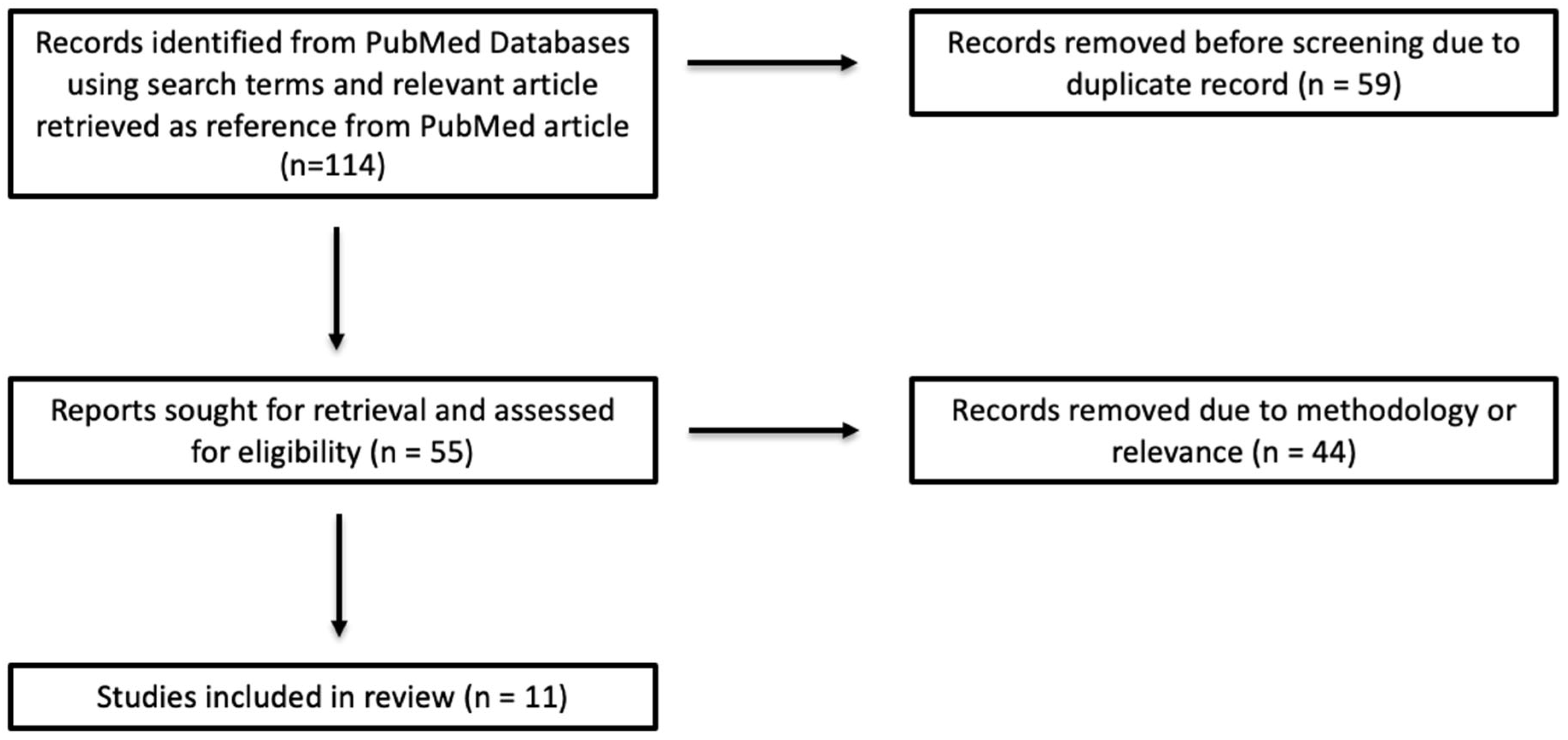
3. Results/Discussion

| Author (Year) | Data Sources | Data Type | # of Pts | Ground Truth | Pre-Process | AUC or HR | Outcome |
|---|---|---|---|---|---|---|---|
| Arasu et al. (2020) [19] | UCSF, San Francisco, CA, USA | T0 (pre-treatment), T1 (early), T2 (during regimen), T3 (pre-surgery) Prospective | 29 pCR 59 non-pCR HER2− stage II or III | Difference in pCR rates | BPE measured via fuzzy-clustering of contralateral breast | Highest cross-validated AUC of 0.81 (95% CI: 0.73–0.90) with combined FTV (pCR prediction value) + HR predictors, adding BPE to FTV + HR models had estimated AUC of 0.82 | pCR |
| Li et al. (2020) [24] | UCSF, San Francisco, CA, USA | Baseline (T0), early (T1), inter-regimen (T2), pre-surgery (T3) | 60 HR+/HER2+,162 HR+/HER2−, 30 HR−/HER2+, 132 HR−/HER2− (TNBC) from I-SPY | pCR after surgery | BPE calculated using automated fuzzy-clustering tissue segmentation | Double positive had highest AUC at 0.76 with just BPE | pCR |
| Nguyen AA et al. (2020) [23] | UCSF, San Francisco, CA, USA | Dynamic contrast-enhanced MRI (DCE-MRI) at baseline (T0), after 3 weeks (T1), after 12 weeks (T2), and after completion of treatment before surgery (T3) | HR+/HER2+: 57 HR+/HER2−: 140 HR−/HER2+: 27 HR−/HER2−: 116 I-SPY data | pCR after surgery | Three bilateral subvolumes analyzed: full, half, center, continuous variable (Mean early enhancement predicted pCR using AUC) | Statistically significant and highest AUC (0.87) in HR−/HER2+ at early timepoint (T1), early half-stack; vs. early full-stack AUC = 0.78 and early center 5 AUC = 0.78 | pCR |
| Moliere et al. (2019) [25] | Department of Women’s Imaging, Strasbourg University Hospital, Strasbourg, France | Pre- and post-NAC | 84 received Epirubicine + 5-Fluoro-uracile + Cyclophosphamide, 51 received additional weekly treatment with taxane and 33 patients received Trastuzumab therapy | Recurrence-free survival (local, regional, or distant); median follow-up 37 months | Semi-automated segmentation, threshold based (Used: 20%, higher post quantitative BPE (measured after NAC, before surgery) predicted recurrence on multivariable Cox) | HR = 6.38, p < 0.05 for post-NAC BPE predicting recurrence | RFS, pCR |
| Rella R et al. (2020) [26] | Fondazione Policlinico Universitario A. Gemelli IRCCS, Rome, Italy | Post-NAC | 228 patients with breast cancer | ROI analysis of enhancement kinetics performed by two radiologists in consensus | Semi-automated contralateral ROI segmentation assessed enhancement change | N/A | pCR |
| Onishi et al. (2021) [20] | UCSF, San Francisco, CA, USA | Baseline (T0), early (T1), inter-regimen (T2), pre-surgery (T3) | 882 from I-SPY (hormone receptor + and—cohorts) HR positive and HR-negative groups analyzed separately | Difference in pCR rates | Fully automated segmentation; central 50% of axial sections of contralateral breast as target volume; fuzzy c-means clustering algorithm for segmentation of FGT; voxel-by-voxel early percent enhancement map (non-contrast and early contrast-enhanced phase) computed as follows: [(SI post – SI pre/SI pre] × 100%. Averaged percent enhancement values for all voxels in masked volume to generate quantitative BPE. | No AUC given; lack of BPE suppression and lower pCR rate in the HR-positive cohort at T2 (p = 0.02) and T3 (p = 0.003) | pCR |
| Rella R. et al. (2022) [28] | Fondazione Policlinico Universitario A. Gemelli IRCCS, University Cattolica Sacro Cuore, Rome, Italy. | Baseline MRI was performed within four weeks before NAC‚ post-NAC MRI within two weeks after completion of chemotherapy. | 30 pCR, 198 non-pCR | pCR defined as absence of residual invasive cancer cells in the breast and ipsilateral lymph nodes | FGT manually segmented, BPE rate calculated as [(SIpostCM − SIpreCM)/SIpreCM] × 100%. | Higher stage (HR 3.6), Ki-67 (1.9), ypN (2.0) worsen survival | OS, DFS |
| You et al. (2017) [21] | Fudan University Cancer Center, Shanghai, China | Baseline (T0), after 2nd, 4th, 6th NAC | 90 unilateral breast cancer 25 pCR/65 non-pCR | pCR after surgery | Fully auto whole breast segmentation → FGT segmentation → enhanced FGT segmentation | AUC = 0.726 for ΔBPE predicting pCR highest at early timepoint (after 2nd NAC) Larger magnitude for change in BPE in HR-negative group | pCR |
| Chen et al. (2015) [22] | University of California, Irvine, USA | Pre-treatment MRI, 2 follow-up MRIs during ongoing NAC | 46 total 24 pCR/22 non-pCR | pCR on surgical pathology | Auto averaged enhanced FGT segmentation | AUC N/A Higher pre-treatment BPE if pCR. Compared to baseline, BPE at F/U-1 significantly decreased in pCR. Subgroup analysis by age: only seen in the younger group (<55 years old), not in the older group (≥55 years old). Older patients significantly lower pre-treatment BPE. Significantly decreased BPE at F/U-1 only in the ER-negative pCR group but not non-pCR, or ER-positive groups. | pCR |
| Shin G et al. (2019) [27] | Yonsei University College of Medicine, Seoul, Korea | Preoperative DCE-MRI (contralateral breast) | 289 unilateral ER+/HER2−, node-negative invasive breast cancer (>5 mm) | RFS and distant metastasis-free survival | Quantitative BPE (manual ROI-based as well as fully automated segmentation) in addition to qualitative BPE assessment by two radiologists using BI-RADS | AUC N/A Contralateral BPE not associated with RFS or DFS (p > 0.05). Ki-67 expression level was associated with worse survival outcome. | RFS, DFS |
| van der Velden et al. (2018) [14] | Memorial Sloan Kettering Cancer Center, USA | Pre-treatment DCE-MRI (contralateral parenchyma) | Biomarker-assessment study (ER+/HER2− invasive ductal carcinoma) (n = 302) | Invasive DFS (IDFS), OS ER+/HER2− | Auto contralateral parenchyma segmentation → late-phase enhancement computations | Pre-treatment BPE independent biomarker for Survival: IDFS HR = 0.27, OS HR = 0.22 | IDFS, OS |
4. Conclusions
Author Contributions
Funding
Institutional Review Board Statement
Informed Consent Statement
Data Availability Statement
Conflicts of Interest
Abbreviations
| BPE | Background parenchymal enhancement |
| MRI | Magnetic resonance imaging |
| pCR | Pathologic complete response |
| RFS | Recurrence-free survival |
| DFS | Disease-free survival |
| OS | Overall survival |
| NAC | Neoadjuvant chemotherapy |
| BI-RADS | Breast imaging-reporting assessment and data system |
| HR | Hormone receptor |
| HER2 | Human epidermal growth factor receptor |
| FTV | Functional tumor volume |
| FGT | Fibroglandular tissue |
| AUC | Area under the receiver operating characteristic curve |
| ROI | Region of interest |
| BRCA | Breast cancer gene |
| TNBC | Triple-negative breast cancer |
| DCE-MRI | Dynamic contrast-enhanced MRI |
| IDFS | Invasive DFS |
References
- Kim, N.; Adam, R.; Maldjian, T.; Duong, T.Q. Radiomics Analysis of Breast MRI to Predict Oncotype Dx Recurrence Score: Systematic Review. Diagnostics 2025, 15, 1054. [Google Scholar] [CrossRef] [Scilit]
- Khan, N.; Adam, R.; Huang, P.; Maldjian, T.; Duong, T.Q. Deep Learning Prediction of Pathologic Complete Response in Breast Cancer Using MRI and Other Clinical Data: A Systematic Review. Tomography 2022, 8, 2784–2795. [Google Scholar] [CrossRef] [Scilit]
- Hadidchi, R.; Agrawal, A.; Liu, M.Z.; Maldijan, T.; Zhu, Y.; Nguyen, H.Q.; Lu, J.; Makower, D.; Fineberg, S.; Duong, T.Q. A deep learning framework to stratify Nottingham histologic grade 2 breast tumors based on dynamic contrast-enhanced MRI. Eur. Radiol. 2025. Epub ahead of printing. [Google Scholar] [CrossRef] [Scilit]
- Hussain, L.; Huang, P.; Nguyen, T.; Lone, K.J.; Ali, A.; Khan, M.S.; Li, H.; Suh, D.Y.; Duong, T.Q. Machine learning classification of texture features of MRI breast tumor and peri-tumor of combined pre- and early treatment predicts pathologic complete response. Biomed. Eng. Online 2021, 20, 63. [Google Scholar] [CrossRef] [Scilit]
- Nishizawa, T.; Maldjian, T.; Jiao, Z.; Duong, T.Q. Attention-based multimodal deep learning for interpretable and generalizable prediction of pathological complete response in breast cancer. J. Transl. Med. 2025, 23, 774. [Google Scholar] [CrossRef] [Scilit] [PubMed]
- Dell’Aquila, K.; Vadlamani, A.; Maldjian, T.; Fineberg, S.; Eligulashvili, A.; Chung, J.; Adam, R.; Hodges, L.; Hou, W.; Makower, D.; et al. Machine learning prediction of pathological complete response and overall survival of breast cancer patients in an underserved inner-city population. Breast Cancer Res. 2024, 26, 7. [Google Scholar] [CrossRef] [Scilit]
- Dammu, H.; Ren, T.; Duong, T.Q. Deep learning prediction of pathological complete response, residual cancer burden, and progression-free survival in breast cancer patients. PLoS ONE 2023, 18, e0280148. [Google Scholar] [CrossRef] [Scilit] [PubMed]
- Karagiannis, G.S.; Bianchi, A.; Sanchez, L.R.; Ambadipudi, K.; Cui, M.-H.; Anampa, J.M.; Asiry, S.; Wang, Y.; Harney, A.S.; Pastoriza, J.M.; et al. Assessment of MRI to estimate metastatic dissemination risk and prometastatic effects of chemotherapy. npj Breast Cancer 2022, 8, 101. [Google Scholar] [CrossRef] [Scilit]
- Rella, R.; Contegiacomo, A.; Bufi, E.; Mercogliano, S.; Belli, P.; Manfredi, R. Background parenchymal enhancement and breast cancer: A review of the emerging evidences about its potential use as imaging biomarker. Br. J. Radiol. 2021, 94, 20200630. [Google Scholar] [CrossRef] [Scilit]
- Liao, G.J.; Bancroft, L.C.H.; Strigel, R.M.; Chitalia, R.D.; Kontos, D.; Moy, L.; Partridge, S.C.; Rahbar, H. Background parenchymal enhancement on breast MRI: A comprehensive review. J. Magn. Reson. Imaging 2020, 51, 43–61. [Google Scholar] [CrossRef] [Scilit] [PubMed]
- Duong, K.S.; Rubner, R.; Siegel, A.; Adam, R.; Ha, R.; Maldjian, T. Machine Learning Assessment of Background Parenchymal Enhancement in Breast Cancer and Clinical Applications: A Literature Review. Cancers 2024, 16, 3681. [Google Scholar] [CrossRef] [Scilit]
- Niell, B.L.; Abdalah, M.; Stringfield, O.; Raghunand, N.; Ataya, D.; Gillies, R.; Balagurunathan, Y. Quantitative Measures of Background Parenchymal Enhancement Predict Breast Cancer Risk. AJR Am. J. Roentgenol. 2021, 217, 64–75. [Google Scholar] [CrossRef] [Scilit]
- Li, X.; Yan, F. Predictive value of background parenchymal enhancement on breast magnetic resonance imaging for pathological tumor response to neoadjuvant chemotherapy in breast cancers: A systematic review. Cancer Imaging 2024, 24, 35. [Google Scholar] [CrossRef] [Scilit]
- van der Velden, B.H.M.; Sutton, E.J.; Carbonaro, L.A.; Pijnappel, R.M.; Morris, E.A.; Gilhuijs, K.G. Contralateral parenchymal enhancement on dynamic contrast-enhanced MRI reproduces as a biomarker of survival in ER-positive/HER2-negative breast cancer patients. Eur. Radiol. 2018, 28, 4705–4716. [Google Scholar] [CrossRef] [Scilit]
- Gullo, R.L.; Daimiel, I.; Saccarelli, C.R.; Bitencourt, A.; Sevilimedu, V.; Martinez, D.F.; Jochelson, M.S.; Morris, E.A.; Reiner, J.S.; Pinker, K. MRI background parenchymal enhancement, fibroglandular tissue, and mammographic breast density in patients with invasive lobular breast cancer on adjuvant endocrine hormonal treatment: Associations with survival. Breast Cancer Res. 2020, 22, 93. [Google Scholar] [CrossRef] [Scilit]
- Kim, J.Y.; Lee, J.W.; Lee, N.K.; Kim, S.; Nam, K.J.; Lee, K.; Choo, K.S. Are background breast parenchymal features on preoperative breast MRI associated with disease-free survival in patients with invasive breast cancer? Radiol. Med. 2024, 129, 1790–1801. [Google Scholar] [CrossRef] [Scilit]
- Lee, Y.J.; Youn, I.K.; Kim, S.H.; Kang, B.J.; Park, W.-C.; Lee, A. Triple-negative breast cancer: Pretreatment magnetic resonance imaging features and clinicopathological factors associated with recurrence. Magn. Reson. Imaging 2020, 66, 36–41. [Google Scholar] [CrossRef] [Scilit]
- Xu, C.; Yu, J.; Wu, F.; Li, X.; Hu, D.; Chen, G.; Wu, G. High-background parenchymal enhancement in the contralateral breast is an imaging biomarker for favorable prognosis in patients with triple-negative breast cancer treated with chemotherapy. Am. J. Transl. Res. 2021, 13, 4422–4436. [Google Scholar] [PubMed]
- Arasu, V.A.; Kim, P.; Li, W.; Strand, F.; McHargue, C.; Harnish, R.; Newitt, D.C.; Jones, E.F.; Glymour, M.M.; Kornak, J.; et al. Predictive Value of Breast MRI Background Parenchymal Enhancement for Neoadjuvant Treatment Response among HER2- Patients. J. Breast Imaging 2020, 2, 352–360. [Google Scholar] [CrossRef] [Scilit] [PubMed]
- Onishi, N.; Li, W.; Newitt, D.C.; Harnish, R.J.; Strand, F.; Nguyen, A.A.-T.; Arasu, V.A.; Gibbs, J.; Jones, E.F.; Wilmes, L.J.; et al. Breast MRI during Neoadjuvant Chemotherapy: Lack of Background Parenchymal Enhancement Suppression and Inferior Treatment Response. Radiology 2021, 301, 295–308. [Google Scholar] [CrossRef] [Scilit] [PubMed]
- You, C.; Peng, W.; Zhi, W.; He, M.; Liu, G.; Xie, L.; Jiang, L.; Hu, X.; Shen, X.; Gu, Y. Association Between Background Parenchymal Enhancement and Pathologic Complete Remission Throughout the Neoadjuvant Chemotherapy in Breast Cancer Patients. Transl. Oncol. 2017, 10, 786–792. [Google Scholar] [CrossRef] [Scilit]
- Chen, J.H.; Yu, H.J.; Hsu, C.; Mehta, R.S.; Carpenter, P.M.; Su, M.Y. Background Parenchymal Enhancement of the Contralateral Normal Breast: Association with Tumor Response in Breast Cancer Patients Receiving Neoadjuvant Chemotherapy. Transl. Oncol. 2015, 8, 204–209. [Google Scholar] [CrossRef] [Scilit]
- Nguyen, A.A.; Arasu, V.A.; Strand, F.; Li, W.; Onishi, N.; Gibbs, J.; Jones, E.F.; Joe, B.N.; Esserman, L.J.; Newitt, D.C.; et al. Comparison of Segmentation Methods in Assessing Background Parenchymal Enhancement as a Biomarker for Response to Neoadjuvant Therapy. Tomography 2020, 6, 101–110. [Google Scholar] [CrossRef] [Scilit] [PubMed]
- Li, W.; Newitt, D.C.; Gibbs, J.; Wilmes, L.J.; Jones, E.F.; Arasu, V.A.; Strand, F.; Onishi, N.; Nguyen, A.A.-T.; Kornak, J.; et al. Predicting breast cancer response to neoadjuvant treatment using multi-feature MRI: Results from the I-SPY 2 TRIAL. npj Breast Cancer 2020, 6, 63. [Google Scholar] [CrossRef] [Scilit]
- Moliere, S.; Oddou, I.; Noblet, V.; Veillon, F.; Mathelin, C. Quantitative background parenchymal enhancement to predict recurrence after neoadjuvant chemotherapy for breast cancer. Sci. Rep. 2019, 9, 19185. [Google Scholar] [CrossRef] [Scilit] [PubMed]
- Rella, R.; Bufi, E.; Belli, P.; Petta, F.; Serra, T.; Masiello, V.; Scrofani, A.; Barone, R.; Orlandi, A.; Valentini, V.; et al. Association between background parenchymal enhancement and tumor response in patients with breast cancer receiving neoadjuvant chemotherapy. Diagn. Interv. Imaging 2020, 101, 649–655. [Google Scholar] [CrossRef] [Scilit] [PubMed]
- Shin, G.W.; Zhang, Y.; Kim, M.J.; Su, M.; Kim, E.; Moon, H.J.; Yoon, J.H.; Park, V.Y. Role of dynamic contrast-enhanced MRI in evaluating the association between contralateral parenchymal enhancement and survival outcome in ER-positive, HER2-negative, node-negative invasive breast cancer. J. Magn. Reson. Imaging 2018, 48, 1678–1689. [Google Scholar] [CrossRef] [Scilit]
- Rella, R.; Bufi, E.; Belli, P.; Scrofani, A.R.; Petta, F.; Borghetti, A.; Marazzi, F.; Valentini, V.; Manfredi, R. Association between contralateral background parenchymal enhancement on MRI and outcome in patients with unilateral invasive breast cancer receiving neoadjuvant chemotherapy. Diagn. Interv. Imaging 2022, 103, 486–494. [Google Scholar] [CrossRef] [Scilit]
- Dalmış, M.U.; Litjens, G.; Holland, K.; Setio, A.; Mann, R.; Karssemeijer, N.; Gubern-Mérida, A. Using deep learning to segment breast and fibroglandular tissue in MRI volumes. Med. Phys. 2017, 44, 533–546. [Google Scholar] [CrossRef] [Scilit]

Disclaimer/Publisher’s Note: The statements, opinions and data contained in all publications are solely those of the individual author(s) and contributor(s) and not of MDPI and/or the editor(s). MDPI and/or the editor(s) disclaim responsibility for any injury to people or property resulting from any ideas, methods, instructions or products referred to in the content. |
© 2026 by the authors. Licensee MDPI, Basel, Switzerland. This article is an open access article distributed under the terms and conditions of the Creative Commons Attribution (CC BY) license.
Share and Cite
Weiss, J.; Hunt, E.; Zhu, Y.; Duong, T.Q.; Maldjian, T. Review of Prognostic Significance of Quantitative BPE Measurements. Diagnostics 2026, 16, 495. https://doi.org/10.3390/diagnostics16030495
Weiss J, Hunt E, Zhu Y, Duong TQ, Maldjian T. Review of Prognostic Significance of Quantitative BPE Measurements. Diagnostics. 2026; 16(3):495. https://doi.org/10.3390/diagnostics16030495
Chicago/Turabian StyleWeiss, Jeremy, Emily Hunt, Yihui Zhu, Tim Q. Duong, and Takouhie Maldjian. 2026. "Review of Prognostic Significance of Quantitative BPE Measurements" Diagnostics 16, no. 3: 495. https://doi.org/10.3390/diagnostics16030495
APA StyleWeiss, J., Hunt, E., Zhu, Y., Duong, T. Q., & Maldjian, T. (2026). Review of Prognostic Significance of Quantitative BPE Measurements. Diagnostics, 16(3), 495. https://doi.org/10.3390/diagnostics16030495