Article

The Shrinking Blind Spot: How Freeze–Thaw Obscures Microscopic Evidence of Ante-Mortem Ecchymosis


Department of Surgical Pathology, Medical, Molecular, and Critical Area, Institute of Legal Medicine, University of Pisa, 56126 Pisa, Italy
*
Author to whom correspondence should be addressed.
Diagnostics 2026, 16(3), 419; https://doi.org/10.3390/diagnostics16030419

Submission received: 23 December 2025 / Revised: 19 January 2026 / Accepted: 21 January 2026 / Published: 1 February 2026
(This article belongs to the Special Issue Diagnostic Methods in Forensic Pathology, Third Edition)

Abstract

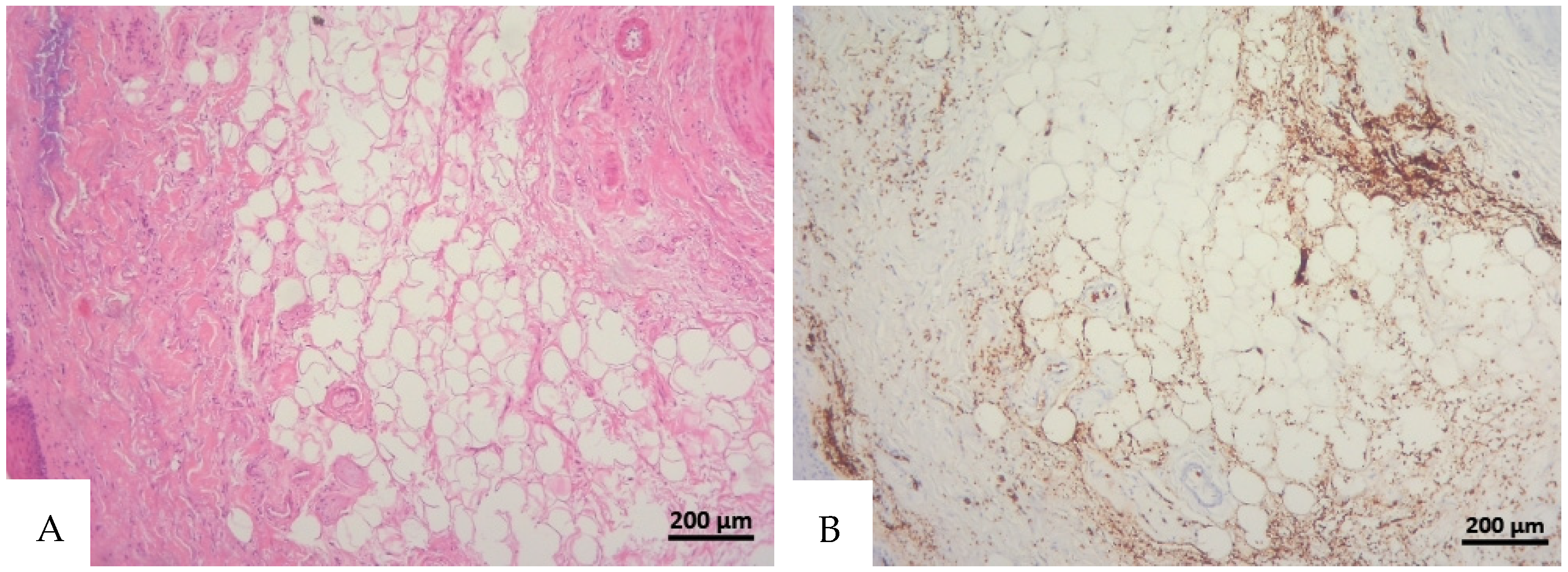
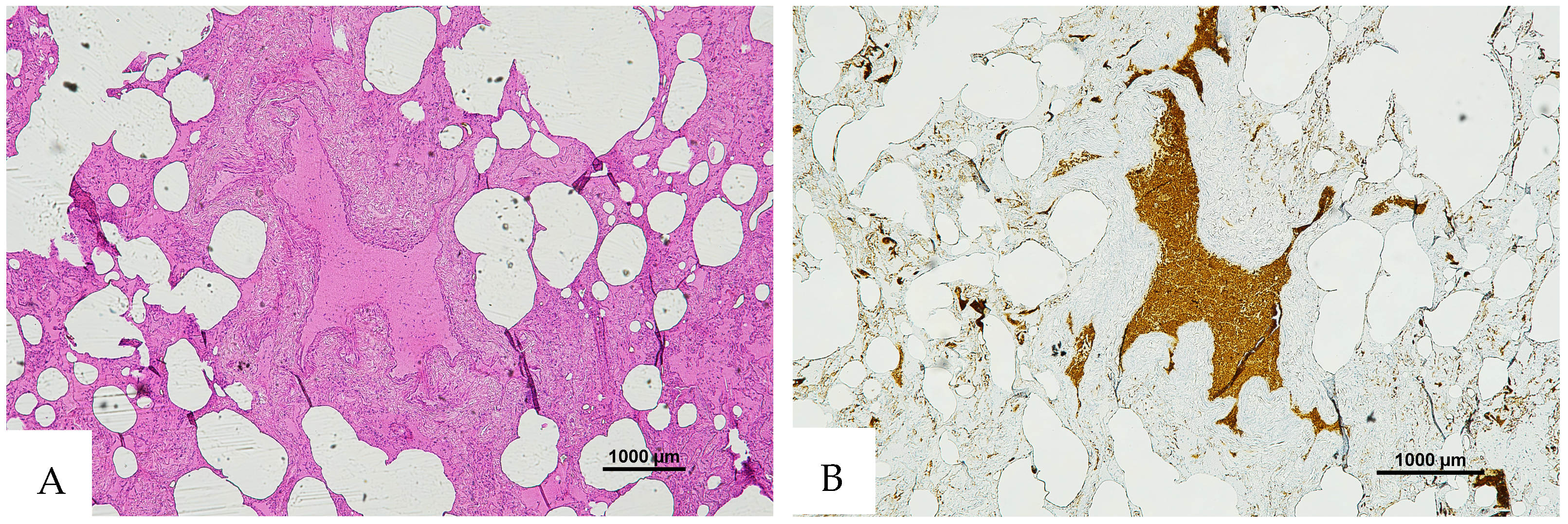
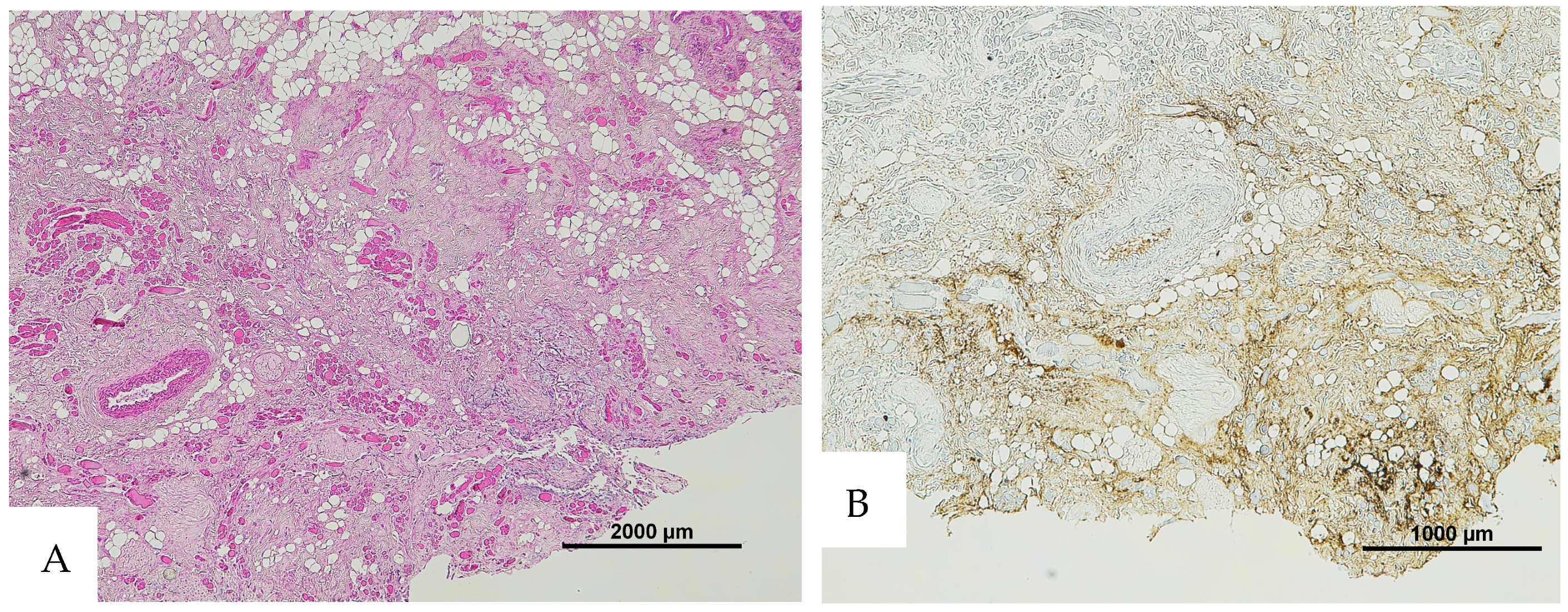
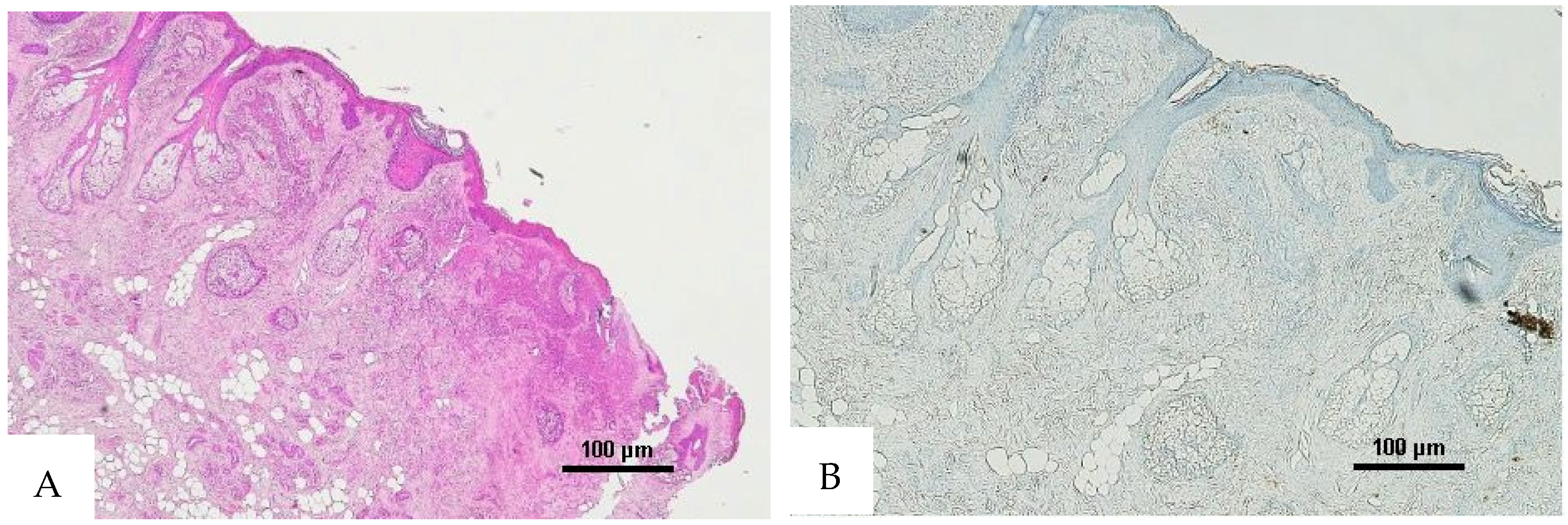
Background/Objectives: Histological examination constitutes a fundamental methodology for establishing the vitality of a lesion. In cases where the corpse is preserved for an extended duration of time prior to the post-mortem evaluation, particularly if the body has undergone freezing and thawing cycles, post-mortem changes may obscure or alter evidence of traumatic injuries. Consequently, the reliability of hematoxylin and eosin (H&E) staining for the reliable detection of intralesional erythrocytes in suspected traumatic fatalities is potentially severely compromised. The primary objective of this study is to rigorously underscore the detrimental influence of freeze–thaw processes on histologic examination and to advocate the indispensable incorporation of immunohistochemical analysis, specifically employing anti-human glycophorin A antibodies, to ascertain the presence of red blood cells. Methods: Skin samples from 10 autopsy cases were subjected to serial freeze–thaw cycles and analyzed using anti-human Glycophorin A (GPA) immunohistochemistry staining to evaluate skin lesion vitality in freeze–thawed tissues compared to fresh controls. Results: Results indicated that while H&E reliability was limited to fresh tissue, anti-GPA staining remained stable across all freeze–thaw cycles. Conclusions: Forensic pathologists must remain acutely cognizant of the potential artifacts produced by freeze–thaw cycles. In these cases, anti-GPA staining proved to be a reliable asset for evaluating the vitality of a lesion.
Keywords: glycophorin; immunohistochemistry; red blood cells; freeze–thaw cycles; autopsy artifacts glycophorin; immunohistochemistry; red blood cells; freeze–thaw cycles; autopsy artifacts