Echocardiographic Assessment of Left Ventricular Diastolic Function in Adults Between Old and New: Progress and Challenges



Abstract
1. Introduction
2. LV Diastolic (Dys)Function: Basic Pathophysiological Considerations and Clinical Implications
3. Echocardiographic Evaluation of LV Diastolic Function by Primary Techniques
3.1. Doppler Ultrasound Techniques
3.2. LA Volume
3.3. LV GLS
4. Echocardiographic Evaluation of LV Diastolic Function by Secondary Techniques
5. Advanced Techniques for LV Diastolic Function Assessment
5.1. LA Strain
5.2. LV MW
5.3. Cardiac THE
6. Gaps in the Evidence and Future Perspectives on LV Diastolic Function Assessment
7. Conclusions
Author Contributions
Funding
Institutional Review Board Statement
Informed Consent Statement
Data Availability Statement
Conflicts of Interest
References
- Nagueh, S.F.; Sanborn, D.Y.; Oh, J.K.; Anderson, B.; Billick, K.; Derumeaux, G.; Klein, A.; Koulogiannis, K.; Mitchell, C.; Shah, A.; et al. Recommendations for the evaluation of left ventricular diastolic function by echocardiography and for heart failure with preserved ejection fraction diagnosis: An update from the American Society of Echocardiography. J. Am. Soc. Echocardiogr. 2025, 38, 537–569. [Google Scholar] [CrossRef]
- Nagueh, S.F.; Smiseth, O.A.; Appleton, C.P.; Byrd, B.F., 3rd; Dokainish, H.; Edvardsen, T.; Flachskampf, F.A.; Gillebert, T.C.; Klein, A.L.; Lancellotti, P.; et al. Recommendations for the evaluation of left ventricular diastolic function by echocardiography: An update from the American Society of Echocardiography and the European Association of Cardiovascular Imaging. J. Am. Soc. Echocardiogr. 2016, 29, 277–314. [Google Scholar] [CrossRef]
- Nagueh, S.F.; Appleton, C.P.; Gillebert, T.C.; Marino, P.N.; Oh, J.K.; Smiseth, O.A.; Waggoner, A.D.; Flachskampf, F.A.; Pellikka, P.A.; Evangelista, A. Recommendations for the evaluation of left ventricular diastolic function by echocardiography. J. Am. Soc. Echocardiogr. 2009, 22, 107–133. [Google Scholar] [CrossRef]
- Mitchell, C.; Rahko, P.S.; Blauwet, L.A.; Canaday, B.; Finstuen, J.A.; Foster, M.C.; Horton, K.; Ogunyankin, K.O.; Palma, R.A.; Velazquez, E.J. Guidelines for performing a comprehensive transthoracic echocardiographic examination in adults: Recommendations from the American Society of Echocardiography. J. Am. Soc. Echocardiogr. 2019, 32, 1–64. [Google Scholar] [CrossRef]
- Mor-Avi, V.; Lang, R.M.; Badano, L.P.; Belohlavek, M.; Cardim, N.M.; Derumeaux, G.; Galderisi, M.; Marwick, T.; Nagueh, S.F.; Sengupta, P.P.; et al. Current and evolving echocardiographic techniques for the quantitative evaluation of cardiac mechanics: ASE/EAE consensus statement on methodology and indications endorsed by the Japanese Society of Echocardiography. J. Am. Soc. Echocardiogr. 2011, 24, 277–313. [Google Scholar] [CrossRef]
- Dell’Angela, L.; Nicolosi, G.L. Shaping the optimal timing for treatment of isolated asymptomatic severe aortic stenosis with preserved left ventricular ejection fraction: The role of non-invasive diagnostics focused on strain echocardiography and future perspectives. J. Imaging 2025, 11, 48. [Google Scholar] [CrossRef] [PubMed]
- Tromp, J.; Seekings, P.J.; Hung, C.L.; Iversen, M.B.; Frost, M.J.; Ouwerkerk, W.; Jiang, Z.; Eisenhaber, F.; Goh, R.S.M.; Zhao, H.; et al. Automated interpretation of systolic and diastolic function on the echocardiogram: A multicohort study. Lancet Digit. Health 2022, 4, e46–e54. [Google Scholar] [CrossRef]
- Carluccio, E.; Cameli, M.; Rossi, A.; Dini, F.L.; Biagioli, P.; Mengoni, A.; Jacoangeli, F.; Mandoli, G.E.; Pastore, M.C.; Maffeis, C.; et al. Left atrial strain in the assessment of diastolic function in heart failure: A machine learning approach. Circ. Cardiovasc. Imaging 2023, 16, e014605. [Google Scholar] [CrossRef] [PubMed]
- Pandey, A.; Kagiyama, N.; Yanamala, N.; Segar, M.W.; Cho, J.S.; Tokodi, M.; Sengupta, P.P. Deep-learning models for the echocardiographic assessment of diastolic dysfunction. JACC Cardiovasc. Imaging 2021, 14, 1887–1900. [Google Scholar] [CrossRef] [PubMed]
- Chao, C.J.; Kato, N.; Scott, C.G.; Lopez-Jimenez, F.; Lin, G.; Kane, G.C.; Pellikka, P.A. Unsupervised machine learning for assessment of left ventricular diastolic function and risk stratification. J. Am. Soc. Echocardiogr. 2022, 35, 1214–1225.e8. [Google Scholar] [CrossRef]
- Dell’Angela, L.; Nicolosi, G.L. Artificial intelligence applied to cardiovascular imaging, a critical focus on echocardiography: The point-of-view from “the other side of the coin”. J. Clin. Ultrasound 2022, 50, 772–780. [Google Scholar] [CrossRef] [PubMed]
- Dell’Angela, L.; Nicolosi, G.L. Artificial intelligence in clinical echocardiography: Many expectations, but deep uncertainties for defining strategies to overcome difficulties and obstacles. J. Am. Soc. Echocardiogr. 2022, 35, 1336. [Google Scholar] [CrossRef]
- Tseng, A.S.; Lopez-Jimenez, F.; Pellikka, P.A. Artificial intelligence in clinical echocardiography: Many expectations, but deep uncertainties for defining strategies to overcome difficulties and obstacles: Authors’ reply. J. Am. Soc. Echocardiogr. 2022, 35, 1336–1337. [Google Scholar] [CrossRef] [PubMed]
- Brutsaert, D.L.; Sys, S.U.; Gillebert, T.C. Diastolic failure: Pathophysiology and therapeutic implications. J. Am. Coll. Cardiol. 1993, 22, 318–325. [Google Scholar] [CrossRef]
- Mihos, C.G.; Liu, J.E.; Anderson, K.M.; Pernetz, M.A.; O’dRiscoll, J.M.; Aurigemma, G.P.; Ujueta, F.; Wessly, P.; on behalf of the American Heart Association Council on Peripheral Vascular Disease; Council on Cardiovascular and Stroke Nursing; et al. Speckle-tracking strain echocardiography for the assessment of left ventricular structure and function: A scientific statement from the American Heart Association. Circulation 2025, 152, e96–e109. [Google Scholar] [CrossRef]
- Thomas, J.D.; Edvardsen, T.; Abraham, T.; Appadurai, V.; Badano, L.; Banchs, J.; Cho, G.Y.; Cosyns, B.; Delgado, V.; Donal, E.; et al. Clinical applications of strain echocardiography: A clinical consensus statement from the American Society of Echocardiography developed in collaboration with the European Association of Cardiovascular Imaging of the European Society of Cardiology. J. Am. Soc. Echocardiogr. 2025, 38, 985–1020. [Google Scholar] [CrossRef]
- Di Virgilio, E.; Monitillo, F.; Santoro, D.; D’alessandro, S.; Guglielmo, M.; Baggiano, A.; Fusini, L.; Memeo, R.; Rabbat, M.G.; Favale, S.; et al. Mid-diastolic events (L events): A critical review. J. Clin. Med. 2021, 10, 5654. [Google Scholar] [CrossRef]
- Fortuni, F.; Biagioli, P.; Myagmardorj, R.; Mengoni, A.; Chua, A.P.; Zuchi, C.; Sforna, S.; Bax, J.; Marsan, N.A.; Ambrosio, G.; et al. Left atrioventricular coupling index: A novel diastolic parameter to refine prognosis in heart failure. J. Am. Soc. Echocardiogr. 2024, 37, 1038–1046. [Google Scholar] [CrossRef]
- Wang, Y.H.; Dong, Y.; Li, G.Y.; Ma, C.-Y. Unveiling the left atrioventricular coupling index: A promising marker for diastolic dysfunction and prognosis. J. Am. Soc. Echocardiogr. 2025, 38, 448. [Google Scholar] [CrossRef] [PubMed]
- Fortuni, F.; Biagioli, P.; Carluccio, E. Reply to unveiling the left atrioventricular coupling index: A promising marker for diastolic dysfunction and prognosis. J. Am. Soc. Echocardiogr. 2025, 38, 448–449. [Google Scholar] [CrossRef]
- Zornitzki, L.; Topilsky, Y. Left atrioventricular coupling index: When minimal left atrial volume is actually ‘more’ than maximal left atrial volume. J. Am. Soc. Echocardiogr. 2024, 37, 1047–1050. [Google Scholar] [CrossRef]
- Anwar, A.M.; Alshammakh, M.S.; Eyaz, M.; Al-Katheri, A.; Abdelfattah, A.N.; Ali, M.A.M.; Albakri, I. Normal reference values of left atrioventricular coupling index on two-dimensional echocardiography. Int. J. Cardiovasc. Imaging 2025, 41, 1589–1595. [Google Scholar] [CrossRef]
- Tran, H.M.; Truong, H.P.; Tran, C.C.; Vo, T.M.; Nguyen, D.N.Q.; Dao, L.T. Analysis of factors related to early left ventricular dysfunction in hypertensive patients with preserved ejection fraction using speckle tracking echocardiography: A cross-sectional study in Vietnam. Diagnostics 2025, 15, 222. [Google Scholar] [CrossRef] [PubMed]
- Sonaglioni, A.; Nicolosi, G.L.; Trevisan, R.; Lombardo, M.; Grasso, E.; Gensini, G.F.; Ambrosio, G. The influence of pectus excavatum on cardiac kinetics and function in otherwise healthy individuals: A systematic review. Int. J. Cardiol. 2023, 381, 135–144. [Google Scholar] [CrossRef] [PubMed]
- Meyer, T.; Wellge, B.; Barzen, G.; Chandia, S.K.; Knebel, F.; Hahn, K.; Elgeti, T.; Fischer, T.; Braun, J.; Tzschätzsch, H.; et al. Cardiac elastography with external vibration for quantification of diastolic myocardial stiffness. J. Am. Soc. Echocardiogr. 2025, 38, 431–442. [Google Scholar] [CrossRef] [PubMed]
- Rodriguez-Sanchez, I.; Villanueva-Benito, I.; Agirre, U.; Onaindia, J.J.; Urkullu, A.; Cacicedo, A.; Ullate, A.; Bravo, I.; Florido, J.; Salcedo, A.; et al. Diastolic function and cardiovascular events in patients with preserved left ventricular ejection fraction. Improving risk stratification with left atrial strain. Front. Cardiovasc. Med. 2025, 12, 1565052. [Google Scholar] [CrossRef]
- Tolvaj, M.; Zhubi Bakija, F.; Fábián, A.; Ferencz, A.; Lakatos, B.; Ladányi, Z.; Szijártó, Á.; Edvi, B.; Kiss, L.; Szelid, Z.; et al. Integrating left atrial reservoir strain into the first-line assessment of diastolic function: Prognostic implications in a community-based cohort with normal left ventricular systolic function. J. Am. Soc. Echocardiogr. 2025, 38, 570–582. [Google Scholar] [CrossRef]
- Smiseth, O.A.; Aalen, J.M. Imaging of left ventricular diastolic function: Do we need both left atrial volume and reservoir strain? J. Am. Soc. Echocardiogr. 2025, 38, 583–585. [Google Scholar] [CrossRef]
- Elhady, F.; Ali, A.A.; Elkholy, N.S.; El-Mnakhly, E.A.; Elkareem, T.S.A.; Galal, A.; Negm, M.A. Left atrial filling index and stiffness index and its correlation to the duration of diabetes in patients with type II diabetes mellitus. Int. J. Cardiovasc. Imaging 2025, 41, 1309–1320. [Google Scholar] [CrossRef]
- Liang, H.; Zhao, X.; Xiao, C.; Sun, L.; Zhang, F. Four-dimensional echocardiographic quantification of left atrial function in metabolic syndrome across different diastolic function states. BMC Cardiovasc. Disord. 2025, 25, 449. [Google Scholar] [CrossRef]
- Mannina, C.; Ito, K.; Jin, Z.; Yoshida, Y.; Russo, C.; Nakanishi, K.; Rundek, T.; Homma, S.; Elkind, M.S.; Di Tullio, M.R. Left atrial function and incident heart failure in older adults. J. Am. Soc. Echocardiogr. 2025, 38, 103–110. [Google Scholar] [CrossRef]
- Gegenava, T.; Nieman, K. Left atrial volumetric/mechanical coupling index: Best of both worlds? J. Am. Soc. Echocardiogr. 2025, 38, 111–114. [Google Scholar] [CrossRef]
- Dell’Angela, L.; Nicolosi, G.L. From ejection fraction, to myocardial strain, and myocardial work in echocardiography: Clinical impact and controversies. Echocardiography 2024, 41, e15758. [Google Scholar] [CrossRef]
- Chilingaryan, A.; Tunyan, L.; Arzumanyan, M.; Balyan, H. Predictive value of left ventricular myocardial constructive work in patients with heart failure with preserved ejection fraction and preclinical diastolic dysfunction. J. Cardiovasc. Imaging 2025, 33, 11. [Google Scholar] [CrossRef]
- Sokratous, S.; Kyriakou, M.; Khattab, E.; Alexandraki, A.; Fotiou, E.L.; Chrysanthou, N.; Papakyriakopoulou, P.; Korakianitis, I.; Constantinidou, A.; Kadoglou, N.P.E. The role of diastolic stress echo and myocardial work in early detection of cardiac dysfunction in women with breast cancer undergoing chemotherapy. Biomedicines 2025, 13, 2341. [Google Scholar] [CrossRef] [PubMed]
- Jasaityte, R.; Bajraktarevic, R.; Blaschke-Waluga, D.; Seeland, U.; Regitz-Zagrosek, V.; Landmesser, U.; Stangl, K.; Knebel, F.; Stangl, V.; Brand, A. Determinants of myocardial work indices in women. Echocardiography 2023, 40, 1196–1204. [Google Scholar] [CrossRef] [PubMed]
- Grapsa, J.; Argulian, E.; Smiseth, O.A. Diastolic dysfunction: A comparison of 2025 ASE, 2024 BSE and 2022 EACVI guidelines. Eur. Heart J. Cardiovasc. Imaging 2025, 26, 1725–1727. [Google Scholar] [CrossRef]
- Taub, C.C.; Stainback, R.F.; Abraham, T.; Forsha, D.; Garcia-Sayan, E.; Hill, J.C.; Hung, J.; Mitchell, C.; Rigolin, V.H.; Sachdev, V.; et al. Guidelines for the standardization of adult echocardiography reporting: Recommendations from the American Society of Echocardiography. J. Am. Soc. Echocardiogr. 2025, 38, 735–774. [Google Scholar] [CrossRef] [PubMed]
- Dell’Angela, L.; Sonaglioni, A.; Nicolosi, G.L. Performing and reporting duration of the optimal echocardiographic exam in current clinical practice: Time to turn the page? J. Am. Soc. Echocardiogr. 2025; online ahead of print. [Google Scholar] [CrossRef]
- Stainback, R.F.; Taub, C.C. Reply to: Performing and reporting duration of the optimal echocardiographic exam in current clinical practice: Time to turn the page? J. Am. Soc. Echocardiogr. 2025; online ahead of print. [Google Scholar] [CrossRef]



| Parameter/Variable | Acquisition | Measurements/Pathophysiological Considerations | Advantages | Limitations |
|---|---|---|---|---|
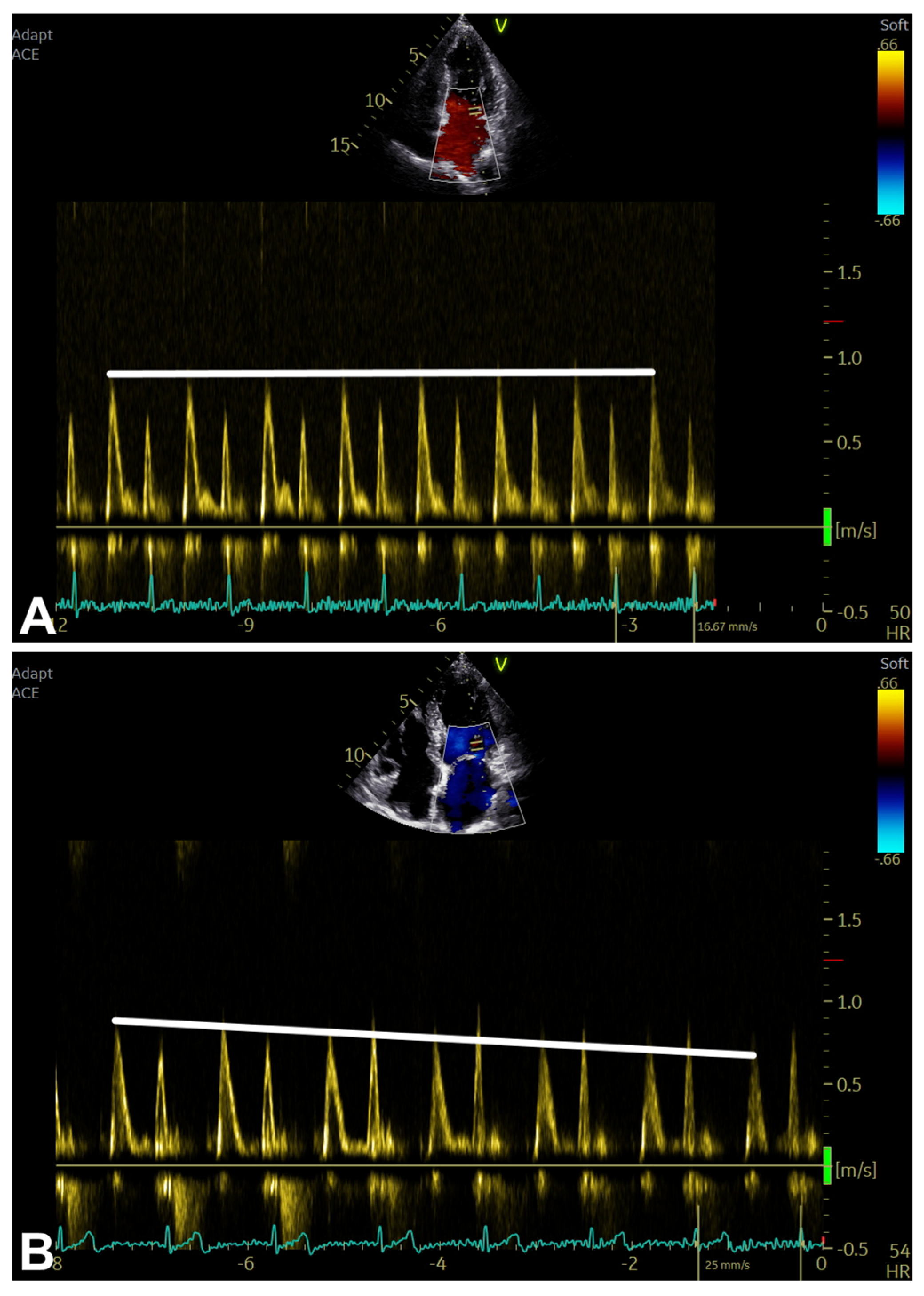
| Transmitral inflow | - Apical 4C view, color and spectral (PW) Doppler imaging. - Preferably, low wall filter setting (100–200 MHz), low signal gain, PW Doppler SV 1–3 mm, and sweep speed at 100 mm/s are needed. | Peak E-wave velocity (or E) (measured in m/s): After ECG T-wave, measure peak early diastolic modal velocity; During early diastole, indicates the LA-to-LV pressure gradient; Affected by LV relaxation and LAP rate modifications. Range (20–39 y): 0.54 (0.52–0.57) to 1.11 (1.07–1.16); Range (40–60 y): 0.47 (0.46–0.49) to 1.02 (0.99–1.05); Range (60–80 y): 0.39 (0.37–0.42) to 0.92 (0.88–0.96). | - Feasible. - Reproducible. - Diagnostic and prognostic information (e.g., peak E-wave velocity and E/A ratio: predictor of clinical outcome in subjects with DCM and reduced LV EF; short DT in reduced LV EF reflects increased LV EDP; in the presence of cardiac disease, short A-wave duration reflects high LV FP). | - Related to age (e.g., both peak E-wave velocity and E/A decrease with age; both DT and peak A-wave velocity increase with age). - Related to preload. - US beam optimal alignment-related. - Challenging in the presence of arrhythmias (e.g., both peak/duration of A-wave velocity and E/A not applicable in AF/AFL; DT not applicable in AFL; peak A-wave velocity possibly not feasible if paced rhythm, sinus tachycardia, and first-degree AV block; A-wave duration not feasible if E-A fusion, sinus arrhythmias, second- and third-degree AV block, and ECG PR interval < 120 ms). - Other: peak E-wave velocity poorly correlates with LV FP in subjects with CAD and/or HCM with LV EF >50%. |
| DT (in ms): Along the deceleration slope, measure TI from peak E-wave velocity to the zero baseline; May be used to determine filling patterns (normal, IR, PN, and RF). Range: in the presence of reduced LV EF, if <140 ms reflects increased LV EDP (high accuracy in both SR and AF). | ||||
| Peak A-wave velocity (or A) (in m/s): After ECG P-wave, measure peak late diastolic modal velocity; During late diastole, indicates the LA-to-LV pressure gradient; Affected by LV compliance and LA contractile function. Range (20–39 y): 0.24 (0.21–0.27) to 0.68 (0.63–0.72); Range (40–60 y): 0.33 (0.32–0.35) to 0.82 (0.80–0.84); Range (60–80 y): 0.43 (0.40–0.45) to 0.97 (0.93–1.00). | ||||
| E/A ratio (or E/A) (adimensional): Peak E-wave velocity divided by peak A-wave velocity; May be used to determine filling patterns (normal, IR, PN, and RF). Range (20–39 y): 0.88 (0.82–0.94) to 2.73 (2.66–2.81); Range (40–60 y): 0.69 (0.66–0.73) to 2.07 (2.03–2.11); Range (60–80 y): 0.50 (0.45–0.56) to 1.40 (1.34–1.47). | ||||
| A-wave duration (in ms): At zero baseline, measure TI from the onset to the offset of the A-wave signal; During late diastole, indicates LV compliance; Particularly helpful if used with pulmonary venous AR duration. Range: in the presence of cardiac disease, if <120 ms reflects increased LV FP. | ||||
| L-wave velocity (or L) (in m/s): In the presence of triphasic transmitral inflow pattern, represents the mid-diastolic flow; During diastasis, indicates the continued LA-to-LV pressure gradient associated with delayed LV relaxation, as well as high LV FP probably; In the presence of LVH/HCM, is specific for high LV FP (but low sensitivity); in the presence of AF, may be related to high LV FP. Range: if (rarely) present in subjects with normal LV diastolic function and bradycardia, it is usually <0.4. | ||||
| TDI (at mitral annulus) | - Apical 4C view with TDI preset optimization *. - Alignment optimization (angle of interrogation should be parallel to annular motion). - Preferably, PW Doppler SV 5–10 mm and sweep speed at 100 mm/s are needed. | Peak e’-wave velocity (or e’) (measured in cm/s): After ECG T-wave, measure peak early diastolic modal velocity; Index of LV relaxation and less load-dependent if compared to other conventional PW Doppler parameters; Affected by LV relaxation, restoring forces and FP; Helpful for differentiation PN from normal transmitral inflow patterns. - Lateral e’: Range (20–39 y): 9.9 (9.4–10.4) to 22.1 (21.5–22.8); Range (40–60 y): 7.5 (7.3–7.8) to 17.5 (17.1–17.9); Range (60–80 y): 5.2 (4.8–5.6) to 13.0 (12.4–13.5). - Septal e’: Range (20–39 y): 7.2 (6.8–7.7) to 16.4 (16.0–16.9); Range (40–60 y): 5.7 (5.4–5.9) to 13.5 (13.2–13.8); Range (60–80 y): 4.1 (3.7–4.5) to 10.6 (10.1–11.0). - Average e’: Range (20–39 y): 8.7 (8.2–9.2) to 19.1 (18.6–19.7); Range (40–60 y): 6.7 (6.4–7.0) to 15.4 (15.1–15.7); Range (60–80 y): 4.7 (4.3–5.1) to 11.7 (11.2–12.2). | - Feasible. - Reproducible. - Diagnostic and prognostic information. | - Related to age (e.g., peak e’-wave velocity decreases with age). - US beam optimal alignment-related. - Challenging in the presence of arrhythmias. - Significantly limited accuracy in some conditions (e.g., relevant MAC, prosthetic mitral ring/valve, pericardial disease, CAD in the presence of regional dysfunction in the sampled segments). |
| Peak a’-wave velocity (or a’) (in cm/s): after ECG P-wave, measure peak late diastolic modal velocity. | ||||
| MV E/TDI e’ ratio (or E/e’) (adimensional): MV peak E-wave velocity (in cm/s) divided by TDI peak e’-wave velocity (in cm/s); Helpful for predicting increased LV FP. Range: generally, if <8 usually indicates normal LV FP, and if >14 usually indicates increased LV FP with high specificity. - Lateral E/e’: Range (20–39 y): 2.5 (2.0–3.0) to 6.3 (5.3–7.2); Range (40–60 y): 3.6 (3.4–3.9) to 9.4 (8.9–10.0); Range (60–80 y): 4.8 (4.5–5.0) to 12.6 (12.0–13.2). - Septal E/e’: Range (20–39 y): 4.0 (3.3–4.7) to 9.1 (8.2–9.9); Range (40–60 y): 4.9 (4.6–5.3) to 12.1 (11.7–12.6); Range (60–80 y): 5.9 (5.5–6.3) to 15.2 (14.7–15.7). - Average E/e’: Range (20–39 y): 4.0 (3.8–4.3) to 9.1 (8.5–9.7); Range (40–60 y):4.6 (4.4–4.8) to 11.5 (11.2–11.9); Range (60–80 y): 5.2 (4.9–5.4) to 14.0 (13.4–14.5). | ||||
| MV E/Average TDI e’ ratio (or Average E/e’) (adimensional): MV peak E-wave velocity (in cm/s) divided by the average of TDI septal peak e’-wave velocity (in cm/s) and TDI lateral e’-wave velocity (in cm/s). |
| Parameter/Variable | Acquisition | Measurements/Pathophysiological Considerations | Advantages | Limitations |
|---|---|---|---|---|
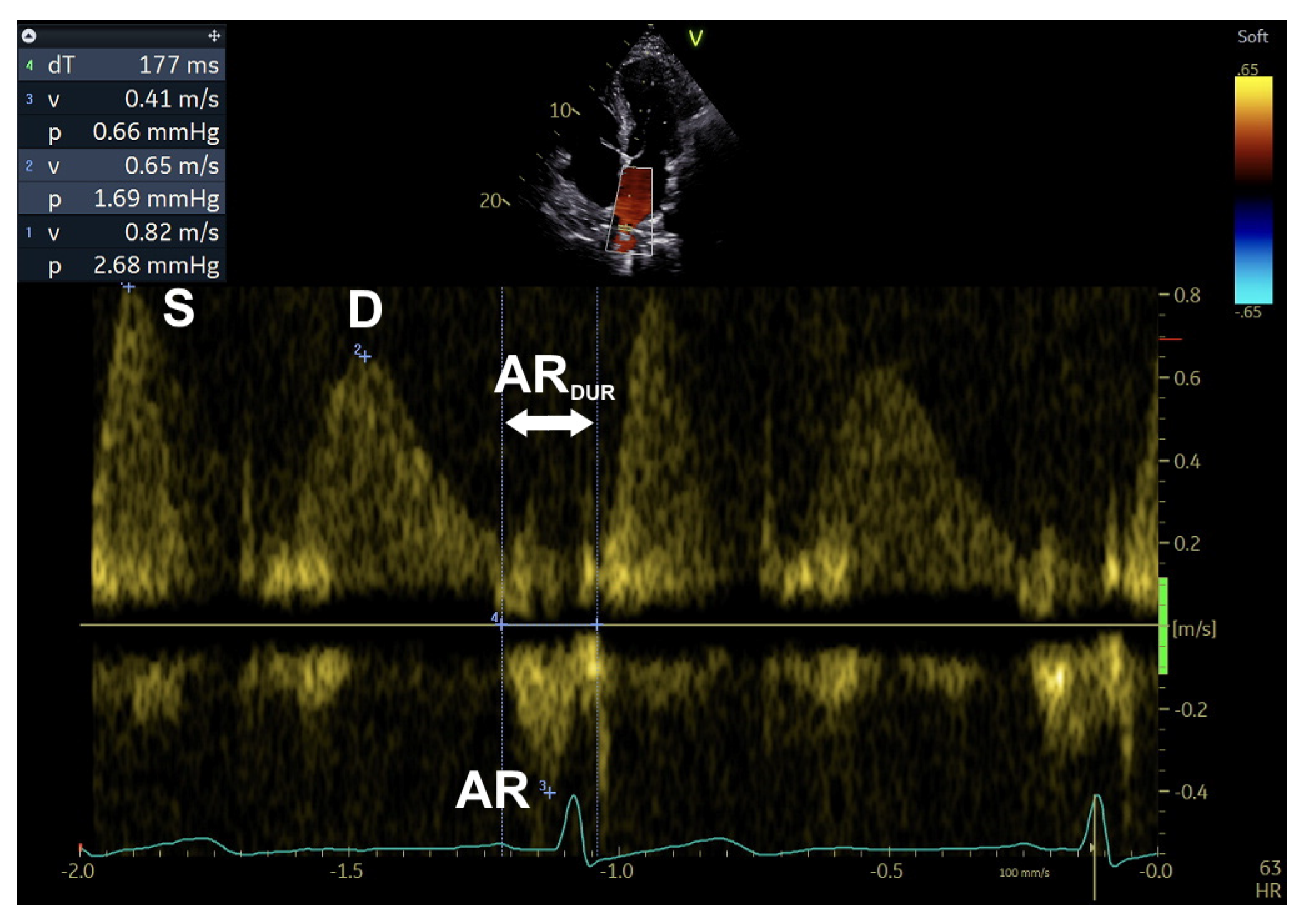
| PV inflow | - Apical 4C view with color/PW Doppler imaging (and reduced NL: high PRF may be needed when peak velocities exceed the NL). - PW Doppler SV (3–5 mm) placed 5–10 mm (generally) into the right upper and/or right lower PV. - Preferably, low wall filter setting (100–200 MHz), low signal gain, and sweep speed at 100 mm/s are needed. | Peak S-wave velocity (or S) (measured in cm/s): At ECG T-wave, measure peak systolic modal velocity (in the presence of two systolic peaks S1 and S2, S2 should be considered for S/D ratio); Affected by LAP, LA contractility/relaxation/stiffness, and LV/RV contractility. Range: in the presence of S/D ratio < 1, SFF < 40%, and reduced LV EF, reduced S indicates high LAP. | - Generally feasible. - Diagnostic and prognostic information. | - Possibly suboptimal in ICU subjects. - (S, D, and S/D) in the presence of preserved LV EF, MV disease, HCM, and AF, less accurate relationship between PV systolic filling fraction and LAP. - Challenging in the presence of arrhythmias (e.g., in the presence of AF, AR velocity is absent; in the presence of heart block, sinus tachycardia, and atrial arrhythmias, Ar-A is not applicable). |
| Peak D-wave velocity (or D) (in cm/s): After ECG T-wave, measure peak early diastolic modal velocity; Affected by both LV relaxation and LAP in early diastole; Changes in parallel with E. Range: in the presence of AF, DT of D-wave velocity ≤ 220 ms indicates high LV FP. | ||||
| S/D ratio (or S/D) (adimensional): Peak S-wave velocity divided by peak D-wave velocity; May be used to determine filling patterns (normal, IR, PN, and RF); Inversely related to LAP. Range: presence of S/D ratio < 1, SFF < 40%, reduced LV EF, and reduced S suggest high LAP. | ||||
| Peak AR-wave velocity (in cm/s): After ECG P-wave, measure peak late diastolic modal velocity; Affected by both LV compliance and LA contractility; Helpful when elevated LV EDP and normal LAP have been suspected (e.g., grade 1 diastolic dysfunction). Range: if >35 cm/s, indicates an increased LV EDP. | ||||
| AR-wave duration (in ms): At zero baseline, measure TI from the onset to the offset of the AR-wave signal; Longer AR duration, increased AR and D velocities, and decreased S velocity are related to both decreased LV compliance and increased LAP. | ||||
| AR-A duration (or Ar-A) (in ms): Time difference between AR-wave and A-wave durations; Age- and LV EF-independent; Related to LV EDP/FP. Range: if ≥30 ms, indicates increased LV EDP/FP (particularly, accurate in the presence of HCM and MR). | ||||
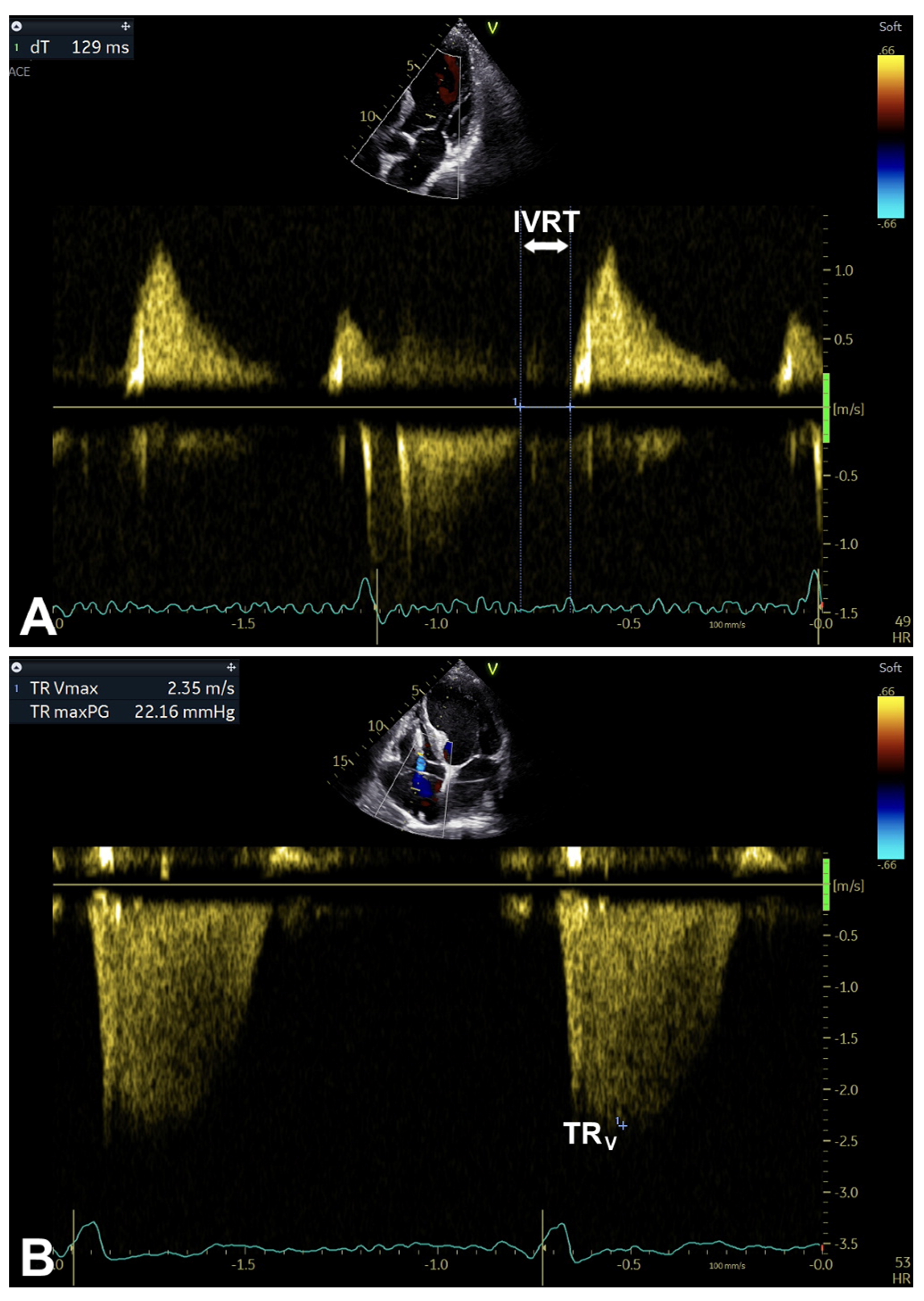
| TR | - From any view that allows correct alignment of US beam in parallel with TR jet (CW Doppler). - Preferably, sweep speed at 50–100 mm/s is needed. | Peak TR velocity (measured in m/s): Averaged over the respiratory cycle; In the absence of PS/RVOT obstruction, estimates the PASP (in the absence of pulmonary disease, high PASP suggests high LAP). Ranges, (20–39 y): 1.3 (1.1–1.5) to 2.7 (2.6–2.7); (40–60 y): 1.5 (1.4–1.6) to 2.7 (2.7–2.7); (60–80 y): 1.7 (1.5–1.8) to 2.8 (2.7–2.8). | - Generally feasible and reproducible. - Diagnostic and prognostic information. | - Indirect LAP estimation. - Related to age. - US beam optimal alignment-related. - Significantly limitations in some conditions (e.g., PS, RVOT obstruction, very severe TR and low systolic RV-RA pressure gradient). |
| IVRT | - Apical 3C (or long-axis) or 5C view with color/CW Doppler imaging (CW Doppler through LVOT in order to simultaneously display the end of aortic ejection and transmitral inflow onset). - Preferably, low wall filter setting (100–200 MHz), low signal gain, and sweep speed at 100 mm/s are needed. | IVRT duration (or IVRT) (measured in ms): At zero baseline, measure TI between AVC and MVO; Determines the TI between AVC and MVO, as well as the crossover between LA and LV pressures; Directly related to LV relaxation and inversely related to LAP; Useful for estimating LV FP (e.g., in the presence of MAC; in the presence of HFrEF, in combination with transmitral inflow measurements; in the presence of MS/MR, in combination with TE-e’). Range: LAP likely normal if >110 ms; if <70 ms, high specificity for elevated LAP in the presence of cardiac disease. | - Generally feasible and reproducible. | - More helpful in combination with other parameters. - Related to preload (tends to normalize with increasing LAP). - Partially affected by HR and AP. - Related to age (shorter in young subjects, lengthens with age). |
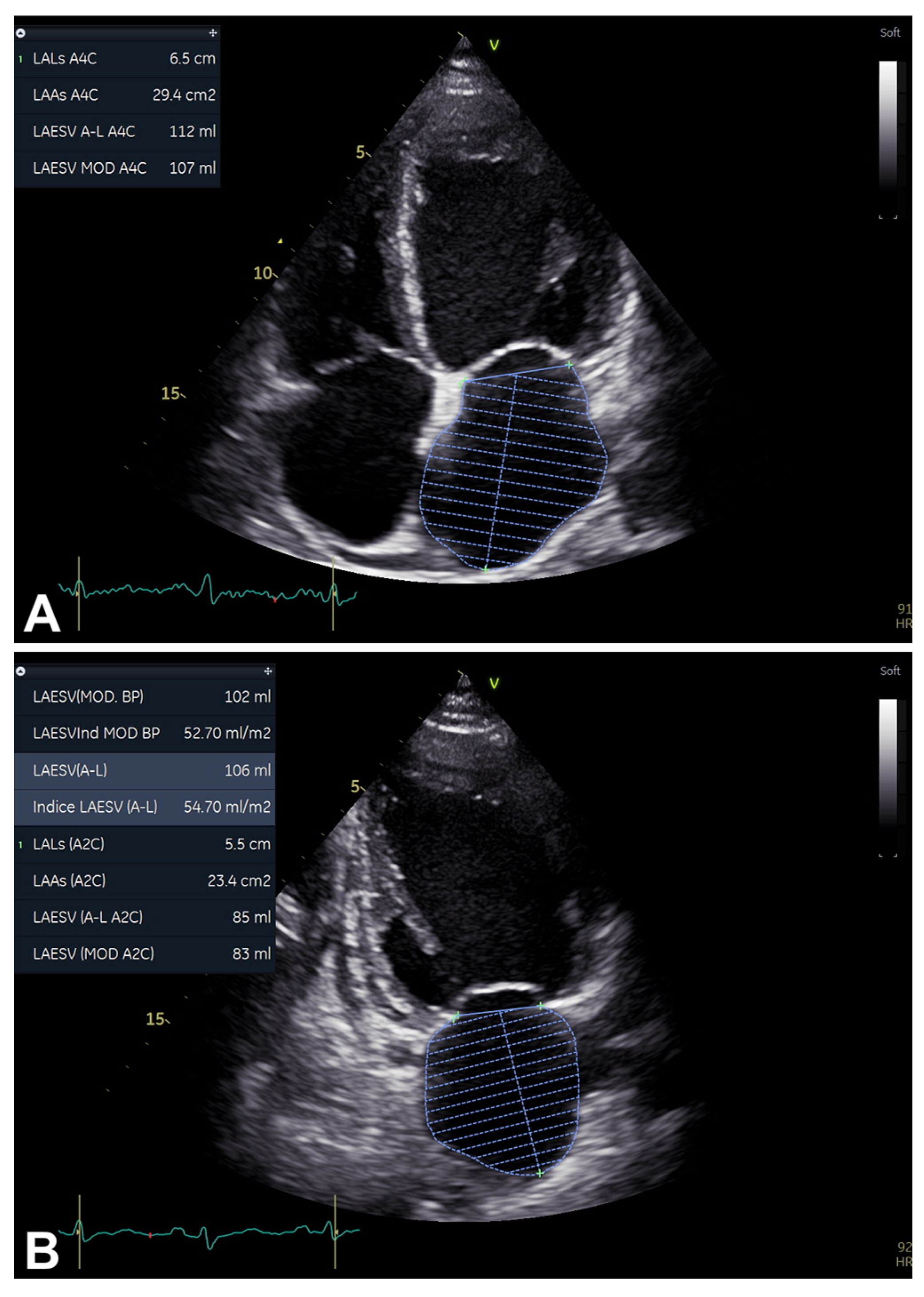
| LAVi | - Apical 2C and 4C views. - Maximize both LA base width and LA long axis. | LAVi (measured in mL/mq): From each view, acquire ES frames and trace LA area (excluding PV and LAA), thus (generally automatic) LAV calculation using the method of disks or area-length method (subsequently divided by BSA); Reflects the increased LV FP effects; LA dilation is an independent predictor of death, HF, AF and ischemic stroke. - LAVi: Range (20–39 y): 12.1 (10.9–13.2) to 39.4 (34.6–44.2); Range (40–60 y): 12.9 (12.2–13.5) to 38.3 (35.4–41.1); Range (60–80 y): 13.7 (12.7–14.6) to 37.1 (33.0–41.3). - LAVi (Simpson’s method): Range (20–39 y): 12.5 (12.0–13.0) to 41.9 (38.1–45.6); Range (40–60 y): 13.3 (13.0–13.6) to 41.0 (38.5–43.4); Range (60–80 y): 14.2 (13.7–14.6) to 40.0 (36.5–43.6). - LAVi (A-L): Range (20–39 y): 8.9 (3.9–13.9) to 20.9 (12.9–28.8); Range (40–60 y): 11.0 (8.9–13.0) to 27.1 (24.0–30.3); Range (60–80 y): 13.0 (9.9–16.0) to 33.4 (28.6–38.2). | - Feasible and reproducible. - Diagnostic and prognostic information. | - Good image quality is needed. - Related to age. |
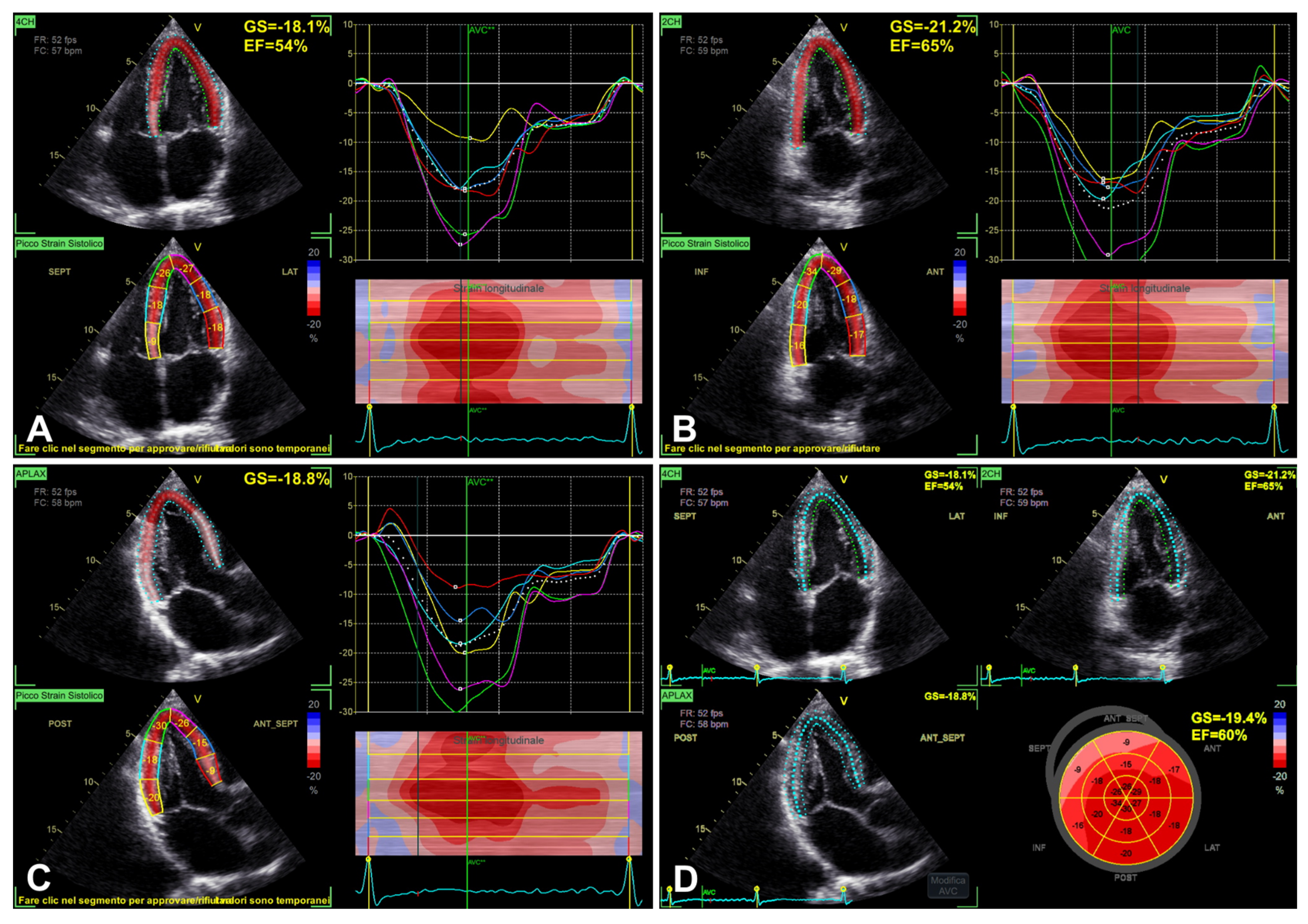
| LV GLS | - 2D apical 4C, 3C, and 2C views. - Optimize 2D gains, FR (40–80 frames/s), ECG. - Acquire 3-to-5 cardiac cycles for each view to obtain similar HRs. | LV GLS (measured in %): Probe in apical zone with 4C, 3C, and 2C views; Dedicated (AI-assisted, if available) strain software automatically tracks LV endocardium and calculates LV GLS; LV systolic function measurement (with strain-derived EF). | - Generally feasible and reproducible (mainly achievable with dedicated software package, adequate operator skills, and a good US window). - Prognostic and predictive value. | - Age- and load-dependent. - Chest-shape-dependent. - Image quality-related. - Inter-vendor and inter-software variability. |
| Parameter/Variable | Acquisition | Measurements/Pathophysiological Considerations | Advantages | Limitations |
|---|---|---|---|---|
| Valsalva maneuver (transmitral inflow) | - Apical 4C view ± color Doppler imaging. - Preferably, low wall filter setting (100–200 MHz), low signal gain, sweep speed ≤ 50 mm/s, and 10-to-12-second continuous recording during patient’s bearing down against a closed glottis (at rest and during peak strain) are needed. - Adequate maneuver if >10% reduction in maximal E-wave velocity from baseline status. | Positive: E/A < 1 or increased A. Negative: E/A > 1. Helpful in distinguishing either normal versus PN pattern, or reversible versus irreversible grade 3 diastolic dysfunction (by reducing preload); Highly specific for increased LV FP, during maneuver, if E/A decreases ≥ 50% or A increases (not caused by E-A fusion). | - If well-performed, good accuracy in diagnosing increased LV FP. | - Not every subject can adequately perform the maneuver. |
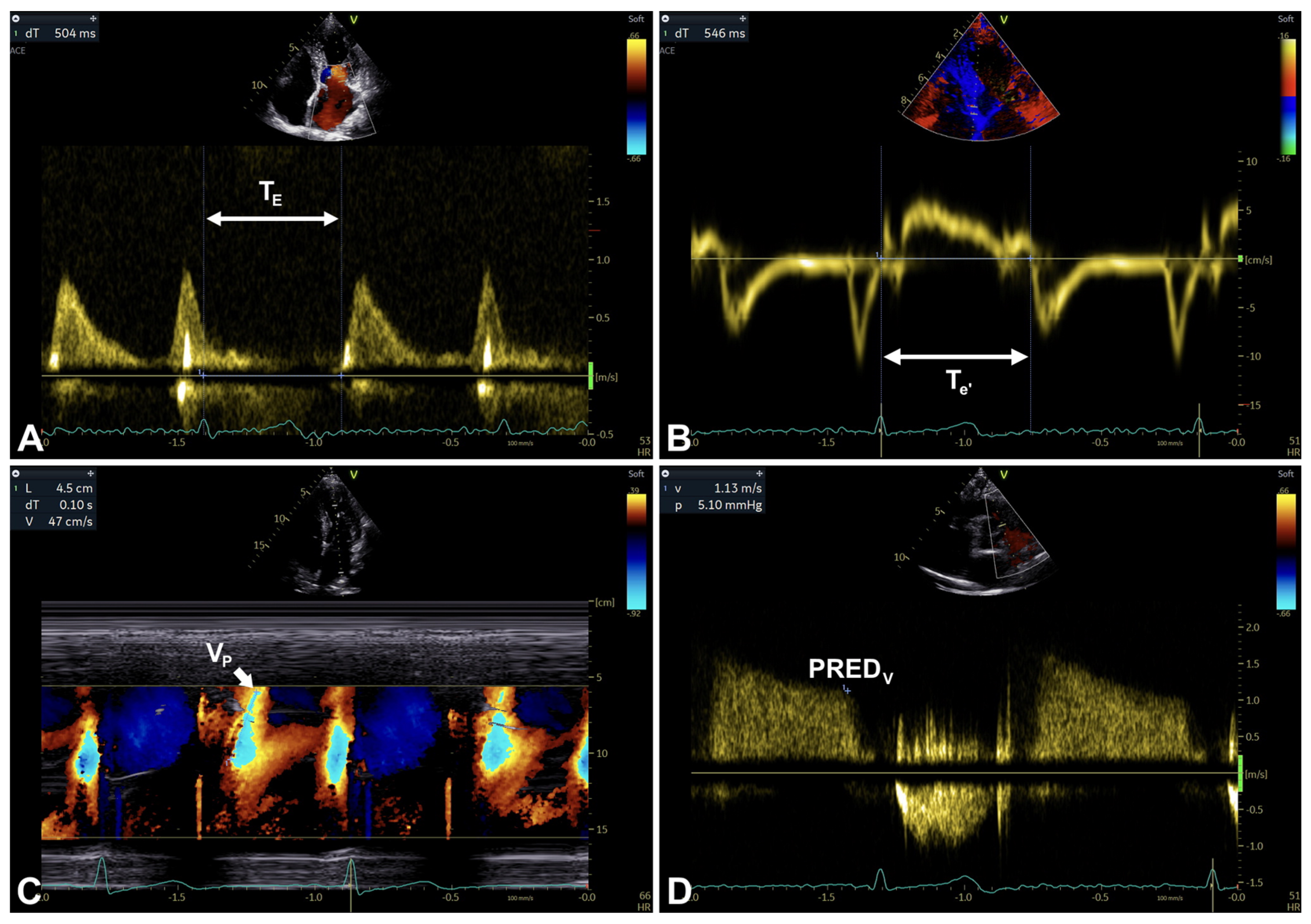
| Color M-mode Vp | - Apical 4C view with color Doppler imaging of transmitral inflow (variance mode off). - Alignment optimization of the M-mode cursor with the path of transmitral inflow. - For enhancing the early diastolic slope, color NL lowering is needed. | Color M-mode Vp (or Vp) (measured in cm/s): Along the early diastolic slope of first aliased velocity (red–blue interface), measure from the level of mitral annulus to 4 cm into the LV cavity; May be helpful in distinguishing normal versus PN pattern; Vp is indirectly associated with Ƭ (the longer it takes for the LV to relax, the slower the Vp), and E/Vp is directly related to LAP. | - Relatively load-independent. - Reliable index in the presence of LV dysfunction/dilation. | - Lower feasibility and reproducibility. - In the presence of normal LV volumes/function and high LV FP, possibly misleading normal Vp. |
| E/Vp ratio (or E/Vp) (adimensional, after converting E velocity in cm/s). Range: in the presence of reduced LV EF, if ≥2.5 predicts PCWP > 15 mmHg with reasonable accuracy. | ||||
| TE-e’-related parameters | For E-wave (transmitral inflow): - Apical 4C view and color/PW Doppler imaging. - Preferably, low wall filter setting (100–200 MHz), low signal gain, PW Doppler SV 1–3 mm, and sweep speed at 100 mm/s are needed. For e’-wave (TDI at mitral annulus): - Apical 4C view with TDI preset optimization *. - Alignment optimization (angle of interrogation should be parallel to annular motion). - Preferably, PW Doppler SV 5–10 mm and sweep speed at 100 mm/s are needed. | TE (measured in ms): TI between the peak R-wave (on ECG) and the onset of E-wave (transmitral inflow). | - Generally feasible. - May be helpful in diagnosing diastolic dysfunction due to delayed e’ onset compared with E onset (e.g., differentiating RC_prolonged TI_versus PC_usually, not-prolonged TI _). | - Non-simultaneous measurements (R-R interval matching is important) in the presence of small TI (thus, increased probability of error). |
| Te’ (measured in ms): TI between the peak R-wave (on ECG) and the onset of e’-wave (TDI). | ||||
| TE-e’ (measured in ms): TI calculated by subtracting TE from Te’ (namely, Te’—TE); May be helpful in distinguishing normal versus PN pattern. In the presence of MS/MR, may be useful for estimating LV FP in combination with IVRT. (IVRT/TE-e’ ratio; adimensional): in the presence of MS, elevated LV FP if IVRT/TE-e’ < 4.2; in the presence of MR, elevated LV FP if IVRT/TE-e’ < 5.6. | ||||
| PR ED velocity | - From any view that allows correct alignment of US beam in parallel with PR jet (CW Doppler). - Preferably, sweep speed at 50–100 mm/s is needed. | Peak PR ED velocity (or PRED) (measured in m/s): Measured at ED; Related to PAEDP and PCWP. In the absence of pulmonary disease, PRED ≥ 2 m/s suggests elevated LAP. | - Generally feasible. - Diagnostic and prognostic information. | - Misleading in the presence of pulmonary disease. |
| Parameter/Variable | Acquisition | Measurements/Pathophysiological Considerations | Advantages | Limitations |
|---|---|---|---|---|
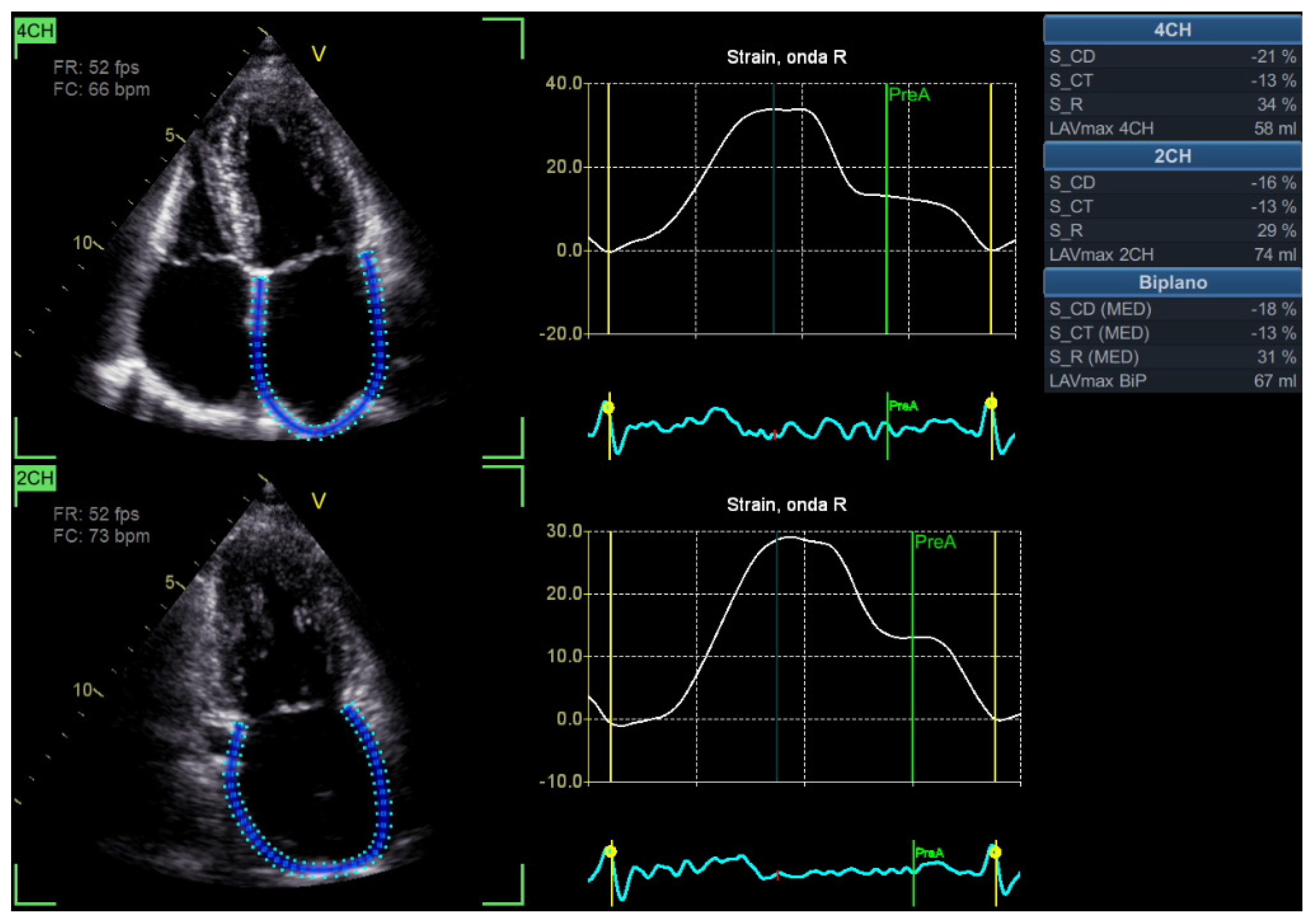
| LA strain-related parameters | - 2D apical 4C and 2C views. - Optimize 2D gains, FR (50–70 frames/s), ECG (well-visible P-waves). - Acquire 3-to-5 cardiac cycles for each view to obtain similar HRs. | LARS (measured in %): Probe in apical zone with 4C and 2C views; Dedicated (AI-assisted, if available) strain software automatically tracks LA wall (excluding PV and LAA) and calculates LARS; During ventricular systole, peak positive strain value; For assessing diastolic function, LA strain primarily focuses on LARS, directly related to diastolic dysfunction, and inversely related to LV FP. - LA strain: Range (20–39 y): 29.5 (27.6–31.3) to 63.2 (59.9–66.5); Range (40–60 y): 26.8 (25.6–28.0) to 57.7 (55.6–59.9); Range (60–80 y): 24.1 (22.2–26.0) to 52.3 (48.9–55.7). - LA strain (TomTec): Range (20–39 y): 29.9 (27.0–32.9) to 60.5 (57.6–63.4); Range (40–60 y): 27.5 (25.7–29.4) to 55.4 (53.6–57.2); Range (60–80 y): 25.1 (22.6–27.6) to 50.3 (47.9–52.7). - LA strain (EchoPAC): Range (20–39 y): 29.5 (27.9–31.1) to 64.9 (59.7–70.2); Range (40–60 y): 25.3 (24.0–26.5) to 61.5 (57.4–65.6); Range (60–80 y): 21.1 (18.7–23.4) to 58.1 (50.3–65.8). | - Generally feasible and reproducible (mainly achievable with dedicated software package, adequate operator skills, and a good US window). - Prognostic and predictive value. | - Age- (LARS decreases with age) and load-dependent. - Challenging or inaccurate in the presence of arrhythmias and other conditions (e.g., in the presence of BBB, R-R gating may be inaccurate; in the presence of some anatomical conditions, including mobile AS, thin-walled LA, and MAC, LA strain may be inaccurate; in the presence of atrial arrhythmias/stunning, significant MR, HT recipients, preserved EF and GLS > 18%, LARS should not be used to evaluate LV FP). - Chest-shape-dependent. - Image quality-related. - Inter-vendor and inter-software variability. |
| LASCT (measured in %): Probe in apical zone with 4C and 2C views; Dedicated (AI-assisted, if available) strain software automatically tracks LA wall (excluding PV and LAA) and calculates LASCT as well; Measured in SR as 0 minus strain value at the onset of atrial contraction (namely, pre-A-wave on ECG); Inversely related to LVEDP. | ||||
| LASCD (measured in %): Probe in apical zone with 4C and 2C views; Dedicated (AI-assisted, if available) strain software automatically tracks LA wall (excluding PV and LAA) and calculates LASCD as well; Measured in SR as 0 minus strain value at atrial contraction). | ||||
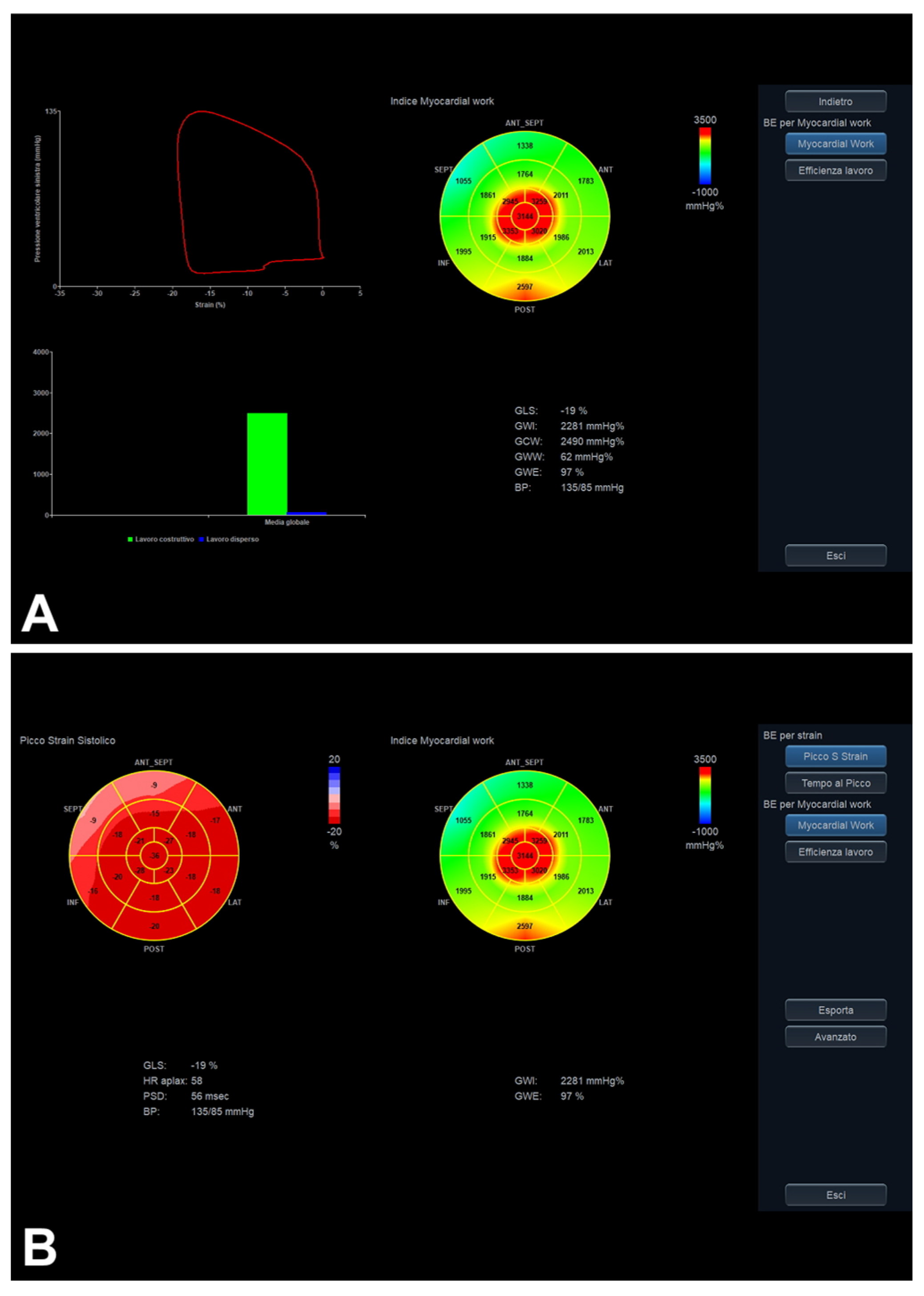
| LV MW | - 2D apical 4C, 3C, and 2C views. - Optimize 2D gains, FR (40–80 frames/s) and ECG. - Acquire 3-to-5 cardiac cycles for each view to obtain similar HRs. - LV GLS calculation and further derived MW indices (by introducing non-invasive AP and dedicated software package). | GCW (MW index measured in mmHg%): positive work evaluated from AVO to AVC and negative work from AVC to MVO. | - Generally feasible and reproducible (mainly achievable with dedicated software package, adequate operator skills, and a good US window). - Prognostic and predictive value. | - GLS-related limits (including image quality). - AP measurement-related limits. - Single available software. |
| GWI (MW index measured in mmHg%): total work evaluated from MVC to MVO. | ||||
| GWE (MW index measured in %): GCW/(GCW + GWW). | ||||
| GWW (MW index measured in mmHg%): positive work evaluated from AVO to AVC and negative work from AVC to MVO. | ||||
| Cardiac THE | - 2D standard medical ultrasound and continuous external harmonic vibration (patients positioned on a vibration bed). - Vibration bed introduces shear waves into the heart, aiming at creating a mechanical stiffness contrast. | - Regionally resolved mapping of diastolic shear wave (as a myocardial stiffness indicator) is performed (to date, LV posterior wall and interventricular septum have generally been chosen from parasternal long-axis view, for higher reproducibility as well). | - Diastolic myocardial stiffness detecting and mapping. - Accurate at greater depths of up to 15 cm, independently of both region selection and body mass index. - Cost-effective technique. | - 2D acquisition/evaluation of diastolic phases only. - Cardiac THE setup relying on a vibrating bed not easily movable to the patient. |
Disclaimer/Publisher’s Note: The statements, opinions and data contained in all publications are solely those of the individual author(s) and contributor(s) and not of MDPI and/or the editor(s). MDPI and/or the editor(s) disclaim responsibility for any injury to people or property resulting from any ideas, methods, instructions or products referred to in the content. |
© 2026 by the authors. Licensee MDPI, Basel, Switzerland. This article is an open access article distributed under the terms and conditions of the Creative Commons Attribution (CC BY) license.
Share and Cite
Dell’Angela, L.; Nicolosi, G.L. Echocardiographic Assessment of Left Ventricular Diastolic Function in Adults Between Old and New: Progress and Challenges. Diagnostics 2026, 16, 235. https://doi.org/10.3390/diagnostics16020235
Dell’Angela L, Nicolosi GL. Echocardiographic Assessment of Left Ventricular Diastolic Function in Adults Between Old and New: Progress and Challenges. Diagnostics. 2026; 16(2):235. https://doi.org/10.3390/diagnostics16020235
Chicago/Turabian StyleDell’Angela, Luca, and Gian Luigi Nicolosi. 2026. "Echocardiographic Assessment of Left Ventricular Diastolic Function in Adults Between Old and New: Progress and Challenges" Diagnostics 16, no. 2: 235. https://doi.org/10.3390/diagnostics16020235
APA StyleDell’Angela, L., & Nicolosi, G. L. (2026). Echocardiographic Assessment of Left Ventricular Diastolic Function in Adults Between Old and New: Progress and Challenges. Diagnostics, 16(2), 235. https://doi.org/10.3390/diagnostics16020235