Structural Retinal Analysis in Toxoplasmic Retinochoroiditis: OCT Follow-Up with Three-Dimensional Reconstruction

 ,
, 
 ,
, 
Abstract
1. Introduction
2. Materials and Methods
2.1. Case Selection
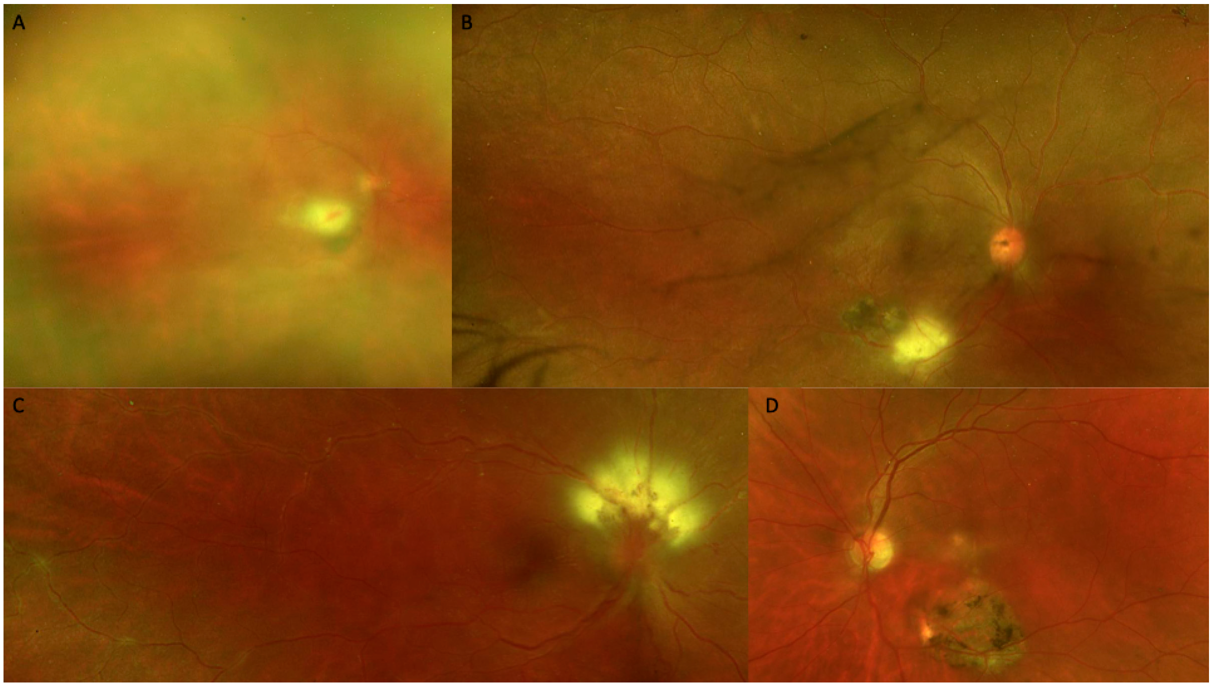
2.2. Diagnosis
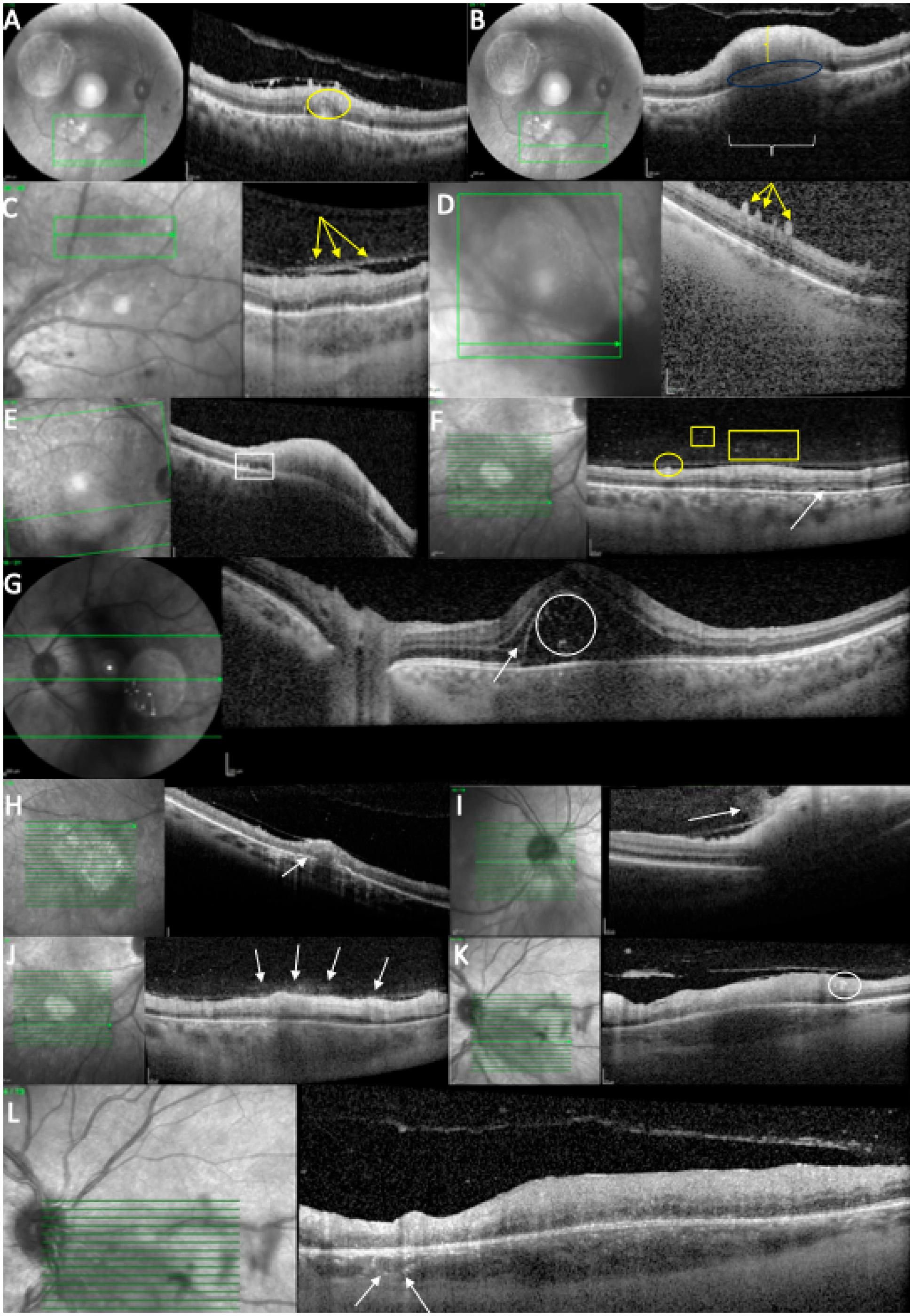
2.3. Optical Coherence Tomography (OCT) Analysis
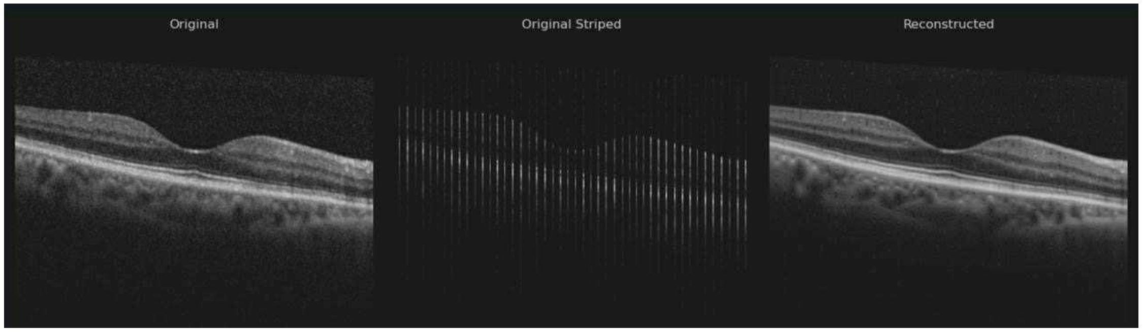
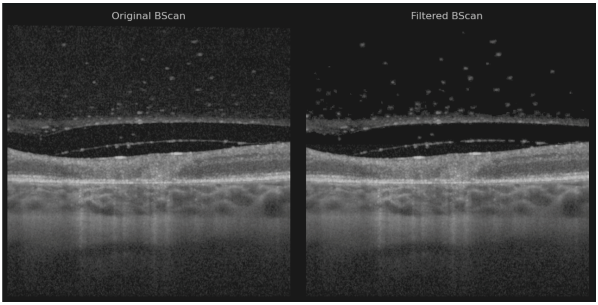
2.4. Volume Reconstruction Technique
3. Results
4. Discussion
5. Conclusions
Supplementary Materials
Author Contributions
Funding
Institutional Review Board Statement
Informed Consent Statement
Data Availability Statement
Conflicts of Interest
Abbreviations
| OCT | Optical coherence tomography |
| PCR | Polymerase chain reaction |
| SD | Standard deviation |
| OPL | Outer plexiform layer |
| ONL | Outer nuclear layer |
| RPE | Retinal pigment epithelium |
| ILM | Internal limiting membrane |
| PVD | Posterior vitreous detachment |
| ELM | External limiting membrane |
| ERM | Epiretinal membrane |
| VA | Visual acuity |
| T. gondii | Toxoplasma gondii |
| AC | Anterior chamber |
| IgM | Immunoglobulin M |
| IgG | Immunoglobulin G |
| EZ | Ellipsoid zone |
| TRC | Toxoplasmic retinal choroiditis |
| CFP | Color fundus photography |
| AMD | Age-related macular degeneration |
| 3D | Three-dimensional |
References
- Ozgonul, C.; Besirli, C.G. Recent Developments in the Diagnosis and Treatment of Ocular Toxoplasmosis. Ophthalmic Res. 2017, 57, 1–12. [Google Scholar] [CrossRef] [Scilit] [PubMed]
- Park, J.H.; Lee, S.Y.; Lee, E.K. Morphological characteristics of ocular toxoplasmosis and its regression pattern on swept-source optical coherence tomography angiography: A case report. BMC Ophthalmol. 2019, 19, 199. [Google Scholar] [CrossRef] [Scilit] [PubMed]
- Kijlstra, A.; Petersen, E. Epidemiology, pathophysiology, and the future of ocular toxoplasmosis. Ocul. Immunol. Inflamm. 2014, 22, 138–147. [Google Scholar] [CrossRef] [Scilit]
- Goldenberg, D.; Goldstein, M.; Loewenstein, A.; Habot-Wilner, Z. Vitreal, retinal, and choroidal findings in active and scarred toxoplasmosis lesions: A prospective study by spectral-domain optical coherence tomography. Graefes Arch. Clin. Exp. Ophthalmol. 2013, 251, 2037–2045. [Google Scholar] [CrossRef] [Scilit]
- Pleyer, U.; Schlüter, D.; Mänz, M. Ocular toxoplasmosis: Recent aspects of pathophysiology and clinical implications. Ophthalmic Res. 2014, 52, 116–123. [Google Scholar] [CrossRef] [Scilit]
- Rothova, A.; Meenken, C.; Buitenhuis, H.J.; Brinkman, C.J.; Baarsma, G.S.; Boen-Tan, T.N.; de Jong, P.T.; Klaassen-Broekema, N.; Schweitzer, C.M.; Timmerman, Z.; et al. Therapy for ocular toxoplasmosis. Am. J. Ophthalmol. 1993, 115, 517–523. [Google Scholar] [CrossRef] [Scilit] [PubMed]
- Oliver, G.F.; Ferreira, L.B.; Vieira, B.R.; Arruda, S.; Araújo, M.; Carr, J.M.; Smith, J.R.; Furtado, J.M. Posterior segment findings by spectral-domain optical coherence tomography and clinical associations in active toxoplasmic retinochoroiditis. Sci. Rep. 2022, 12, 1156. [Google Scholar] [CrossRef] [Scilit]
- Ammar, F.; Mahjoub, A.; Ben Abdesslam, N.; Knani, L.; Ghorbel, M.; Mahjoub, H. Spectral optical coherence tomography findings in patients with ocular toxoplasmosis: A case series study. Ann. Med. Surg. 2020, 54, 125–128. [Google Scholar] [CrossRef] [Scilit]
- Diniz, B.; Regatieri, C.; Andrade, R.; Maia, A. Evaluation of spectral domain and time domain optical coherence tomography findings in toxoplasmic retinochoroiditis. Clin. Ophthalmol. 2011, 5, 645–650. [Google Scholar] [CrossRef] [Scilit]
- Oréfice, J.L.; Costa, R.A.; Scott, I.U.; Calucci, D.; Oréfice, F.; Grupo Mineiro de Pesquisa em Doenças Oculares Inflamatórias (MINAS). Spectral optical coherence tomography findings in patients with ocular toxoplasmosis and active satellite lesions (MINAS Report 1). Acta Ophthalmol. 2013, 91, e41–e47. [Google Scholar] [CrossRef] [Scilit]
- Sofia, O.; Wahyudi, I.N.S.A.; Fitri, L.E.; Prayitnaningsih, S.; Susianti, H. Optical Coherence Tomography Angiography Findings in Ocular Toxoplasmosis with Multiple Recurrences. Int. Med. Case Rep. J. 2023, 16, 35–43. [Google Scholar] [CrossRef] [Scilit]
- Schindelin, J.; Arganda-Carreras, I.; Frise, E.; Kaynig, V.; Longair, M.; Pietzsch, T.; Preibisch, S.; Rueden, C.; Saalfeld, S.; Schmid, B.; et al. Fiji: An open-source platform for biological-image analysis. Nat. Methods 2012, 9, 676–682. [Google Scholar] [CrossRef] [Scilit]
- Schmitt, J.M.; Xiang, S.H.; Yung, K.M. Speckle in optical coherence tomography. J. Biomed. Opt. 1999, 4, 95–100. [Google Scholar] [CrossRef] [Scilit]
- Babin, D.; Vansteenkiste, E.; Pizurica, A.; Philips, W. Segmentation of brain blood vessels using projections in 3-d ct angiography images. Annu. Int. Conf. IEEE Eng. Med. Biol. Soc. 2011, 2011, 8475–8478. [Google Scholar]
- Pop, A.; Damian, I.; Groza, A.; Nicoară, S.D. Blood Vessel Segmentation and 3D Reconstruction of Neovascularization in Age-Related Macular Degeneration from OCTA Scans. In 9th International Conference on Advancements of Medicine and Health Care Through Technology; MEDITECH 2024. IFMBE Proceedings; Springer: Cham, Switzerland, 2025; Volume 130. [Google Scholar]
- Mayer, M.A.; Hornegger, J.; Mardin, C.Y.; Tornow, R.P. Retinal Nerve Fiber Layer Segmentation on FD-OCT Scans of Normal Subjects and Glaucoma Patients. Biomed. Opt. Express 2010, 1, 1358–1383. [Google Scholar] [CrossRef] [Scilit]
- Kermany, D.; Zhang, K.; Goldbaum, M. Labeled Optical Coherence Tomography (OCT) and Chest X-Ray Images for Classification. Mendeley Data 2018, V2. [Google Scholar] [CrossRef]
- Rozhyna, A.; Somfai, G.M.; Atzori, M.; DeBuc, D.C.; Saad, A.; Zoellin, J.; Müller, H. Exploring Publicly Accessible Optical Coherence Tomography Datasets: A Comprehensive Overview. Diagnostics 2024, 14, 1668. [Google Scholar] [CrossRef] [Scilit] [PubMed]
- Nussenblatt, R.B.; Belfort, R., Jr. Ocular toxoplasmosis: An old disease revisited. JAMA 1994, 271, 304–307. [Google Scholar] [CrossRef] [Scilit] [PubMed]
- Kharel Sitaula, R.; Joshi, S.N.; Sah, R.; Khadka, S.; Khatri Kc, A.; Pokharel, B.M. Toxoplasma gondii bradyzoites and tachyzoites isolation from vitreous of atypical necrotizing retinitis. J. Ophthal Inflamm. Infect. 2018, 8, 8. [Google Scholar] [CrossRef] [Scilit]
- Monnet, D.; Averous, K.; Delair, E.; Brézin, A.P. Optical coherence tomography in ocular toxoplasmosis. Int. J. Med. Sci. 2009, 6, 137–138. [Google Scholar] [CrossRef] [Scilit]
- Miyagaki, M.; Zong, Y.; Yang, M.; Zhang, J.; Zou, Y.; Ohno-Matsui, K.; Kamoi, K. Ocular Toxoplasmosis: Advances in Toxoplasma gondii Biology, Clinical Manifestations, Diagnostics, and Therapy. Pathogens 2024, 13, 898. [Google Scholar] [CrossRef] [Scilit]
- Saxena, S.; Rastogi, R.A.; Vishvkarma, K.; Hansraj, S. Meyer CH.Spectral-domain optical coherence tomography in healed ocular toxoplasmosis. J. Ocul. Biol. Dis. Infor. 2010, 3, 109–111. [Google Scholar] [CrossRef] [Scilit] [PubMed]
- Frizziero, L.; Midena, G.; Danieli, L.; Torresin, T.; Perfetto, A.; Parrozzani, R.; Pilotto, E.; Midena, E. Hyperreflective Retinal Foci (HRF): Definition and Role of an Invaluable OCT Sign. J. Clin. Med. 2025, 14, 3021. [Google Scholar] [CrossRef] [Scilit] [PubMed]
- Duraffour, P.; Mehanna, C.; Hoogewoud, F.; Touboul, A.; Monnet, D.; Brézin, A.P. Comparison between the areas of scarred and active toxoplasmic retinochoroiditis. Eye 2021, 35, 2733–2739. [Google Scholar] [CrossRef] [Scilit]
- Dubielzig, R.R.; Ketring, K.; McLellan, G.J.; Albert, D.M. The uvea. Vet. Ocul. Pathol. 2010, 28, 245–322. [Google Scholar]
- Ksiaa, I.; Khochtali, S.; Mefteh, M.; Ben Fredj, M.; Ben Amor, H.; Abroug, N.; Khairallah, M. Distinguishing swept-source optical coherence tomography findings in active toxoplasmic retinochoroiditis. Eye 2022, 36, 1222–1230. [Google Scholar] [CrossRef] [Scilit] [PubMed]
- Brancato, R.; Trabucchi, G. Fluorescein and Indocyanine green angiography in vascular chorioretinal diseases. Semin. Ophthalmol. 1998, 13, 190–198. [Google Scholar] [CrossRef] [Scilit]
- Atmaca, L.S.; Simsek, T.; Atmaca Sonmez, P.; Sonmez, K. Fluorescein and indocyanine green angiography in ocular toxoplasmosis. Graefes Arch. Clin. Exp. Ophthalmol. 2006, 244, 1688–1691. [Google Scholar] [CrossRef] [Scilit]
- Auer, C.; Bernasconi, O.; Herbort, C.P. Indocyanine green angiography features in toxoplasmic retinochoroiditis. Retina 1999, 19, 22–29. [Google Scholar] [CrossRef] [Scilit]
- Khanifar, A.A.; Espiritu, M.J.; Myung, J.S.; Aaker, G.D.; Schuetz, A.N.; D’Amico, D.J.; Chan, R.V. Three-dimensional spectral domain optical coherence tomography and light microscopy of an intravitreal parasite. J. Ophthal Inflamm. Infect. 2015, 5, 33. [Google Scholar] [CrossRef] [Scilit]
- Saxena, S.; Singhal, V.; Akduman, L. Three-dimensional spectral domain optical coherence tomography imaging of the retina in choroidal tuberculoma. BMJ Case Rep. 2013, 2013, bcr2012008156. [Google Scholar] [CrossRef] [Scilit]
- Koizumi, H.; Spaide, R.F.; Fisher, Y.L.; Freund, K.B.; Klancnik, J.M., Jr.; Yannuzzi, L.A. Three-dimensional evaluation of vitreomacular traction and epiretinal membrane using spectral-domain optical coherence tomography. Am. J. Ophthalmol. 2008, 145, 509–517. [Google Scholar] [CrossRef] [Scilit] [PubMed]
- Ahn, S.E.; Oh, J.; Oh, J.H.; Oh, I.K.; Kim, S.W.; Huh, K. Three-Dimensional Configuration of Subretinal Fluid in Central Serous Chorioretinopathy. Investig. Ophthalmol. Vis. Sci. 2013, 54, 5944–5952. [Google Scholar] [CrossRef] [Scilit] [PubMed]
- Iwasaki, T.; Miura, M.; Matsushima, C.; Yamanari, M.; Makita, S.; Yasuno, Y. Three-dimensional optical coherence tomography of proliferative diabetic retinopathy. Br. J. Ophthalmol. 2008, 92, 713. [Google Scholar] [CrossRef] [Scilit] [PubMed]
- Saxena, S.; Manisha Meyer, C.H. Three-dimensional spectral domain optical coherence tomography in X linked foveal retinoschisis. BMJ Case Rep. 2013, 2013, bcr2012007661. [Google Scholar] [CrossRef] [Scilit]


| Characteristics | Unit (%) | |
|---|---|---|
| Sex | female | 6/12 (50) |
| male | 6/12 (50) | |
| Age | years | 39.17 ± 17.69 |
| Laterality | right eye | 7/12 (58.30) |
| left eye | 5/12 (41.67) | |
| Disease type | recurrent | 2/12 (16.7) |
| primary | 10/12 (83.3) | |
| Symptom duration until diagnosis | days | 16.2 |
| Symptoms | visual acuity decrease | 6/12 (50) |
| myodesopsia | 3/12 (25) | |
| blurred vision | 2/12 (16.7) | |
| central scotoma | 1/12 (8.35) | |
| ocular pain | 2/12 (16.7) | |
| Baseline VA | ≥20/40 | 6/12 |
| ≤20/50 | 3/12 | |
| ≤20/200 | 3/12 | |
| Serology | T. gondii IgM-positive | 1 (8.35) |
| T. gondii IgG-positive | 5 (41.67) | |
| PCR AC tap | T. gondii-positive | 3/12 (25) |
| Systemic treatment | trimethoprim + sulfamethoxazole | 11/12 |
| sulfadiazine + pyrimethamine | 1/12 | |
| oral prednisone | 9/12 | |
| Intravitreal treatment | clindamycin | 3/12 |
| Topical treatment | corticosteroid | 3/12 |
| IOP-lowering drops | 3/12 |
| Biomicroscopic Findings | Signs | Unit (%) |
|---|---|---|
| anterior segment | keratic precipitates | 5/12 (41.67) |
| Tyndall | 5/12 (41.67) | |
| vitreous | vitreous cells | 11/12 (91.67) |
| snowballs | 1/12 (8.35) | |
| posterior segment | ERM | 1/12 (8.35) |
| macular edema | 1/12 (8.35) | |
| white retinal lesion | 12/12 (100) | |
| chorioretinal scar | 2/12 (16.7) | |
| associated vasculitis near the lesion | 2/12 (16.7) | |
| hemorrhage near the lesion | 2/12 (16.7) | |
| perilesional exudation | 2/12 (16.7) | |
| macular star | 1/12 (8.35) | |
| Kyrieleis arteritis | 1/12 (8.35) | |
| optic disc edema | 1/12 (8.35) | |
| lesion involving the optic disc | 2/12 (16.7) | |
| macular hole | 1/12 (8.35) |
| SD-OCT Features at Baseline | Unit (%) |
|---|---|
| mean lesion height (μm) ± −SD | 522.77 ± 293.5 |
| Retinal Lesion | |
| thickened retina | 12/12 (100) |
| full-thickness retinal hyperreflectivity | 11/12 (91.67) |
| intraretinal OPL and ONL hyperreflective dots | 11/12 (91.67) |
| periarteriolar hyperreflective dots | 4/12 (33.3) |
| intraretinal fluid with ONL or OPL cysts | 3/12 (25) |
| subretinal fluid | 4/12 (33.3) |
| subretinal hyperreflective dots | 2/12 (16.7) |
| disorganized retinal layers adjacent to lesion | 10/12 (83.3) |
| macular hole | 1/12 (8.35) |
| thickened RPE | 6/12 (50) |
| RPE bumps | 2/12 (16.7) |
| bowing of retina–RPE–Bruch’s membrane | 7/12 (58.30) |
| liquefactive necrosis | 3/12 (25) |
| Choroid | |
| choroidal hyporeflectivity | 11/12 (91.67) |
| choroidal thickening | 11/12 (91.67) |
| hyperreflective dots around choroidal vessels | 4/12 (33.3) |
| choroidal thickness under the lesion (μm) ±SD | 514.1 ± 267.9 |
| Vitreous | |
| posterior hyaloid thickening | 8/12 (66.66) |
| hyperreflective vitreous dots | 8/12 (66.66) |
| hyperreflective vitreous dots over a blood vessel | 3/12 (25) |
| hyperreflective ILM deposits | 11/12 (91.67) |
| hyperreflective deposits on the posterior hyaloid | 8/12 (66.66) |
| incomplete PVD | 4/12 (33.3) |
| Biomicroscopic Findings | Signs | Unit (%) |
|---|---|---|
| anterior segment | keratic precipitates | 2 (16.7) |
| Tyndall | 2 (16.7) | |
| vitreous | vitreous cells | 5 (41.67) |
| snowballs | 1 (8.35) | |
| posterior segment | ERM | 3 (25) |
| macular edema | 0 | |
| chorioretinal scar | 6 (50) | |
| associated vasculitis near the lesion | 0 | |
| hemorrhage near the lesion | 0 | |
| perilesional exudation | 0 | |
| macular star | 0 | |
| Kyrieleis | 0 | |
| optic disc edema | 2 (16.7) | |
| lesion involving the optic disc | 2 (16.7) | |
| macular hole | 0 |
| SD-OCT Features at Follow-Up | Unit (%) |
|---|---|
| mean lesion height (μm) ±-SD | 286.76 ± 163.3 |
| Retina | |
| thickened retina | 4/12 (33.3) |
| full-thickness retinal hyperreflectivity | 12/12 (100) |
| intraretinal OPL and ONL hyperreflective dots | 5/12 (41.67) |
| periarteriolar hyperreflective dots | 0 |
| intraretinal fluid with ONL or OPL cysts | 2/12 (16.7) |
| subretinal fluid | 0 |
| subretinal hyperreflective dots | 0 |
| disorganized retinal layers adjacent to lesion | 12/12 (100) |
| macular hole | 1/12 (8.35) |
| thickened RPE | 0 |
| RPE bumps | 0 |
| bowing of retina–RPE–Bruch’s membrane | 3/12 (25) |
| RPE atrophy and hypertransmission | 9/12 (75) |
| liquefactive necrosis | 8/12 (66.66) |
| coagulative necrosis | 4/12 (33.3) |
| Choroid | |
| choroidal hyporeflectivity | 9/12 (75) |
| choroidal thickening | 4/12 (33.3) |
| hyperreflective dots around choroidal vessels | 4/12 (33.3) |
| hyperreflective dots in the choroid | 9/12 (75) |
| choroidal thickness under the lesion (μm) ± SD | 261.1 ± 111.3 |
| Vitreous | |
| posterior hyaloid thickening | 5/12 (41.67) |
| hyperreflective vitreous dots | 5/12 (41.67) |
| hyperreflective vitreous dots over a blood vessel | 0 |
| hyperreflective ILM deposits | 7/12 (58.30) |
| hyperreflective deposits on the posterior hyaloid | 6/12 (50) |
| incomplete PVD | 7/12 (58.30) |
Disclaimer/Publisher’s Note: The statements, opinions and data contained in all publications are solely those of the individual author(s) and contributor(s) and not of MDPI and/or the editor(s). MDPI and/or the editor(s) disclaim responsibility for any injury to people or property resulting from any ideas, methods, instructions or products referred to in the content. |
© 2025 by the authors. Licensee MDPI, Basel, Switzerland. This article is an open access article distributed under the terms and conditions of the Creative Commons Attribution (CC BY) license (https://creativecommons.org/licenses/by/4.0/).
Share and Cite
Damian, I.; Pop, A.; Groza, A.; Miserocchi, E.; Nicoară, S.D. Structural Retinal Analysis in Toxoplasmic Retinochoroiditis: OCT Follow-Up with Three-Dimensional Reconstruction. Diagnostics 2025, 15, 3091. https://doi.org/10.3390/diagnostics15233091
Damian I, Pop A, Groza A, Miserocchi E, Nicoară SD. Structural Retinal Analysis in Toxoplasmic Retinochoroiditis: OCT Follow-Up with Three-Dimensional Reconstruction. Diagnostics. 2025; 15(23):3091. https://doi.org/10.3390/diagnostics15233091
Chicago/Turabian StyleDamian, Ioana, Adrian Pop, Adrian Groza, Elisabetta Miserocchi, and Simona Delia Nicoară. 2025. "Structural Retinal Analysis in Toxoplasmic Retinochoroiditis: OCT Follow-Up with Three-Dimensional Reconstruction" Diagnostics 15, no. 23: 3091. https://doi.org/10.3390/diagnostics15233091
APA StyleDamian, I., Pop, A., Groza, A., Miserocchi, E., & Nicoară, S. D. (2025). Structural Retinal Analysis in Toxoplasmic Retinochoroiditis: OCT Follow-Up with Three-Dimensional Reconstruction. Diagnostics, 15(23), 3091. https://doi.org/10.3390/diagnostics15233091