Review

Multimodality Imaging in Apical Hypertrophic Cardiomyopathy: Can Echocardiography Learn from Cardiac Magnetic Resonance?

1
Cardiology Department, General Regional Hospital F. Miulli, 70021 Acquaviva delle Fonti, Italy
2
Radiology Department, General Regional Hospital F. Miulli, 70021 Acquaviva delle Fonti, Italy
3
Cardiology Department, Ospedale Accreditato Villa dei Fiori, 80011 Acerra, Italy
4
Cardiology Department, Ospedale Policlinico di Bari, Università degli Studi di Bari, 70121 Bari, Italy
5
Cardiology Department, Ospedali Riuniti Foggia, 71122 Foggia, Italy
6
Radiology Department, Ospedali Riuniti Foggia, 71122 Foggia, Italy
7
Cardiology Department, Azienda Ospedaliero Universitaria, 43126 Parma, Italy
8
Cardiology Section, Hospital “S. Paolo”, 70123 Bari, Italy
9
Imaging Diagnostics Center, 73048 Nardò, Italy
10
Department of Cardiology, Roper/St Francis Hospital, Charleston, SC 29414, USA
*
Author to whom correspondence should be addressed.
Diagnostics 2025, 15(23), 3013; https://doi.org/10.3390/diagnostics15233013
Submission received: 28 October 2025 / Revised: 18 November 2025 / Accepted: 23 November 2025 / Published: 26 November 2025
(This article belongs to the Special Issue Advances in Non-Invasive Diagnostic Technologies for Heart Diseases)

Abstract

Apical hypertrophic cardiomyopathy is a distinctive and often under-recognized variant of hypertrophic cardiomyopathy, characterized by predominant thickening of the apical segments of the left ventricle. Echocardiography and cardiac magnetic resonance imaging represent the two principal modalities for diagnosis and morphological assessment. While transthoracic echocardiography remains the first-line imaging technique, it may underestimate apical involvement, particularly when image foreshortening or poor endocardial/epicardial delineation occurs. Cardiac magnetic resonance has become the reference standard for defining apical morphology, quantifying hypertrophy, and characterizing myocardial tissue and perfusion. Beyond its diagnostic role, magnetic resonance serves as a research platform for the identification of new apical-centric criteria which, after appropriate validation, may be translated into echocardiographic practice. Echocardiography, however, retains unique strengths through its real-time evaluation of cardiac dynamics, ready-to-use approach to diastolic function assessment, and its ability to identify subtle apical or para-apical obstructive gradients that may raise the initial diagnostic suspicion. This review underscores the complementary roles of the two modalities and the multiple domains in which transthoracic echocardiography can derive substantial methodological and conceptual benefit from cardiac magnetic resonance imaging, both in imaging methodology and in the refinement of diagnostic evaluation.

1. Introduction

Hypertrophic cardiomyopathy (HCM) is the most common genetically determined cardiomyopathy, affecting approximately 1/500 to 1/200 of the general population [1]. It is characterized by an increase in the wall thickness of the left ventricle (LV) that cannot be explained by conditions of hemodynamic overload. In most cases, it results from pathogenic mutations in sarcomeric genes. Current diagnostic criteria for hypertrophic cardiomyopathy rely on cardiac imaging–based wall thickness assessment, defining the condition by a maximal left ventricular wall thickness ≥ 15 mm, or ≥13 mm in the context of a positive family history [2]. From a pathogenetic perspective, hypertrophic cardiomyopathy (HCM) encompasses more than the macroscopically defined myocardial hypertrophy, extending to a spectrum of microscopically detectable abnormalities including myocyte disarray, interstitial fibrosis, microvascular dysfunction, and alterations in energy metabolism. These pathological changes collectively contribute to the remodeling of the left ventricle (LV), affecting both its mechanical performance and electrical stability [3]. The concepts outlined above, together with the intricate interplay of genetic, epigenetic, and environmental determinants, collectively shape the profound pathogenic complexity of HCM, a complexity that ultimately manifests as striking clinical heterogeneity, with some individuals remaining asymptomatic for decades, while others develop exertional dyspnea, angina, palpitations, or syncope, and a susceptible minority progressing to sudden cardiac death [4]. Compared to the past, the prognosis of HCM has improved due to more structured clinical pathways, increased awareness of the need for family screening, and, most notably, a more extensive use of multimodality imaging [1,5,6,7]. There are several phenotypic variants of HCM, distinguished by the location and degree of wall thickening. Among these, the apical HCM (ApHCM) is a distinctive subtype, but also the most frequently underdiagnosed. This review critically explores the role of imaging in the diagnosis and management of the apical variant, focusing on the two most commonly used methods in clinical practice and recommended for this pathology: transthoracic echocardiography (TTE) and cardiac magnetic resonance imaging (CMRi). Additionally, it illustrates the different possible domains in which TTE can derive substantial methodological and conceptual benefit from CMRi, both in terms of imaging methodology and in the refinement of diagnostic evaluation.

2. Overview of ApHCM

Within the phenotypic spectrum, ApHCM represents a variant first described in the late 1970s by Yamaguchi [8], now explicitly recognized in a recent EACVI Clinical Consensus as a distinct morphological subtype [9]. Typical and classically described features include the presence of deep negative T waves on the electrocardiogram (ECG) [10], apical wall thickening with obliteration during systole, and in more advanced forms, the development of an apical aneurysm [11,12]. Epidemiology varies according to ethnicity and imaging methodology: in Asian cohorts, the proportion of ApHCM among all HCM can reach 25% [6,13], whereas in Europe and North America, it is more often between 1 and 10% [6,14,15]. For a long time, ApHCM was considered a relatively “benign” variant [14]. However, recent evidence over the past decade has updated this perception: atrial dilation, myocardial fibrosis, apical aneurysm, and mid-ventricular systolic obstruction are elements that influence risk and explain less reassuring clinical trajectories in specific subgroups [12,16,17], with an overall mortality rate reported between 0.5% and 3.5% [6]. Guo et al. recommend distinguishing the overall apical phenotype from its risk subgroups, providing a less stereotypical and more clinically relevant picture; indeed, in a study on a Chinese cohort of nearly 500 patients, including over 100 with ApHCM, it has been shown that, compared to non-apical forms, ApHCM patients are generally older at diagnosis, show higher ejection fraction (EF), and a lower long-term risk of events; within the apical phenotype, age and EF remain independent predictors of outcomes [6]. In diagnostic workup, TTE is the first-line method, while advanced imaging involves CMRi, which remains the gold standard for morphological study and tissue characterization, with complementary roles for CT and nuclear imaging [18,19,20,21]. In septal phenotypes, the presence of a dynamic gradient in left ventricular outflow tract (LVOT) has a well-established prognostic significance and guides treatment options such as myectomy or septal alcohol ablation in cases refractory to medication [22]. In ApHCM, the dynamic of obstruction is generally not localized to the LVOT but occurs in the mid-cavity and apex, where mid-ventricular systolic obstruction compartmentalizes the ventricle into proximally and distally separated hemodynamic regions, increasing apical pressure and predisposing to aneurysm formation [16]. The relationship between mid-ventricular systolic obstruction and aneurysm has been frequently documented in dedicated case series [12]. From an electrocardiographic, morphological, and tissue characterization perspective, the apical variant exhibits typical features such as deep, sometimes giant negative T waves [10], the LV cavity can assume the characteristic “sword-shaped” profile during systole, and fibrous substitution with a typical pattern often more intense at the apex, along with left atrial remodeling [2,23,24]. In normal hearts, basal segments are thicker than apical ones; therefore, reaching the conventional 15 mm cutoff requires a greater degree of thickening in the apical segments than in the basal regions. Consequently, universally applying a 15 mm threshold regardless of the segment considered may not be appropriate. Indeed, there is still no consensus on the definition of ApHCM, and some true cases of apical involvement may go unrecognized. In clinical practice, ECG alterations and underdiagnosis via TTE often lead clinicians to suspect ischemic etiology, prompting further investigation with CMRi after ruling out coronary artery disease. Beyond the mere apical wall thickening, novel criteria have been proposed, mostly based on CMRi studies, which may increase the likelihood of identifying this pathology through imaging [25]. The main epidemiological, electrocardiographic, and imaging features of classic versus apical HCM are summarized in Table 1. Recent evidence highlights the importance of distinguishing between pure-apical and distal-dominant forms of ApHCM, as they likely represent distinct clinical and genetic entities with different morphological evolution and prognostic implications. In classic ApHCM, hypertrophy is typically confined to the apex below the level of the papillary muscles; however, particularly in Western populations, a distal-dominant variant has been described, characterized by wall thickening that extends to the midventricular segments, most frequently the midventricular septum, thereby blurring the anatomical boundary between apical and midventricular phenotypes. Sugiura et al. [26] recently demonstrated that patients with distal-dominant ApHCM show a higher prevalence of sarcomeric mutations, greater apical wall stress, and a more adverse arrhythmic substrate compared with pure-apical forms, highlighting the clinical relevance of refined imaging-based phenotyping.

3. Multimodal Imaging: TTE and CMRi as Main Actors

The recent European and American guidelines explicitly recommend a multimodal approach for the assessment of HCM [2,22]. A recent review describes the role of different methodologies, including computed tomography (CT) and nuclear medicine [18]. Currently, in clinical practice, the two most used examinations are TTE and CMRi. TTE is the initial examination due to its wide availability, relatively short duration, and repeatability. CMRi represents the standard for morphological and kinetic studies of the walls, for accurate quantification of volumes and ventricular mass [18,19,27,28]; additionally, CMRi is today the gold standard for tissue characterization (LGE for fibrotic replacement, T1/T2 mapping and ECV for diffuse fibrosis/edema). CT and nuclear imaging can also play a role. Cardiac CT, especially in dual-energy or spectral iterations, allows high-quality cine-morphology and, in selected cases, tissue characterization via “late iodine enhancement” sequences as a surrogate for scar tissue when CMRi is contraindicated. Single photon emission computed tomography (SPECT) and positron emission tomography (PET) perfusion imaging add a pathophysiological dimension: ApHCM can display a characteristic apical perfusion pattern (the so-called “solar polar” on SPECT), while stress PET quantifies a selective reduction in apical flow consistent with the “almost universal apical ischemia” observed in CMRi studies [18,19]. However, because patients are not typically referred for second-level imaging unless suspicion is first raised on TTE, it is essential, indeed, the central purpose of this review, that echocardiography be able at least to identify those cases in which ApHCM should be suspected.

4. Echocardiography

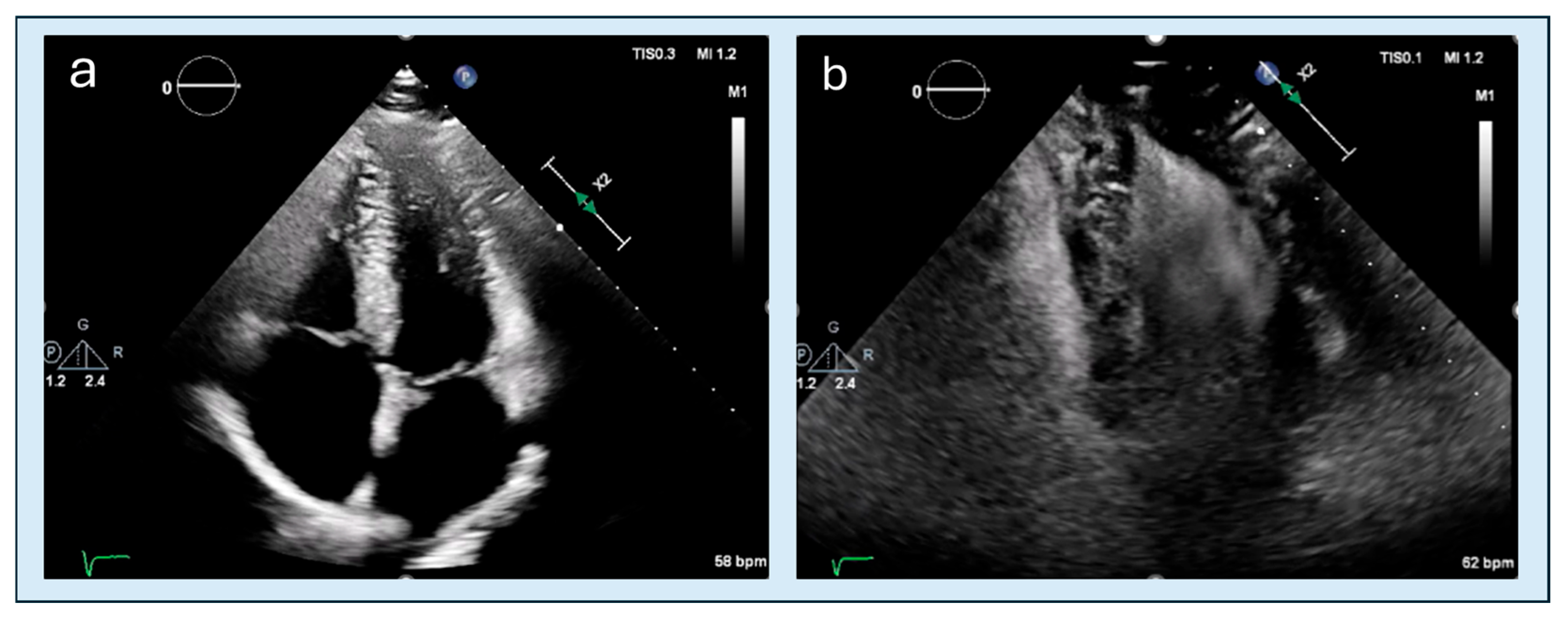
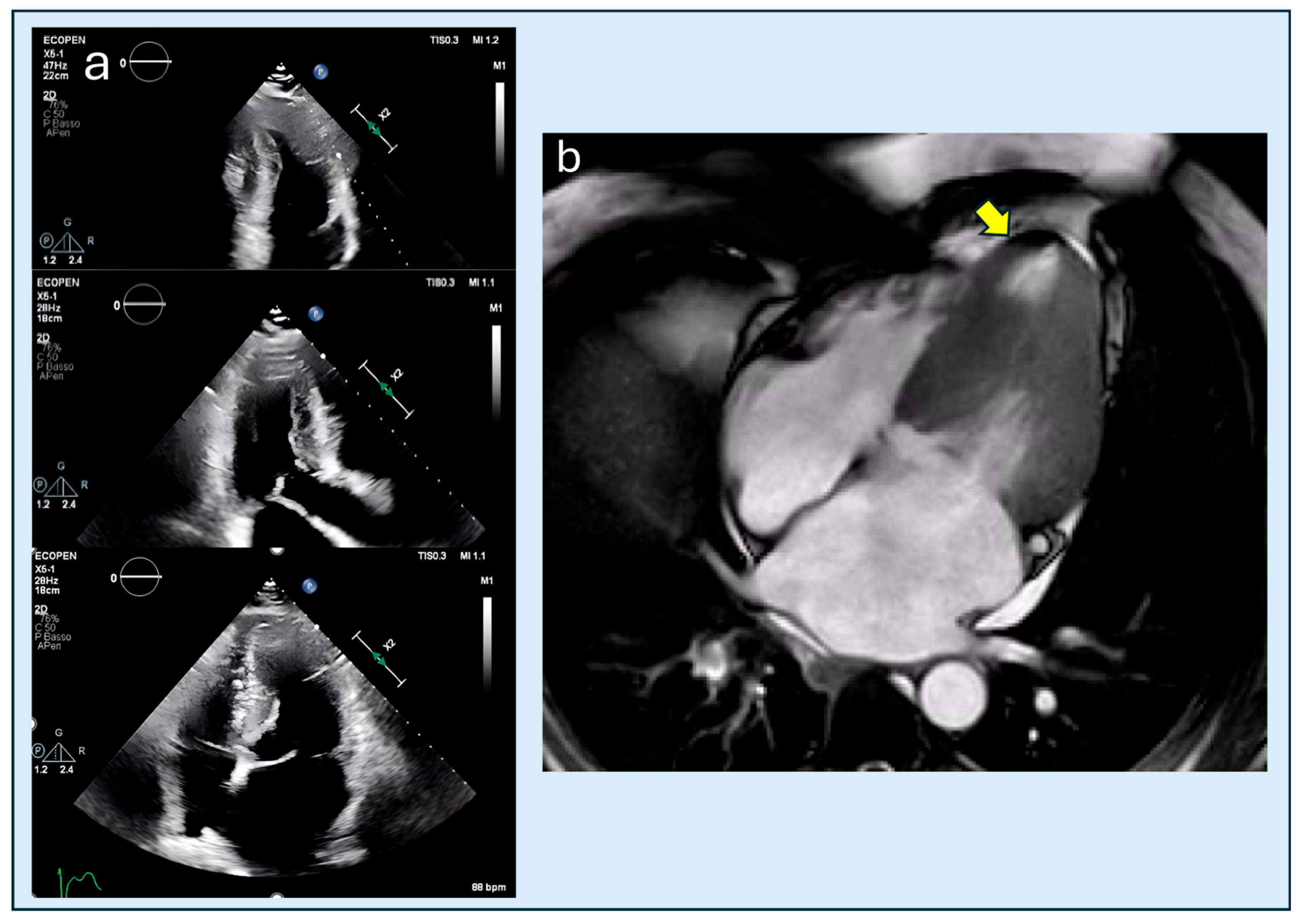
TTE is the first line imaging examination in most settings [1,6]. Its strength lies in availability and in dynamic assessment, i.e., the ability to measure gradients and capture physiological and hemodynamic phenomena in real-time; its limitations include intra/inter-operator variability, a tendency to underestimate volumes and overestimate EF compared to CMRi and other second-level methods, and a lower signal-to-noise ratio, which limits morphological evaluation in some contexts [24]. A general limitation of TTE in wall thickness assessment is the reduced signal-to-noise ratio at the blood–endocardium interface, compared to CMRi [7], which becomes particularly relevant at the apex; at this level, there is typically a greater trabecular expression and a relatively reduced cavity; these elements cause endocardial delineation to be frequently suboptimal, and wall thickness is systematically underestimated unless contrast is employed. Another limitation lies in the execution of the exam, where the most insidious error is the foreshortening of apical views: a window that is too high artificially shortens the ventricle, reduces volumes, alters the EF estimate, and, most importantly, can make the true apex ‘disappear,’ replacing it with a fictitious apex. The two possible consequences are either an overestimation of the wall thickness at the apex due to signal overlap or an underestimation of apical hypertrophy with distortion of strain measurements [29]. Hence, it is important to maximize the longitudinal extension of the LV in views 2, 3, and 4 chambers and to carefully check the probe orientation (Figure 1). The parasternal long axis remains the starting point for measuring septal and posterior wall thicknesses in diastole, with the beam perpendicular to the septum to avoid systematic errors [30]. Optimizing parameters is crucial: excessively high gain can generate artifacts in the cavity and simulate an apical obliteration that does not exist; too low gain can erase the endocardial profile precisely where it needs to be delineated better, namely at the apex margins. A balanced setting allows distinguishing a hypokinetic apex with residual cavity from a truly hypertrophic and obliterated apex in systole [31]. It is advisable to consider long-axis views, specifically the apical four-, two-, and three-chamber views as the primary reference for measuring left ventricular wall thickness, as they provide optimal alignment with the ventricular axis and a more reliable depiction of apical segment morphology. Whenever feasible, these measurements should be corroborated by short-axis views, which often allow clearer visualization of trabeculations and help distinguish true hypertrophy from trabecular interference. This combined approach ensures a more accurate and anatomically robust assessment of all left ventricular segments. When endocardial delineation is imperfect in two or more segments, the EACVI/ASE recommendations suggest using contrast media [32]. In ApHCM, contrast usually highlights the ‘spade’ profile clearly (Figure 2) and more generally allows detection of aneurysms and thrombi that unenhanced TTE might miss; a clinical-imaging review from 2025 reaffirmed that TTE, if not rigorously performed or without contrast, may fail to detect apical aneurysm in up to 43% of patients [20]. Transesophageal echocardiography, although useful in septal forms for studying the left ventricular outflow tract (LVOT) and the mechanism and grading of mitral regurgitation, generally has limited utility in the apical variant: the difficulty in visualizing the apex with TEE and the low frequency of mitral anomalies in pure forms reduce the clinical impact of this technique in ApHCM.

4.1. Obstruction

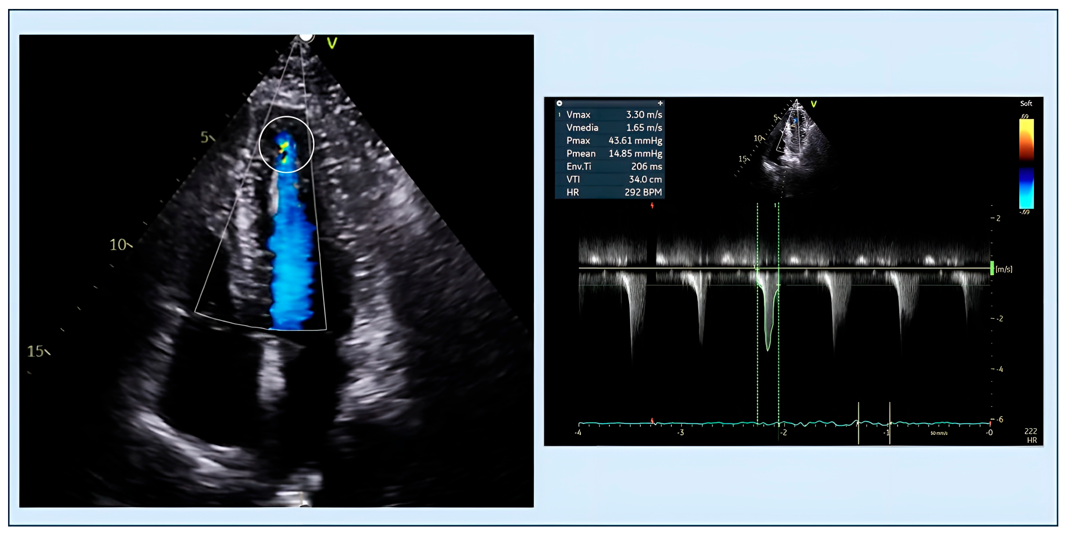
One of the strengths of echocardiography is real-time dynamic assessment. Provocative maneuvers such as the Valsalva maneuver, squat-to-stand, and even the simple post-prandial state can reveal latent dynamic gradients at the LVOT and, in ApHCM, especially in the mid-cavity region [33]. When obstruction is marked, continuous Doppler reveals a ‘mid-systolic signal-void,’ a black zone in the CW trace indicating transient cessation of flow during mid-systole, effectively indicating compartmentalization of the LV [34]. In rarer cases, obstruction may localize at the level of the purely apical cavity (Figure 3); since normally no gradients are expected at the apex, this finding should raise suspicion of ApHCM, regardless of the absolute wall thickness. Because in ApHCM the relevant hemodynamics occur away from the outflow, systematic investigation for mid-ventricular obstruction is crucial: aligning the beam along the cavity axis, ‘catching’ the apical jet, and quantifying the gradient are essential steps in a laboratory truly interested in this phenotype. A practical echocardiographic issue is distinguishing truly mid-ventricular obstruction (MVO) from mere changes in the CW Doppler configuration recorded at a similar mid-ventricular level in the presence of left ventricular outflow tract obstruction (LVOTO). In true mid-ventricular obstruction (this concept also applies to apical obstruction), two-dimensional imaging typically shows systolic “hour-glass” cavity narrowing at the mid-ventricular level, with color aliasing originating at this site and CW Doppler demonstrating a high-velocity jet (usually ≈2.5–4.0 m/s) and late-peaking or even double-peaked envelopes that may extend into isovolumic relaxation or early diastole, in keeping with a persistent apex-to-base intraventricular gradient [35]. By contrast, in LVOTO with only mid-ventricular flow configuration changes (for example due to trabeculation, apical remodeling, or cavity tapering), the principal obstruction and highest velocities are confined to the subaortic outflow tract, yielding the classic dagger-shaped late-systolic CW profile at the LVOT, whereas any mid-ventricular signal is of lower velocity, strictly systolic, and not associated with clear mid-cavity obliteration or diastolic apex-to-base flow reversal [36].

4.2. Apical Obliteration

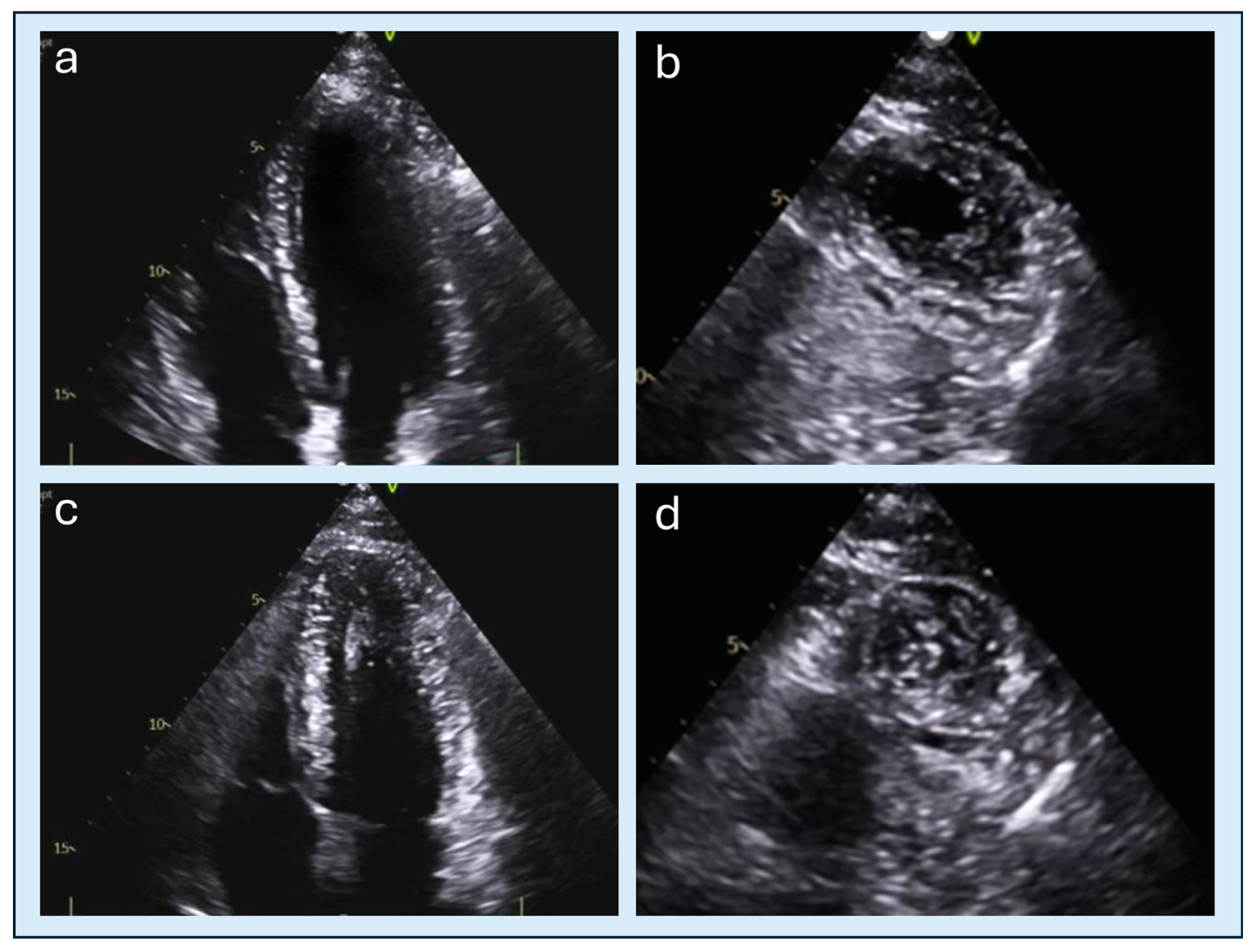
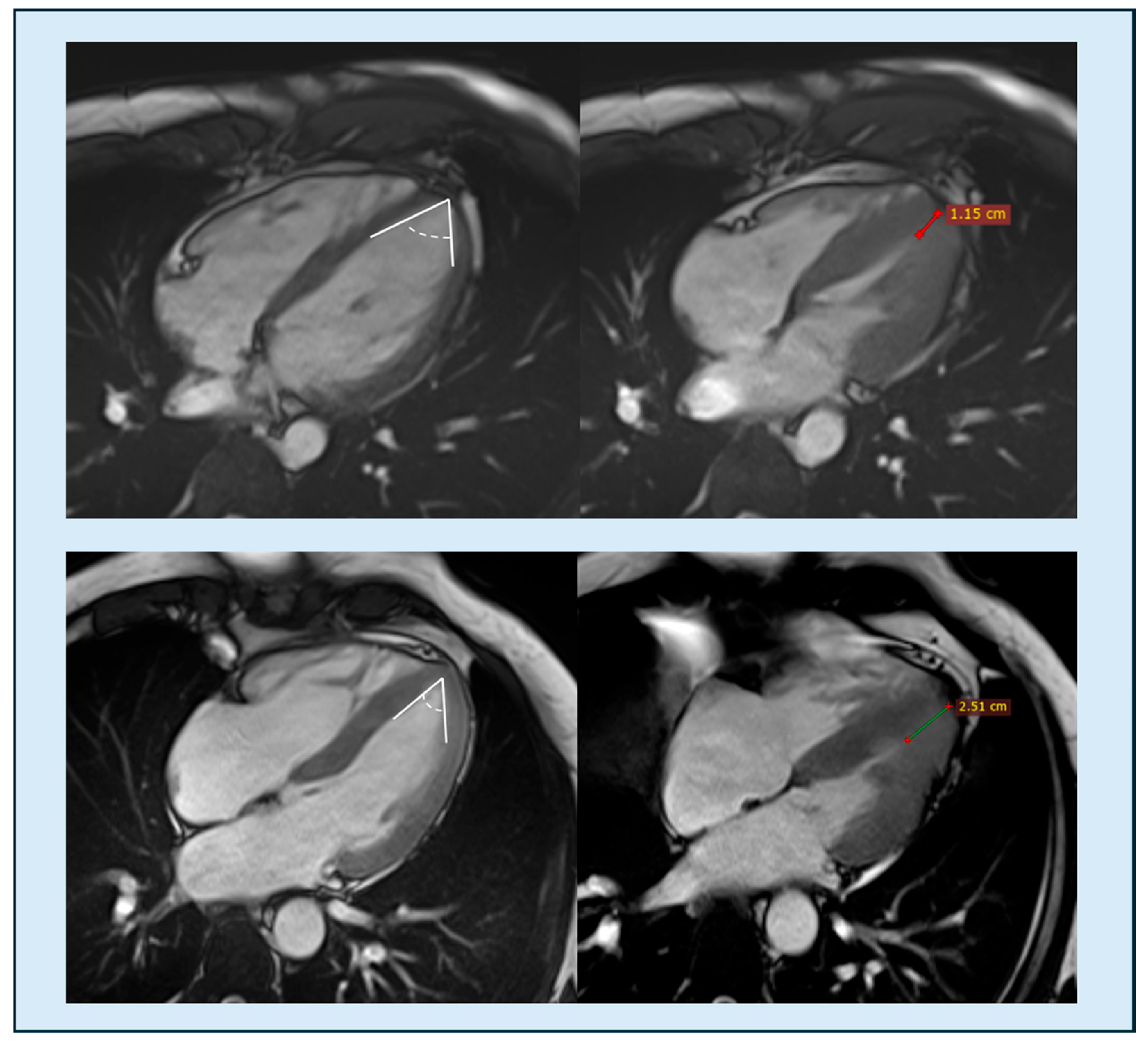
Apical obliteration represents one of the characteristic morphologic features of ApHCM and reflects the extreme systolic apposition of the apical walls, often accompanied by mid-ventricular cavity compartmentalization. The reference views for identifying apical obliteration are the apical long-axis views (Figure 1); however, performing a focused short-axis section on the apex can also be helpful in showing apical obliteration, especially when apical sections are insufficient (Figure 4). For many years, its evaluation remained purely qualitative, based on visual identification of complete systolic closure of the apical cavity. More recently, however, attempts have been made to quantify the extent of apical obliteration, introducing geometric ratios and standardized measurements (see the criterion described in Section 9). This quantitative approach allows a more reproducible assessment and may help identify patients at greater risk of adverse events associated with significant apical compartmentalization.

4.3. Speckle Tracking Imaging

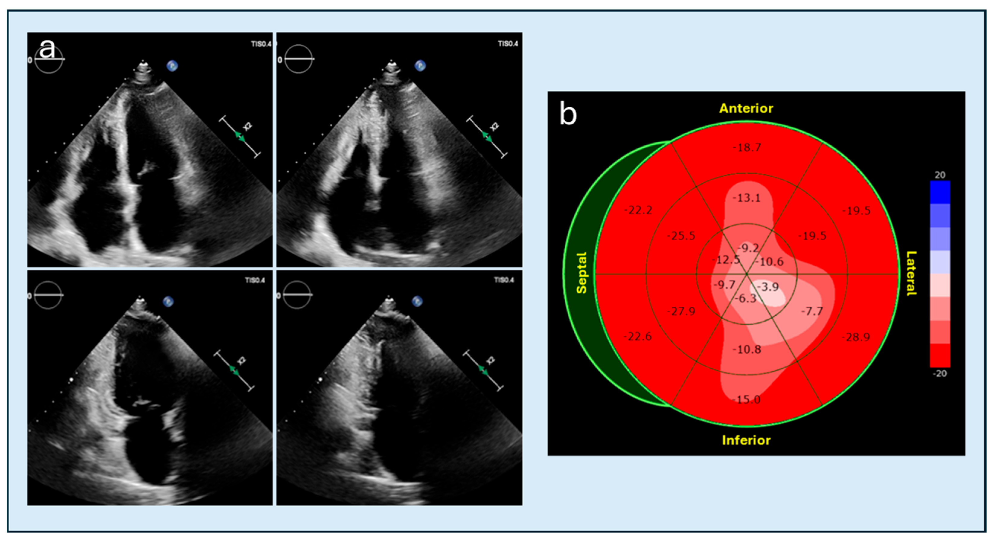
Global longitudinal strain (GLS) and segmental strain allow the detection of regional dysfunction even when the EF is normal. In ApHCM, the contraction of apical segments is frequently mechanically constrained by systolic apposition of the apical walls. This phenomenon results in a characteristic pattern of apical “shrinkage” on longitudinal strain analysis, with the bull’s-eye map demonstrating reduced apical strain values relative to the mid- and basal-ventricular segments [37] (Figure 5). Associated with this are alterations in apical rotation and global twist: the apex loses its traditional rotational dominance, and the baso-apical gradient flattens [18]. Myocardial work, integrating strain and blood pressure, promises to offer additional prognostic signals in HCM [38]. Finally, 3D echocardiography helps reduce inter-operator variability and provides more reliable volumetry, especially in apical regions, with automated models that quickly return measurements of mass, shape, volumes, and EF [39]. In this context, a 3D ‘mass dispersion index’ has been proposed as an indicator of hypertrophic asymmetry; it is an intriguing concept validated for HCM but still requiring specific validation for ApHCM [18].

4.4. Perfusion

Alongside hemodynamics, a ‘microcirculation’ perspective is emerging with contrast-enhanced perfusion imaging. Low-mechanical-index contrastography, at rest and under stress, can reveal segmental perfusion differences that often favor the apex in ApHCM [40]. This is an evolving field, but convergence with SPECT/PET and CMRi perfusion, which consistently indicate almost universal apical ischemia in the ApHCM phenotype, makes this line of investigation particularly promising [17,19].

5. Differential Diagnosis and Interpretative Pitfalls

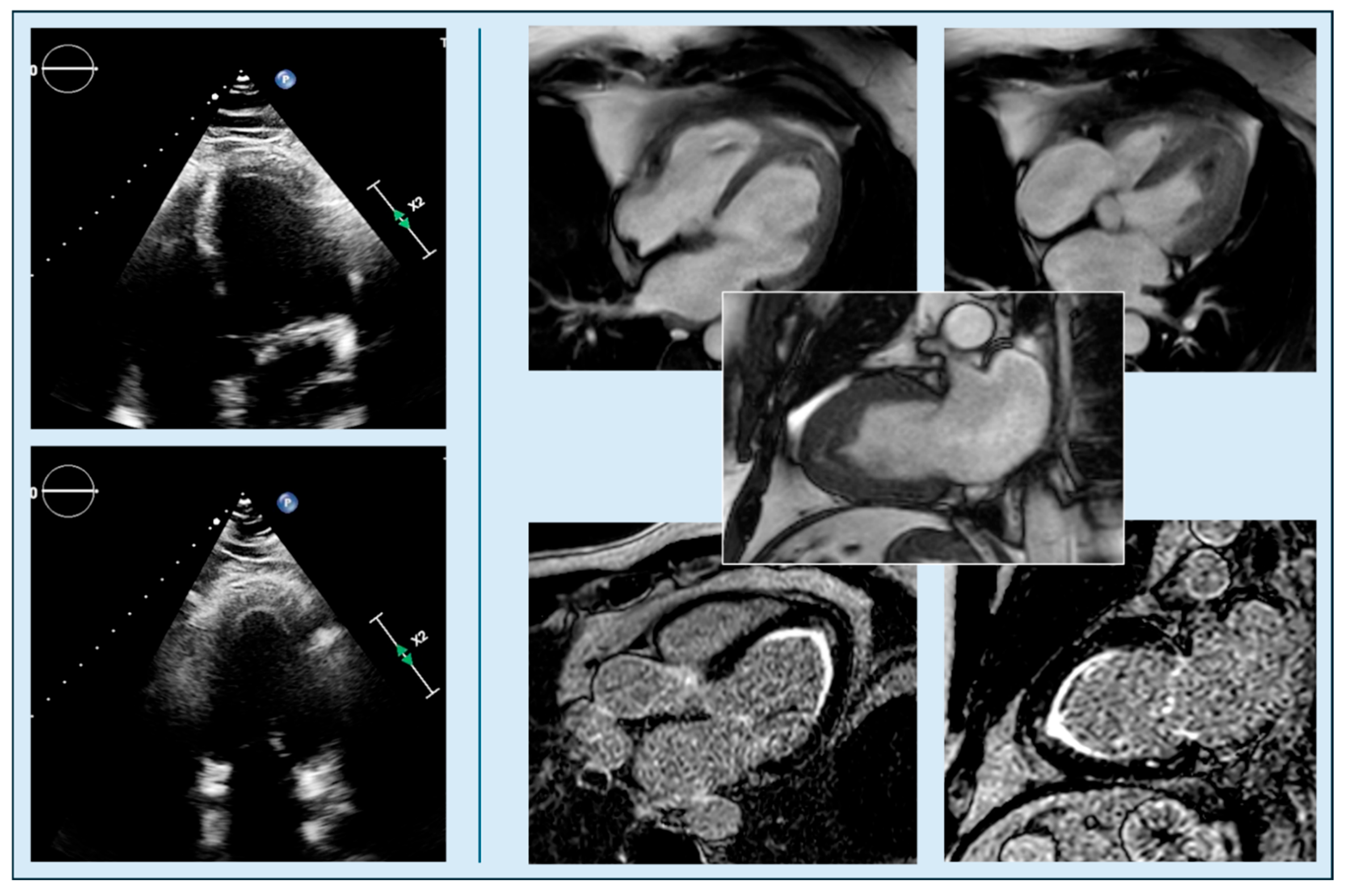
The first pitfall lies in the failure to recognize the disease itself, leading to underdiagnosis. When wall thickening is detected, a further challenge is represented by the potential difficulty in accurately determining the precise level from which hypertrophy originates. This limitation may hinder a reliable differential diagnosis between the pure-apical and distal-dominant forms of ApHCM. Furthermore, ApHCM shares numerous morphological and functional characteristics with other cardiac conditions that can make differential diagnosis complex, especially when hypertrophy is mild or confined to the apex. Major alternative diagnoses include physiological hypertrophy in athletes, secondary hypertrophy due to pressure or volume overload, non-sarcomeric forms, such as cardiac amyloidosis or Fabry disease. Another condition that can mimic ApHCM is eosinophilic myocarditis, which shares apical involvement with it. In the subacute phase, fibrosis and endocardial thickening produce a pseudo-hypertrophic echocardiographic appearance; CMRi allows differentiation between the two entities thanks to the presence of diffuse subendocardial Late Gadolinium Enhancement (LGE) and segmental edema, findings not typical of ApHCM (Figure 6). An additional diagnostic pitfall is the potential misdiagnosis of apical hypo-akinesia in patients with apical myocardial hypertrophy. The presence of hypertrophic segments contracting against themselves at the apex can cause mechanical limitation of cardiac movement, mimicking hypokinesia (Figure 5). The distinction between these two conditions is crucial: in ApHCM, the reduction in contractility is caused by mechanical limitation and is persistent, often associated with cavity obliteration while hypo-akinesia is usually associated with an increase in the volume of the apical cavity. Furthermore, conditions of absolute or relative hypovolemia and hyperdynamic states can simulate ApHCM due to the tendency for systolic obliteration, which is generally more evident at the apex in these conditions. To provide guidance in clinical scenarios where transthoracic echocardiographic findings prove insufficiently definitive, a modality-specific and progressive interpretative strategy can contribute to a more accurate diagnostic process. When image quality is limited by inadequate acoustic windows or by apical foreshortening, the first step is to repeat the acquisition with meticulous optimization of apical views. If uncertainty persists, contrast-enhanced TTE is particularly helpful because it improves endocardial border delineation and allows a more reliable reassessment of apical wall thickness, tapering and systolic obliteration. If contrast-enhanced TTE still does not clarify whether true apical hypertrophy is present, CMRi represents the subsequent investigation. CMRi is also valuable when apparent apical hypokinesia on TTE may instead reflect mechanical restriction from hypertrophic segments. Cine imaging and strain analysis can help distinguish constrained motion from true systolic impairment. When an infiltrative or inflammatory condition such as amyloidosis, Fabry disease or eosinophilic myocarditis is suspected, CMRi provides essential diagnostic information. In patients presenting with hypovolemia or hyperdynamic states, repeating TTE after hemodynamic stabilization is recommended, because physiological cavity reduction may accentuate apical obliteration and mimic pathological findings. In summary, a rigorous echocardiographic evaluation combined with the selective use of CMRi when uncertainty persists allows clinicians to navigate atypical or complex presentations more confidently and reduces the risk of interpretative pitfalls in the assessment of apical pathology. The subsequent section will address in detail the role and potential of CMRi in this context.

6. Cardiac Magnetic Resonance Imaging

In HCM, current ESC and AHA/ACC guidelines recommend CMRi at the initial stage of the diagnostic workup, when tissue characterization is required, particularly in the suspicion of non-sarcomeric forms, when transthoracic echocardiography is inconclusive, or when additional information on fibrosis, aneurysms, thrombi, apical morphology, or perfusion is needed [2,22].

6.1. Cine-MR and Morphology

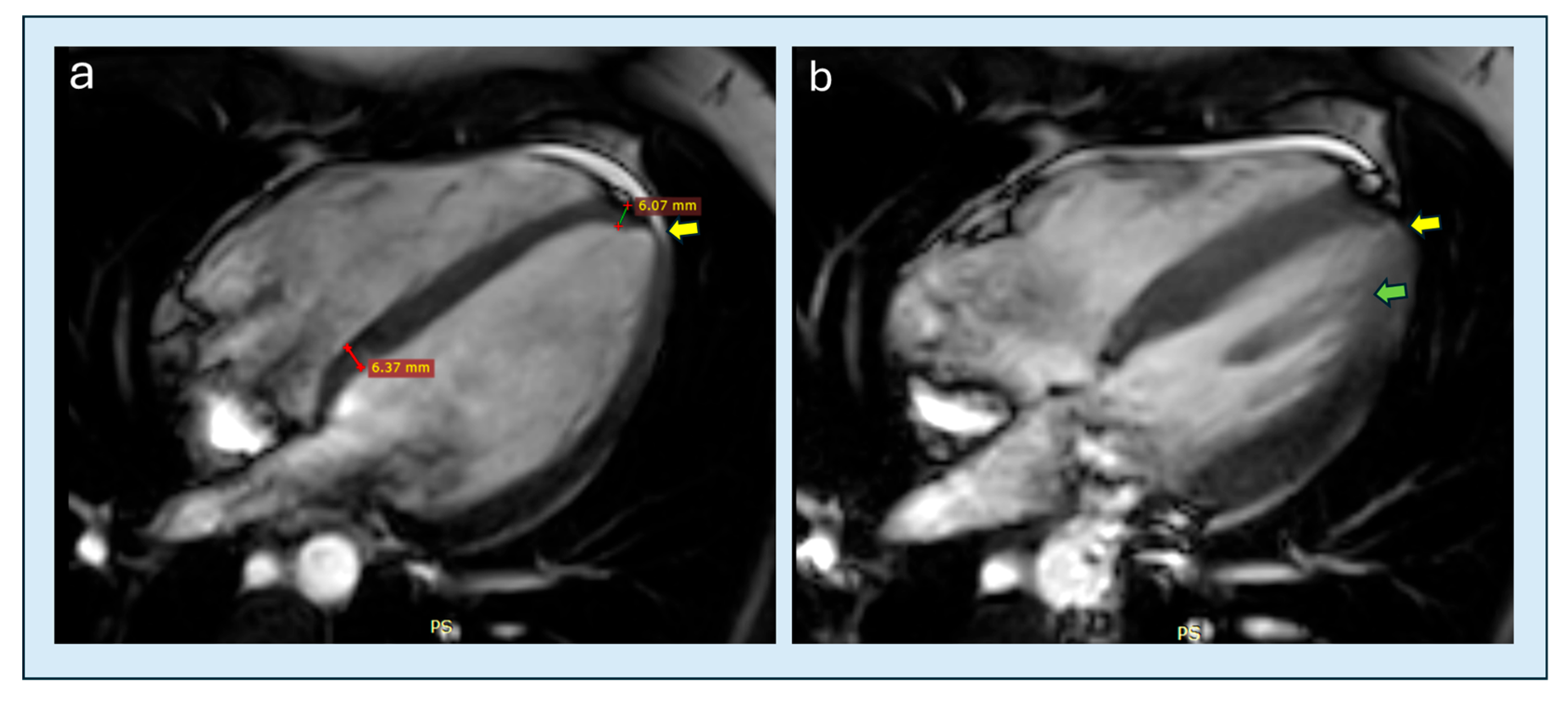
Sequences such as SSFP cine form the basis of morphological and functional assessment: the high signal-to-noise ratio and optimal blood-myocardium contrast allow accurate measurement of volumes, mass, and ventricular function, especially in difficult regions like the apex and lateral wall [41,42]. Compared to TTE, CMRi can uncover “occult” or limited hypertrophies, reliably recognize apical aneurysms (Figure 7), and describe atypical papillary-trabecular geometries (including crypts), all elements supporting diagnosis even when wall thickness does not reach traditional thresholds [9,10,42]. Furthermore, CMRi allows for accurate identification of the level at which apical thickening begins and extends distally and proximally consequently permitting a clearer distinction between pure-apical and distal-dominant variants of ApHCM [26].
In CMRi, obstruction can be identified: areas of high-velocity turbulence generate typical “dephasing” artifacts (signal void) on gradient-echo sequences, serving as visual indicators of obstruction, most often, but not exclusively, at the LVOT [43,44]. Quantitative analysis of the gradient remains complex due to the dynamic geometry of outflow and the limitations of provoking maneuvers within the scanner, which is why stress-dependent hemodynamic quantification remains the domain of TTE. Conversely, CMRi provides a robust assessment of mitral apparatus behavior, including SAM presence and quantification of mitral regurgitation through combined volumetric cine and phase-contrast flow mapping [45].

6.2. Feature Tracking and Strain

Myocardial tissue tracking on cine images adds important functional and prognostic information: early reductions in left ventricular and left atrial strain are detectable even when the ejection fraction is still preserved and have been shown to correlate with circulating markers of myocardial injury and wall stress, specifically high-sensitivity cardiac troponins and N-terminal pro–B-type natriuretic peptide. Moreover, alterations in diastolic strain rates have been associated with adverse clinical outcomes, including death from any cause, death related to heart failure, death related to stroke, and documented episodes of exertional or rest-related decompensation. Impaired circumferential strain has also been linked to an increased burden of ventricular arrhythmias, such as non-sustained and sustained ventricular tachycardia, as well as resuscitated cardiac arrest, while non-contrast tissue tracking parameters have emerged as potential indicators of underlying myocardial fibrosis. Advanced techniques, including myocardial tagging and three-dimensional approaches, further improve sensitivity for detecting subtle regional dysfunction and for characterizing remodeling after therapeutic interventions [46,47,48,49]. Advanced techniques like tagging and 3D approaches further extend sensitivity for regional function and post-treatment remodeling.

6.3. Tissue Characterization

6.3.1. T1 Mapping and ECV

T1 mapping and extracellular volume (ECV) quantify diffuse interstitial fibrosis and complement LGE, which identifies replacement fibrosis. Inversion recovery-based techniques (MOLLI, ShMOLLI) show high reproducibility and diagnostic sensitivity [50,51,52]. In HCM, native T1 and ECV increase with phenotypic severity, correlate with LV mass and wall thickness, and help differentiate sarcomeric HCM from hypertensive hypertrophy [49,50,51]. Their elevation reflects extracellular expansion from interstitial fibrosis and myocyte disarray. In ApHCM, evidence is more limited, though recent work confirms the diagnostic and prognostic value of parametric mapping without variant-specific cut-offs [53,54]. In mixed HCM cohorts, apical segments show higher native T1 and ECV than basal ones, consistent with greater microvascular vulnerability. T1 mapping also aids differential diagnosis from apical mimickers (e.g., Takotsubo syndrome, eosinophilic myocarditis), where normalized T1/ECV at follow-up indicates transient edema rather than fibrosis [55]. Reporting should include protocol, field strength, vendor, and local reference ranges following EACVI-SCMR recommendations [27,28].

6.3.2. T2 Mapping

T2 mapping quantifies myocardial edema and sensitively detects inflammatory or ischemic activity. In HCM, increased T2 occurs even in non-hypertrophied segments and is associated with reduced strain and systolic thickening [54]. In ApHCM, the pattern is typically apice-centric, reflecting chronic wall stress and distal microvascular dysfunction. Combined elevation of T2 and ECV identifies a transitional phase from reversible edema to emerging fibrosis, offering value for disease monitoring [19,56].
Across all mapping techniques, the apical phenotype presents inherent technical limitations related to increased susceptibility to motion artifacts, partial-volume contamination from trabeculations, and field inhomogeneities, and current evidence remains limited because relatively few mapping data have been specifically validated in apical forms.

6.3.3. Late Gadolinium Enhancement

LGE is the gold standard for assessing replacement fibrosis. After gadolinium administration, inversion-recovery sequences, preferably in PSIR mode (Phase-Sensitive Inversion Recovery), allow nulling of normal myocardium signal and visualization of fibrotic areas [57,58]. Optimal acquisitions involve complete short-axis coverage with voxel sizes around 1.5 mm and a post-contrast delay of 10–15 min. Dark-blood sequences improve endocardial definition and thrombus detection, while wide-band variants reduce artifacts in device carriers [41]. In HCM, replacement fibrosis is detected in more than half of patients, typically exhibiting a patchy mid-wall distribution within hypertrophied myocardial segments; however, other patterns have also been described such as subepicardial or diffuse mid-myocardial fibrosis, whereas a subendocardial pattern has been reported in association with ischemic imbalance due to microvascular dysfunction [18,57,59,60,61]; an extension ≥15% of LV mass strongly predicts ventricular arrhythmias and sudden death [59,60,61,62]. In ApHCM, LGE prevalence ranges from 60% to 80% [11,63]. The pattern is mainly apical or peri-apical, with possible extension to mid-distal septal or inferior wall segments (Figure 8). Yang et al. (2020) reported LGE in 87% of patients with ApHCM and apical aneurysm, compared to 52% without aneurysm; its extent correlated with ventricular arrhythmias and major cardiovascular events [11]. Li et al. (2021) [64] documented a 67% prevalence of LGE in a cohort of ApHCM, with an average extent of 4.9% of LV mass and an independent prognostic value for cardiovascular events when ≥5%. Studies also showed progressive increase in apical fibrotic burden during follow-up, associated with aneurysm development and increased arrhythmic risk [65]. Co-localization of LGE and perfusion defects on CMRI supports the pathophysiological model of microvascular ischemia leading to fibrosis leading to apical aneurysm [19].

6.4. Perfusion and Microvascularity

CMRi perfusion is the most sensitive method for detecting microvascular dysfunction, a key aspect of HCM. Rest perfusion defects correlate with mass and wall thickness, while adenosine stress reveals abnormalities in 40–50% of patients, often associated with extensive LGE and non-sustained ventricular tachycardia [66,67]. Myocardial Blood Flow (MBF) and Myocardial Perfusion Reserve (MPR) quantification shows reductions even in non-hypertrophied segments, suggesting microvascular disease may precede overt hypertrophy [68,69]. In ApHCM, microvascular dysfunction is nearly universal, with selective apical flow reduction and heterogeneous perfusion reflecting apico-basal pressure gradients and high apical fibrosis [19,68]. Combined analysis of perfusion, mapping, and LGE helps distinguish reversible ischemia from fibrosis, supporting prognosis and therapy.

Intracavitary Dynamics and 4D-Flow

4D-FLOW CMRi enables three-dimensional and temporal representation of intracavitary flow, quantifying vortices, pressure gradients, kinetic energy, and energy losses. In HCM, this technique has demonstrated flow inefficiencies even in the absence of outflow gradients, which correlate with wall deformation and fibrosis [70,71]. In ApHCM, the middle-apical compartmentalization and “funnel-shaped” geometry cause persistent turbulence and vortices, increasing wall stress and energy loss. These phenomena, also detectable with echocardiographic Vector Flow Mapping (VFM), contribute to understanding the relationship between morphology and hydrodynamic inefficiency [72].
The integration of 4D-FLOW, Feature Tracking, and tissue mapping represents the most promising approach for quantitative hemodynamic characterization of ApHCM and for identifying new risk biomarkers.

6.5. Practical Limitations and Technological Developments

CMRi is limited by acquisition times, availability, and costs while technical issues include claustrophobia and presence of non-conditional devices. New sequences such as PSIR, dark-blood, wide-band, and accelerated acquisitions with Compressed Sensing have improved sensitivity, resolution, and feasibility of LGE imaging [58]. The integration of Artificial Intelligence and Machine Learning in automatic segmentation, LGE quantification, and 4D-FLOW processing will potentially enable the development of phenotype-specific risk scores based on the integration of morphology, tissue, and function [72,73].

7. Diagnosis and Novel Diagnostic Criteria

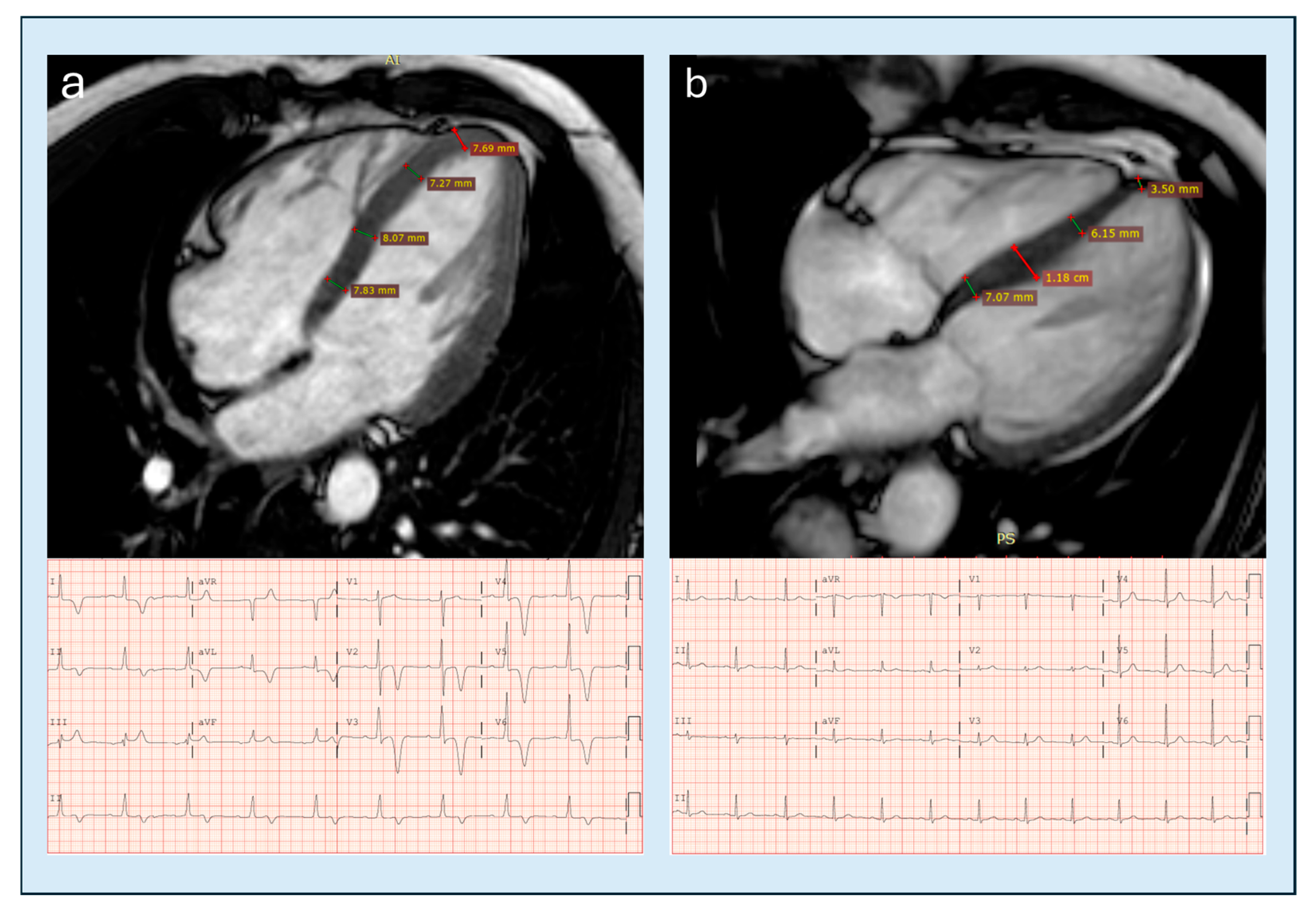
As also emphasized in the 2025 EACVI Clinical Consensus [9], classical diagnostic thresholds based solely on absolute apical wall thickness (≥15 mm) have limited sensitivity, particularly in early or “relative” ApHCM, where hypertrophy may be subtle or confined to small segments. Over the past decade, several apical-centric diagnostic criteria derived primarily from CMR have improved phenotype recognition and may provide conceptual models to refine echocardiographic assessment. Because these criteria originate from CMR cohorts, understanding their methodological background is essential to evaluate their potential transferability to TTE, which remains the first-line imaging modality. One of the most relevant developments is the body-surface-area–indexed apical thickness proposed by Hughes et al. [24], who demonstrated that an indexed threshold of 5.2–5.6 mm/m2 (≈11 mm unindexed) identifies almost all overt and most relative ApHCM cases while maintaining high specificity. Although this index has not yet been validated for echocardiography, it highlights how geometric normalization, widely used in CMR, may guide the future development of standardized TTE cut-offs. Similarly, apical angle measurement, introduced by Li et al. [64], has shown discriminatory value in distinguishing apical from non-apical forms (76° vs. 89°, p = 0.016) (Figure 9). Its use in echocardiography is conceptually feasible but limited by the risk of foreshortening, and dedicated acquisition protocols and validation studies are needed before routine application. Other CMR-derived geometric markers exhibit stronger alignment with echocardiographic assessment. The apical-to-basal wall thickness ratio, for example, is traditionally set at >1.3 and more recently considered diagnostic even when >1 [74], can often be evaluated on optimized apical TTE views, especially when contrast is used. The base-to-apex tapering pattern, well-illustrated by CMR [25], is also recognizable by echocardiography and helps distinguish true apical hypertrophy from hypertensive remodeling (Figure 10). Among the most promising indices is the indexed apical obliteration ratio, originally defined by Kim et al. [75] in a large echocardiographic cohort. A ratio >0.5 identifies significant apical compartmentalization and correlates with atrial fibrillation, stroke, heart failure and cardiovascular mortality. The absolute length of apical obliteration, with thresholds of >20 mm (high specificity) and >10 mm (overlap with hypertensive cardiopathy) [75], is also measurable with focused apical and short-axis TTE views, particularly when contrast is used (Figure 9). Recent literature has also identified apical papillary muscle displacement as a potential early morpho-functional precursor of ApHCM [76] (Figure 11). Although first described in CMR cohorts, this feature is frequently visible on high-quality TTE and may help identify individuals at risk before the development of overt hypertrophy. Despite the proliferation of apical-centric markers, none of these criteria has yet undergone sufficient large-scale, modality-specific validation, and their transferability to routine TTE varies widely. For this reason, it is essential to organize them within a structured framework that clarifies their level of evidence and practical applicability. Overall, these developments illustrate how CMR-derived geometric and morphologic insights can support a more nuanced and standardized echocardiographic approach to ApHCM. As validation studies expand and multimodality datasets grow, it is likely that selected CMR-derived criteria will progressively inform and refine TTE-based diagnosis, contributing to a more harmonized framework for apical phenotype identification. The 2025 EACVI Clinical Consensus [9] defines the diagnosis of ApHCM not through rigid cut-offs of individual measurements, but rather through a core set of qualitative imaging features such as loss of physiological basal-to-apical tapering, true apical hypertrophy, systolic apical cavity obliteration, mid-ventricular obstruction, apical aneurysm, and typical apical or periapical LGE patterns. Although papillary muscle displacement is not included as a formal diagnostic criterion, the consensus acknowledges apically displaced or morphologically abnormal papillary muscles as a recurrent anatomical feature of the apical phenotype, contributing to its distinctive geometry and potentially influencing distal flow disturbances or mid-cavity obstruction. The consensus does not yet incorporate the newer quantitative apical-centric indices proposed in recent CMR-based studies, such as indexed apical wall thickness, apical angle, and refined apical-to-basal ratios, which remain conceptually aligned with these descriptive features but still require dedicated validation before they can be integrated into formal diagnostic frameworks. Table 2 summarizes the main novel apical-centric diagnostic criteria proposed for ApHCM, including their proposed cut-offs, validation cohorts, strength of evidence, and estimated feasibility in real-world echocardiographic practice.

8. What Echocardiography Can Learn from CMRi and Future Perspectives

While CMRi remains the most accurate and comprehensive technique for defining ventricular morphology and tissue characterization, it also serves as the reference standard for research and for the development of new diagnostic criteria. Conversely, TTE represents the first-line imaging tool in clinical practice and therefore must be capable of raising diagnostic suspicion in the majority of cases. The domains in which TTE can derive substantial methodological and conceptual benefit from CMRi can be broadly articulated across different areas. The first domain is operational. The reason CMRi is considered the gold standard for morphological evaluation lies in its intrinsically high signal-to-noise ratio and excellent blood–myocardium contrast, which together provide superior spatial definition. Although such levels of contrast resolution are unlikely to be matched by echocardiography, significant improvement in accuracy can be made by optimizing image acquisition, selecting the best acoustic windows, adjusting parameters to obtain the clearest delineation of the endocardial border, and employing contrast agents to enhance endocardial delineation. The systematic optimization of echocardiographic settings and apical views can therefore minimize foreshortening, improve geometric accuracy, and increase the visibility of apical obliteration or aneurysmal remodeling. The second domain concerns the translation of novel CMR-derived criteria into echocardiographic practice. Even if direct cut-off values identified by magnetic resonance are not yet validated for echocardiography, these criteria can already be applied, at least for a visual/qualitative evaluation to improve morphological interpretation of the apex and guide diagnostic reasoning. Through this conceptual borrowing, echocardiography can progressively align its morphological assessment with that of magnetic resonance, enhancing consistency and diagnostic sensitivity. The third domain is prospective and research-oriented. Future comparative studies between TTE and CMRi may be helpful to evaluate and eventually confirm the concordance and reproducibility of emerging apical-centric indices and to validate their implementation within standardized echocardiographic protocols. A fourth domain lies in the study of intracavitary flow dynamics. The introduction of VFM and BSI now allows TTE to explore intracavitary hemodynamics in ways previously limited to CMRi. These techniques provide quantitative visualization of vortices, flow direction, and energy loss within the ventricular cavity, offering potential functional correlates to the geometric and morphological abnormalities typical of the apical variant. Integration of these approaches may enable the identification of specific flow signatures suggestive of disease even before the development of overt hypertrophy, thereby opening a new frontier in early diagnosis and pathophysiological understanding. The application of CMR-derived apical-centric criteria to TTE must be approached with caution, as none of these parameters have yet been validated through sufficient large-scale, modality-specific studies. Their integration into routine TTE therefore requires a graded and selective interpretation rather than direct adoption. As summarized in Table 2, different CMR-based indices show varying degrees of potential transferability to echocardiography. Some qualitative elements, such as apical obliteration and “spade” appearance, tend to demonstrate a closer alignment between the two modalities and may be more feasible to interpret on TTE, while further validation remains essential, especially regarding quantitative parameters. Other measures, including BSA-indexed apical thickness or the apical angle, appear promising but still require dedicated echocardiographic standardization before they can be reliably incorporated into clinical protocols. Overall, further studies are needed to validate the most promising apical-centric metrics within echocardiography and to assess their reproducibility across different ultrasound platforms and operators. The progressive integration of CMR-informed geometric concepts with echocardiographic strain analysis and emerging flow-based tools (VFM, BSI) may ultimately enhance early detection of apical remodeling and support more refined multimodality risk stratification in ApHCM.

9. Clinical Implications

Multimodality imaging in ApHCM has important implications for clinical management, beginning with the diagnostic phase. The combined use of echocardiography and CMRi helps differentiate sarcomeric HCM from infiltrative or metabolic phenocopies. This distinction is clinically relevant, since sarcomeric disease carries different patterns of progression and guides genetic counseling and family screening, while conditions such as amyloidosis or Fabry disease require specific therapies and benefit from early recognition. Imaging also contributes substantially to prognostic assessment. Echocardiography and CMRi together provide the variables needed for the ESC HCM SCD risk calculator, and CMRi refines risk stratification through the quantification of myocardial fibrosis. An LGE burden greater than 15% of LV mass is considered an important risk modifier that may influence ICD indication in selected patients [2,9]. The identification of an apical aneurysm, particularly when associated with extensive LGE, thrombus or significant systolic obliteration, further informs therapeutic decisions and often supports anticoagulation and closer rhythm monitoring [2]. More broadly, multimodality imaging assists in clarifying the presence and significance of ventricular obstruction, guiding individualized medical therapy and, when appropriate, referral to specialized centers. Once the diagnosis of HCM is established, imaging also supports family screening and genetic counseling across all phenotypic variants, enabling early detection and structured follow-up in accordance with current guideline recommendations.

10. Conclusions

ApHCM is a distinctive phenotype often underdiagnosed by TTE. CMRi remains the cornerstone of diagnosis, providing superior characterization of morphology, tissue, and perfusion. Beyond its diagnostic role, CMRi serves as a true reference platform from which TTE can learn to provide methodological rigor, quantitative morphology, and tissue-based insight that can be progressively adapted to echocardiographic practice. In fact, CMRi has driven the development of novel diagnostic criteria that hold promise for translation into echocardiographic use. By applying CMRi-derived principles such as apical-centric acquisition, optimized contrast use, and geometric indices, TTE can refine its ability to detect apical hypertrophy and improve diagnostic reproducibility. Although it may never reach the image quality of CMRi, echocardiography can evolve through this process of translational learning, turning research-derived parameters into clinically applicable tools. The integration of novel hemodynamic metrics from 4D-flow CMRi, VFM, and BSI further enhances this cross-modality synergy, consolidating a multimodal and physiologically informed approach to ApHCM where TTE becomes not only complementary to but conceptually enriched by CMRi.

Author Contributions

Conceptualization, F.M.; methodology, F.M.; resources, F.M., F.S. and R.C.; data curation, F.M., F.S. and M.G. (Massimo Grimaldi); validation, F.M., R.C., M.G. (Massimo Grimaldi), F.S., S.Q., S.D., G.C. (Grazia Casavecchia), G.C. (Gaetano Citarelli), M.G. (Matteo Gravina), A.D.M., V.B., L.S. and S.S.; investigation, F.M., F.S., V.B., L.S. and S.S.; writing—original draft preparation, F.M.; writing—review and editing, F.M., R.C., M.G. (Massimo Grimaldi) and R.W.W.B.; visualization, F.M. and E.F.; supervision, R.C.; project administration, F.M. All authors have read and agreed to the published version of the manuscript.

Funding

This research received no external funding.

Institutional Review Board Statement

Not applicable.

Informed Consent Statement

Not applicable.

Data Availability Statement

The examinations from which the images were taken are always available on the portal of the institute to which the corresponding author belongs.

Acknowledgments

All intellectual and scientific content was conceived, written, and verified by the authors. Artificial intelligence tools were used only for language refinement and grammatical correction.

Conflicts of Interest

The authors declare no conflicts of interest.

Abbreviations

The following abbreviations are used in this manuscript:
HCMhypertrophic cardiomyopathy
LVleft ventricle
ApHCMapical hypertrophic cardiomyopathy
TTEtransthoracic echocardiography
CMRicardiac magnetic resonance imaging
ECGelectrocardiogram
EFejection fraction
LVOTleft ventricular outflow tract
MVOmid-ventricular obstruction
LVOTOleft ventricular outflow tract obstruction
CTcomputed tomography
SPECTsingle-photon emission computed tomography
PETpositron emission tomography
EACVIEuropean Association of Cardiovascular Imaging
ASEAmerican Society of Echocardiography
TEEtransesophageal echocardiography
CWcontinuous-wave
GLSglobal longitudinal strain
3Dthree-dimensional
SSFPsteady-state free-precession
SAMsystolic anterior motion
LGElate gadolinium enhancement
ECVextracellular volume
PSIRphase-sensitive inversion recovery
MBFmyocardial blood flow
MPRmyocardial perfusion reserve
4D-FLOWfour-dimensional flow
VFMvector flow mapping
BSIblood speckle

References

  1. Maron, B.J.; Desai, M.Y.; Nishimura, R.A.; Spirito, P.; Rakowski, H.; Towbin, J.A.; Rowin, E.J.; Maron, M.S.; Sherrid, M.V. Diagnosis and Evaluation of Hypertrophic Cardiomyopathy: JACC State-of-the-Art Review. J. Am. Coll. Cardiol. 2022, 79, 372–389. [Google Scholar] [CrossRef]
  2. Arbelo, E.; Protonotarios, A.; Gimeno, J.R.; Arbustini, E.; Barriales-Villa, R.; Basso, C.; Bezzina, C.R.; Biagini, E.; Blom, N.A.; de Boer, R.A.; et al. 2023 ESC Guidelines for the management of cardiomyopathies. Eur. Heart J. 2023, 44, 3503–3626. [Google Scholar] [CrossRef]
  3. Coleman, J.A.; Ashkir, Z.; Raman, B.; Bueno-Orovio, A. Mechanisms and prognostic impact of myocardial ischaemia in hypertrophic cardiomyopathy. Int. J. Cardiovasc. Imaging 2023, 39, 1979–1996. [Google Scholar] [CrossRef]
  4. Aro, A.L.; Nair, S.G.; Reinier, K.; Jayaraman, R.; Stecker, E.C.; Uy-Evanado, A.; Rusinaru, C.; Jui, J.; Chugh, S.S. Population Burden of Sudden Death Associated with Hypertrophic Cardiomyopathy. Circulation 2017, 136, 1665–1667. [Google Scholar] [CrossRef] [PubMed]
  5. Dicorato, M.M.; Citarelli, G.; Mangini, F.; Alemanni, R.; Albanese, M.; Cicco, S.; Greco, C.A.; Forleo, C.; Basile, P.; Carella, M.C.; et al. Integrative Approaches in the Management of Hypertrophic Cardiomyopathy: A Comprehensive Review of Current Therapeutic Modalities. Biomedicines 2025, 13, 1256. [Google Scholar] [CrossRef] [PubMed]
  6. Guo, M.; Liu, C.; Ye, J.; Liu, J. Clinical characteristics and long-term outcomes in patients with apical hypertrophic cardiomyopathy. ESC Heart Fail. 2025, 12, 2887–2897. [Google Scholar] [CrossRef] [PubMed]
  7. Dentamaro, I.; Dicorato, M.M.; Falagario, A.; Cicco, S.; Dentamaro, S.; Correale, M.; Manuppelli, V.; Citarelli, G.; Mangini, F.; Fiore, C.; et al. The Evolving Role of Cardiac Imaging in Hypertrophic Cardiomyopathy: Diagnosis, Prognosis, and Clinical Practice. Biomedicines 2025, 13, 2138. [Google Scholar] [CrossRef]
  8. Yamaguchi, H.; Ishimura, T.; Nishiyama, S.; Nagasaki, F.; Nakanishi, S.; Takatsu, F.; Nishijo, T.; Umeda, T.; Machii, K. Hypertrophic nonobstructive cardiomyopathy with giant negative T waves (apical hypertrophy): Ventriculographic and echocardiographic features in 30 patients. Am. J. Cardiol. 1979, 44, 401–412. [Google Scholar] [CrossRef]
  9. Cardim, N.; Haugaa, K.; Mohiddin, S.A.; Hinojar, R.; Hirsch, A.; Szabo, L.; Podlesnikar, T.; Dall’ARmellina, E.; Cameli, M.; Mandoli, G.E.; et al. Role of multimodality cardiac imaging in the management of patients with hypertrophic cardiomyopathy in 2025. A Clinical Consensus Statement of the European Association of Cardiovascular Imaging (EACVI) of the ESC. Eur. Heart J. Cardiovasc. Imaging 2025, jeaf282. [Google Scholar] [CrossRef]
  10. Sakamoto, T. Apical hypertrophic cardiomyopathy (apical hypertrophy): An overview. J. Cardiol. 2001, 37 (Suppl. 1), 161–178. [Google Scholar]
  11. Yang, K.; Song, Y.Y.; Chen, X.Y.; Wang, J.X.; Li, L.; Yin, G.; Zheng, Y.C.; Wei, M.D.; Lu, M.J.; Zhao, S.H. Apical hypertrophic cardiomyopathy with left ventricular apical aneurysm: Prevalence, cardiac magnetic resonance characteristics, and prognosis. Eur. Heart J. Cardiovasc. Imaging 2020, 21, 1341–1350. [Google Scholar] [CrossRef]
  12. Rowin, E.J.; Maron, B.J.; Haas, T.S.; Garberich, R.F.; Wang, W.; Link, M.S.; Maron, M.S. Hypertrophic Cardiomyopathy with Left Ventricular Apical Aneurysm: Implications for Risk Stratification and Management. J. Am. Coll. Cardiol. 2017, 69, 761–773. [Google Scholar] [CrossRef]
  13. Kitaoka, H.; Doi, Y.; Casey, S.A.; Hitomi, N.; Furuno, T.; Maron, B.J. Comparison of prevalence of apical hypertrophic cardiomyopathy in Japan and the United States. Am. J. Cardiol. 2003, 92, 1183–1186. [Google Scholar] [CrossRef]
  14. Eriksson, M.J.; Sonnenberg, B.; Woo, A.; Rakowski, P.; Parker, T.G.; Wigle, E.; Rakowski, H. Long-term outcome in patients with apical hypertrophic cardiomyopathy. J. Am. Coll. Cardiol. 2002, 39, 638–645. [Google Scholar] [CrossRef] [PubMed]
  15. Paluszkiewicz, J.; Krasinska, B.; Milting, H.; Gummert, J.; Pyda, M. Apical hypertrophic cardiomyopathy: Diagnosis, medical and surgical treatment. Kardiochir. Torakochirurgia Pol. 2018, 15, 246–253. [Google Scholar] [CrossRef] [PubMed]
  16. Sherrid, M.V.; Bernard, S.; Tripathi, N.; Patel, Y.; Modi, V.; Axel, L.; Talebi, S.; Ghoshhajra, B.B.; Sanborn, D.Y.; Saric, M.; et al. Apical Aneurysms and Mid-Left Ventricular Obstruction in Hypertrophic Cardiomyopathy. JACC Cardiovasc. Imaging 2023, 16, 591–605. [Google Scholar] [CrossRef] [PubMed]
  17. Lee, D.Z.J.; Montazeri, M.; Bataiosu, R.; Hoss, S.; Adler, A.; Nguyen, E.T.; Rakowski, H.; Chan, R.H. Clinical Characteristics and Prognostic Importance of Left Ventricular Apical Aneurysms in Hypertrophic Cardiomyopathy. JACC Cardiovasc. Imaging 2022, 15, 1696–1711. [Google Scholar] [CrossRef] [PubMed]
  18. Hoey, E.T.; Teoh, J.K.; Das, I.; Ganeshan, A.; Simpson, H.; Watkin, R.W. The emerging role of cardiovascular MRI for risk stratification in hypertrophic cardiomyopathy. Clin. Radiol. 2014, 69, 221–230. [Google Scholar] [CrossRef]
  19. Hughes, R.K.; Augusto, J.B.; Knott, K.; Davies, R.; Shiwani, H.; Seraphim, A.; Malcolmson, J.W.; Khoury, S.; Joy, G.; Mohiddin, S.; et al. Apical Ischemia Is a Universal Feature of Apical Hypertrophic Cardiomyopathy. Circ. Cardiovasc. Imaging 2023, 16, e014907. [Google Scholar] [CrossRef]
  20. Hamza, I.; Odigie-Okon, E.; Xie, T.; Ahmad, M. Apical Hypertrophic Cardiomyopathy: A Clinical & Multimodality Imaging Assessment. Echocardiography 2025, 42, e70235. [Google Scholar] [CrossRef]
  21. Pradeep Kundur, S.; Malik, A.; Sivalokanathan, S. The Utility of Nuclear Imaging in Hypertrophic Cardiomyopathy: A Narrative Review. J. Clin. Med. 2025, 14, 2183. [Google Scholar] [CrossRef]
  22. Ommen, S.R.; Mital, S.; Burke, M.A.; Day, S.M.; Deswal, A.; Elliott, P.; Evanovich, L.L.; Hung, J.; Joglar, J.A. 2020 AHA/ACC Guideline for the Diagnosis and Treatment of Patients with Hypertrophic Cardiomyopathy: A Report of the American College of Cardiology/American Heart Association Joint Committee on Clinical Practice Guidelines. Circulation 2020, 142, e558–e631. [Google Scholar] [CrossRef]
  23. Hughes, R.K.; Knott, K.D.; Malcolmson, J.; Augusto, J.B.; Mohiddin, S.A.; Kellman, P.; Moon, J.C.; Captur, G. Apical Hypertrophic Cardiomyopathy: The Variant Less Known. J. Am. Heart Assoc. 2020, 9, e015294. [Google Scholar] [CrossRef] [PubMed]
  24. Grothues, F.; Smith, G.C.; Moon, J.C.; Bellenger, N.G.; Collins, P.; Klein, H.U.; Pennell, D.J. Comparison of interstudy reproducibility of cardiovascular magnetic resonance with two-dimensional echocardiography in normal subjects and in patients with heart failure or left ventricular hypertrophy. Am. J. Cardiol. 2002, 90, 29–34. [Google Scholar] [CrossRef] [PubMed]
  25. Hughes, R.K.; Shiwani, H.; Rosmini, S.; Augusto, J.B.; Burke, L.; Jiang, Y.; Pierce, I.; Joy, G.; Castelletti, S.; Orini, M.; et al. Improved Diagnostic Criteria for Apical Hypertrophic Cardiomyopathy. JACC Cardiovasc. Imaging 2024, 17, 501–512. [Google Scholar] [CrossRef] [PubMed]
  26. Sugiura, K.; Kubo, T.; Inoue, S.; Nomura, S.; Yamada, T.; Tobita, T.; Kuramoto, Y.; Miyashita, Y.; Asano, Y.; Ochi, Y.; et al. Unveiling Clinical and Genetic Distinctions in Pure-Apical Versus Distal-Dominant Apical Hypertrophic Cardiomyopathy. J. Am. Heart Assoc. 2025, 14, e038208. [Google Scholar] [CrossRef]
  27. Schulz-Menger, J.; Bluemke, D.A.; Bremerich, J.; Flamm, S.D.; Fogel, M.A.; Friedrich, M.G.; Kim, R.J.; von Knobelsdorff-Brenkenhoff, F.; Kramer, C.M.; Pennell, D.J.; et al. Standardized image interpretation and post-processing in cardiovascular magnetic resonance—2020 update: Society for Cardiovascular Magnetic Resonance (SCMR): Board of Trustees Task Force on Standardized Post-Processing. J. Cardiovasc. Magn. Reson. 2020, 22, 19. [Google Scholar] [CrossRef]
  28. Kramer, C.M.; Barkhausen, J.; Bucciarelli-Ducci, C.; Flamm, S.D.; Kim, R.J.; Nagel, E. Standardized cardiovascular magnetic resonance imaging (CMR) protocols: 2020 update. J. Cardiovasc. Magn. Reson. 2020, 22, 17. [Google Scholar] [CrossRef]
  29. Voigt, J.U.; Pedrizzetti, G.; Lysyansky, P.; Marwick, T.H.; Houle, H.; Baumann, R.; Pedri, S.; Ito, Y.; Abe, Y.; Metz, S.; et al. Definitions for a common standard for 2D speckle tracking echocardiography: Consensus document of the EACVI/ASE/Industry Task Force to standardize deformation imaging. Eur. Heart J. Cardiovasc. Imaging 2015, 16, 1–11. [Google Scholar] [CrossRef]
  30. Lang, R.M.; Badano, L.P.; Mor-Avi, V.; Afilalo, J.; Armstrong, A.; Ernande, L.; Flachskampf, F.A.; Foster, E.; Goldstein, S.A.; Kuznetsova, T.; et al. Recommendations for cardiac chamber quantification by echocardiography in adults: An update from the American Society of Echocardiography and the European Association of Cardiovascular Imaging. Eur. Heart J. Cardiovasc. Imaging 2015, 16, 233–270. [Google Scholar] [CrossRef]
  31. Porter, T.R.; Mulvagh, S.L.; Abdelmoneim, S.S.; Becher, H.; Belcik, J.T.; Bierig, M.; Choy, J.; Gaibazzi, N.; Gillam, L.D.; Janardhanan, R.; et al. Clinical Applications of Ultrasonic Enhancing Agents in Echocardiography: 2018 American Society of Echocardiography Guidelines Update. J. Am. Soc. Echocardiogr. 2018, 31, 241–274. [Google Scholar] [CrossRef] [PubMed]
  32. Porter, T.R.; Abdelmoneim, S.; Belcik, J.T.; McCulloch, M.L.; Mulvagh, S.L.; Olson, J.J.; Porcelli, C.; Tsutsui, J.M.; Wei, K. Guidelines for the cardiac sonographer in the performance of contrast echocardiography: A focused update from the American Society of Echocardiography. J. Am. Soc. Echocardiogr. 2014, 27, 797–810. [Google Scholar] [CrossRef] [PubMed]
  33. Saccheri, M.C.; Cianciulli, T.F.; Morita, L.A.; Méndez, R.J.; Beck, M.A.; Guerra, J.E.; Cozzarin, A.; Puente, L.J.; Balletti, L.R.; Lax, J.A. Speckle tracking echocardiography to assess regional ventricular function in patients with apical hypertrophic cardiomyopathy. World J. Cardiol. 2017, 9, 363–370. [Google Scholar] [CrossRef] [PubMed]
  34. Po, J.R.; Kim, B.; Aslam, F.; Arabadjian, M.; Winson, G.; Cantales, D.; Kushner, J.; Kornberg, R.; Sherrid, M.V. Doppler Systolic Signal Void in Hypertrophic Cardiomyopathy: Apical Aneurysm and Severe Obstruction without Elevated Intraventricular Velocities. J. Am. Soc. Echocardiogr. 2015, 28, 1462–1473. [Google Scholar] [CrossRef]
  35. Nakamura, T.; Kitamura, H.; Furukawa, K.; Matsubara, K.; Katahira, T.; Okamuro, S.; Tsuji, Y.; Takahashi, T.; Kunishige, H.; Katsume, H. Intraventricular flow dynamics in hypertrophic cardiomyopathy with midventricular obstruction investigated by Doppler echocardiography. J. Cardiol. 1989, 19, 455–471. [Google Scholar]
  36. Rakowski, H.; Sasson, Z.; Wigle, E.D. Echocardiographic and Doppler assessment of hypertrophic cardiomyopathy. J. Am. Soc. Echocardiogr. 1988, 1, 31–47. [Google Scholar] [CrossRef]
  37. Mihos, C.G.; Horvath, S.A.; Fernandez, R.; Escolar, E. Left ventricular strain and myocardial work in apical hypertrophic cardiomyopathy. J. Thorac. Dis. 2023, 15, 3197–3207. [Google Scholar] [CrossRef]
  38. Hiemstra, Y.L.; van der Bijl, P.; El Mahdiui, M.; Bax, J.J.; Delgado, V.; Marsan, N.A. Myocardial Work in Nonobstructive Hypertrophic Cardiomyopathy: Implications for Outcome. J. Am. Soc. Echocardiogr. 2020, 33, 1201–1208. [Google Scholar] [CrossRef]
  39. Hoffmann, R.; von Bardeleben, S.; Cate, F.T.; Borges, A.C.; Kasprzak, J.; Firschke, C.; Lafitte, S.; Al-Saadi, N.; Kuntz-Hehner, S.; Engelhardt, M.; et al. Assessment of systolic left ventricular function: A multi-centre comparison of cineventriculography, cardiac magnetic resonance imaging, unenhanced and contrast-enhanced echocardiography. Eur. Heart J. 2005, 26, 607–616. [Google Scholar] [CrossRef]
  40. Pathan, F.; Marwick, T.H. Myocardial perfusion imaging using contrast echocardiography. Prog. Cardiovasc. Dis. 2015, 57, 632–643. [Google Scholar] [CrossRef]
  41. White, R.D.; Obuchowski, N.A.; Gunawardena, S.; Lipchik, E.O.; Lever, H.M.; Van Dyke, C.W.; Lytle, B.W. Left ventricular outflow tract obstruction in hypertrophic cardiomyopathy: Presurgical and postsurgical evaluation by computed tomography magnetic resonance imaging. Am. J. Card. Imaging 1996, 10, 1–13. [Google Scholar] [PubMed]
  42. Chu, L.C.; Porter, K.K.; Corona-Villalobos, C.P.; Gülsün, M.A.; Shea, S.M.; Markl, M.; Abraham, T.P.; Bluemke, D.A.; Kamel, I.R.; Zimmerman, S.L. Evaluation of Left Ventricular Outflow Tract Obstruction with Four-Dimensional Phase Contrast Magnetic Resonance Imaging in Patients with Hypertrophic Cardiomyopathy-A Pilot Study. J. Comput. Assist. Tomogr. 2016, 40, 937–940. [Google Scholar] [CrossRef] [PubMed]
  43. Hansen, M.W.; Merchant, N. MRI of hypertrophic cardiomyopathy: Part I, MRI appearances. Am. J. Roentgenol. 2007, 189, 1335–1343. [Google Scholar] [CrossRef]
  44. Vasquez, N.; Ostrander, B.T.; Lu, D.Y.; Ventoulis, I.; Haileselassie, B.; Goyal, S.; Greenland, G.V.; Vakrou, S.; Olgin, J.E.; Abraham, T.P.; et al. Low Left Atrial Strain Is Associated with Adverse Outcomes in Hypertrophic Cardiomyopathy Patients. J. Am. Soc. Echocardiogr. 2019, 32, 593–603.e1. [Google Scholar] [CrossRef]
  45. Mangini, F.; Scarcia, M.; Biederman, R.W.W.; Calbi, R.; Spinelli, F.; Casavecchia, G.; Brunetti, N.D.; Gravina, M.; Fiore, C.; Suma, S.; et al. Cardiac magnetic resonance imaging in the evaluation and management of mitral valve prolapse—A comprehensive review. Echocardiography 2024, 41, e15894. [Google Scholar] [CrossRef]
  46. Cavus, E.; Muellerleile, K.; Schellert, S.; Schneider, J.; Tahir, E.; Chevalier, C.; Jahnke, C.; Radunski, U.K.; Adam, G.; Kirchhof, P.; et al. CMR feature tracking strain patterns and their association with circulating cardiac biomarkers in patients with hypertrophic cardiomyopathy. Clin. Res. Cardiol. 2021, 110, 1757–1769. [Google Scholar] [CrossRef]
  47. Li, Z.L.; He, S.; Xia, C.C.; Peng, W.L.; Li, L.; Liu, K.L.; Zhang, J.G.; Pu, J.; Guo, Y.K. Global longitudinal diastolic strain rate as a novel marker for predicting adverse outcomes in hypertrophic cardiomyopathy by cardiac magnetic resonance tissue tracking. Clin. Radiol. 2021, 76, e19–e78. [Google Scholar] [CrossRef]
  48. Pu, C.; Fei, J.; Lv, S.; Wu, Y.; He, C.; Guo, D.; Mabombo, P.U.; Chooah, O.; Hu, H. Global Circumferential Strain by Cardiac Magnetic Resonance Tissue Tracking Associated with Ventricular Arrhythmias in Hypertrophic Cardiomyopathy Patients. Front. Cardiovasc. Med. 2021, 8, 670361. [Google Scholar] [CrossRef]
  49. Sharifian, M.; Rezaeian, N.; Asadian, S.; Mohammadzadeh, A.; Nahardani, A.; Kasani, K.; Toloueitabar, Y.; Farahmand, A.M.; Hosseini, L. Efficacy of Novel Noncontrast Cardiac Magnetic Resonance Methods in Indicating Fibrosis in Hypertrophic Cardiomyopathy. Cardiol. Res. Pract. 2021, 2021, 9931136. [Google Scholar] [CrossRef]
  50. Nakamori, S.; Dohi, K.; Ishida, M.; Goto, Y.; Imanaka-Yoshida, K.; Omori, T.; Goto, I.; Kumagai, N.; Fujimoto, N.; Ichikawa, Y.; et al. Native T1 Mapping and Extracellular Volume Mapping for the Assessment of Diffuse Myocardial Fibrosis in Dilated Cardiomyopathy. JACC Cardiovasc. Imaging 2018, 11, 48–59. [Google Scholar] [CrossRef]
  51. Gravina, M.; Casavecchia, G.; Mangini, F.; Mautone, F.; Ruggeri, D.; Guglielmi, G.; Macarini, L.; Brunetti, N.D. Magnetic resonance mapping for the assessment of cardiomyopathies and myocardial disease. Int. J. Cardiol. 2024, 415, 132440. [Google Scholar] [CrossRef]
  52. Forleo, C.; Carella, M.C.; Basile, P.; Mandunzio, D.; Greco, G.; Napoli, G.; Carulli, E.; Dicorato, M.M.; Dentamaro, I.; Santobuono, V.E.; et al. The Role of Magnetic Resonance Imaging in Cardiomyopathies in the Light of New Guidelines: A Focus on Tissue Mapping. J. Clin. Med. 2024, 13, 2621. [Google Scholar] [CrossRef]
  53. Xu, J.; Zhuang, B.; Sirajuddin, A.; Li, S.; Huang, J.; Yin, G.; Song, L.; Jiang, Y.; Zhao, S.; Lu, M. MRI T1 Mapping in Hypertrophic Cardiomyopathy: Evaluation in Patients Without Late Gadolinium Enhancement and Hemodynamic Obstruction. Radiology 2020, 294, 275–286. [Google Scholar] [CrossRef] [PubMed]
  54. Hughes, R.K.; Knott, K.D.; Malcolmson, J.; Augusto, J.B.; Kellman, P.; Moon, J.C.; Captur, G. Advanced Imaging Insights in Apical Hypertrophic Cardiomyopathy. JACC Cardiovasc. Imaging 2020, 13 Pt 2, 624–630. [Google Scholar] [CrossRef]
  55. Maurizi, N.; Antiochos, P.; Monney, P. Takotsubo cardiomyopathy mimicking an apical hypertrophic cardiomyopathy. Int. J. Cardiovasc. Imaging 2024, 40, 1389–1391. [Google Scholar] [CrossRef]
  56. Huang, L.; Ran, L.; Zhao, P.; Tang, D.; Han, R.; Ai, T.; Xia, L.; Tao, Q. MRI native T1 and T2 mapping of myocardial segments in hypertrophic cardiomyopathy: Tissue remodeling manifested prior to structure changes. Br. J. Radiol. 2019, 92, 20190634. [Google Scholar] [CrossRef] [PubMed]
  57. Kellman, P.; Arai, A.E.; McVeigh, E.R.; Aletras, A.H. Phase-sensitive inversion recovery for detecting myocardial infarction using gadolinium-delayed hyperenhancement. Magn. Reson. Med. 2002, 47, 372–383. [Google Scholar] [CrossRef] [PubMed]
  58. Holtackers, R.J.; Van De Heyning, C.M.; Chiribiri, A.; Wildberger, J.E.; Botnar, R.M.; Kooi, M.E. Dark-blood late gadolinium enhancement cardiovascular magnetic resonance for improved detection of subendocardial scar: A review of current techniques. J. Cardiovasc. Magn. Reson. 2021, 23, 96. [Google Scholar] [CrossRef]
  59. Basile, P.; Soldato, N.; Pedio, E.; Siena, P.; Carella, M.C.; Dentamaro, I.; Khan, Y.; Baggiano, A.; Mushtaq, S.; Forleo, C.; et al. Cardiac magnetic resonance reveals concealed structural heart disease in patients with frequent premature ventricular contractions and normal echocardiography: A systematic review. Int. J. Cardiol. 2024, 412, 132306. [Google Scholar] [CrossRef]
  60. Liu, J.; Zhao, S.; Yu, S.; Wu, G.; Wang, D.; Liu, L.; Song, J.; Zhu, Y.; Kang, L.; Wang, J.; et al. Patterns of Replacement Fibrosis in Hypertrophic Cardiomyopathy. Radiology 2022, 302, 298–306. [Google Scholar] [CrossRef]
  61. Chan, R.H.; Maron, B.J.; Olivotto, I.; Pencina, M.J.; Assenza, G.E.; Haas, T.; Lesser, J.R.; Gruner, C.; Crean, A.M.; Rakowski, H.; et al. Prognostic value of quantitative contrast-enhanced cardiovascular magnetic resonance for the evaluation of sudden death risk in patients with hypertrophic cardiomyopathy. Circulation 2014, 130, 484–495. [Google Scholar] [CrossRef]
  62. Dicorato, M.M.; Basile, P.; Naccarati, M.L.; Carella, M.C.; Dentamaro, I.; Falagario, A.; Cicco, S.; Forleo, C.; Guaricci, A.I.; Ciccone, M.M.; et al. Predicting New-Onset Atrial Fibrillation in Hypertrophic Cardiomyopathy: A Review. J. Clin. Med. 2025, 14, 2018. [Google Scholar] [CrossRef]
  63. Wang, J.; Yang, S.; Ma, X.; Zhao, K.; Yang, K.; Yu, S.; Yin, G.; Dong, Z.; Song, Y.; Cui, C.; et al. Assessment of late gadolinium enhancement in hypertrophic cardiomyopathy improves risk stratification based on current guidelines. Eur. Heart J. 2023, 44, 4781–4792. [Google Scholar] [CrossRef] [PubMed]
  64. Li, S.; He, J.; Xu, J.; Zhuang, B.; Wu, B.; Wei, B. Patients who do not fulfill criteria for hypertrophic cardiomyopathy but have unexplained giant T-wave inversion: A cardiovascular magnetic resonance mid-term follow-up study. J. Cardiovasc. Magn. Reson. Off. J. Soc. Cardiovasc. Magn. Reson. 2021, 23, 67. [Google Scholar] [CrossRef] [PubMed]
  65. Habib, M.; Adler, A.; Fardfini, K.; Hoss, S.; Hanneman, K.; Rowin, E.J.; Maron, M.S.; Maron, B.J.; Rakowski, H.; Chan, R.H. Progression of Myocardial Fibrosis in Hypertrophic Cardiomyopathy: A Cardiac Magnetic Resonance Study. JACC Cardiovasc. Imaging 2021, 14, 947–958. [Google Scholar] [CrossRef] [PubMed]
  66. Chiribiri, A.; Leuzzi, S.; Conte, M.R.; Bongioanni, S.; Bratis, K.; Olivotti, L.; De Rosa, C.; Lardone, E.; Di Donna, P.; Villa, A.; et al. Rest perfusion abnormalities in hypertrophic cardiomyopathy: Correlation with myocardial fibrosis and risk factors for sudden cardiac death. Clin. Radiol. 2015, 70, 495–501. [Google Scholar] [CrossRef] [PubMed]
  67. Kim, E.K.; Lee, S.C.; Chang, S.A.; Jang, S.Y.; Kim, S.M.; Park, S.J.; Choi, J.O.; Park, S.W.; Jeon, E.S.; Choe, Y.H. Prevalence and clinical significance of cardiovascular magnetic resonance adenosine stress-induced myocardial perfusion defect in hypertrophic cardiomyopathy. J. Cardiovasc. Magn. Reson. 2020, 22, 30. [Google Scholar] [CrossRef]
  68. Ismail, T.F.; Hsu, L.Y.; Greve, A.M.; Gonçalves, C.; Jabbour, A.; Gulati, A. Coronary microvascular ischemia in hypertrophic cardiomyopathy—A pixel-wise quantitative cardiovascular magnetic resonance perfusion study. J. Cardiovasc. Magn. Reson. Off. J. Soc. Cardiovasc. Magn. Reson. 2014, 16, 49. [Google Scholar] [CrossRef]
  69. Camaioni, C.; Knott, K.D.; Augusto, J.B.; Seraphim, A.; Rosmini, S.; Ricci, F.; Boubertakh, R.; Xue, H.; Hughes, R.; Captur, G.; et al. Inline perfusion mapping provides insights into the disease mechanism in hypertrophic cardiomyopathy. Heart 2020, 106, 824–829. [Google Scholar] [CrossRef]
  70. Crandon, S.; Elbaz, M.S.M.; Westenberg, J.J.M.; van der Geest, R.J.; Plein, S.; Garg, P. Clinical applications of intra-cardiac four-dimensional flow cardiovascular magnetic resonance: A systematic review. Int. J. Cardiol. 2017, 249, 486–493. [Google Scholar] [CrossRef]
  71. El Mathari, S.; van Ooij, P.; Merton, R.; Schrauben, E.; Hopman, L.; Nederveen, A.; Götte, M.; Kluin, J. Feasibility of 4D-flow CMR for haemodynamic characterization in hypertrophic cardiomyopathy after septal myectomy with and without anterior mitral valve leaflet extension. Interdiscip. Cardiovasc. Thorac. Surg. 2024, 40, ivae210. [Google Scholar] [CrossRef]
  72. Zhao, K.; Zhu, Y.; Chen, X.; Yang, S.; Yan, W.; Yang, K.; Song, Y.; Cui, C.; Xu, X.; Zhu, Q.; et al. Machine Learning in Hypertrophic Cardiomyopathy: Nonlinear Model From Clinical and CMR Features Predicting Cardiovascular Events. JACC Cardiovasc. Imaging 2024, 17, 880–893. [Google Scholar] [CrossRef]
  73. Sun, X.; Cheng, L.H.; Plein, S.; Garg, P.; van der Geest, R.J. Deep learning based automated left ventricle segmentation and flow quantification in 4D flow cardiac MRI. J. Cardiovasc. Magn. Reson. 2024, 26, 100003. [Google Scholar] [CrossRef]
  74. Flett, A.S.; Maestrini, V.; Milliken, D.; Fontana, M.; Treibel, T.A.; Harb, R. Diagnosis of apical hypertrophic cardiomyopathy: T-wave inversion and relative but not absolute apical left ventricular hypertrophy. Int. J. Cardiol. 2015, 183, 143–148. [Google Scholar] [CrossRef]
  75. Kim, H.; Park, J.H.; Won, K.B.; Yoon, H.J.; Park, H.S.; Cho, Y.K. Significance of apical cavity obliteration in apical hypertrophic cardiomyopathy. Heart 2016, 102, 1215–1220. [Google Scholar] [CrossRef]
  76. Filomena, D.; Vandenberk, B.; Dresselaers, T.; Willems, R.; Van Cleemput, J.; Olivotto, I.; Robyns, T.; Bogaert, J. Apical papillary muscle displacement is a prevalent feature and a phenotypic precursor of apical hypertrophic cardiomyopathy. Eur. Heart J. Cardiovasc. Imaging 2023, 24, 1009–1016. [Google Scholar] [CrossRef]
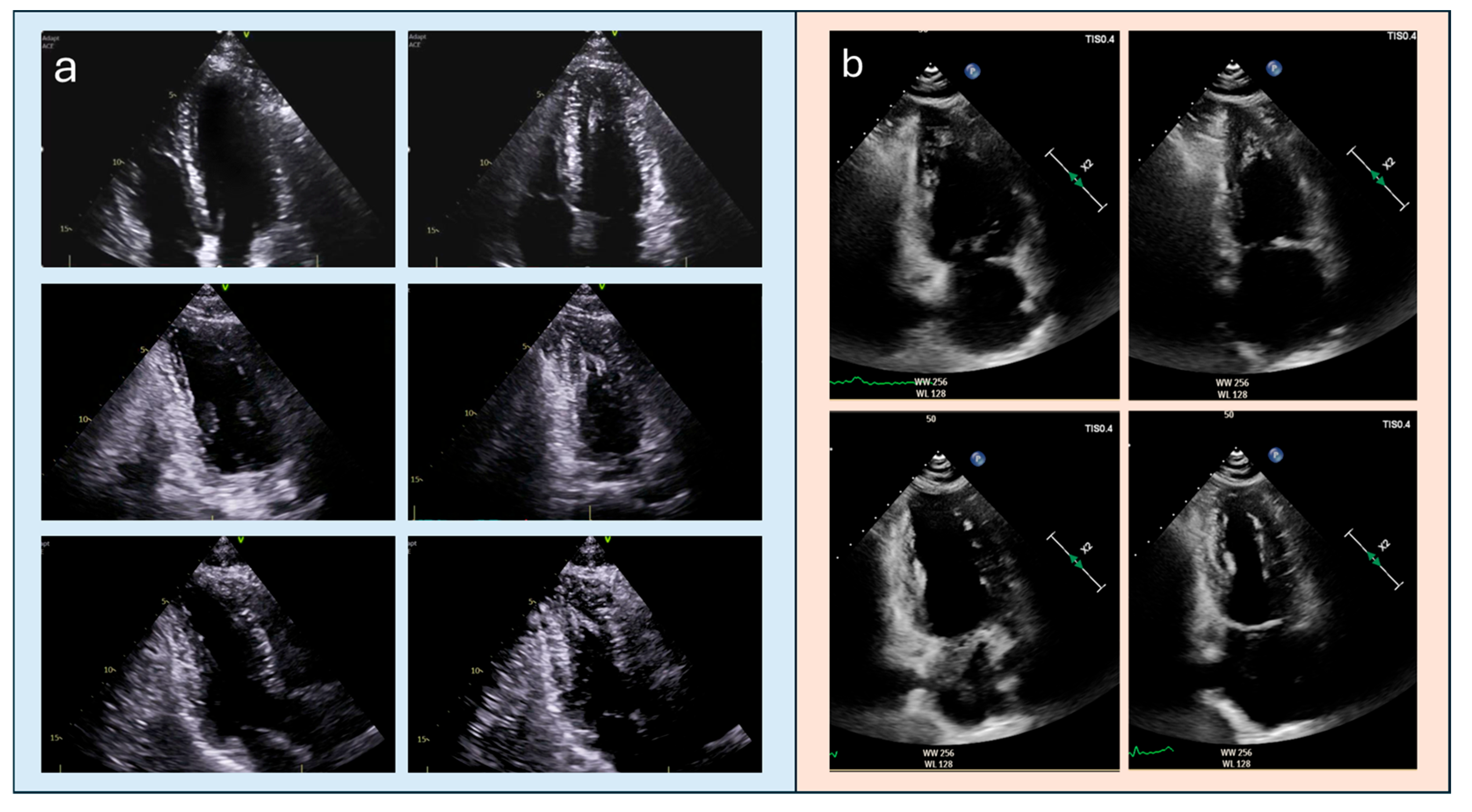
Figure 1. TTE assessment of long-axis views. (a): TTE long-axis views in a patient with ApHCM: correctly acquired apical four-, two-, and three-chamber views at end-diastole (right) and end-systole (left), demonstrating appropriate visualization of the true apex. (b) TTE long-axis views in a normal patient: example of apical foreshortening in the apical two-chamber view (upper row), which may falsely suggest apical hypertrophy; the correctly obtained view (lower row) restores the true apical geometry and demonstrates normal apical morphology. TTE, transthoracic echocardiography; ApHCM, apical hypertrophic cardiomyopathy.
Diagnostics 15 03013 g001
Figure 2. (a) Apical four-chamber echocardiographic view without the use of an intracavitary contrast agent. (b) Apical four-chamber echocardiographic view with the use of an intracavitary contrast agent. Contrast enhancement markedly improves endocardial border delineation and reveals the characteristic “spade-like” apical configuration, which is poorly defined on unenhanced imaging.
Diagnostics 15 03013 g002
Figure 3. A 38-year-old male patient with ApHCM. Color Doppler imaging shows apical aliasing artifact (circled), suggesting localized obstruction. Continuous-wave Doppler aligned with the same jet confirms an apical systolic gradient, consistent with obstruction in ApHCM. ApHCM, Apical hypertrophic cardiomyopathy.
Diagnostics 15 03013 g003
Figure 4. TTE assessment of apical obliteration. (a) apical four-chamber long-axis view in diastole, showing a preserved apical cavity. (b) focused short-axis view in diastole, confirming a residual apical lumen. (c) apical four-chamber long-axis view in systole, demonstrating near-complete obliteration of the apical cavity. (d) focused short-axis section in systole, showing circular endocardial apposition consistent with apical obliteration.
Diagnostics 15 03013 g004
Figure 5. Speckle-tracking echocardiography in a patient with ApHCM. (a) apical four- and two-chamber views showing systolic apposition of the apical walls with marked reduction in the apical cavity. (b) bull’s-eye longitudinal strain map demonstrating reduced apical strain values compared with mid- and basal segments, simulating reduced contractility and reflecting the mechanical constraint caused by apical segments contracting against each other during systole. ApHCM, apical hypertrophic cardiomyopathy.
Diagnostics 15 03013 g005
Figure 6. A 41-year-old male patient showing non-specific ECG abnormalities; case initially interpreted as ApHCM on TTE (left: apical four-chamber view, top; two-chamber view, bottom), but subsequently identified as eosinophilic myocarditis on CMRi (right). In fact, cine CMRi images (upper panels) reveal that the apparent apical thickening is due to non-myocardial material (fibrosis/thrombus), with the characteristic “flying V” sign (central inset). Late gadolinium enhancement sequences (lower panels) confirm the diagnosis, showing diffuse subendocardial enhancement of the mid-to-apical segments, typical of eosinophilic. CMRi, cardiac magnetic resonance imaging; TTE, transthoracic echocardiography, ApHCM, Apical hypertrophic cardiomyopathy.
Diagnostics 15 03013 g006
Figure 7. 54-year-old male patient with HCM. (a) TTE failed to detect the apical aneurysm, likely due to limitations related to suboptimal acoustic windows and reverberation artifact. (b) CMRi clearly demonstrates the early aneurysmal remodeling of the apical septum (yellow arrow). HCM, hypertrophic cardiomyopathy; TTE, transthoracic echocardiography; CMRi, cardiac magnetic resonance imaging.
Diagnostics 15 03013 g007
Figure 8. Late gadolinium enhancement sequences in ApHCM. Patchy mid-wall fibrosis is visible within the most hypertrophic apical segments (left and middle images, yellow arrows), but it may also involve non-hypertrophied regions (middle image, green arrow) and papillary muscles (middle image, blue arrow). In addition, a subepicardial enhancement pattern (right image, yellow arrow) and an ischemic subendocardial pattern (right image, blue arrow) have also been described in ApHCM. LGE, late gadolinium enhancement; ApHCM, apical hypertrophic cardiomyopathy.
Diagnostics 15 03013 g008
Figure 9. Comparison of two patients with a base-to-apex wall thickness ratio of 1. Top row: patient with hypertensive heart disease showing a normal apical angle (right) and limited apical obliteration (left) of 11 mm (<20 mm); base-to-apex wall thickness tapering appears preserved. Bottom row: patient with confirmed apical hypertrophic cardiomyopathy (ApHCM), characterized by a reduced apical angle (left) and marked apical obliteration (right) of 25 mm (>20 mm); base-to-apex wall thickness ratio is approximately 1. ApHCM, apical hypertrophic cardiomyopathy.
Diagnostics 15 03013 g009
Figure 10. Base-to-apex wall thickness tapering by CMRi and corresponding 12-lead ECG in two different patients. (a) a patient with apical hypertrophic cardiomyopathy (ApHCM), showing a maximal apical wall thickness of 8 mm without Base-to-apex tapering, associated with striking electrocardiographic abnormalities, namely deep negative T waves. (b) a patient with hypertensive heart disease, with a greater absolute wall thickness (12 mm), preserved basal–apical tapering and a normal ECG finding. CMRi, cardiac magnetic resonance imaging, ApHCM, apical hypertrophic cardiomyopathy; ECG, electrocardiogram.
Diagnostics 15 03013 g010
Figure 11. A 49-year-old female patient with documented ApHCM (a) diastole: the patient exhibits base-to-apex tapering ratio of approximately 1 and diastolic thinning of the apical cap (yellow arrow). (b) systole: the same patient exhibits and an initial, focal tendency toward apical aneurysmal evolution (yellow arrow); in addition, there is apical displacement of the posterolateral papillary muscle (green arrow, right). ApHCM, apical hypertrophic cardiomyopathy.
Diagnostics 15 03013 g011
Table 1. Comparison between classic HCM and Apical HCM. The apical variant represents a smaller but clinically relevant subset, with distinct epidemiology, imaging challenges, and prognostic factors compared with the classic phenotype. HCM, hypertrophic cardiomyopathy.
FeatureClassic HCM Apical HCM
EpidemiologyPrevalence: 1/500–1/2001–10% in Europe/North America; up to 25% in Asian cohorts
PrognosisOverall annual mortality ≈ 1%Overall annual mortality 0.5–3.5%
Electrocardiographic featuresLV hypertrophy criteria, nonspecific T-wave inversion or repolarization abnormalitiesDeep or “giant” negative T waves (10–15 mm)
Echocardiographic DetectionGood sensitivity, especially for septal hypertrophy; color-Doppler useful for LVOT obstructionOften underestimated without contrast; up to 43% of apical aneurysms may be missed by TTE
Advanced Imaging (CMRi)Confirms diagnosis; differentiates non-sarcomeric causes; quantifies fibrosis (LGE)Gold standard for apical wall thickness and aneurysm; differentiates non-sarcomeric etiologies; quantifies fibrosis (LGE)
Table 2. Overview of the main apical-centric diagnostic criteria proposed for ApHCM, including their proposed cut-offs, validation cohorts, diagnostic accuracy metrics (AUC, sensitivity, specificity, false-positive rate when available), strength of evidence, and estimated feasibility in real-world transthoracic echocardiographic practice. Strength of evidence is a qualitative assessment based on study design, cohort size, degree of external confirmation, and consistency with literature. Feasibility in TTE: “+” = limited or technically demanding; “++” = feasible with careful acquisition; “+++” = readily applicable in routine echocardiographic practice. CMRi, cardiac magnetic resonance imaging; ApHCM, apical hypertrophic cardiomyopathy; AUC, area under the curve; FP, false positive; Sens, sensitivity; TTE, transthoracic echocardiography; HCM, hypertrophic cardiomyopathy; LV, left ventricle.
CriterionModalityCut-Off/DefinitionValidation CohortDiagnostic AccuracyEvidenceTTE FeasibilityReference
Indexed apical maximum wall thicknessCMRi≥5.2–5.6 mm/m2>4000 controls +104 ApHCM ptsAUC 0.94; Sens 99/78%; FP 3%High++Hughes 2024 [25]
Loss of basal–apical taperingCMRi Apical ≥ basal22 relative ApHCM vs. controlsSupportive; no AUCModerate+++Flett 2015 [74]
Apical–basal thickness ratioCMRi>1 (prev ≥1.3)Mixed CMR cohortsNot availableModerate+++Flett 2015 [74]
Apical angleCMRi≤75–76°71 ptsAUC 0.77Low–mod++Li 2021 [64]
Apical thickness progressionCMRiMean ≥7.6 mm; Max ≥9.5 mm71 ptsAUC 0.87–0.898Moderate++Li 2021 [64]
Indexed apical obliteration lengthTTE>0.5~180–190 ApHCMPrognosticModerate+++Kim 2016 [75]
Absolute apical obliterationTTE>20 mm~188 ApHCMSpecificModerate+++Kim 2016 [75]
Apical papillary displacementCMRiApical beyond mid-LV>150 HCM + ctrlsSupportiveModerate+Filomena 2023 [76]
Apical cavity obliteration severityTTE/CMRiEnd-systolic closureEcho/CMR cohortsPrognosticModerate+++Hamza 2025 [20]
Apical aneurysm detectionCMRi→TTEDyskinesis + scarCMR aneurysm cohortsEcho misses 43%High+Hamza 2025 [20]
Disclaimer/Publisher’s Note: The statements, opinions and data contained in all publications are solely those of the individual author(s) and contributor(s) and not of MDPI and/or the editor(s). MDPI and/or the editor(s) disclaim responsibility for any injury to people or property resulting from any ideas, methods, instructions or products referred to in the content.