Article

Association Between Microcalcification Patterns in Mammography and Breast Tumors in Comparison to Histopathological Examinations


by
Iqbal Hussain Rizuana
1,2,
Ming Hui Leong
1,2,
Geok Chin Tan
2,3,* and
Zaleha Md. Isa
2,4
1
Department of Radiology, Faculty of Medicine, Universiti Kebangsaan Malaysia Medical Center, Jalan Yaakob Latif, Kuala Lumpur 56000, Malaysia
2
Hospital Canselor Tuanku Muhriz, Jalan Yaakob Latif, Bandar Tun Razak, Kuala Lumpur 56000, Malaysia
3
Department of Pathology, Faculty of Medicine, Universiti Kebangsaan Malaysia Medical Center, Jalan Yaakob Latif, Kuala Lumpur 56000, Malaysia
4
Department of Community Health, Faculty of Medicine, Universiti Kebangsaan Malaysia Medical Center, Jalan Yaakob Latif, Kuala Lumpur 56000, Malaysia
*
Author to whom correspondence should be addressed.
Diagnostics 2025, 15(13), 1687; https://doi.org/10.3390/diagnostics15131687

Submission received: 17 January 2025 / Revised: 24 June 2025 / Accepted: 27 June 2025 / Published: 2 July 2025
(This article belongs to the Special Issue Recent Advances in Breast Cancer Imaging)

Abstract

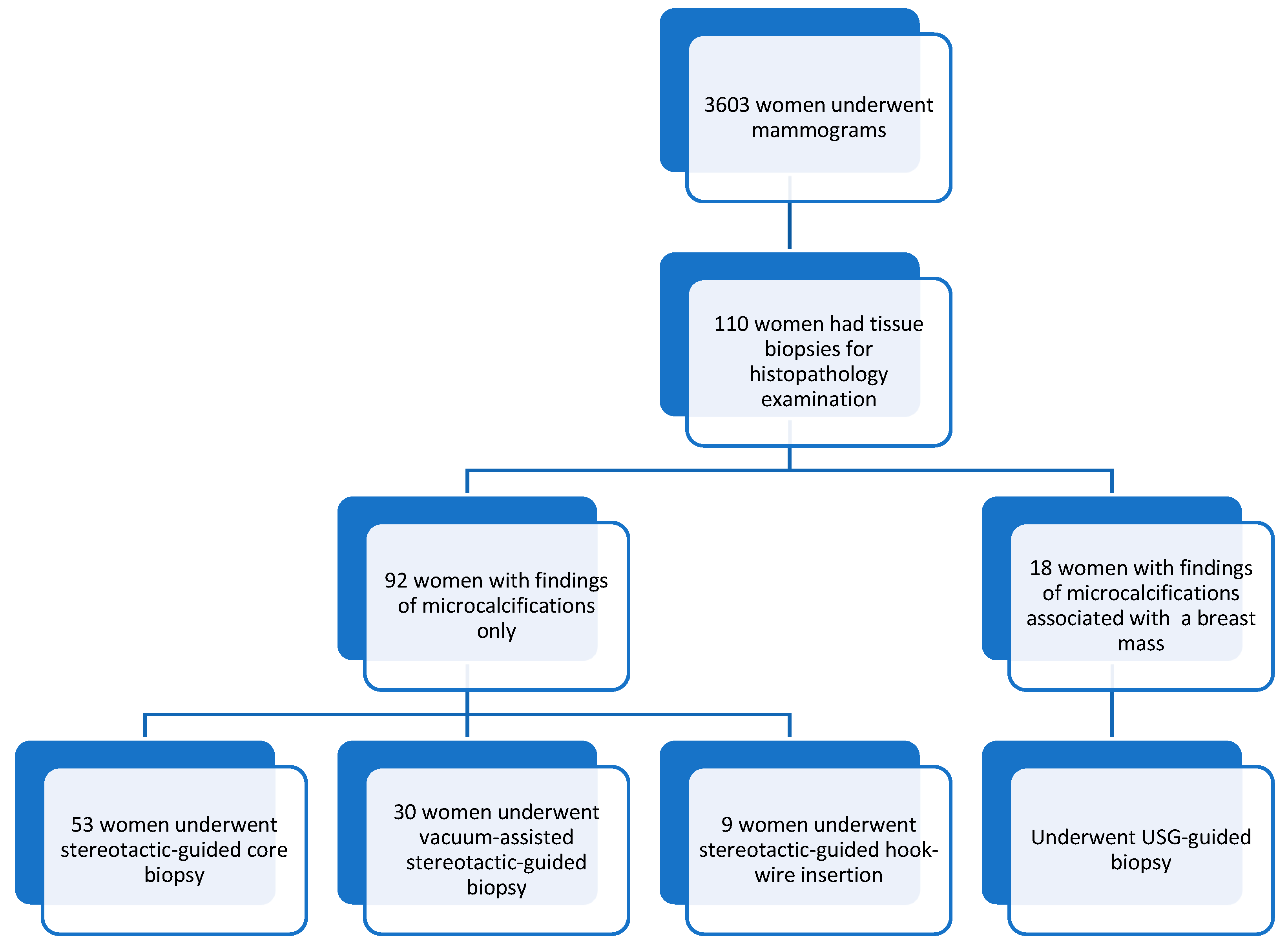
Background/Objectives: Accurately correlating mammographic findings with corresponding histopathologic features is considered one of the essential aspects of mammographic evaluation, guiding the next steps in cancer management and preventing overdiagnosis. The objective of this study was to evaluate patterns of mammographic microcalcifications and their association with histopathological findings related to various breast tumors. Methods: 110 out of 3603 women had microcalcification of BIRADS 3 or higher and were subjected to stereotactic/ultrasound (USG) guided biopsies, and hook-wire localization excision procedures. Ultrasound and mammography images were reviewed by experienced radiologists using the standard American College of Radiology Breast-Imaging Reporting and Data System (ACR BI-RADS). Results: Our study showed that features with a high positive predictive value (PPV) of breast malignancy were heterogeneous (75%), fine linear/branching pleomorphic microcalcifications (66.7%), linear (100%), and segmental distributions (57.1%). Features that showed a higher risk of association with ductal carcinoma in situ (DCIS) were fine linear/branching pleomorphic (odds ratio (OR): 3.952), heterogeneous microcalcifications (OR: 3.818), segmental (OR: 5.533), linear (OR: 3.696), and regional (OR: 2.929) distributions. Furthermore, the features with higher risks associated with invasive carcinoma had heterogeneous (OR: 2.022), fine linear/branching pleomorphic (OR: 1.187) microcalcifications, linear (OR: 6.2), and regional (OR: 2.543) distributions. The features of associated masses in mammograms that showed a high PPV of malignancy had high density (75%), microlobulation (100%), and spiculated margins (75%). Conclusions: We concluded that specific patterns and distributions of microcalcifications were indeed associated with a higher risk of malignancy. Those with fine linear or branching pleomorphic and segmental distribution were at a higher risk of DCIS, whereas those with heterogeneous morphology with a linear distribution were at a higher risk of invasive carcinoma.
Keywords: mammogram; microcalcifications; mass; histopathology; breast malignancy; ductal carcinoma in situ; invasive carcinoma mammogram; microcalcifications; mass; histopathology; breast malignancy; ductal carcinoma in situ; invasive carcinoma