A Multi-Stage Approach for Cardiovascular Risk Assessment from Retinal Images Using an Amalgamation of Deep Learning and Computer Vision Techniques
Abstract
1. Introduction
2. Materials and Methods
2.1. Research Design
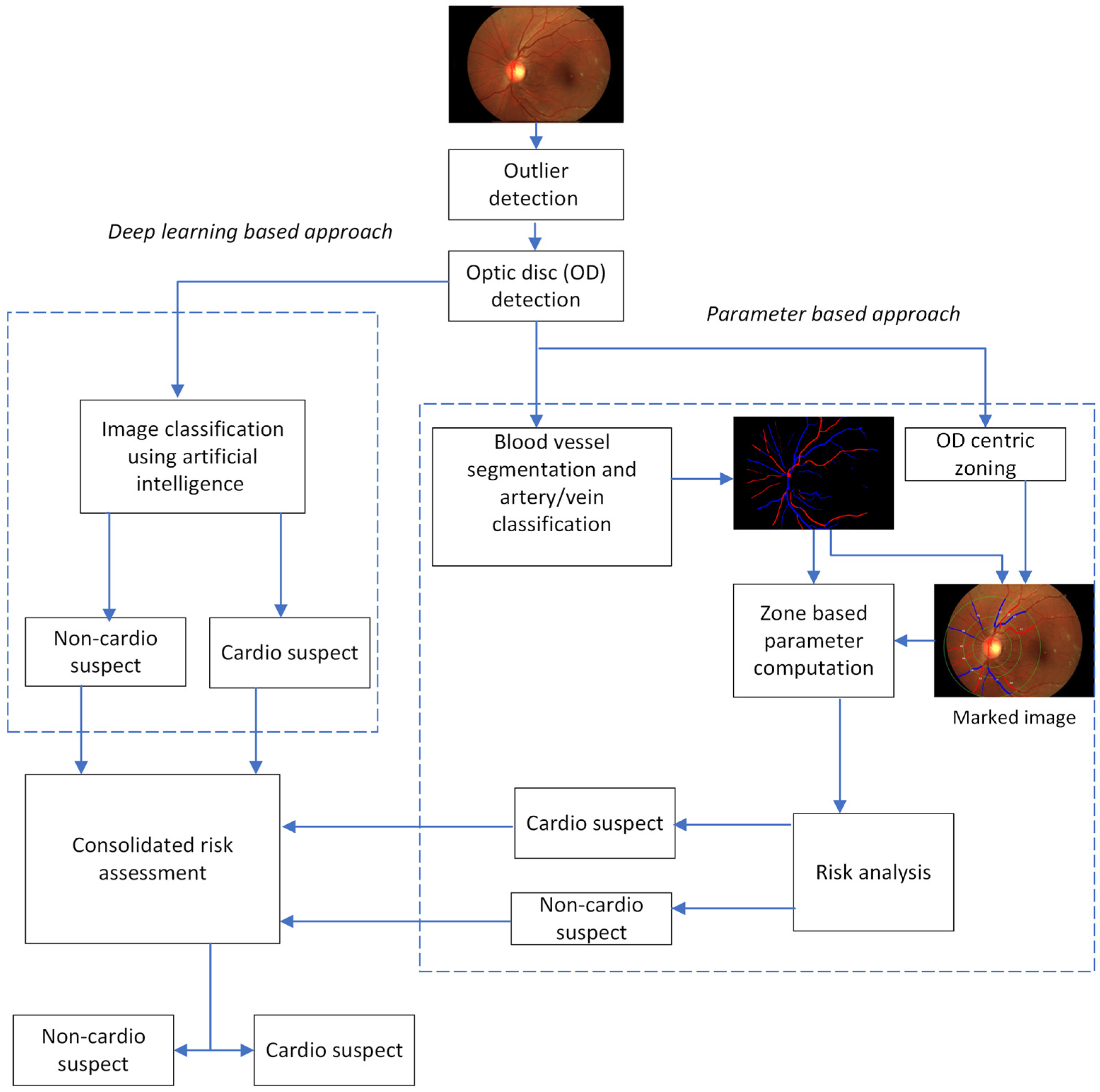
2.2. Implementation Approach
2.2.1. Parameter-Based Approach
- Branching coefficient
- Junction exponent
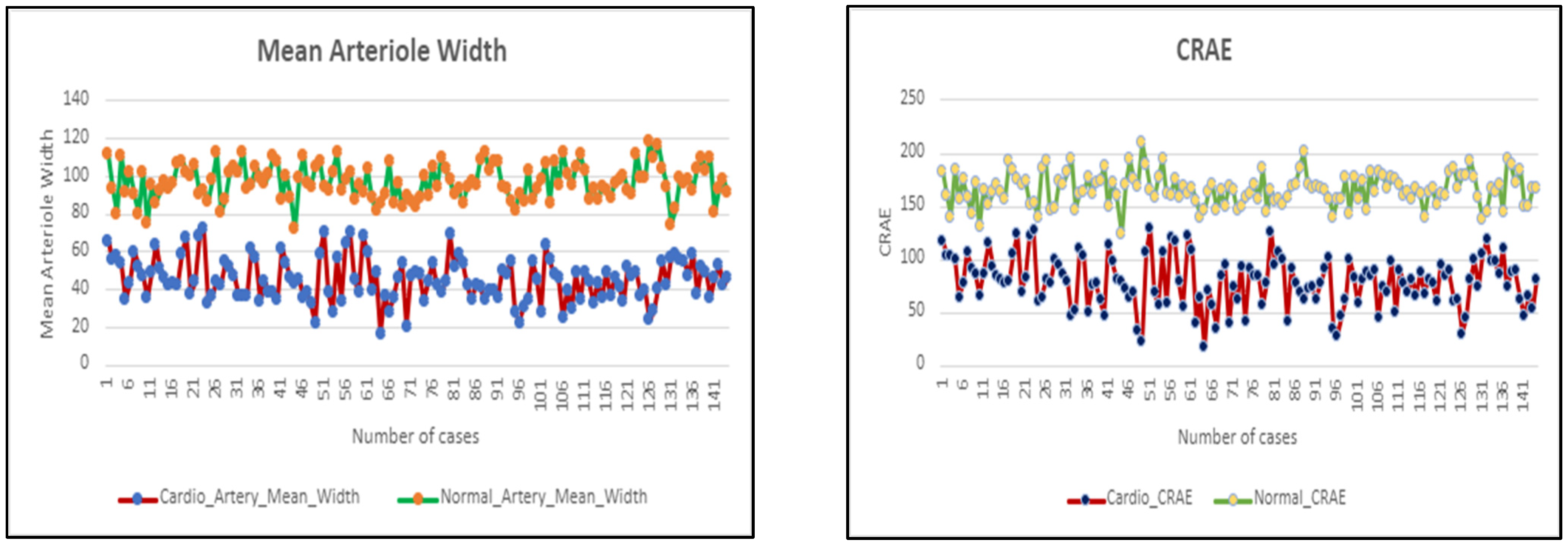
- Mean artery/vein width
- Optimality ratio
- Path length
- Simple tortuosity
- Vessel diameter reduction
- CRAE
- CRVE
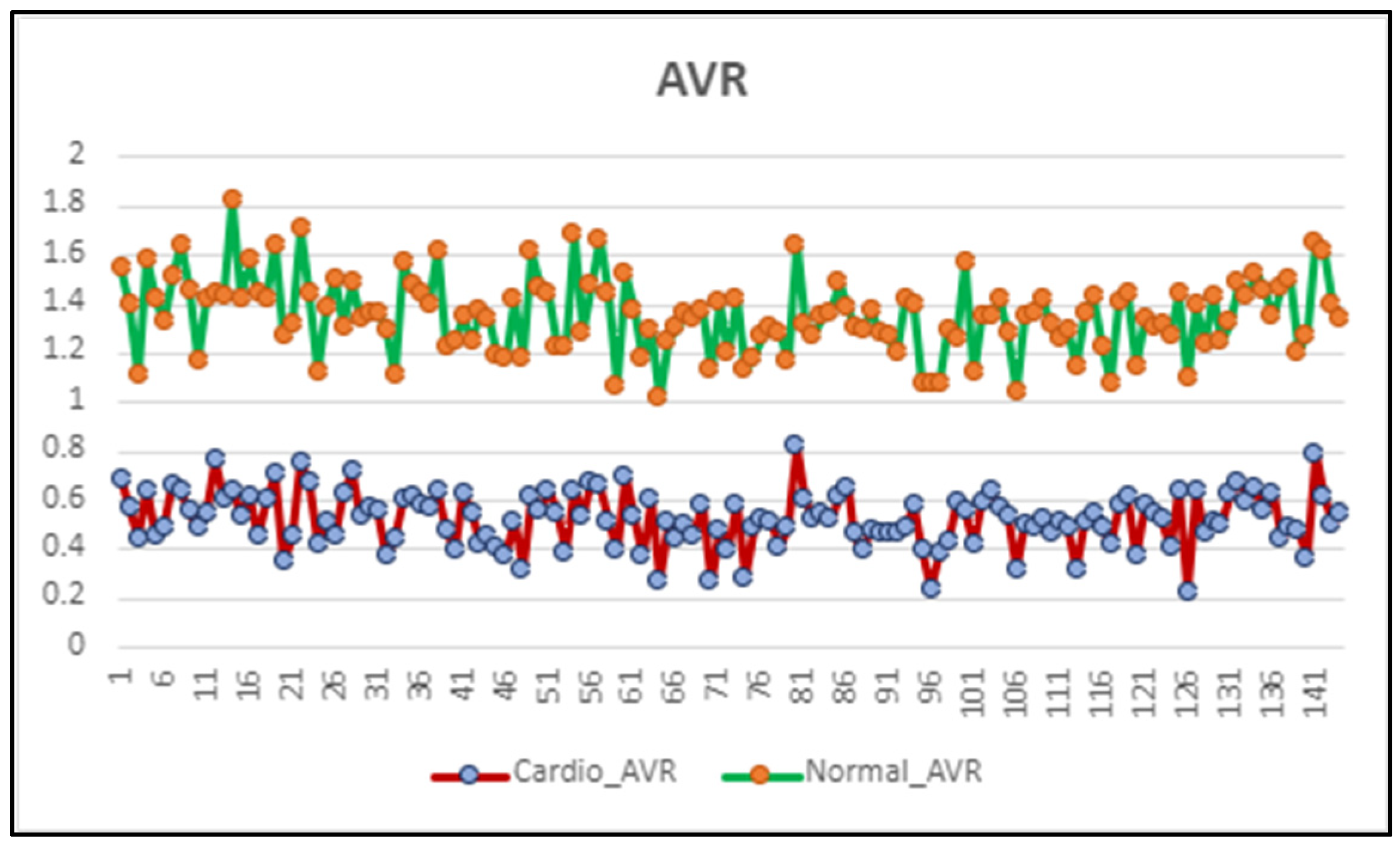
- Arteriovenous ratio
- Fractal dimension
- —Store parent vessel width
- —Store first and second daughter branch vessel width.
- Calculate and squares of all
- —Store parent vessel width
- —Store th daughter branch vessel width = 1 to n
- Consider a vessel centerline pixel for selecting a cross-section.
- Search an edge pixel from a certain distance (r) and angle (a).
- Each time an edge pixel is found, obtain the opposite edge pixel by adding 180° to (a) and varying the distance (r) from the centerline pixel.
- Calculate the width by measuring the minimum Euclidean distance between two opposite edge points.
- —Store parent vessel width
- —Store first and second daughter branch vessel width.
- Calculate and cubes of all
- Finally, calculate OR as OR =
- Get a stable retinal vascular tree.
- Find the single largest external path length in the tree.
- Find the total sum of external path lengths in the tree.
- Find the total number of exterior–interior path lengths in the tree.
- —Store parent vessel width
- —Store first and second daughter branch vessel width.
- Deviation of vessels from the normal diameter measure
- Calculate widths of all veins and arterioles.
- Rank them in decreasing order.
- Select the top 6 veins and arteries.
- Calculate the CRAE and CRVE using the following formulae given in Equations (5) and (6):
- —width of narrower branch
- —width of wider branch
- 5.
- Repeat step 4 till a single value remains for both CRVE and CRAE.
- Find CRAE and CRVE.
- Use the formula given in Equation (7) to calculate AVR:
- Cover image by a sequence of grids of descending sizes.
- Record:
- The number of square boxes intersected by the image, N(s).
- The side length of the squares, s.
- Plot .
- Get the regression slope D.
- Find the fractal dimension from Equation (8) as:
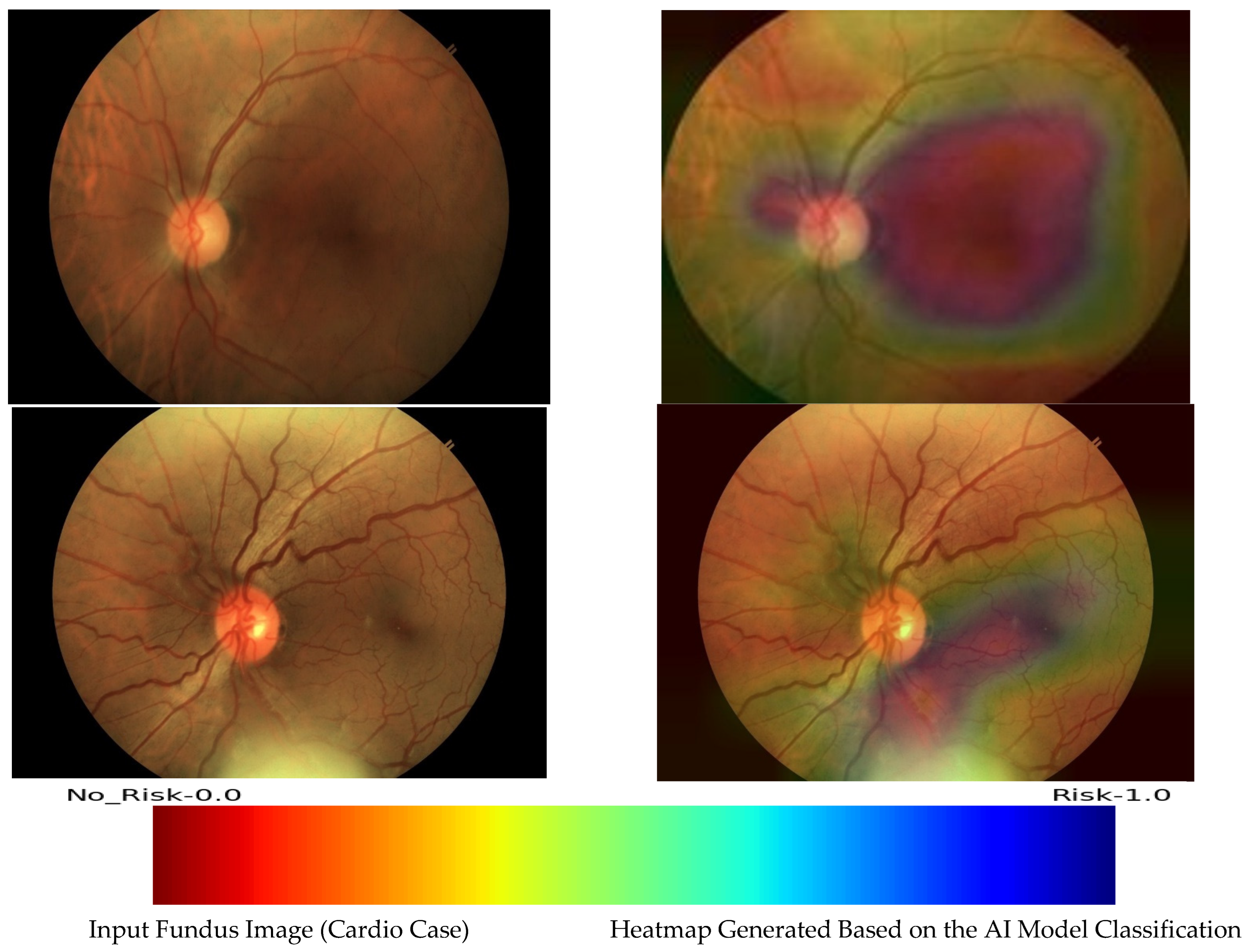
2.2.2. AI-Based CVD Analysis
- Depth-wise Separable Convolution:
- Inverted Residual Blocks:
- Global Average Pooling (GAP):
- Scaling Coefficients:
- Input Image Size:
- Number of Parameters:
- Data Preprocessing:
- Import the retinal images dataset.
- Resize images to match the input size expected by EfficientNetB0 i.e., (224, 224).
- Normalize pixel values to ensure consistent input to the model.
- The black border from the retinal images is removed to remove the unnecessary portion of the fundus image not required for training.
- Architecture Customization:
- Append additional layers to the base model.
- A batch normalization layer, drop out layer, regularization layer, and a fully connected dense layer are appended to the base model.
- Transfer Learning:
- Load the pre-trained EfficientNetB0 model, which has already learned features from a large dataset.
- Use the pre-trained weights from the initial layers of the network to retain the learned features.
- Customize the final layers to adapt the model for binary classification.
- Training:
- Split the dataset into training and validation sets.
- Train the model on the training set, fine-tuning the final layers for the specific task.
- Validate the model on the validation set to monitor performance.
- Evaluation:
- Assess the model’s performance using metrics such as accuracy, precision, recall, and F1 score.
- Use a separate test set to evaluate the model’s generalization to unseen data.
3. Results
4. Discussion
4.1. Implications of the Hybrid Approach
- Customized Solution for CVD Risk Evaluation: This work presents a bespoke approach for assessing the risk of CVD. By customizing the methodology, it aims to provide a more precise assessment of cardiovascular health.
- Hybrid Architectural Framework: Introducing a hybrid architecture, this research combines multiple methodologies leveraging both handcrafted features and AI-driven retinal vascular pattern analysis. This amalgamation enhances the robustness and efficacy of the proposed system.
- Superior Performance in Clinical Trial Validation: Through meticulous clinical trial validation, this research establishes the superiority of its methodology over existing systems. By showcasing improved performance metrics, it underscores the potential clinical utility and reliability of the developed approach.
4.2. Comparison with Existing Solutions in CVD Risk Assessment
5. Conclusions
Challenges and Future Directions
Supplementary Materials
Author Contributions
Funding
Institutional Review Board Statement
Informed Consent Statement
Data Availability Statement
Conflicts of Interest
References
- WHO. Cardiovascular Diseases (CVDs). 2021. Available online: https://www.who.int/en/news-room/fact-sheets/detail/cardiovascular-diseases-(cvds) (accessed on 20 November 2022).
- Wang, H.; Naghavi, M.; Allen, C.; Barber, R.M.; Bhutta, Z.A.; Carter, A.; Casey, D.C.; Charlson, F.J.; Chen, A.Z.; Coates, M.M.; et al. Global, regional, and national life expectancy, all-cause mortality, and cause-specific mortality for 249 causes of death, 1980–2015: A systematic analysis for the Global Burden of Disease Study 2015. Lancet 2016, 388, 1459–1544. [Google Scholar] [CrossRef] [PubMed]
- Goff, D.C., Jr.; Lloyd-Jones, D.M.; Bennett, G.; Coady, S.; D’Agostino, R.B.; Gibbons, R.; Greenland, P.; Lackland, D.T.; Levy, D.; O’Donnell, C.J.; et al. 2013 ACC/AHA guideline on the assessment of cardiovascular risk: A report of the American College of Cardiology/American Heart Association Task Force on Practice Guidelines. Circulation 2013, 129, S49–S73. [Google Scholar] [CrossRef] [PubMed]
- Huang, L.; Aris, I.M.; Teo, L.L.Y.; Wong, T.Y.; Chen, W.; Koh, A.S.; Li, L. Exploring associations between cardiac structure and retinal vascular geometry. J. Am. Heart Assoc. 2020, 9, e014654. [Google Scholar] [CrossRef] [PubMed]
- McGeechan, K.; McGeechan, M.K.; Liew, M.G.; Macaskill, P.; Irwig, M.L.; Klein, R.; Klein, B.E.; Wang, M.J.J.; Mitchell, P.; Vingerling, J.R.; et al. Meta-analysis: Retinal vessel caliber and risk for coronary heart disease. Ann. Intern. Med. 2009, 151, 404–413. [Google Scholar] [CrossRef]
- Seidelmann, S.B.; Claggett, B.; Bravo, P.E.; Gupta, A.; Farhad, H.; Klein, B.E.; Klein, R.; Di Carli, M.; Solomon, S.D. Retinal Vessel Calibers in Predicting Long-Term Cardiovascular Outcomes: The Atherosclerosis Risk in Communities Study. Circulation 2016, 134, 1328–1338. [Google Scholar] [CrossRef] [PubMed]
- Arnould, L.; Binquet, C.; Guenancia, C.; Alassane, S.; Kawasaki, R.; Daien, V.; Tzourio, C.; Kawasaki, Y.; Bourredjem, A.; Bron, A.; et al. Association between the retinal vascular network with Singapore “I” Vessel Assessment (SIVA) software, cardiovascular history and risk factors in the elderly: The montrachet study, population-based study. PLoS ONE 2018, 13, e0194694. [Google Scholar] [CrossRef] [PubMed]
- Danielescu, C.; Dabija, M.G.; Nedelcu, A.H.; Lupu, V.V.; Lupu, A.; Ioniuc, I.; Gîlcă-Blanariu, G.-E.; Donica, V.-C.; Anton, M.-L.; Musat, O. Automated Retinal Vessel Analysis Based on Fundus Photographs as a Predictor for Non-Ophthalmic Diseases—Evolution and Perspectives. J. Pers. Med. 2024, 14, 45. [Google Scholar] [CrossRef] [PubMed]
- Arnould, L.; Meriaudeau, F.; Guenancia, C.; Germanese, C.; Delcourt, C.; Kawasaki, R.; Cheung, C.Y.; Creuzot-Garcher, C.; Grzybowski, A. Using Artificial Intelligence to Analyse the Retinal Vascular Network: The Future of Cardiovascular Risk Assessment Based on Oculomics? A Narrative Review. Ophthalmol. Ther. 2023, 12, 657–674. [Google Scholar] [CrossRef]
- Hu, W.; Yii, F.S.L.; Chen, R.; Zhang, X.; Shang, X.; Kiburg, K.; Woods, E.; Vingrys, A.; Zhang, L.; Zhu, Z.; et al. A Systematic Review and Meta-Analysis of Applying Deep Learning in the Prediction 778 of the Risk of Cardiovascular Diseases From Retinal Images. Trans. Vis. Sci. Tech. 2023, 12, 14. [Google Scholar] [CrossRef]
- Early Signs of Heart Disease Appear in the Eyes. Available online: https://www.aao.org/eye-health/news/eye-stroke-heart-disease-vision-exam-retina-oct#:~:text=A%20new%20study%20finds%20that,a%20retinal%20ischemic%20perivascular%20lesion (accessed on 10 October 2023).
- Chaikijurajai, T.; Ehlers, J.P.; Tang, W.W. Retinal Microvasculature: A Potential Window into Heart Failure Prevention. JACC Heart Fail. 2022, 10, 785–791. [Google Scholar] [CrossRef]
- Fetit, A.E.; Doney, A.S.; Hogg, S.; Wang, R.; MacGillivray, T.; Wardlaw, J.M.; Doubal, F.N.; McKay, G.J.; McKenna, S.; Trucco, E. A multimodal approach to cardiovascular risk stratification in patients with type 2 diabetes incorporating retinal, genomic and clinical features. Sci. Rep. 2019, 9, 3591. [Google Scholar] [CrossRef] [PubMed]
- Venkataramani, D.; Veeranan, J.; Pitchai, L. Fractal analysis of retinal vasculature in relation with retinal diseases—An machine learning approach. Nonlinear Eng. 2022, 11, 411–419. [Google Scholar] [CrossRef]
- Menon, S.; Menon, G. Role of Retinal Vessel Caliber Assessment in Predicting Hemorrhagic Stroke—Emerging Concepts. J. Stroke Med. 2022, 5, 7–13. [Google Scholar] [CrossRef]
- Hanssen, H.; Streese, L.; Vilser, W. Retinal vessel diameters and function in cardiovascular risk and disease. Prog. Retin. Eye Res. 2022, 91, 101095. [Google Scholar] [CrossRef]
- Cheung, N.; Bluemke, D.A.; Klein, R.; Sharrett, A.R.; Islam, F.M.; Cotch, M.F.; Klein, B.E.; Criqui, M.H.; Wong, T.Y. Retinal arteriolar narrowing and left ventricular remodeling: The multi-ethnic study of atherosclerosis. J. Am. Coll. Cardiol. 2007, 50, 48–55. [Google Scholar] [CrossRef] [PubMed]
- Chandra, A.; Seidelmann, S.B.; Claggett, B.L.; Klein, B.E.; Klein, R.; Shah, A.M.; Solomon, S.D. The association of retinal vessel calibres with heart failure and long-term alterations in cardiac structure and function: The Atherosclerosis Risk in Communities (ARIC) Study. Eur. J. Heart Fail. 2019, 21, 1207–1215. [Google Scholar] [CrossRef] [PubMed]
- Fathalla, K.M.; Ekárt, A.; Seshadri, S.; Gherghel, D. Cardiovascular risk prediction based on Retinal Vessel Analysis using machine learning. In Proceedings of the 2016 IEEE International Conference on Systems, Man, and Cybernetics (SMC), Budapest, Hungary, 9–12 October 2016; IEEE Press: Piscataway, NJ, USA, 2016; pp. 880–885. [Google Scholar] [CrossRef]
- Rudnicka, A.R.; Welikala, R.; Barman, S.; Foster, P.J.; Luben, R.; Hayat, S.; Khaw, K.T.; Whincup, P.; Strachan, D.; Owen, C.G. Artificial intelligence-enabled retinal vasculometry for prediction of circulatory mortality, myocardial infarction and stroke. Br. J. Ophthalmol. 2022, 106, 1722–1729. [Google Scholar] [CrossRef] [PubMed]
- Fraz, M.; Welikala, R.; Rudnicka, A.; Owen, C.; Strachan, D.; Barman, S. QUARTZ: Quantitative Analysis of Retinal Vessel Topology and size—An automated system for quantification of retinal vessels morphology. Expert Syst. Appl. 2015, 42, 7221–7234. [Google Scholar] [CrossRef]
- Poplin, R.; Varadarajan, A.V.; Blumer, K.; Liu, Y.; McConnell, M.V.; Corrado, G.S.; Peng, L.; Webster, D.R. Prediction of cardiovascular risk factors from retinal fundus photographs via deep learning. Nat. Biomed. Eng. 2018, 2, 158–164. [Google Scholar] [CrossRef]
- Diaz-Pinto, A.; Ravikumar, N.; Attar, R.; Suinesiaputra, A.; Zhao, Y.; Levelt, E.; Dall’armellina, E.; Lorenzi, M.; Chen, Q.; Keenan, T.D.L.; et al. Predicting myocardial infarction through retinal scans and minimal personal information. Nat. Mach. Intell. 2022, 4, 55–61. [Google Scholar] [CrossRef]
- Peng, Q.; Qian, Y.; Xu, X.; Tham, Y.C.; Yu, M.; Yong, L.; Cheng, C.Y. Prediction of future cardiovascular diseases from retinal images using a deep-learning-based hybrid model. Investig. Ophthalmol. Vis. Sci. 2023, 64, 1876. [Google Scholar]
- Lee, C.J.; Rim, T.H.; Kang, H.G.; Kim, S.S.; Lee, G.; Yu, M.; Park, S.H. Deep Learning-Based Retinal Imaging for Predicting Cardiovascular Disease Events in Prediabetic and Diabetic Patients: A Study Using the UK Biobank. Circulation 2023, 148 (Suppl. S1), A17817. [Google Scholar] [CrossRef]
- Zekavat, S.M.; Raghu, V.K.; Trinder, M.; Ye, Y.; Koyama, S.; Honigberg, M.C.; Yu, Z.; Pampana, A.; Urbut, S.; Haidermota, S.; et al. Deep learning of the retina enables phenome-and genome-wide analyses of the microvasculature. Circulation 2022, 145, 134–150. [Google Scholar] [CrossRef] [PubMed]
- Ramesh Cardiology Clinic, Bengaluru, India. Available online: https://www.rameshclinic.com/ (accessed on 22 March 2024).
- Available online: https://www.forushealth.com/3nethra-classic-hd.html (accessed on 10 March 2023).
- Arnould, L.; Guenancia, C.; Bourredjem, A.; Binquet, C.; Gabrielle, P.H.; Eid, P.; Baudin, F.; Kawasaki, R.; Cottin, Y.; Creuzot-Garcher, C.; et al. Prediction of Cardiovascular Parameters With Supervised Machine Learning From Singapore “I” Vessel Assessment and OCT-Angiography: A Pilot Study. Transl. Vis. Sci. Technol. 2021, 10, 20. [Google Scholar] [CrossRef] [PubMed] [PubMed Central]
- Available online: https://lmb.informatik.uni-freiburg.de/people/ronneber/u-net/ (accessed on 29 March 2023).
- Available online: https://keras.io/api/applications/efficientnet/ (accessed on 15 March 2023).
- ImageNet. Available online: http://www.image-net.org (accessed on 15 March 2023).
- Jeena, R.S.; Sukesh Kumar, A.; Mahadevan, K. A Novel Method for Stroke Prediction from Retinal Images Using HoG Approach. In Advances in Signal Processing and Intelligent Recognition Systems; Thampi, S., Marques, O., Krishnan, S., Li, K.C., Ciuonzo, D., Kolekar, M., Eds.; SIRS 2018; Communications in Computer and Information Science; Springer: Singapore, 2019; Volume 968. [Google Scholar] [CrossRef]
- Heitmar, R.; Kalitzeos, A.A.; Panesar, V. Comparison of Two Formulas Used to Calculate Summarized Retinal Vessel Calibers. Optom. Vis. Sci. 2015, 92, 1085–1091. [Google Scholar] [CrossRef] [PubMed]
- Tan, M.; Le, Q.V. EfficientNet: Rethinking Model Scaling for Convolutional Neural Networks. arXiv 2019, arXiv:abs/1905.11946. [Google Scholar]
- Selvaraju, R.R.; Cogswell, M.; Das, A.; Vedantam, R.; Parikh, D.; Batra, D. Grad-CAM: Visual Explanations from Deep Networks via Gradient-Based Localization. In Proceedings of the 2017 IEEE International Conference on Computer Vision (ICCV), Venice, Italy, 22–29 October 2017; pp. 618–626. [Google Scholar] [CrossRef]


| Classes | Training (Number of Images) | Validation (Number of Images) |
|---|---|---|
| Non-fundus and poor quality fundus images | 904 | 300 |
| Fundus image | 956 | 300 |
| Classes | Training (Number of Images—After Augmentation) | Validation (Number of Images—Original) |
|---|---|---|
| Cardio cases | 1500 | 33 |
| Non-cardio cases | 1500 | 33 |
| Condition | Total Screenings (No. of Subjects) |
|---|---|
| Hypertension ~ | 145 |
| Cardio specific # | 234 |
| Healthy * | 140 |
| Actual | |||
|---|---|---|---|
| Predicted | Cardio | Healthy | |
| Cardio | 28 | 5 | |
| Healthy | 4 | 29 | |
| Actual | |||
|---|---|---|---|
| Predicted | Risk | No-Risk | |
| Risk | 40 | 4 | |
| No-Risk | 9 | 35 | |
Disclaimer/Publisher’s Note: The statements, opinions and data contained in all publications are solely those of the individual author(s) and contributor(s) and not of MDPI and/or the editor(s). MDPI and/or the editor(s) disclaim responsibility for any injury to people or property resulting from any ideas, methods, instructions or products referred to in the content. |
© 2024 by the authors. Licensee MDPI, Basel, Switzerland. This article is an open access article distributed under the terms and conditions of the Creative Commons Attribution (CC BY) license (https://creativecommons.org/licenses/by/4.0/).
Share and Cite
Prasad, D.K.; Manjunath, M.P.; Kulkarni, M.S.; Kullambettu, S.; Srinivasan, V.; Chakravarthi, M.; Ramesh, A. A Multi-Stage Approach for Cardiovascular Risk Assessment from Retinal Images Using an Amalgamation of Deep Learning and Computer Vision Techniques. Diagnostics 2024, 14, 928. https://doi.org/10.3390/diagnostics14090928
Prasad DK, Manjunath MP, Kulkarni MS, Kullambettu S, Srinivasan V, Chakravarthi M, Ramesh A. A Multi-Stage Approach for Cardiovascular Risk Assessment from Retinal Images Using an Amalgamation of Deep Learning and Computer Vision Techniques. Diagnostics. 2024; 14(9):928. https://doi.org/10.3390/diagnostics14090928
Chicago/Turabian StylePrasad, Deepthi K., Madhura Prakash Manjunath, Meghna S. Kulkarni, Spoorthi Kullambettu, Venkatakrishnan Srinivasan, Madhulika Chakravarthi, and Anusha Ramesh. 2024. "A Multi-Stage Approach for Cardiovascular Risk Assessment from Retinal Images Using an Amalgamation of Deep Learning and Computer Vision Techniques" Diagnostics 14, no. 9: 928. https://doi.org/10.3390/diagnostics14090928
APA StylePrasad, D. K., Manjunath, M. P., Kulkarni, M. S., Kullambettu, S., Srinivasan, V., Chakravarthi, M., & Ramesh, A. (2024). A Multi-Stage Approach for Cardiovascular Risk Assessment from Retinal Images Using an Amalgamation of Deep Learning and Computer Vision Techniques. Diagnostics, 14(9), 928. https://doi.org/10.3390/diagnostics14090928


