Article

Evaluating Cellularity Estimation Methods: Comparing AI Counting with Pathologists’ Visual Estimates

1
Healthcare Life Science Division, NEC Corporation, Tokyo 108-8556, Japan
2
Department of Machine Learning, NEC Laboratories America, Princeton, NJ 08540, USA
3
Center for Development of Advanced Diagnostics (C-DAD), Hokkaido University Hospital, Sapporo 060-8648, Japan
4
Department of Pathology, Kanagawa Cancer Center, Yokohama 241-8515, Japan
5
Department of Pathology, Kansai Medical University, Osaka 573-1010, Japan
6
Department of Pathology, Saitama Cancer Center, Saitama 362-0806, Japan
7
Department of Pathology, Oji General Hospital, Tomakomai 053-8506, Japan
8
Department of Surgical Pathology, Hokkaido University Hospital, Sapporo 060-8648, Japan
9
Department of Pathology, Teine Keijinkai Hospital, Sapporo 006-0811, Japan
10
Department of Pathology, KKR Sapporo Medical Center, Sapporo 062-0931, Japan
11
Department of Pathology, NTT Medical Center Sapporo, Sapporo 060-0061, Japan
12
Department of Pathology, Sapporo City General Hospital, Sapporo 060-8604, Japan
13
Department of Surgical Pathology, Sapporo Medical University Hospital, Sapporo 060-8543, Japan
14
Department of Pathology, Sapporo Tokushukai Hospital, Sapporo 004-0041, Japan
15
Department of Diagnostic Pathology, Asahikawa Medical University Hospital, Asahikawa 078-8510, Japan
*
Authors to whom correspondence should be addressed.
Diagnostics 2024, 14(11), 1115; https://doi.org/10.3390/diagnostics14111115

Submission received: 18 April 2024 / Revised: 17 May 2024 / Accepted: 21 May 2024 / Published: 28 May 2024
(This article belongs to the Topic AI in Medical Imaging and Image Processing)

Abstract

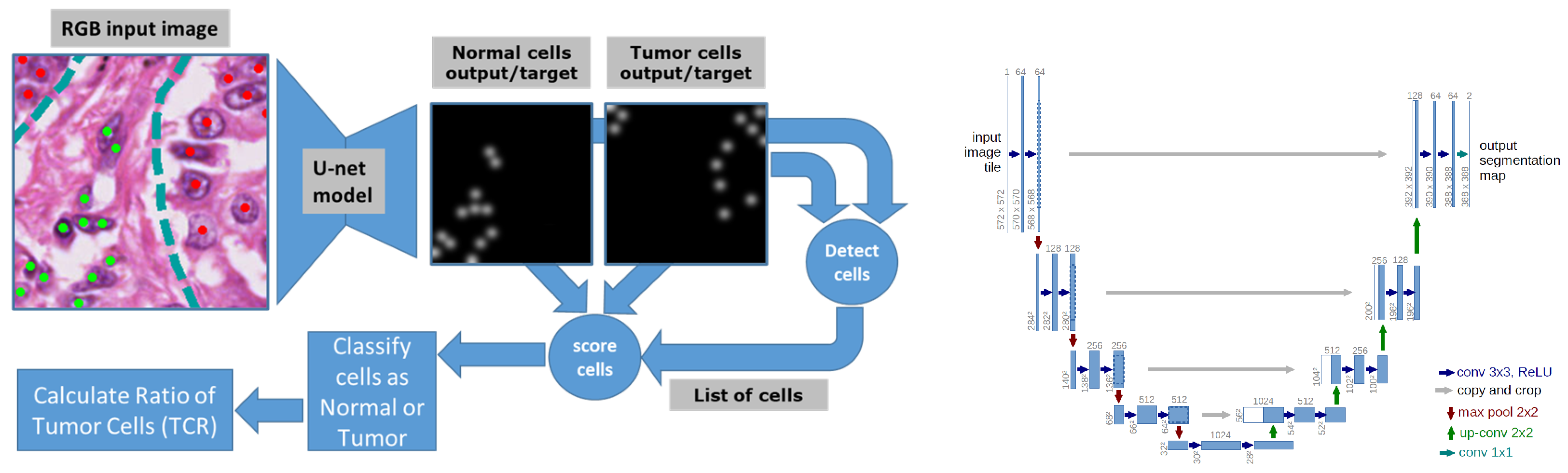
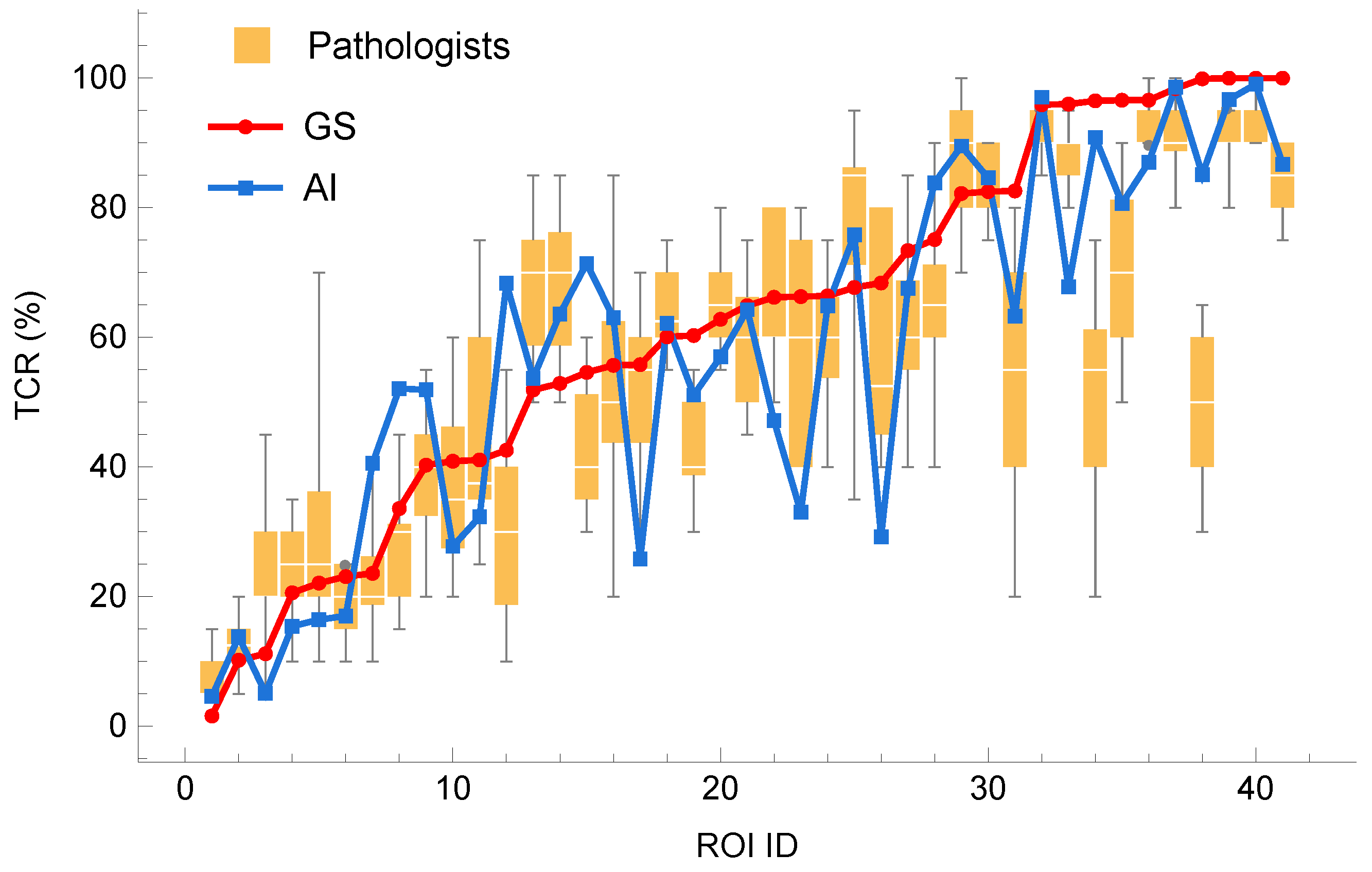
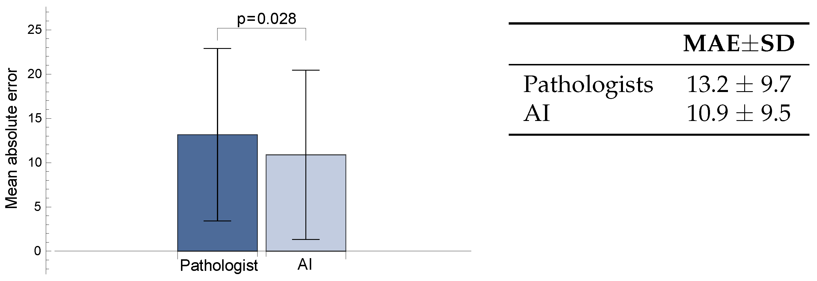
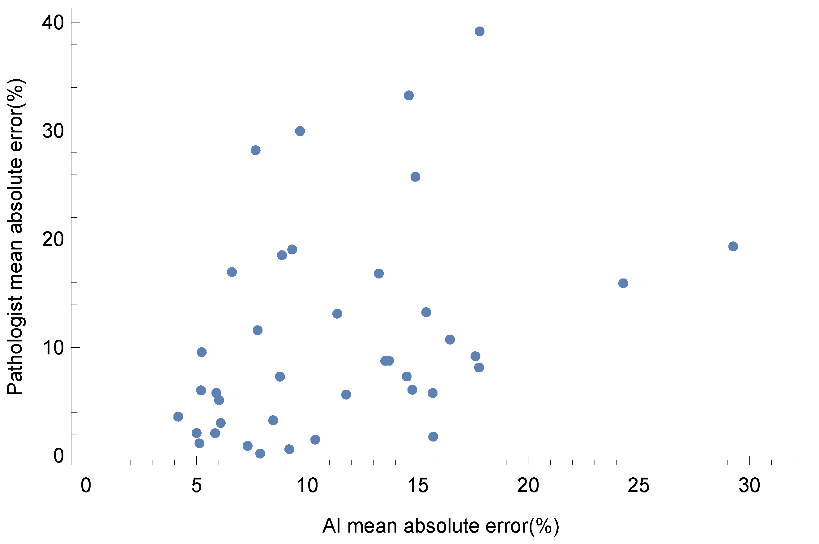
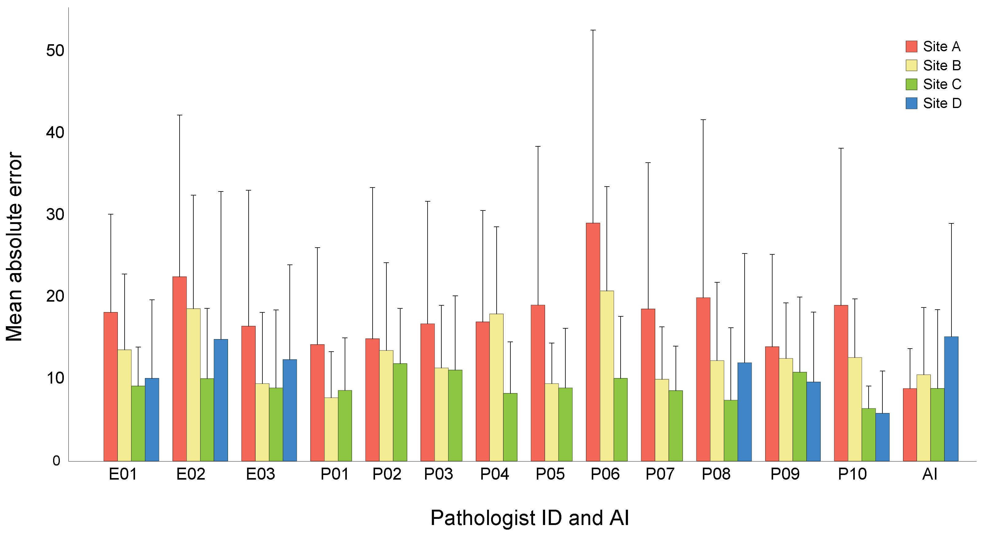
The development of next-generation sequencing (NGS) has enabled the discovery of cancer-specific driver gene alternations, making precision medicine possible. However, accurate genetic testing requires a sufficient amount of tumor cells in the specimen. The evaluation of tumor content ratio (TCR) from hematoxylin and eosin (H&E)-stained images has been found to vary between pathologists, making it an important challenge to obtain an accurate TCR. In this study, three pathologists exhaustively labeled all cells in 41 regions from 41 lung cancer cases as either tumor, non-tumor or indistinguishable, thus establishing a “gold standard” TCR. We then compared the accuracy of the TCR estimated by 13 pathologists based on visual assessment and the TCR calculated by an AI model that we have developed. It is a compact and fast model that follows a fully convolutional neural network architecture and produces cell detection maps which can be efficiently post-processed to obtain tumor and non-tumor cell counts from which TCR is calculated. Its raw cell detection accuracy is 92% while its classification accuracy is 84%. The results show that the error between the gold standard TCR and the AI calculation was significantly smaller than that between the gold standard TCR and the pathologist’s visual assessment (p<0.05). Additionally, the robustness of AI models across institutions is a key issue and we demonstrate that the variation in AI was smaller than that in the average of pathologists when evaluated by institution. These findings suggest that the accuracy of tumor cellularity assessments in clinical workflows is significantly improved by the introduction of robust AI models, leading to more efficient genetic testing and ultimately to better patient outcomes.
Keywords: tumor content ratio (TCR); next generation sequencing (NGS); artificial intelligence (AI); U-Net model; domain shift; site dependency tumor content ratio (TCR); next generation sequencing (NGS); artificial intelligence (AI); U-Net model; domain shift; site dependency