Vascular Neuroembryology: The Genesis of the Intracranial Arterial System and the Circle of Willis
 , , , , , , , , and
Abstract
1. Introduction
1.1. The Circle of Willis
1.1.1. Embryological Development of the Circle of Willis
1.1.2. Aim of This Study
2. Materials and Methods
2.1. Experimental Research Methods
2.1.1. Human Specimens
2.1.2. Image Acquisition and Processing
2.1.3. Alignment, Segmentation, and Visualization in Amira
2.1.4. Interactive 3D-PDF
2.2. Literature Search Strategy
3. Results
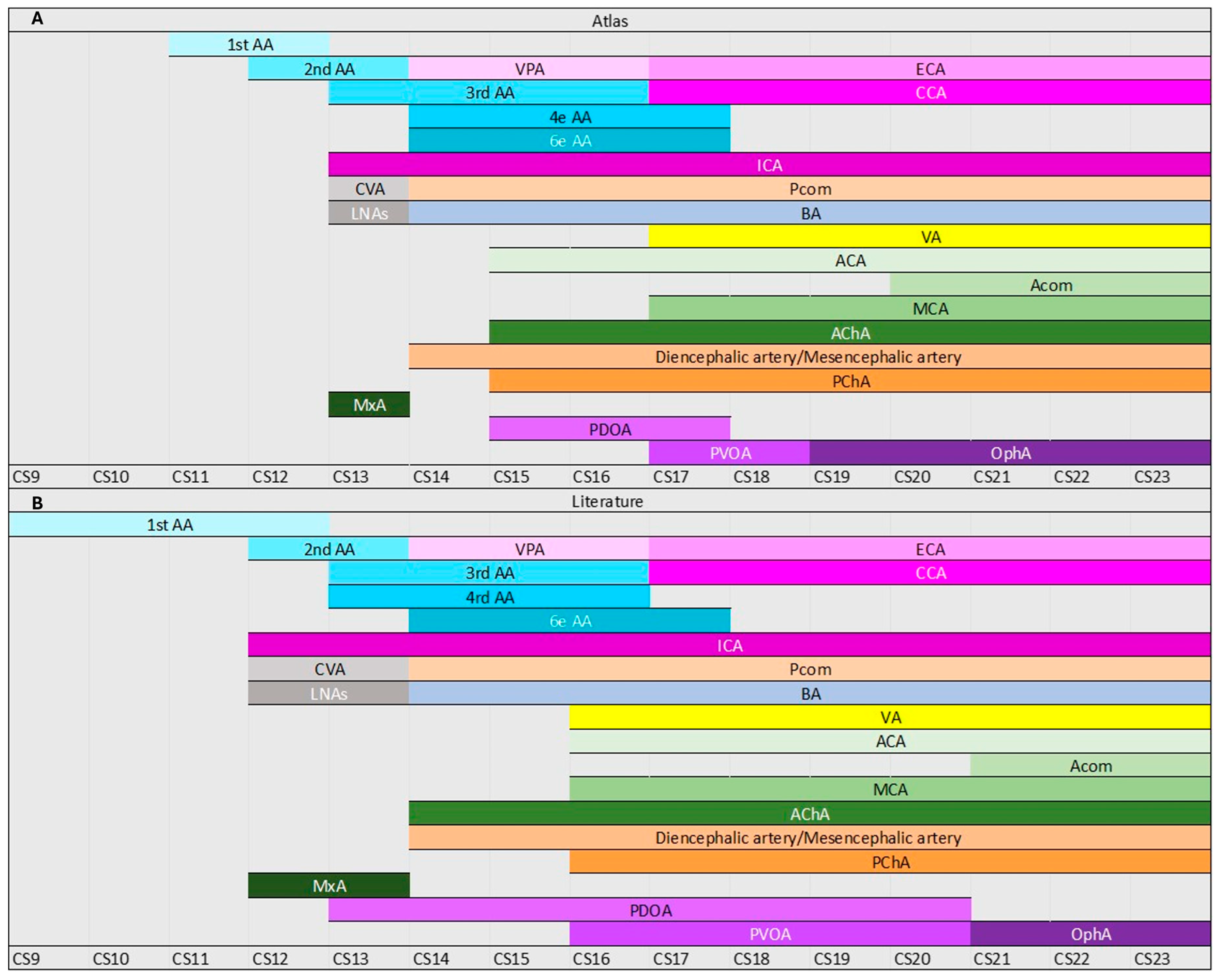
3.1. Aortic Arches
3.2. Carotid Arteries
3.3. Carotid–Vertebral Anastomoses
3.4. Ophthalmic Artery
3.5. Posterior Divisions of the ICA
3.6. Anterior Divisions of the ICA
3.7. Choroidal Arteries
3.8. Vertebral and Basilar Artery
4. Discussion
4.1. Pharyngeal Arch Arteries Development
4.2. Intracranial Arteries and the Trigeminal Artery
4.3. Role of the Notochord in Fusion of the LNAs
4.4. Ophthalmic Artery Development
4.5. Timing and Variability
4.6. Clinical Relevance
4.7. Limitations
5. Conclusions
Supplementary Materials
Author Contributions
Funding
Institutional Review Board Statement
Informed Consent Statement
Data Availability Statement
Acknowledgments
Conflicts of Interest
Abbreviations
| Abbreviation | Definition |
| AA | Aortic Arches |
| ACA | Anterior Cerebral Artery |
| AChA | Anterior Choroidal Artery |
| Acom | Anterior Communicating Artery |
| AICA | Anterior Inferior Cerebellar Artery |
| AS | Aortic Sac |
| BA | Basilar Artery |
| CCA | Common Carotid Artery |
| CD | Carotid Duct |
| CoW | Circle of Willis |
| CRL | Crown–Rump Length |
| CS | Carnegie Stage |
| CVA | Carotid–Vertebral Anastomosis |
| DA | Dorsal Aortae |
| DiA | Diencephalic Artery |
| DV | Diencephalic Vesicle/Diencephalon |
| ECA | External Carotid Artery |
| HA | Heubner Artery (Distal medial striate artery) |
| HpA | Hypoglossal Artery |
| HyA | Hyoid Artery |
| ICA | Internal Carotid Artery |
| LNA | Longitudinal Neural Artery |
| MCA | Middle Cerebral Artery |
| MeA | Mesencephalic Artery |
| MtV | Metencephalic Vesicle/Metencephalon |
| MV | Mesencephalic Vesicle/Mesencephalon |
| MxA | Maxillary Artery |
| MyV | Myelencephalic Vesicle/Myelencephalon |
| OA | Ophthalmic Artery |
| OlfA | Olfactory Artery |
| OpticA | Optic Artery |
| OV | Otic Vesicle |
| PA | Proatlantal Artery (type 1/type 2) |
| PCA | Posterior Cerebral Artery |
| PChA | Posterior Choroidal Artery |
| Pcom | Posterior Communicating Artery |
| PDOA | Primitive Dorsal Ophthalmic Artery |
| PICA | Posterior Inferior Cerebellar Artery |
| PV | Prosencephalic Vesicle/Prosencephalon |
| PVOA | Primitive Ventral Ophthalmic Artery |
| RP | Rathke’s Pouch |
| RV | Rhombencephalic Vesicle/Rhombencephalon |
| SC | Spinal Cord |
| SCA | Superior Cerebellar Artery |
| SpA | Spinal Artery |
| StA | Stapedial Artery |
| TrA | Trigeminal Artery |
| TV | Telencephalic Vesicle/Telencephalon |
| VA | Vertebral Artery |
| VPA | Ventral Pharyngeal Artery |
| I | Olfactory Nerve |
| II | Optic Nerve |
| III | Oculomotor Nerve |
| IV | Trochlear Nerve |
| V | Trigeminal Nerve |
| VI | Abducens Nerve |
| VII | Facial Nerve |
| VIII | Vestibulocochlear Nerve |
| IX | Glossopharyngeal Nerve |
| X | Vagus Nerve |
| XI | Accessory Nerve |
| XII | Hypoglossal Nerve |
References
- Feigin, V.L.; Stark, B.A.; Johnson, C.O.; Roth, G.A.; Bisignano, C.; Abady, G.G.; Abbasifard, M.; Abbasi-Kangevari, M.; Abd-Allah, F.; Abedi, V.; et al. Global, regional, and national burden of stroke and its risk factors, 1990–2019: A systematic analysis for the Global Burden of Disease Study 2019. Lancet Neurol. 2021, 20, 795–820. [Google Scholar] [CrossRef] [PubMed]
- Oumer, M.; Alemayehu, M.; Muche, A. Association between circle of Willis and ischemic stroke: A systematic review and meta-analysis. BMC Neurosci. 2021, 22, 3. [Google Scholar] [CrossRef] [PubMed]
- Ayre, J.R.; Bazira, P.J.; Abumattar, M.; Makwana, H.N.; Sanders, K.A. A new classification system for the anatomical variations of the human circle of Willis: A systematic review. J. Anat. 2022, 240, 1187–1204. [Google Scholar] [CrossRef] [PubMed]
- Rosner, J.; Reddy, V.; Lui, F. Neuroanatomy, Circle of Willis; StatPearls Publishing: Treasure Island, FL, USA, 2024. [Google Scholar]
- Iqbal, S. A comprehensive study of the anatomical variations of the circle of Willis in adult human brains. J. Clin. Diagn. Res. 2013, 7, 2423–2427. [Google Scholar] [CrossRef] [PubMed]
- van Seeters, T.; Hendrikse, J.; Biessels, G.J.; Velthuis, B.K.; Mali, W.P.T.M.; Kappelle, L.J.; van der Graaf, Y. Completeness of the circle of Willis and risk of ischemic stroke in patients without cerebrovascular disease. Neuroradiology 2015, 57, 1247–1251. [Google Scholar] [CrossRef] [PubMed]
- Campos, J.; Fox, A.J.; Vinuela, F.; Lylyk, P.; Ferguson, G.G.; Drake, C.G.; Peerless, S.J. Saccular Aneurysms in Basilar Artery Fenestration. Am. J. Neuroradiol. 1987, 8, 233–236. [Google Scholar] [PubMed]
- Laborde, D.V.; Mason, A.M.; Riley, J.; Dion, J.E.; Barrow, D.L. Aneurysm of a duplicate middle cerebral artery. World Neurosurg. 2012, 77, 201.e1–201.e4. [Google Scholar] [CrossRef] [PubMed]
- Bijari, P.B.; Wasserman, B.A.; Steinman, D.A. Carotid bifurcation geometry is an independent predictor of early wall thickening at the carotid bulb. Stroke 2014, 45, 473–478. [Google Scholar] [CrossRef] [PubMed]
- Thomas, J.B.; Antiga, L.; Che, S.L.; Milner, J.S.; Steinman, D.A.H.; Spence, J.D.; Rutt, B.K.; Steinman, D.A. Variation in the carotid bifurcation geometry of young versus older adults: Implications for geometric risk of atherosclerosis. Stroke 2005, 36, 2450–2456. [Google Scholar] [CrossRef] [PubMed]
- Kathuria, S.; Gregg, L.; Chen, J.; Gandhi, D. Normal cerebral arterial development and variations. Semin. Ultrasound CT MRI 2011, 32, 242–251. [Google Scholar] [CrossRef] [PubMed]
- Celebi, I.; Oz, A.; Yildirim, H.; Bankeroglu, H.; Basak, M. A case of an aberrant internal carotid artery with a persistent stapedial artery: Association of hypoplasia of the A1 segment of the anterior cerebral artery. Surg. Radiol. Anat. 2012, 34, 665–670. [Google Scholar] [CrossRef] [PubMed]
- Patel, J.J.; Metes, S.E.; Noujaim, R.; Silbergleit, D.J.; Quint, B.A.; Mehta, S.C. The Persistent Stapedial Artery. Am. J. Neuroradiol. 2000, 21, 572–577. [Google Scholar]
- De Bakker, B.S.; De Jong, K.H.; Hagoort, J.; De Bree, K.; Besselink, C.T.; De Kanter, F.E.C.; Veldhuis, T.; Bais, B.; Schildmeijer, R.; Ruijter, J.M.; et al. An interactive three-dimensional digital atlas and quantitative database of human development. Science 2016, 354, aag0053. [Google Scholar] [CrossRef] [PubMed]
- Vilela, P. Cranial Vessel Embryology and Imaging Anatomy. In Clinical Neuroradiology; Springer International Publishing: Cham, Switzerland, 2019; pp. 95–135. [Google Scholar] [CrossRef]
- Byrne, J.V. Tutorials in Endovascular Neurosurgery and Interventional Neuroradiology; Springer International Publishing: Cham, Switzerland, 2017. [Google Scholar] [CrossRef]
- Alastruey, J.; Parker, K.H.; Peiró, J.; Byrd, S.M.; Sherwin, S.J. Modelling the circle of Willis to assess the effects of anatomical variations and occlusions on cerebral flows. J. Biomech. 2007, 40, 1794–1805. [Google Scholar] [CrossRef] [PubMed]
- van Raamt, A.F.; Mali, W.P.T.M.; van Laar, P.J.; van der Graaf, Y. The fetal variant of the circle of Willis and its influence on the cerebral collateral circulation. Cerebrovasc. Dis. 2006, 22, 217–224. [Google Scholar] [CrossRef] [PubMed]
- Takakuwa, T.; Koike, T.; Muranaka, T.; Uwabe, C.; Yamada, S. Formation of the circle of Willis during human embryonic development. Congenit Anom. 2016, 56, 233–236. [Google Scholar] [CrossRef] [PubMed]
- Jung, S.; Wiest, R.; Gralla, J.; McKinley, R.; Mattle, H.; Liebeskind, D. Relevance of the cerebral collateral circulation in ischaemic stroke: Time is brain, but collaterals set the pace. Swiss Med. Wkly. 2017, 147, w14538. [Google Scholar] [CrossRef] [PubMed]
- Vrselja, Z.; Brkic, H.; Mrdenovic, S.; Radic, R.; Curic, G. Function of circle of Willis. J. Cereb. Blood Flow Metab. 2014, 34, 578–584. [Google Scholar] [CrossRef] [PubMed]
- Streeter, G.L. Developmental horizons in human embryos. Contrib. Embryol. 1942, 541, 211–245. [Google Scholar]
- O’Rahilly, R.; Müller, F. Developmental stages in human embryos: Revised and new measurements. Cells Tissues Organs 2010, 192, 73–84. [Google Scholar] [CrossRef] [PubMed]
- Congdon, E.D. Transformation of the aortic-arch system during the development of the human embryo. Contrib. Embryol. 1922, 14, 47–110. [Google Scholar]
- Rana, M.S.; Sizarov, A.; Christoffels, V.M.; Moorman, A.F.M. Development of the human aortic arch system captured in an interactive three-dimensional reference model. Am. J. Med. Genet A 2014, 164, 1372–1383. [Google Scholar] [CrossRef] [PubMed]
- Padget, D.H. The Development of the Cranial Arteries in the Human Embryo. Contrib. Embryol. 1948, 212, 205–262. [Google Scholar]
- De Bree, K.; De Bakker, B.S.; Oostra, R.J. The development of the human notochord. PLoS ONE 2018, 13, e0205752. [Google Scholar] [CrossRef] [PubMed]
- Sato, Y. Dorsal aorta formation: Separate origins, lateral-to-medial migration, and remodeling. Dev. Growth Differ. 2013, 55, 113–129. [Google Scholar] [CrossRef] [PubMed]
- Berry, A.D.; Kepes, J.J.; Wetzel, M.D. Segmental Duplication of the Basilar Artery with Thrombosis. Stroke 1988, 19, 256–260. [Google Scholar] [CrossRef] [PubMed]
- Gao, L.-Y.; Guo, X.; Zhou, J.-J.; Zhang, Q.; Fu, J.; Chen, W.-J.; Yang, Y.-J. Basilar artery fenestration detected with CT angiography. Eur. Radiol. 2013, 23, 2861–2867. [Google Scholar] [CrossRef] [PubMed]
- Shroff, M.; Blaser, S.; Jay, V.; Chitayat, D.; Armstrong, D. Basilar Artery Duplication Associated with Pituitary Duplication: A New Finding. Am. J. Neuroradiol. 2003, 24, 956–961. [Google Scholar] [PubMed]
- de Bakker, B.S.; Driessen, S.; Boukens, B.J.D.; van den Hoff, M.J.B.; Oostra, R.J. Single-site neural tube closure in human embryos revisited. Clin. Anat. 2017, 30, 988–999. [Google Scholar] [CrossRef] [PubMed]
- Burke, M.; Zinkovsky, S.; Abrantes, M.-A.; Riley, W. Duplication of the Hypophysis. Pediatr. Neurosurg. 2000, 33, 95–99. [Google Scholar] [CrossRef] [PubMed]
- Kollias, S.S.; Ball, W.S.; Prenger, E.C.; Ball, W.S.; Prenger, E.C. Review of the Embryologic Development of the Pituitary Gland and Report of a Case of Hypophyseal Duplication Detected by MRI; Springer: Berlin/Heidelberg, Germany, 1995; Volume 37. [Google Scholar]
- Bonasia, S.; Bojanowski, M.; Robert, T. Embryology and anatomical variations of the ophthalmic artery. Neuroradiology 2020, 62, 139–152. [Google Scholar] [CrossRef] [PubMed]
- Gabriele, O.F.; Bell, D. Ophthalmic Origin of the Middle Meningeal Artery. Radiology 1967, 89, 841–844. [Google Scholar] [CrossRef] [PubMed]
- Kimball, D.; Kimball, H.; Shane Tubbs, R.; Loukas, M. Variant middle meningeal artery origin from the ophthalmic artery: A case report. Surg. Radiol. Anat. 2015, 37, 105–108. [Google Scholar] [CrossRef] [PubMed]
- Lasjaunias, P.; Berenstein, A.; ter Brugge, K.G. Surgical Neuroangiography, 2nd ed.; Springer: Berlin, Germany, 2001; Volume 1. [Google Scholar]
- Varga, A.; Di Leo, G.; Banga, P.V.; Csobay-Novák, C.; Kolossváry, M.; Maurovich-Horvat, P.; Hüttl, K. Multidetector CT angiography of the Circle of Willis: Association of its variants with carotid artery disease and brain ischemia. Eur. Radiol. 2019, 29, 46–56. [Google Scholar] [CrossRef] [PubMed]
- Banga, P.V.; Varga, A.; Csobay-Novák, C.; Kolossváry, M.; Szántó, E.; Oderich, G.S.; Entz, L.; Sótonyi, P. Incomplete circle of Willis is associated with a higher incidence of neurologic events during carotid eversion endarterectomy without shunting. J. Vasc. Surg. 2018, 68, 1764–1771. [Google Scholar] [CrossRef] [PubMed]
- Jones, J.D.; Castanho, P.; Bazira, P.; Sanders, K. Anatomical variations of the circle of Willis and their prevalence, with a focus on the posterior communicating artery: A literature review and meta-analysis. Clin. Anat. 2021, 34, 978–990. [Google Scholar] [CrossRef] [PubMed]


Disclaimer/Publisher’s Note: The statements, opinions and data contained in all publications are solely those of the individual author(s) and contributor(s) and not of MDPI and/or the editor(s). MDPI and/or the editor(s) disclaim responsibility for any injury to people or property resulting from any ideas, methods, instructions or products referred to in the content. |
© 2026 by the authors. Licensee MDPI, Basel, Switzerland. This article is an open access article distributed under the terms and conditions of the Creative Commons Attribution (CC BY) license.
Share and Cite
Jacobs, K.; Linnenbank, C.M.; Van Den Berg, R.; Langenbach, G.; de Bree, K.; Hagoort, J.; Meijer, B.; Oostra, R.J.; Lobbezoo, F.; Volovici, V.; et al. Vascular Neuroembryology: The Genesis of the Intracranial Arterial System and the Circle of Willis. Life 2026, 16, 1153. https://doi.org/10.3390/life16071153
Jacobs K, Linnenbank CM, Van Den Berg R, Langenbach G, de Bree K, Hagoort J, Meijer B, Oostra RJ, Lobbezoo F, Volovici V, et al. Vascular Neuroembryology: The Genesis of the Intracranial Arterial System and the Circle of Willis. Life. 2026; 16(7):1153. https://doi.org/10.3390/life16071153
Chicago/Turabian StyleJacobs, Karl, Chelsey Marjana Linnenbank, Rene Van Den Berg, Geerling Langenbach, Karel de Bree, Jaco Hagoort, Berrie Meijer, Roelof Jan Oostra, Frank Lobbezoo, Victor Volovici, and et al. 2026. "Vascular Neuroembryology: The Genesis of the Intracranial Arterial System and the Circle of Willis" Life 16, no. 7: 1153. https://doi.org/10.3390/life16071153
APA StyleJacobs, K., Linnenbank, C. M., Van Den Berg, R., Langenbach, G., de Bree, K., Hagoort, J., Meijer, B., Oostra, R. J., Lobbezoo, F., Volovici, V., & de Bakker, B. S. (2026). Vascular Neuroembryology: The Genesis of the Intracranial Arterial System and the Circle of Willis. Life, 16(7), 1153. https://doi.org/10.3390/life16071153