


Abstract
Background: The number of patients with transthyretin amyloid cardiomyopathy (ATTR-CM) has been increasing recently, and the early diagnosis and treatment of it are important. 99mTc pyrophosphate scintigraphy (99mTc-PYP) plays a key role in the early diagnosis of ATTR-CM. In patients who underwent 99mTc-PYP, the early diagnosis of ATTR-CM by echocardiography was evaluated, focusing on left ventricular myocardial form and left ventricular wall thickness. Methods: The present study was conducted on 144 patients who underwent 99mTc-PYP between February 2020 and March 2024. A comparison was made between the 99mTc-PYP positive (P) and negative (N) groups, and significant factors were subjected to multivariate analysis. Results: 17 of 144 patients were positive (14.9%), and 15 patients were diagnosed with ATTR-CM by myocardial or skin (fat) biopsy. Other positive patients were also clinically considered to have ATTR-CM based on findings such as poor cardiac function and cerebral hemorrhage. 99mTc-PYP positive had a significantly larger CTR (60.3% in the P group vs. 53.9% in the N group, p = 0.007) and a larger left atrial diameter (42.8 mm in the P group vs. 40.0 mm in the N group, p = 0.047). On the other hand, the mean LV wall thickness was significantly thicker (15.7 mm in the P group vs. 12.8 mm in the N group, p < 0.001); however the LV end-diastolic diameter was smaller (41.9 mm in the P group vs. 48.4 mm in the P group, p < 0.001). The LV mass was similar in both groups, thus the relative left ventricular wall thickness (RWT), which indicates relative wall thickening, was significantly higher in the P group (0.85 in the P group vs. 0.52 mm in the N group, p < 0.001). The receiver operating characteristic curve of RWT for assessing 99mTc-PYP positivity had a cut-off value of 0.717 (area under the curve 0.862, 95%CI 0.763–0.961). Conclusions: The evaluation of wall thickness and RWT on echocardiography is important for diagnosing ATTR-CM.
1. Introduction
Wild-type transthyretin amyloid cardiomyopathy (ATTR-CM) has a poor prognosis, with a reported 4-year survival rate of approximately 50% [1]. Although no significant improvement was observed over time during this study period, the efficacy of tafamidis for ATTR-CM was reported in 2018 [2], marking a major advance in treatment. Currently, in Japan, the oral tetrameric (TTR) protein stabilizers tafamidis and acoramidis, as well as the injectable RNAi vutrisiran, which inhibits TTR protein production, are approved for the treatment of ATTR-CM. The ATTR-ACT trial reported that tafamidis improved the 30-month survival rate of patients with ATTR-CM from approximately 60% to 70% [3]. However, these medications (disease-modifying medications) for ATTR-CM only slow the progression of the disease, not reverse it. Therefore, the early diagnosis of ATTR-CM and the early administration of disease-modifying medications are necessary. Our hospital also experienced a patient in which it took two years for tafamidis to be administered due to a delay in biopsy, which was essential for the diagnosis at the time [4]. During this time, this patient developed atrial fibrillation and complete atrioventricular block and died early after the administration of tafamidis. This patient may have had a different outcome if earlier diagnosis had enabled the earlier administration of tafamidis. According to the current Japanese statement, biopsy is not necessarily required; however, cardiac accumulation by 99mTc pyrophosphate scintigraphy (99mTc-PYP) is required. Therefore, it is important to appropriately perform 99mTc-PYP in patients with a high possibility of ATTR-CM. We have previously reported on the detection of such patients based on medical history and electrocardiogram changes [5]. In clinical practice, echocardiography is often performed before 99mTc-PYP, following blood examinations, electrocardiograms, and chest X-rays. In the present study, we investigated the usefulness of echocardiographic findings in detecting positive patients of 99mTc-PYP.
2. Materials and Methods
The present study was a single-center, retrospective observational study conducted in accordance with the Declaration of Helsinki. The Ethics Committee of Toho University Omori Medical Center approved the present study (approval number: M24196 23100). Information about the present study was posted on the websites of Toho University Omori Medical Center and the Department of Cardiovascular Medicine. An opt-out form was used, and study subjects were given the opportunity to decline the use of their data (a waiver of informed consent from study participants).
2.1. Study Participants
The present study was conducted on 144 patients (aged 23–89 years) who underwent 99mTc-PYP between February 2020 and March 2024. 99mTc-PYP was performed on patients who had left ventricular (LV) wall thickness ≥12 mm or were suspected to have ATTR-CM by the attending physician based on clinical findings including the red flag sign [6]. These patients primarily had a history of hospitalization for heart failure (HF) and were candidates for disease-modifying medications for ATTR-CM.
2.2. Study Outcomes
A comparison was made between the 99mTc-PYP positive (P) and negative (N) groups. Multivariate analysis was performed for factors that showed significant differences between two groups. The aim of the present study was to identify factors associated with 99mTc-PYP positive patients, with particular focus on echocardiographic findings.
2.3. Clinical Findings
The baseline characteristics, medical history/comorbidities, and physical findings of subjects were evaluated using electronic medical records. As the baseline characteristics, we investigated age, gender, height, weight, body mass index and New York Heart Association (NYHA) classification, as well as, the presence or absence of hypertension (HT), diabetes mellitus (DM), and chronic kidney disease (CKD) as medical history and comorbidities. We assessed HT, DM, and CKD through medication history or respective guidelines. The severity of HF was assessed with NYHA classification and brain natriuretic peptide (BNP) on laboratory findings.
2.4. Physiological Examinations
We evaluated 12-lead electrocardiogram and transthoracic echocardiographic findings (echocardiography) as physiological examinations. These examinations were assessed by two physicians and technicians who were blinded to the present study. Twelve-lead electrocardiograms were evaluated for the presence or absence of low voltage, which is one of red flag sign [6], and intraventricular conduction disorders. We assessed automatically measured conduction time (PQ interval, QRS interval, QTc interval) and block (the presence or absence of first-degree atrioventricular block (PQ duration of 200 ms or more) and left bundle branch block) as intraventricular conduction disorders. We calculated QTc intervals, which were calculated using the correction formula of the Bazett method [7]. Echocardiography assessed cardiac morphology and function, and LV ejection fraction (EF) was used to assess cardiac systolic function. The proportion of patients with HF preserved EF (HFpEF) (LVEF 50% or higher) [8] was also compared. We calculated LVEF using the modified Simpson method (apical two- or four-chamber view) or the Teichholz method (parasternal long-axis view) [9]. Left atrial (LA) volume (LAV) and LV mass (LVM) were also measured, and indexes were calculated based on height and weight. Relative wall thickening (RWT) was also assessed as a pattern of LV hypertrophy. RWT was calculated with LV posterior wall thickness (LVPWT) × 2/LV diastolic diameter [10]. To evaluate RWT, we used LVPWT included in the Kumamoto criteria, which are useful for the diagnosing ATTR-CM in Japan [11].
2.5. Other Examination Findings
In addition, 99mTc-PYP, laboratory data and chest X-rays were also investigated. in 99mTc-PYP, planar and SPECT images were obtained 1 and 3 h after injection of pyrophosphate. Cardiac accumulation was confirmed on SPECT images, and the heart-to-contralateral chest (H/CL) ratios and grade classification were assessed on planar images. From laboratory results, we evaluated C-reactive protein (CRP), electrolytes (sodium and potassium), liver function (total bilirubin (T-Bil), aspartate aminotransferase (AST), alanine aminotransferase (ALT), and lactate dehydrogenase (LDH)), renal function (eGFR, blood urea nitrogen, and creatinine), white blood cell (WBC), hemoglobin, and brain natriuretic peptide (BNP). The eGFR was calculated using the Japanese Society of Nephrology criteria, as follows: eGFR = 194 × Cr − 1.094 × age − 0.287 for men and 194 × Cr − 1.094 × age − 0.287 × 0.739 for women [12]. Moreover, we compared cardiothoracic ratio (CTR) in chest X-rays.
2.6. Statistical Analysis
Data were presented as median or percentage. We used the Mann–Whitney U test to compare the groups. In all analyses, differences with p < 0.05 were considered statistically significant. Factors that had significant differences in the between-group comparison were evaluated using multivariate analysis. Additionally, the ROC curves for assessing ATTR-CM were created for factor that were significant in the multivariate analysis. EZR (Saitama Medical Center, Jichii Medical University), which is a graphical user interface for R (version 2.13.0, The R Foundation for Statistical Computing, Vienna, Austria) was used for statistical analyses.
3. Results
3.1. ATTR-CM Diagnosis in the Present Study
Of 144 patients who underwent 99mTc-PYP, 17 patients (11.9%) were positive. The positive rate was approximately 10%, similar to the previous reports from our hospital [5]. ATTR-CM was diagnosed in 15 patients by skin or myocardial biopsy, while the remaining two patients had negative or no biopsy results. To diagnose ATTR-CM using cardiologic nuclear examinations, blood and urine examinations must not detect M protein, and nuclear examinations must confirm cardiac accumulation of Grade 2 or more. Both patients tested negative for M protein, and cardiac accumulation was confirmed on SPECT imaging with 99mTc-PYP. From on the current revised statement, both patients are diagnosed with ATTR-CM. The H/CL ratio measured by 99mTc-PYP was significantly higher in the P group (N group: 1.13, P group: 1.94, p < 0.001). Grade classification of the P group also significantly higher than that of N group (N group: 1, P group: 3, p < 0.001).
Furthermore, the prevalence of HT was high in approximately 83% of all subjects. There was no significant difference between the two groups; however, blood pressure (BP) was higher in the N group. Patient characteristics are summarized in Table 1. Positive patients were significantly older and smaller, but no gender differences were observed.
Table 1.
Patient characteristics and 99mTc-PYP scintigraphy between groups.
3.2. Laboratory Examinations and the Severity of Heart Failure
Laboratory data was shown in Table 2. T-Bil, AST, and LDH were significantly higher in the P group. ALT tended to be higher in the P group; however, no significant difference was observed. Renal function was similar between the two groups, and no differences were observed in CRP and WBC, which indicate inflammation. There were also no differences in BNP between the two groups, and no differences in the NHYA classification, which evaluates subjective symptoms. Therefore, it was considered that the severity of HF is roughly equivalent.
Table 2.
Laboratory findings between the groups.
3.3. Intraventricular Conduction Disorders
Table 3 summarizes electrocardiogram findings. Low voltage, which is a red flag sign, showed no significant differences between the two groups. On the other hand, intraventricular conduction functions, as measured by PQ, QRS, and QTc intervals, were impaired in the P group, with all indices significantly prolonged in the P group. Left bundle branch block is also a common comorbidity [13]; however, no statistical differences were observed between the two groups. First-degree atrioventricular block was significantly more common in the P group, indicating a higher prevalence of intraventricular conduction disorders in the P group, overall.
Table 3.
ATTR-CM has intraventricular conduction defects.
3.4. Cardiac Structural Changes and the Pattern of Left Ventricular Hypertrophy
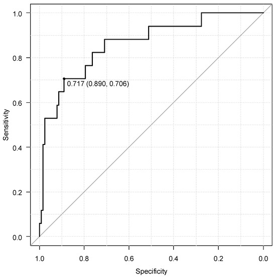
CTR, as shown by chest X-ray, was significantly bigger in the P group. From the echocardiographic results, the LA diameter was significantly longer in the P group; however, there was no difference in the LAV index calculated from body surface area. The LV end-diastolic diameter (LVEDD) was significantly longer in the N group; however, there was no difference in the end-systolic diameter. EF was higher in the N group, on the other hand, there was no statistically significant difference in the percentage of HFpEF. Both the interventricular septum thickness (IVST) and posterior LV wall thickness (PWT) were significantly thicker in the P group (IVST; N group: 13.2 mm, P group: 15.6 mm, p = 0.010, PWT; N group: 12.1 mm, P group: 15.6 mm, p < 0.001). There was no statistically significant difference in the LVM index (LVMI). RWT, which indicates a cardiac hypertrophy pattern, was significantly higher in the P group. These results are summarized in Table 4. Multivariate analysis was performed focusing on RWT. In all three models, RWT significantly and independently predicted for 99mTc-PYP positive patients (Table 5). The cut-off value of RWT from the receiver operating characteristic curve was 0.717 (area under the curve: 0.862, 95% confidence interval: 0.763–0.961) (Figure 1).
Table 4.
Heart morphology in both groups.
Table 5.
Multivariate analysis for assessing ATTR-CM.
Figure 1.
The receiver operating characteristic of relative left ventricular wall thickness for assessing ATTR-CM.
4. Discussion
4.1. ATTR-CM Diagnosis in Patients Who Underwent 99mTc-PYP
Excluding AL amyloidosis, the diagnostic specificity and positive predictive value of ATTR-CM using 99mTc-PYP has been reported to be 100% [14]. The diagnosis of AL amyloidosis requires the detection of M protein in blood and/or urine; however, M protein was not detected in all subjects in the present study. The subjects in the present study were M-protein-negative and showed cardiac accumulation with 99mTc-PYP, and according to the current statement in Japan, ATTR-CM can be diagnosed. In the present study, ATTR-CM was diagnosed in approximately 10% of patients who underwent 99mTc-PYP, which is not different from previous reports from our hospital [5]. It has also been reported that approximately 15% of Japanese HFpEF patients were 99mTc-PYP positive [15]. From this result, 10% of the ATTR-CM diagnostic rate of the present study was reasonable given the unique nature of this disease, which requires early diagnosis and treatment. On the other hand, in the present study, the H/CL ratio in the 99mTc-PYP positive group was 1.94; however, since the H/CL ratio of >1.6 has been reported to be an independent poor prognostic factor, it may be necessary to consider even earlier diagnosis [16]. Furthermore, because wild-type ATTR-CM is more common in elderly patients, it has been reported that it can take more than a year from onset to diagnosis [17]. The aim of the present study was to identify positive cases in patients who underwent 99mTc-PYP; RWT, which indicates the thickening pattern of the ventricular wall, was useful for detecting them.
4.2. Echocardiographic Findings of the ATTR-CM
The Japanese guidelines for cardiac amyloidosis also include information on echocardiography. Two-dimensional method, M-mode method, Doppler method, tissue Doppler method, transesophageal echocardiography, strain Doppler method, and speckle tracking method are each classified as Class I [18]. However, some of these methods cannot be performed at all hospitals. Therefore, an LV wall thickness of 12 mm or more is often used as the initial screening criterion [19]. On the other hand, because diagnosing ATTR-CM based on LV wall thickness alone is difficult, carpal tunnel syndrome and other red flag signs are also used. In echocardiography, in addition to LV wall thickness, the evaluations of LV hypertrophy (LVH) patterns, LV diastolic function, apical sparing and the IWT score were also recommended [20]. This score includes RWT, the ratio between early mitral inflow velocity and mitral annular early diastolic velocity (E/e’), LV longitudinal strain (LS), septal systolic apex-to-base ratio (SAB), and tricuspid annular plane systolic excursion (TAPSE). Among these, RWT was given a higher score. In the present study, RWT was also found to be independently useful for diagnosis, however in this retrospective study, other parameters of IWT score were not assessed in all patients. Therefore, we think it would be useful to combine LV wall thickening with RWT—which is one of the methods used to evaluate LVH—to provide a good indicator that is easily applicable in everyday clinical practice.
In the present study, LVEDD was significantly lower in the P group. Because RWT is calculated from LVEDD and LV wall thickness, it is possible that it may be appropriate to evaluate LVEDD and LVH as with RWT and LVH. In ATTR-CM, LVEDD increases as the disease progresses from transition to HFpEF to HFrEF [21]. In the subjects of the present study, who were diagnosed with an early stage of ATTR-CM, the LVEDD in the P group was prior to enlargement. On the other hand, because LV wall thickness in hypertensive patients is also affected by the number of years they have had HT [22], it is considered that patients with LVH of 12 mm or more, such as those in the present study, had a long history of HT. When high systolic BP due to a long history of HT is accumulated, the higher the cumulative systolic BP, the more advanced the structural changes in the heart, such as LVEDD enlargement [23]. Therefore, differences in RWT, LVEDD, and LV wall thickness occurred in the present study, which captured the early stages of ATTR-CM. Even when evaluation is performed after the disease has progressed, the LV wall progressively hypertrophies in ATTR-CM, and the wall thickness is thicker than in hypertension. In diagnosing ATTR-CM, unlike LVEDD, the diagnostic ability of RWT is maintained regardless of the stage of the disease, and therefore RWT in the IWT score is thought to be high. Other indicators included in the IWT score, such as E/e’, LS, SAB, and TAPSE, were not evaluated in all patients in the present study. These examinations are often difficult for practicing physicians to perform. Therefore, focusing on RWT as a first step in suspecting ATTR-CM from echocardiography is beneficial.
Furthermore, while the cut-off value for RWT in the IWT score is 0.8, the cut-off value in the present study was 0.717. The normal value for RWT is often considered to be below 0.42, and it is important to be aware of cut-off values of 0.7 to 0.8 when proceeding with various examinations such as 99mTc-PYP from both studies.
4.3. Left Ventricular Hypertrophy Pattern and ATTR-CM
In the VALLANT sub-study, LV hypertrophy (LVH) was classified into three patterns based on LVMI and RWT [24]. These three patterns are concentric remodeling (normal LVMI and increased RWT), eccentric LVH (eLVH) (increased LVMI and normal RWT), and concentric LVH (cLVH) (increased both LVMI and RWT). A normal pattern without LVH is when the LVMI does not increase and RWT is normal. The present study demonstrated the progression of concentric remodeling in the PYP-positive group. In the present study, LVMI tended to be lower in the P group; however, this result may be due to the small number of subjects. ATTR-CM is often associated with cLVH [25]; however, a similar patient of ATTR-CM with concentric remodeling has been reported [26], and concentric remodeling has also been shown to be useful for diagnosing ATTR-CM in patients hospitalized for decompensated HF [27]. Based on these results, ATTR-CM exhibits an LVH pattern with increased RWT. On the other hand, HT, one of the most common causes of LVH, often exhibits patterns of increased LVMI (eLVH and cLVH), indicating the pattern of LVH is different [28]. In ATTR-CM, a TTR protein produced in the liver dissociates into monomers, forming amyloid fibrils and accumulating in the heart [29,30]. In conditions with LV afterload, such as HT and aortic valve stenosis, cardiomyocyte hypertrophy occurs. These differences in pathology are thought to be reflected in the different patterns of LVH. Similarly, in Fabry disease, which exhibits accumulation cardiomyopathy, cLVH is the most common, with concentric remodeling and eLVH both occurring in approximately 20% of patients [31]. Therefore, focusing on RWT may be useful in patients of cardiomyopathy characterized by LVH. Although strain echocardiography was not performed in the present study, previous studies have shown that apical strain gradients are useful for distinguishing cardiac amyloidosis on cLVH [32].
4.4. Study Limitations
The main limitation of the present study is a small sample size, which is a single-center retrospective study. There were 144 patients in total, with only 17 in the P group. Since the P group consisted of only 17 patients, the multivariate analysis had to be divided into three models. This also meant that we could not include a large number of factors. Therefore, the possibility of overestimation cannot be ruled out. Additionally, LVH was assessed only by echocardiography, and imaging examinations such as MRI and CT were not included. These examinations have also been reported to be useful in diagnosing ATTR-CM [33]. Although strain echocardiography and other methods have been shown to be useful in differentiating cLVH from ATTR-CM, this retrospective study did not evaluate these methods during echocardiography, which is another limitation of the present study. To address these limitations, prospective studies involving a larger number of patients are needed.
5. Conclusions
In patients with LVH, focusing on RWT on echocardiography may aid in the diagnosis of ATTR-CM.
Author Contributions
Y.M. and S.K. contributed to the study concept and the acquisition of the data. All authors reviewed the raw data and contributed to the analysis and interpretation. S.K. drafted the manuscript and S.H., H.H., Y.M. and T.I. revised it. All authors have read and agreed to the published version of the manuscript.
Funding
The present study received no external funding.
Institutional Review Board Statement
The present study was approved by the Ethics Committee of Toho University Omori Medical Center (approval number: M24196 23100, approval date: 4 March 2025), and been conducted in accordance with the Declaration of Helsinki.
Informed Consent Statement
We had posted the details about the present study in opt-out format were disclosed on the website of our institution and our department (Department of Cardiovascular Medicine) (granted a waiver of informed consent from study participants), and the subjects of the present study were given the opportunity to decline to be enrolled.
Data Availability Statement
The datasets used and/or analyzed during the current study available from the corresponding author on reasonable request.
Acknowledgments
We would like to thank all the members who contributed to the present study.
Conflicts of Interest
The authors declare no conflicts of interest for the present study.
References
- Grogan, M.; Scott, C.G.; Kyle, R.A.; Zeldenrust, S.R.; Gertz, M.A.; Lin, G.; Klarich, K.W.; Miller, W.L.; Maleszewski, J.J.; Dispenzieri, A. Natural History of Wild-Type Transthyretin Cardiac Amyloidosis and Risk Stratification Using a Novel Staging System. J. Am. Coll. Cardiol. 2016, 68, 1014–1020. [Google Scholar] [CrossRef] [Scilit] [PubMed]
- Maurer, M.S.; Schwartz, J.H.; Gundapaneni, B.; Elliott, P.M.; Merlini, G.; Waddington-Cruz, M.; Kristen, A.V.; Grogan, M.; Witteles, R.; Damy, T.; et al. ATTR-ACT Study Investigators. N. Engl. J. Med. 2018, 379, 1007–1016. [Google Scholar] [CrossRef] [Scilit] [PubMed]
- Elliott, P.; Drachman, B.M.; Gottlieb, S.S.; Hoffman, J.E.; Hummel, S.L.; Lenihan, D.J.; Ebede, B.; Gundapaneni, B.; Li, B.; Sultan, M.B.; et al. Long-term Survival With Tafamidis in Patients With Transthyretin Amyloid Cardiomyopathy. Circ. Heart Fail. 2022, 15, e008193. [Google Scholar] [CrossRef] [Scilit] [PubMed]
- Hosono, K.; Kiuchi, S.; Ikeda, T. Cardiac Amyloidosis Patient With Cardiac Conduction Disturbances. J. Clin. Med. Res. 2023, 15, 456–460. [Google Scholar] [CrossRef] [Scilit]
- Kiuchi, S.; Hisatake, S.; Hashimoto, H.; Murakami, Y.; Ikeda, T. Electrocardiographic Features in Transthyretin Cardiac Amyloidosis. Diagnostics 2026, 16, 65. [Google Scholar] [CrossRef] [Scilit]
- Inomata, T.; Tahara, N.; Nakamura, K.; Endo, J.; Ueda, M.; Ishii, T.; Kitano, Y.; Koyama, J. Diagnosis of wild-type transthyretin amyloid cardiomyopathy in Japan: Red-flag symptom clusters and diagnostic algorithm. ESC Heart Fail. 2021, 8, 2647–2659. [Google Scholar] [CrossRef] [Scilit]
- Roguin, A. Henry Cuthbert Bazett (1885–1950)—The man behind the QT interval correction formula. Pacing Clin. Electrophysiol. 2011, 34, 384–388. [Google Scholar] [CrossRef] [Scilit]
- Sokolow, M.; Lyon, T.P. The ventricular complex in left ventricular hypertrophy as obtained by unipolar precordial and limb leads. Am. Heart J. 1949, 37, 161–186. [Google Scholar] [CrossRef] [Scilit]
- Schiller, N.B.; Shah, P.M.; Crawford, M.; DeMaria, A.; Devereux, R.; Feigenbaum, H.; Gutgesell, H.; Reichek, N.; Sahn, D.; Schnittger, I.; et al. Recommendations for quantitation of the left ventricle by two-dimensional echocardiography. J. Am. Soc. Echocardiogr. 1989, 2, 358–367. [Google Scholar] [CrossRef] [Scilit]
- Conrady, A.O.; Rudomanov, O.G.; Zaharov, D.V.; Krutikov, A.N.; Vahrameeva, N.V.; Yakovleva, O.I.; Alexeeva, N.P.; Shlyakhto, E.V. Prevalence and determinants of left ventricular hypertrophy and remodelling patterns in hypertensive patients: The St. Petersburg study. Blood Press. 2004, 13, 101–109. [Google Scholar] [CrossRef] [Scilit]
- Marume, K.; Takashio, S.; Nishi, M.; Hirakawa, K.; Yamamoto, M.; Hanatani, S.; Oda, S.; Utsunomiya, D.; Shiraishi, S.; Ueda, M.; et al. Combination of Commonly Examined Parameters Is a Useful Predictor of Positive 99mTc-Labeled Pyrophosphate Scintigraphy Findings in Elderly Patients With Suspected Transthyretin Cardiac Amyloidosis. Circ. J. 2019, 83, 1698–1708. [Google Scholar] [CrossRef] [Scilit] [PubMed]
- Matsuo, S.; Imai, E.; Horio, M.; Yasuda, Y.; Tomita, K.; Nitta, K.; Yamagata, K.; Tomino, Y.; Yokoyama, H.; Hishida, A.; et al. Collaborators developing the Japanese equation for estimated GFR. Revised equations for estimated GFR from serum creatinine in Japan. Am. J. Kidney Dis. 2009, 53, 982–992. [Google Scholar] [CrossRef] [Scilit] [PubMed]
- Sekijima, Y.; Yazaki, M.; Ueda, M.; Koike, H.; Yamada, M.; Ando, Y. First nationwide survey on systemic wild-type ATTR amyloidosis in Japan. Amyloid 2018, 25, 8–10. [Google Scholar] [CrossRef] [Scilit] [PubMed]
- Gillmore, J.D.; Maurer, M.S.; Falk, R.H.; Merlini, G.; Damy, T.; Dispenzieri, A.; Wechalekar, A.D.; Berk, J.L.; Quarta, C.C.; Grogan, M.; et al. Nonbiopsy Diagnosis of Cardiac Transthyretin Amyloidosis. Circulation 2016, 133, 2404–2412. [Google Scholar] [CrossRef] [Scilit]
- Naito, T.; Nakamura, K.; Abe, Y.; Watanabe, H.; Sakuragi, S.; Katayama, Y.; Kihara, H.; Okizaki, A.; Kawai, Y.; Yoshikawa, M.; et al. ATTR-HFpEF registry investigators. Prevalence of transthyretin amyloidosis among heart failure patients with preserved ejection fraction in Japan. ESC Heart Fail. 2023, 10, 1896–1906. [Google Scholar] [CrossRef] [Scilit]
- Castano, A.; Haq, M.; Narotsky, D.L.; Goldsmith, J.; Weinberg, R.L.; Morgenstern, R.; Pozniakoff, T.; Ruberg, F.L.; Miller, E.J.; Berk, J.L.; et al. Multicenter Study of Planar Technetium 99m Pyrophosphate Cardiac Imaging: Predicting Survival for Patients With ATTR Cardiac Amyloidosis. JAMA Cardiol. 2016, 1, 880–889. [Google Scholar] [CrossRef] [Scilit]
- González-López, E.; Gagliardi, C.; Dominguez, F.; Quarta, C.C.; de Haro-Del Moral, F.J.; Milandri, A.; Salas, C.; Cinelli, M.; Cobo-Marcos, M.; Lorenzini, M.; et al. Clinical characteristics of wild-type transthyretin cardiac amyloidosis: Disproving myths. Eur. Heart J. 2017, 38, 1895–1904. [Google Scholar] [CrossRef] [Scilit]
- Kitaoka, H.; Izumi, C.; Izumiya, Y.; Inomata, T.; Ueda, M.; Kubo, T.; Koyama, J.; Sano, M.; Sekijima, Y.; Tahara, N.; et al. Japanese Circulation Society Joint Working Group. JCS 2020 Guideline on Diagnosis and Treatment of Cardiac Amyloidosis. Circ. J. 2020, 84, 1610–1671. [Google Scholar] [CrossRef] [Scilit]
- Aimo, A.; Tomasoni, D.; Porcari, A.; Vergaro, G.; Castiglione, V.; Passino, C.; Adamo, M.; Bellicini, M.G.; Lombardi, C.M.; Nardi, M.; et al. Left ventricular wall thickness and severity of cardiac disease in women and men with transthyretin amyloidosis. Eur. J. Heart Fail. 2023, 25, 510–514. [Google Scholar] [CrossRef] [Scilit]
- Boldrini, M.; Cappelli, F.; Chacko, L.; Restrepo-Cordoba, M.A.; Lopez-Sainz, A.; Giannoni, A.; Aimo, A.; Baggiano, A.; Martinez-Naharro, A.; Whelan, C.; et al. Multiparametric Echocardiography Scores for the Diagnosis of Cardiac Amyloidosis. JACC Cardiovasc. Imaging 2020, 13, 909–920. [Google Scholar] [CrossRef] [Scilit]
- Achten, A.; Muller, S.A.; Wijk, S.S.-V.; van der Meer, M.G.; van der Harst, P.; van Tintelen, P.; Riele, A.S.T.; van Empel, V.; Oerlemans, M.I.; Knackstedt, C. Diversity of heart failure phenotypes in transthrretin amyloid cardiomyopathy. More than just heart failure with preserved ejection fraction. Ann. Med. 2024, 56, 2418965. [Google Scholar] [CrossRef] [Scilit]
- Scantlebury, D.C.; Kane, G.C.; Wiste, H.J.; Bailey, K.R.; Turner, S.T.; Arnett, D.K.; Devereux, R.B.; Mosley, T.H.; Hunt, S.C.; Weder, A.B.; et al. Left ventricular hypertrophy after hypertensive pregnancy disorders. Heart 2015, 101, 1584–1590. [Google Scholar] [CrossRef] [Scilit] [PubMed]
- Teramoto, K.; Nadruz Junior, W.; Matsushita, K.; Claggett, B.; John, J.E.; Skali, H.; Solomon, S.; Cheng, S.; Shah, A.M. Mid- to Late-Life Time-Averaged Cumulative Blood Pressure and Late-Life Cardiac Structure, Function, and Heart Failure. Hypertension 2020, 76, 808–818. [Google Scholar] [CrossRef] [Scilit] [PubMed]
- Akatsuka, T.; Fujimoto, N.; Ishiyama, M.; Nakamori, S.; Imanaka-Yoshida, K.; Dohi, K. Trajectory of left ventricular geometry and diastolic dysfunction in hereditary transthyretin cardiac amyloidosis. ESC Heart Fail. 2021, 8, 3422–3426. [Google Scholar] [CrossRef] [Scilit] [PubMed]
- Shinada, K.; Endo, J.; Tsuruta, H.; Matsumura, A.; Shirakawa, K.; Okada, M.; Ikura, H.; Kitakata, H.; Moriyama, H.; Ieda, M. Mechanism of mitral valve tethering caused by apical sparing contractility pattern in transthyretin amyloid cardiomyopathy. J. Cardiol. 2025, 86, 552–560. [Google Scholar] [CrossRef] [Scilit]
- Verma, A.; Meris, A.; Skali, H.; Ghali, J.K.; Arnold, J.M.O.; Bourgoun, M.; Velazquez, E.J.; McMurray, J.J.; Kober, L.; Pfeffer, M.A.; et al. Prognostic implications of left ventricular mass and geometry following myocardial infarction: The VALIANT (VALsartan In Acute myocardial iNfarcTion) Echocardiographic Study. JACC Cardiovasc. Imaging 2008, 1, 582–591. [Google Scholar] [CrossRef] [Scilit]
- Melero Polo, J.; Roteta Unceta-Barrenechea, A.; Revilla Martí, P.; Pérez-Palacios, R.; Gutiérrez, A.G.; Juana, E.B.; Gracia, A.A.; Ayala, S.A.; Arregui, M.Á.A. Echocardiographic Markers of Cardiac Amyloidosis in Patients with Heart Failure and Left Ventricular Hypertrophy. Cardiol. J. 2023, 30, 266–275. [Google Scholar] [CrossRef] [Scilit]
- Ganau, A.; Devereux, R.B.; Roman, M.J.; de Simone, G.; Pickering, T.G.; Saba, P.S.; Vargiu, P.; Simongini, I.; Laragh, J.H. Patterns of left ventricular hypertrophy and geometric remodeling in essential hypertension. J. Am. Coll. Cardiol. 1992, 19, 1550–1558. [Google Scholar] [CrossRef] [Scilit]
- Tschöpe, C.; Elsanhoury, A.; Kristen, A.V. Transthyretin Amyloid Cardiomyopathy-2025 Update: Current Diagnostic Approaches and Emerging Therapeutic Options. J. Clin. Med. 2025, 14, 4785. [Google Scholar] [CrossRef] [Scilit]
- Schwarting, S.K.; Bieber, T.; Davies, D.R.; Siepen, F.A.D.; Schwarting, J.; Grabmaier, U.; Massberg, S.; Maurer, M.S.; Kääb, S. Guideline-Directed Medical Therapy for Heart Failure in Transthyretin Amyloid Cardiomyopathy. Circ. Heart Fail. 2025, 18, e011796. [Google Scholar] [CrossRef] [Scilit]
- Meucci, M.C.; Lillo, R.; Del Franco, A.; Monda, E.; Di Brango, C.; Cianci, A.; Lombardo, A.; Ditaranto, R.; Parisi, V.; Biagini, E.; et al. Patterns of Left Ventricular Remodeling and Outcomes in Fabry Disease. J. Am. Soc. Echocardiogr. 2025, 39, 318–320. [Google Scholar] [CrossRef] [Scilit]
- Liu, D.; Hu, K.; Niemann, M.; Herrmann, S.; Cikes, M.; Störk, S.; Gaudron, P.D.; Knop, S.; Ertl, G.; Bijnens, B.; et al. Effect of Combined Systolic and Diastolic Functional Parameter Assessment for Differentiation of Cardiac Amyloidosis from Other Causes of Concentric Left Ventricular Hypertrophy. Circ. Cardiovasc. Imaging 2013, 6, 1066–1072. [Google Scholar] [CrossRef] [Scilit]
- Yamasaki, H.; Kondo, H.; Shiroo, T.; Iwata, N.; Masuda, T.; Makita, T.; Iwabuchi, Y.; Tanazawa, K.; Takahashi, M.; Ono, Y.; et al. Efficacy of Computed Tomography-Based Evaluation of Myocardial Extracellular Volume Combined With Red Flags for Early Screening of Concealed Cardiac Amyloidosis in Patients With Atrial Fibrillation. Circ. J. 2024, 88, 1167–1175. [Google Scholar] [CrossRef] [Scilit]


Disclaimer/Publisher’s Note: The statements, opinions and data contained in all publications are solely those of the individual author(s) and contributor(s) and not of MDPI and/or the editor(s). MDPI and/or the editor(s) disclaim responsibility for any injury to people or property resulting from any ideas, methods, instructions or products referred to in the content. |
© 2026 by the authors. Licensee MDPI, Basel, Switzerland. This article is an open access article distributed under the terms and conditions of the Creative Commons Attribution (CC BY) license.