1. Introduction
The concept of diet, derived from the Greek word
díaita (“way of life”), reflects the deep interconnection between dietary habits, lifestyle, and health. Dietary patterns are shaped by geographical, cultural, social, and economic factors and exert profound effects on quality of life, disease risk, and longevity [
1].
In recent decades, global dietary habits have undergone a marked transition, characterized not only by changes in macronutrient composition but by a fundamental structural transformation of the food matrix itself. This nutritional transition has coincided with a dramatic rise in non-communicable diseases (NCDs), including diabetes, cardiovascular disease, cancer, and chronic respiratory conditions, which now account for the majority of global mortality [
2].
Diet represents a major modifiable risk factor underlying this burden, particularly the shift from traditional, minimally processed diets toward modern dietary patterns dominated by ultra-processed foods (UPFs) [
3,
4].
These foods are characterized by refined substrates, low dietary fiber content, altered physical structures, and the widespread use of synthetic additives such as emulsifiers, artificial sweeteners, preservatives, and colorants.
Increasing evidence indicates that UPFs represent a biologically distinct nutritional paradigm, with health effects that extend beyond caloric density or macronutrient ratios. A central mediator of these effects is the gut microbiota, a metabolically active microbial ecosystem increasingly recognized as a key regulator of host physiology. The gut microbiota plays a crucial role in nutrient metabolism, immune system development, maintenance of mucosal barrier integrity, metabolic signaling, and inflammatory regulation [
5].
Diet is one of the strongest modulators of gut microbiota composition and function, capable of inducing rapid and reproducible changes in microbial communities [
5].
A healthy gut microbiota, characterized by diversity, functional redundancy, and ecological stability (eubiosis), supports immune tolerance and metabolic homeostasis, whereas dysbiosis—marked by reduced diversity, altered community structure, and functional imbalance—has been linked to a wide range of chronic diseases, including obesity, type 2 diabetes (T2D), inflammatory bowel disease, cardiovascular disease, and cancer [
5,
6].
While the adult human gut microbiota is typically dominated by the phyla
Firmicutes and
Bacteroidetes, its composition remains dynamic and highly responsive to dietary, environmental, and lifestyle factors throughout life [
7]. Diets rich in UPFs, low in fiber, and high in saturated fats, refined sugars, and food additives have been consistently associated with reduced microbial diversity, depletion of beneficial taxa, enrichment of pro-inflammatory or opportunistic species, impaired intestinal barrier integrity, and chronic low-grade inflammation [
5].
Importantly, emerging evidence suggests that the effects of modern processed diets are not limited to isolated nutrients or single additives. Rather, chronic exposure to additive-rich UPFs induces cumulative, multi-layered disruptions of the gut ecosystem, affecting microbial composition, metabolic output, mucosal integrity, and host immune responses. These effects may help explain the strong epidemiological links between Western dietary patterns and metabolic dysfunction, inflammatory diseases, and colorectal cancer [
5].
Crucially, susceptibility to diet-induced disruption of the microbiota is not uniform across the lifespan. Early life is characterized by high microbial plasticity and critical windows of immune and metabolic imprinting, rendering the developing microbiome particularly sensitive to dietary exposures. In adulthood, cumulative exposure to processed foods may progressively erode microbial resilience, while ageing is associated with reduced functional redundancy, immune senescence, and diminished capacity for recovery following perturbation. A life-course perspective, therefore, provides a more mechanistically coherent framework for understanding how processed dietary environments shape long-term disease risk.
Rather than providing a purely descriptive synthesis of additive-specific findings, this review proposes a conceptual framework integrating structural barrier disruption, microbial metabolic reprogramming, and immune activation within a life-course-dependent resilience model. By situating multi-additive exposure within a systems-level ecological perspective, we aim to reframe ultra-processed dietary environments as coordinated perturbation fields rather than isolated chemical effects.
2. Ultra-Processed Diets and Additive Exposure: Mechanistic Disruption of the Gut Ecosystem
The NOVA defines UPFs as industrially made items intended for convenience, enhanced palatability, and extended shelf life; they often include food-derived chemicals as well as cosmetic additives [
8].
The modern Western dietary environment is characterized by sustained consumption of UPFs, which expose the gut ecosystem to chronic, low-dose mixtures of emulsifiers, artificial sweeteners, preservatives, colorants, and emerging environmental contaminants. Epidemiological data link additive-rich dietary patterns to colon cancer, inflammatory bowel disease, obesity, insulin resistance, and metabolic-associated fatty liver disease [
9,
10]. Importantly, real-world dietary exposure does not involve isolated compounds but rather continuous multi-additive co-exposure embedded within structurally altered, fiber-depleted food matrices. Rather than acting independently, these exposures converge on three interrelated pathogenic layers: structural disruption, metabolic reprogramming, and immune activation.
A growing body of mechanistic literature indicates that selected additive classes commonly found in UPFs—including emulsifiers, non-nutritive sweeteners, and antimicrobial preservatives—can perturb the gut ecosystem (microbiota composition/function, mucus barrier properties, epithelial integrity, and immune–metabolic signaling), particularly in experimental models [
11,
12,
13]. Because real-world UPF exposure usually involves chronic, low-dose co-exposure to multiple additives, emerging mixture-focused epidemiology supports evaluating combined additive patterns as a distinct exposure paradigm relevant to metabolic disease risk.
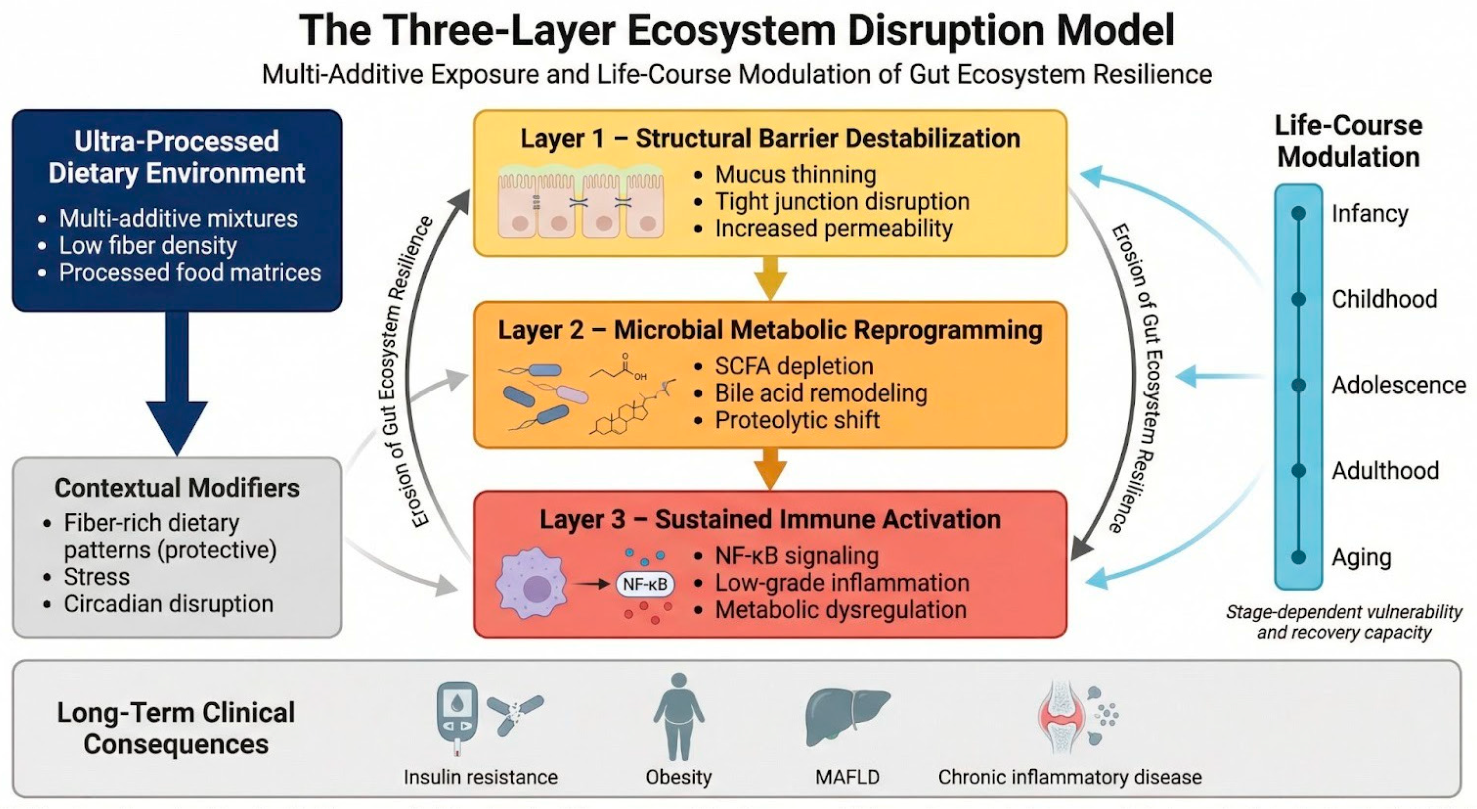
We propose the Three-Layer Ecosystem Disruption Model (TLED Model) (
Figure 1) in which chronic multi-additive exposure embedded within ultra-processed dietary matrices induces:
Structural barrier destabilization;
Microbial metabolic reprogramming;
Sustained immune activation.
These effects collectively lead to erosion of gut ecosystem resilience across the life course.
2.1. Additive-Driven Structural Dysfunction: Mucus Erosion and Tight-Junction Destabilization
The first mechanistic layer of ultra-processed food-associated gut disruption involves impairment of intestinal barrier architecture, including mucus integrity and epithelial tight-junction organization. In the colon, the mucus barrier normally limits direct microbial contact with host tissues; the inner mucus layer is dense and largely impenetrable to bacteria under homeostatic conditions, preserving epithelial sterility at the luminal interface. Goblet cells synthesize and secrete the gel-forming mucin MUC2, which is essential for mucus barrier integrity and mucosal immune homeostasis. Disruption of mucus production or altered mucus physicochemical properties increases susceptibility to inflammation by permitting closer microbe–epithelium interactions [
14,
15].
In preclinical models, common dietary emulsifiers such as carboxymethylcellulose (CMC) and polysorbate-80 (P80) have been shown to alter microbiota composition and promote features consistent with impaired mucus–epithelial spatial organization, including increased microbial encroachment toward the epithelium [
9,
11,
16,
17]. These structural alterations are accompanied by expansion of mucolytic and pro-inflammatory taxa—including
Proteobacteria and
Ruminococcus gnavus—which are capable of penetrating the mucus barrier, alongside depletion of protective organisms such as
Akkermansia muciniphila and members of Bacteroidetes [
18].
In murine systems, emulsifier exposure has been associated with low-grade inflammation, metabolic syndrome–like phenotypes, and exacerbation of colitis in susceptible models, supporting a microbiota- and barrier-linked mechanism [
11]. More recent experimental syntheses continue to cite mucus thinning, increased epithelial permeability, and altered lipopolysaccharide (LPS)-related signaling as plausible downstream pathways in emulsifier-driven host responses, while emphasizing that strength of evidence varies across compounds, doses, and models [
12,
19].
It is important to acknowledge that much of the mechanistic evidence derives from murine models in which administered concentrations may exceed average human intake on a body-weight basis. Although several studies aim to approximate regulatory acceptable daily intake (ADI) levels, interspecies differences in microbiota composition, metabolism, and exposure patterns complicate direct extrapolation. Notably, a randomized controlled-feeding human study evaluating dietary CMC demonstrated measurable perturbations in gut microbiota composition and fecal metabolomic profiles during short-term exposure within ranges relevant to processed food consumption [
20,
21]
Within that trial, a subset of participants exhibited microbiota encroachment into the normally bacteria-sparse inner mucus layer, indicating inter-individual susceptibility to mucus–microbe spatial disruption under emulsifier/thickener exposure. Commentary accompanying these findings underscores the importance of evaluating ubiquitous additives under real-world dietary conditions and highlights biological heterogeneity in host response. Nevertheless, the extent to which chronic low-dose mixture exposure in humans recapitulates murine findings remains incompletely defined. Beyond emulsifiers, in vitro intestinal epithelial models demonstrate that artificial sweeteners such as sucralose, saccharin, and aspartame increase paracellular permeability through downregulation and mislocalization of tight-junction proteins, including claudin-3, occludin, and ZO-1 [
22,
23]. These effects are mediated, at least in part, by oxidative stress-dependent signaling and activation of NF-κB pathways. Saccharin exposure promotes NF-κB-mediated downregulation and degradation of claudin-1, directly compromising epithelial barrier integrity [
13,
24].
Additional structural stressors within ultra-processed dietary environments include certain food colorants, nanoparticulate additives, and emerging contaminants such as micro- and nanoplastics. Experimental studies suggest that these compounds can activate inflammasomes, alter mucosal immune responses, and increase intestinal permeability [
25]. In murine models, polylactic acid (PLA) microplastics have been shown to induce gut dysbiosis, including expansion of Lachnospiraceae, and to contribute to hepatotoxicity [
26].
Collectively, emulsifier-induced mucus erosion, tight-junction destabilization, additive-driven oxidative stress, and contaminant-associated epithelial injury converge to reduce barrier compartmentalization and facilitate translocation of microbial products into the lamina propria and systemic circulation. This structural destabilization constitutes the first pathogenic layer linking ultra-processed dietary exposure to downstream metabolic and immune dysregulation.
2.2. Metabolic Shift: SCFA Depletion, Bile Acid Reconfiguration, and Proteolytic Fermentation
The second pathogenic layer reflects functional reprogramming of microbial metabolic output under additive-rich dietary conditions. At the community level, emulsifier- and sweetener-rich environments suppress short-chain fatty acid (SCFA)-producing taxa, including butyrate-producing Firmicutes, while favoring Gram-negative, lipopolysaccharide-producing bacteria [
11,
16,
17]. Reduced SCFA availability compromises colonocyte energy metabolism, mucin synthesis, and regulatory T-cell differentiation, thereby weakening epithelial immune tolerance and metabolic homeostasis [
27,
28,
29].
Recent experimental evidence further demonstrates that CMC exposure promotes expansion of potentially pathogenic genera, including
Clostridium and
Escherichia–Shigella, while reducing beneficial taxa such as
Lactobacillus and
Akkermansia [
30].
Artificial sweeteners exert parallel metabolic effects. Bian et al. reported sex-specific dysbiosis following acesulfame-K exposure in CD-1 mice, with enrichment of
Sutterella and
Bacteroides in males and depletion of
Clostridium and
Lactobacillus in females [
31,
32].
Prolonged sucralose intake has been associated with reduced luminal butyrate levels and enrichment of
Blautia spp. and
Bacteroides, indicating altered microbial metabolic signaling and substrate utilization [
9,
24].
Beyond SCFA depletion, additive-induced dysbiosis is characterized by altered bile acid metabolism and increased proteolytic fermentation, shifting microbial output toward secondary bile acids and other pro-inflammatory metabolites [
16,
30].
Such changes reflect not only taxonomic shifts, but also reconfigurations of host–microbe metabolic cross-talk, with downstream consequences for insulin sensitivity, lipid metabolism, and inflammatory tone.
2.3. Immune Reprogramming: Sustained Low-Grade Inflammatory Tone
Structural barrier compromise combined with enrichment of Gram-negative taxa enhances systemic exposure to lipopolysaccharides (LPSs), promoting metabolic endotoxemia. Elevated circulating LPS levels have been mechanistically linked to insulin resistance, adipose tissue inflammation, and chronic low-grade immune activation [
16,
33].
Experimental studies demonstrate that emulsifier exposure induces colitis, metabolic syndrome-like features, and tumorigenesis in murine models, supporting a microbiota-dependent inflammatory mechanism [
9,
16,
18].
Sucralose exacerbates DSS-induced colitis through activation of the TLR5–MyD88–NF-κB axis and may contribute to colitis-associated carcinogenesis [
9,
24].
Nanoparticulate additives activate inflammasomes, while micro- and nanoplastics have been shown to alter immune cell populations and cytokine profiles in preclinical models [
26,
34,
35].
Complementing mechanistic findings, large prospective cohorts such as the NutriNet-Santé study report associations between higher emulsifier consumption—including carrageenans (E407), guar gum (E412), and xanthan gum (E415)—and increased risk of T2D, providing epidemiological support for additive-related metabolic dysregulation [
36].
Taken together, structural barrier disruption, metabolic reprogramming, and immune activation operate as a self-reinforcing triad linking additive-rich dietary environments to chronic inflammatory and metabolic disease.
Collectively, these perturbations reduce ecosystem resilience—defined as the capacity of the gut microbiota to resist, adapt to, and recover from environmental stressors. Within ultra-processed dietary environments, chronic multi-additive exposure may therefore compromise not only microbial composition and function but also the adaptive stability of the gut ecosystem itself.
2.4. Multi-Additive Synergy and the Mixture Paradigm
Most mechanistic studies of additives historically tested single additives (e.g., one emulsifier or one sweetener) under controlled conditions, which may not represent real-world UPF consumption patterns where multiple additives are repeatedly co-ingested [
37]. From a risk-assessment perspective, combined exposure to multiple chemicals is increasingly recognized as a relevant framework for food-related chemical mixtures [
38].
EFSA highlights the need for harmonized methodologies to assess chemical mixtures and combined exposure, supporting the scientific rationale for mixture-aware approaches in food additive evaluation [
39]. In this context, synergy refers to the situation where co-exposure to multiple additives produces a gut-ecosystem effect larger (or qualitatively different) than expected from the individual effects alone.
Synergy is plausible in the gut because additives can target distinct but connected layers: (i) mucus barrier structure, (ii) microbial composition and metabolism, and (iii) epithelial/immune signaling, which together determine resilience vs. dysbiosis [
40].
Mice exposed to common emulsifiers such as P80 and CMC showed an increase in metabolic syndrome-like symptoms and low-grade inflammation. On top of that, the inadequate spatial barrier structure was linked to the microbial penetration of the epithelium caused by these emulsifiers.
The major additive classes, their associated microbiota alterations, and their mechanistic effects across the three-layer model are summarized in
Table 1.
A randomized controlled-feeding trial in humans reported that adding CMC to an otherwise additive-free diet perturbed microbiota composition and metabolomic profiles, with microbiota encroachment observed in a subset of participants, highlighting heterogeneity in susceptibility [
20].
If emulsifiers/thickeners weaken mucus structure or increase microbe–epithelium proximity, they may sensitize the host to other concurrently ingested additives by increasing exposure of epithelial and immune sensors to microbial products.
Antimicrobial preservatives can act as selective ecological pressures, inhibiting susceptible organisms and potentially reshaping community structure in ways that alter function (e.g., fermentation capacity, colonization resistance) [
41].
A systematic in vivo evaluation of antimicrobial preservatives reported that preservative exposure can impact gut microbiota and host outcomes in experimental settings, supporting the idea that repeated preservative intake contributes to community restructuring. When combined with barrier-altering emulsifiers, preservative-driven selective pressure may amplify dysbiosis by reducing beneficial competitors while barrier disruption increases the inflammatory consequences of the altered community. Reviews synthesizing additive research found that multiple additive classes can influence microbial metabolic outputs and host-relevant pathways (e.g., barrier signaling and inflammation), indicating that co-exposures may converge on shared downstream mechanisms. Because different additives can alter distinct microbial processes (growth suppression, stress responses, substrate utilization), co-exposure can plausibly destabilize microbial networks and reduce resilience even if each additive alone only produces modest shifts [
42].
In the NutriNet-Santé cohort analysis, investigators empirically identified food additive mixture patterns reflecting real-world co-consumption and examined their associations with incident T2D. An increased incidence of T2D was positively associated with two of the five detected combination patterns. Two of the combination patterns that were seen included sweeteners for acidity management and associated chemicals, and the other contained various textural agents such modified starches, pectin, E412, E407, and E415 [
37].
A PLOS Medicine cohort paper explicitly notes the need for experimental studies to clarify underlying mechanisms, including potential synergistic/antagonistic effects within mixtures, reinforcing multi-additive synergy as a testable hypothesis rather than a rhetorical claim [
37].
Public research communications summarizing the same findings report that the observed mixtures align with common UPF categories (e.g., dairy desserts/fats/sauces and artificially sweetened beverages), supporting ecological validity of mixture patterns [
37].
Multi-additive synergy is a biologically plausible and increasingly evidence-motivated paradigm because UPF diets generate chronic co-exposures and because the gut ecosystem integrates additive effects across barrier structure, microbial ecology, and host signaling. Regulatory-science frameworks already recognize the need to assess combined chemical exposures, supporting the translation of mixture thinking into food additive research and safety assessment [
39].
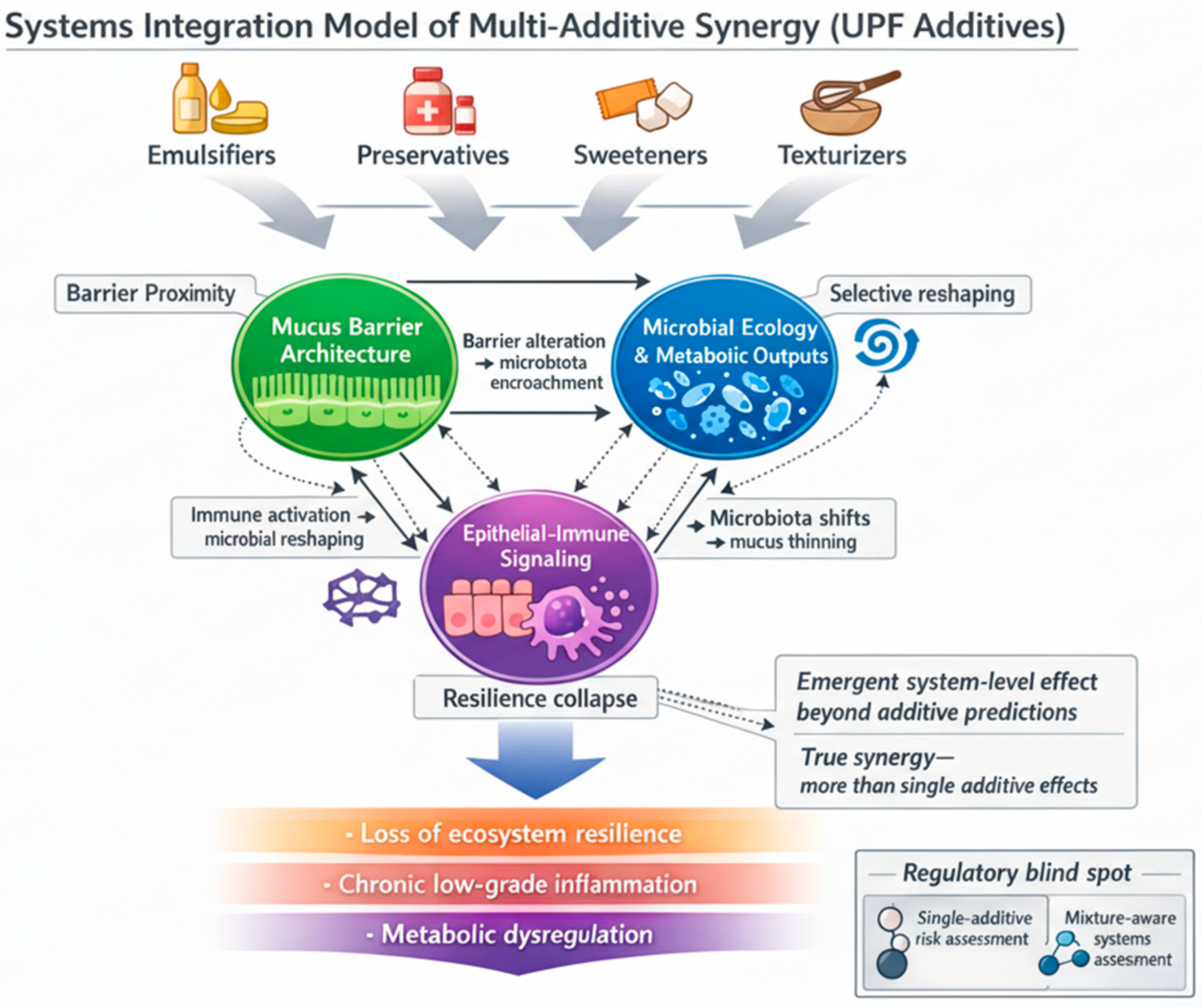
While the TLED Model conceptualizes the coordinated structural, metabolic, and immune cascade induced by ultra-processed dietary environments, real-world exposure rarely involves single compounds. Instead, multiple additive classes are chronically co-ingested within processed food matrices. To mechanistically contextualize this multi-additive exposure paradigm, we provide an additional systems-level framework (
Figure 2) illustrating how simultaneous perturbations across barrier architecture, microbial ecology, and epithelial–immune signaling may generate non-linear amplification beyond single-additive effects. This figure does not represent a separate model, but rather a mechanistic extension of the TLED framework, emphasizing mixture-driven system-level interactions and potential limitations of single-additive risk assessment approaches.
3. Dietary Context as Modifier of Additive Effects: Protective Roles of Mediterranean, Vegetarian and Fiber-Rich Dietary Patterns on Gut Microbiota
Modern dietary patterns rich in UPFs expose the gut microbiota to multiple food additives whose long-term biological effects were historically underestimated. Emerging evidence indicates that emulsifiers, artificial sweeteners, preservatives, and synthetic dyes can alter microbial diversity, compromise mucosal integrity, reduce short-chain fatty acid (SCFA) production, and promote low-grade inflammation. However, the overall dietary context appears to significantly modify these additive-related effects.
Mediterranean diet, vegetarian diets, and fiber-rich nutritional models—may buffer or counteract additive-induced dysbiosis. We synthesize mechanistic, experimental, and clinical evidence demonstrating that plant-forward, fiber-dense dietary environments enhance microbial diversity, increase SCFA-producing taxa, and strengthen epithelial barrier function. We propose that dietary context acts as a critical modifier of additive impact, influencing whether microbial perturbations translate into metabolic and inflammatory pathology.
Modern Western dietary patterns, characterized by UPFs, refined sugars, saturated fats, and industrial additives, are strongly associated with microbial dysbiosis and metabolic dysfunction [
43].
Increasingly, research suggests that not only macronutrient composition but also additive exposure contributes to microbiome disruption [
44,
45]. Importantly, dietary context may determine whether these perturbations persist or are mitigated. While additive exposure may promote dysbiosis, the surrounding dietary matrix—particularly fiber content, plant diversity, and polyphenol intake—can substantially modify these effects. Low-carbohydrate and high-protein diets illustrate the importance of distinguishing macronutrient distribution from food quality. When low-carbohydrate regimens preserve plant-derived fiber, microbial richness can be maintained or even increased. In contrast, extreme animal-based, low-fiber patterns favor bile-tolerant and proteolytic taxa, reduce butyrate-producing bacteria, and shift microbial metabolism toward branched-chain fatty acids and potentially deleterious metabolites.
Thus, microbial outcomes depend less on carbohydrate restriction per se and more on fiber preservation and food structural complexity. This distinction is essential when interpreting microbiota responses to dietary interventions.
3.1. Mediterranean Diet and Microbial Resilience
The Mediterranean diet, characterized by high intake of dietary fibre, polyphenols, and unsaturated fatty acids, is consistently associated with increased microbial richness and enrichment of short-chain fatty acid (SCFA)-producing taxa. Intervention studies demonstrate increased abundance of genera such as
Faecalibacterium,
Roseburia, Bifidobacterium, and
Prevotella, alongside reductions in pro-inflammatory markers including CRP and IL-17 [
46,
47,
48].
These effects are likely mediated by sustained provision of fermentable substrates and bioactive compounds that reinforce mucosal integrity, promote SCFA production, and support ecological stability within the gut microbiome. Rather than acting through a single nutrient, the Mediterranean diet appears to enhance microbial resilience through its structural and compositional complexity.
Studies report increased abundance of
Bacteroides,
Bifidobacterium,
Prevotella,
Roseburia,
Lactobacillus,
Faecalibacterium, and
Clostridium clusters under Mediterranean adherence, alongside reduced
Proteobacteria [
47,
49,
50,
51]. Increased
Prevotella-to-
Bacteroides ratios suggest enhanced fiber fermentation capacities [
49,
50].
Pagliai et al. demonstrated significant microbiome remodeling after a 3-month Mediterranean intervention, with increased SCFA production and shifts toward beneficial taxa [
51]. Nagpal et al. (2018) demonstrated that a Mediterranean diet in non-human primates promotes a more diverse and functionally beneficial gut microbiome, enriched in SCFA-producing taxa, whereas a Western diet induces dysbiosis, reduced diversity, and a pro-inflammatory microbial profile associated with metabolic dysfunction [
52]. Ghosh et al., in a 12-month multicenter study (
n = 612), observed reductions in inflammatory markers including CRP, IL-17, and IL-2, alongside increased IL-10 [
53].
Mechanistically, high fiber intake promotes butyrate production, strengthens tight junction integrity, enhances mucus synthesis, and modulates immune responses—potentially counteracting additive-induced epithelial damage.
3.2. Vegetarian Diets and Plant-Based Diets
Vegetarian diets exclude meat and fish and are typically rich in dietary fiber, plant proteins, and polyphenols while low in saturated fat and heme iron [
54]. Cross-sectional and cohort studies demonstrate that vegetarians exhibit greater microbial richness compared to omnivores [
55].
Plant-based diets consistently increase
Prevotella,
Ruminococcus, Lactobacillus,
Eubacterium, and SCFA-producing
Clostridium clusters [
54,
56,
57,
58]. Large-scale analyses (>20,000 individuals) reveal distinct microbial “signatures” associated with vegetarian and vegan patterns, characterized by enrichment of fiber-fermenting bacteria and reductions in bile-tolerant, inflammation-associated taxa such as
Bilophila wadsworthia [
59,
60].
Functional metagenomic analyses indicate enhanced carbohydrate fermentation pathways in vegetarian and vegan microbiomes [
57]. The higher fiber substrate availability likely buffers against additive-induced microbial depletion by promoting fermentative resilience and ecological redundancy.
3.3. Low-Carbohydrate Diets: The Role of Fiber Preservation
Dietary fiber is the primary substrate for SCFA-producing bacteria. Butyrate, acetate, and propionate support epithelial energy metabolism, reinforce tight-junction proteins, regulate immune tolerance, and suppress inflammatory signaling [
61,
62].
High-protein, low-fiber regimens reduce butyrate producers such as
Roseburia spp. and
Eubacterium rectale and increase proteolytic fermentation products, including branched-chain fatty acids and N-nitroso compounds [
63]. Conversely, fiber-inclusive low-carbohydrate diets can preserve microbial richness and promote favorable phylum-level shifts [
64].
Thus, fiber availability appears to be the key determinant in whether dietary modifications enhance or compromise microbial resilience. When “low-carb” diets become “low-plant, high-animal, low-fiber” diets, microbial diversity and butyrogenic capacity decline [
60,
63].
4. Lifestyle Amplifiers of Gut Ecosystem Disruption
Although ultra-processed dietary exposure represents the central perturbation discussed in this review, lifestyle factors such as chronic stress and circadian misalignment converge on the same structural, metabolic, and immune pathways described in the TLED Model. Rather than acting independently, these factors may amplify additive-driven barrier dysfunction, microbial reprogramming, and inflammatory activation, thereby lowering ecosystem resilience.
4.1. Chronic Stress as a Convergent Amplifier of Barrier and Immune Dysregulation
Chronic psychological or physiological stress disrupts gut homeostasis, primarily through sustained activation of the hypothalamic–pituitary–adrenal (HPA) axis and the sympathetic nervous system. Persistent elevation of cortisol and catecholamines alters epithelial integrity, mucosal immunity, and microbial ecology [
65,
66]
Experimental and in vivo studies demonstrate that chronic stress increases intestinal permeability through disruption of tight-junction proteins and altered mucosal immune signaling [
67,
68]. Stress exposure is associated with reduced production of antimicrobial peptides and decreased secretory IgA, weakening mucosal defense and barrier compartmentalization [
69,
70]. Increased permeability facilitates translocation of microbial products, including lipopolysaccharides (LPSs), into systemic circulation.
Sympathetic neurotransmitters such as norepinephrine directly enhance bacterial growth, iron acquisition, motility, and biofilm formation in opportunistic taxa including
Escherichia coli and other
Enterobacteriaceae [
71,
72]. Chronic stress is consistently associated with reduced abundance of beneficial commensals such as
Lactobacillus and decreased overall microbial diversity, weakening colonization resistance [
69,
73]. Stress-induced inflammatory changes increase luminal nitrate availability, further favoring
Proteobacteria expansion [
74,
75].
Stress-mediated increases in TNF-α and IL-6 promote low-grade inflammatory tone and may exacerbate metabolic endotoxemia [
76,
77].
Within additive-rich dietary environments, chronic stress may therefore lower the threshold for barrier destabilization and immune activation, amplifying the three-layer cascade and accelerating resilience erosion.
4.2. Circadian Rhythm Disruption and Oscillatory Instability of the Gut Ecosystem
The gut microbiota exhibits pronounced diurnal oscillations in composition and metabolic output, coordinated by host circadian clock genes including CLOCK, BMAL1, PER, and CRY [
65,
78,
79,
80,
81,
82,
83,
84]. Feeding timing acts as a primary synchronizing cue for microbial rhythmicity.
Circadian regulation influences bile acid synthesis, antimicrobial peptide secretion, epithelial turnover, and immune receptor expression, including rhythmic Toll-like receptor (TLR) activity [
79,
85,
86,
87,
88]. Microbial metabolites reciprocally influence host clock gene expression via G-protein-coupled receptors and nuclear receptors such as REV-ERBα [
79,
85,
86].
Circadian misalignment—due to night-shift work, irregular eating patterns, sleep deprivation, or jet lag—is associated with reduced microbial diversity, altered
Firmicutes:Bacteroidetes ratios, increased
Proteobacteria, increased intestinal permeability, and systemic inflammation [
79,
84,
89,
90,
91,
92,
93,
94,
95]. Persistent circadian disruption has been linked to metabolic syndrome, obesity, T2D, and inflammatory bowel disease [
9,
79,
90,
96].
Within the TLED framework, circadian disruption affects the gut on multiple interconnected levels. When normal day–night rhythms are disturbed, intestinal permeability tends to increase and epithelial repair processes become less efficient, weakening the physical barrier between microbes and host tissues. At the same time, irregular feeding patterns alter nutrient flow and bile acid cycling, reshaping microbial metabolism and shifting the functional output of the gut microbiota. On the immune side, disrupted circadian control of TLR activity and inflammatory signaling lowers the threshold for immune activation, making the gut more reactive to both microbial products and dietary exposures. Together, these changes can amplify additive-driven perturbations and further erode ecosystem resilience. Thus, circadian misalignment may sensitize the gut ecosystem to additive-driven perturbation by destabilizing oscillatory equilibrium and reducing recovery capacity following dietary stress.
4.3. The Combined Impact of Diet and Lifestyle on Gut Ecosystem Resilience
Chronic stress and circadian disruption do not introduce independent pathogenic pathways; rather, they converge mechanistically with additive exposure on barrier integrity, microbial metabolism, and immune regulation. In the context of ultra-processed dietary environments, these lifestyle factors may act as amplifiers of structural destabilization and inflammatory activation.
This convergence supports a broader conceptualization of modern lifestyle as a coordinated perturbation field in which dietary additives, psychosocial stressors, and chronobiological disruption collectively shape gut ecosystem stability across the life course. Collectively, these dietary models demonstrate that microbial resilience is enhanced when diets provide structural complexity, fermentable substrates, and minimal additive exposure. In contrast, ultra-processed diets combine low fiber availability with multi-additive co-exposure, creating a permissive environment for mucus degradation, barrier dysfunction, and inflammatory dysbiosis.
Importantly, protective dietary patterns do not merely “increase good bacteria”; they reinforce ecosystem-level stability, functional redundancy, and host–microbe homeostasis. This resilience may buffer against inflammatory triggers and metabolic stressors, particularly during sensitive life stages such as early childhood and aging.
By integrating stress biology and chronobiology into the TLED Model, the present framework underscores that resilience is determined not only by dietary composition but also by the cumulative interaction between environmental exposures and host regulatory systems.
5. Life-Course Modulation of Diet-Induced Dysbiosis
The Three-Layer Model operates across the life course, but susceptibility and recovery capacity vary according to developmental stage, reflecting stage-specific microbial plasticity and host vulnerability.
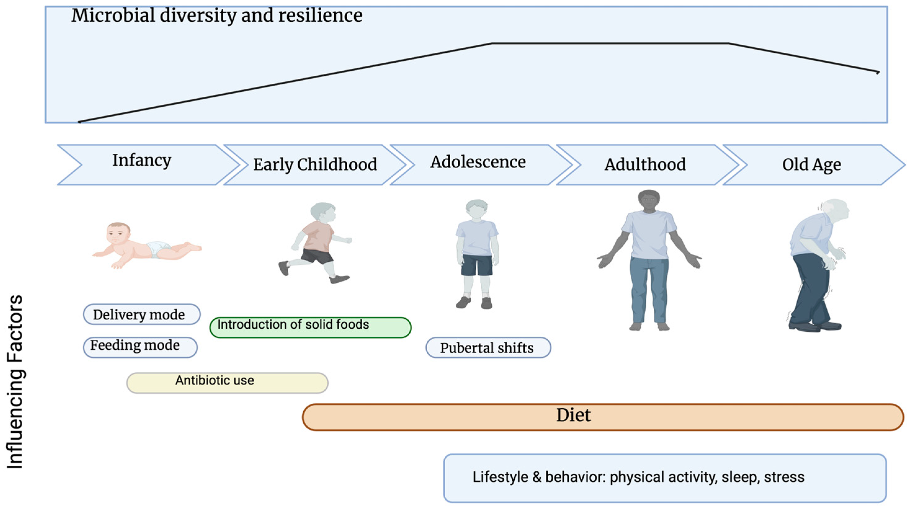
The TLED Model operates across the life course; however, susceptibility and recovery capacity vary according to developmental stage. The gut microbiota evolves from low-diversity colonization at birth, to relative stability in adulthood, and toward reduced resilience in older age [
97]. At each stage, dietary exposures interact with stage-specific microbial plasticity, immune maturation, and metabolic capacity (
Figure 3).
Importantly, microbial perturbations acquired early in life may persist as latent functional imprints—what can be conceptualized as microbiological scarring—shaping microbiome responsiveness to later-life stressors, including ultra-processed dietary environments [
98,
99].
5.1. Infancy—Plasticity and Developmental Programming
At birth, the human gut is rapidly colonized by microorganisms, which vary depending on the delivery mode, antibiotic use and feeding mode (breast milk or formula).
Infancy represents a period of high microbial plasticity and tight integration between microbiota, immune education, and metabolic maturation. Delivery mode, feeding practices, and antibiotic exposure strongly influence early colonization patterns [
100]. The gut microbiota of infants born via vaginal delivery is primarily colonized by maternal vaginal and fecal microbiota, dominated by genera such as
Lactobacillus and
Prevotella and high levels of
Bacteroides and
Bifidobacterium. Infants born via cesarean section have a lower microbial diversity, and the gut is colonized by microorganisms from the maternal skin and hospital environment. Key genera associated with C-sections are
Staphylococcus,
Streptococcus,
Corynebacterium, and
Propionibacterium, as well as increased
Enterococcus and
Klebsiella [
101,
102].
In addition, exclusive breastfeeding promotes a gut community dominated by
Bifidobacteria due to the selective effect of human milk oligosaccharides (HMOs), while formula-feeding is associated with a more diverse, adult-like profile containing higher levels of
Clostridiales and
Proteobacteria [
103].
During this window, additive-driven structural disruption (e.g., mucus thinning, altered SCFA signaling) may interfere with barrier maturation and regulatory T-cell calibration. Because foundational host–microbe interactions are being established, even transient dysbiosis may produce durable functional imprinting, persisting beyond apparent taxonomic recovery [
104,
105,
106].
5.2. Childhood—Metabolic Imprinting
Although the microbiota becomes increasingly adult-like during early childhood, it remains functionally immature [
107,
108]. The administration of broad-spectrum antibiotics in early infancy acts as a major perturbation, causing a temporary decrease in overall microbial richness and a significant reduction in beneficial taxa like
Bifidobacterium, with the community taking a prolonged period to normalize [
109].
Within the first two years of life, the gut microbiome undergoes dramatic shifts primarily induced by the introduction of solid foods. Taxonomic similarity does not imply adult-equivalent metabolic capacity; pathways related to vitamin biosynthesis, oxidative phosphorylation, and immune modulation continue to mature through childhood and adolescence [
110,
111]. While some studies suggest that the gut microbiota reaches an “adult-like” configuration by 3 years, other authors support the idea that maturation continues through childhood and even adolescence [
98,
107,
112]. For instance, children’s microbiota is shown to be enriched in
Bifidobacterium,
Faecalibacterium, and members of
Lachnospiraceae, whereas adults have higher abundances of
Bacteroides.
Disruptions during this extended developmental window—including antibiotic exposure, low-fiber intake, or early introduction of additive-rich foods—may alter microbial network consolidation and metabolic programming. Epidemiological data link early-life dysbiosis to later risk of allergic disease, obesity, metabolic dysfunction, and immune-mediated conditions. Derrien et al. [
107] report that while the gut microbiota of infants (<3 years) and adults has been described, there is little data on pre-school (3–6 years), school-age (6–12 years) and adolescence (12–18 years) stages. However, research shows that the microbiome does not fully stabilize at this stage; instead, co-occurrence networks continue to consolidate, β-diversity gradually diminishes, and functional attributes continue shifting [
98]. Several studies link early-life dysbiosis with increased risk for allergic diseases, asthma, obesity, metabolic disorders, autoimmunity, and even neurodevelopmental and behavioral outcomes [
100,
113,
114,
115]. The introduction of UPFs, synthetic additives (e.g., emulsifiers, artificial sweeteners), low dietary fiber, or other modern dietary patterns during complementary feeding may derail optimal microbial colonization—leading to dysfunctional interactions between the microbes and the host from early on [
8,
13,
16,
116].
Thus, the early period of life, from infancy to early childhood, represents a window of vulnerability but also an opportunity: dietary modulation (breast feeding, high intake of minimally processed, fiber-rich foods) could support healthy microbiota establishment and long-term disease prevention [
106,
117,
118,
119].
Within the TLED framework, structural and metabolic perturbations during childhood may recalibrate inflammatory and metabolic set points that persist into adulthood.
5.3. Adolescence—Transitional Vulnerability
Following childhood metabolic imprinting, adolescence represents a second developmental recalibration phase characterized by hormonal and immune remodeling. Metabolic shifts—including reduced SCFA production and altered bile acid signaling—may influence adiposity trajectories, insulin sensitivity, and inflammatory tone during this transitional stage. Structural and immune perturbations may interact with pubertal immune modulation, amplifying long-term cardiometabolic risk.
Adolescence also represents a period marked by physiological changes—hormonal maturation, neurodevelopment, growth and immune modulation. The interplay between diet, the microbiota, and the host during this period may therefore have unique consequences.
In adolescence, the microbiota tends towards an adult-like composition, but it continues to mature and is strongly influenced by external factors such as diet, lifestyle and environmental exposures [
98].
One study showed that a 4-week lifestyle and diet intervention in adolescents positively influenced the gut microbiota, increasing the microbial diversity and the abundance of health-promoting bacterial species such as
Lachnospira,
Alistipes,
Barnesiella. Functional predictions showed upregulation of genes related to energy production and metabolism [
112].
A review on dietary patterns and mental health in adolescents highlights how high-sugar, low-fibre diets can lead to disruptions in the gut microbiota composition and contribute to mental health disorders such as mood disorders, anxiety, ADHD and depression via the gut–brain axis [
120]. This is also supported by animal studies, which show that high-fat diets in adolescence induce long-lasting changes in the gut microbiota of rodents and expression of neuroimmune and neurotransmission-related genes in adulthood, suggesting that adolescent diets may have enduring effects on brain function via microbiota-mediated pathways [
121].
In addition, adolescence may represent a vulnerable period for shaping metabolic health: diet-induced microbiota alterations during adolescence could influence energy balance, insulin sensitivity, inflammatory tone, and risk of obesity or metabolic syndrome in later life. A systematic review on microbiota–adiposity associations in children and adolescents reported inconsistent but suggestive associations between early-life microbial composition and later adiposity.
Because adolescence also represents a critical period in the development of the gut microbiome and thus general health, dietary and lifestyle interventions may still “course-correct” trajectories of dysbiosis. The improvements in microbial diversity and beneficial taxa seen after a short-term dietary change in adolescents show that these alterations can still be reversed [
122].
Nevertheless, current evidence on microbiome-based interventions (prebiotics, probiotics, SCFAs) for pediatric obesity is still of low certainty. A 2025 Cochrane-style review concluded that in adolescents (10–19 years), microbiome-based interventions may lead to small reductions in BMI or waist circumference, but evidence is limited by small sample sizes, short follow-ups, and methodological heterogeneity [
123].
Given the magnitude of dietary shifts many adolescents undergo (e.g., increased processed food consumption, “Western” diet patterns), more longitudinal, controlled studies are needed—especially to assess long-term metabolic, immunological, and neurobehavioral outcomes.
5.4. Adulthood—Cumulative Burden
In adulthood, the microbiome exhibits relative stability but remains modifiable under sustained dietary pressure. The gut microbiota generally attains a relatively stable, differentiated composition, usually dominated by the major bacterial phyla
Firmicutes,
Bacteroidetes, and
Actinobacteria [
97,
100]. By young adulthood, the relative stability and intra-individual variation over time is modest: some studies estimate that approximately 20–30% of microbiota compositional variation is explained by intra-individual factors over time, emphasizing resilience under constant lifestyle and diet [
100].
Within this stage, the Three-Layer cascade manifests as a cumulative burden: persistent barrier stress, reduced SCFA production, and chronic low-grade inflammatory activation. Long-term consumption of additive-rich, low-fiber diets is associated with depletion of SCFA-producing taxa (e.g.,
Akkermansia muciniphila,
Faecalibacterium prausnitzii), increased permeability, and metabolic dysregulation [
124].
Over decades, this “slow-burn” erosion of resilience may contribute to insulin resistance, fatty liver disease, cardiovascular risk, and inflammatory disorders [
124,
125].
Given the fact that adulthood often spans over decades, the cumulative effects of high intake of processed food consumption may have substantial outcomes. A recent review summarized how UPFs—defined as foods with low fiber, synthetic additives, high glycemic loads, and processing-related structural changes—are consistently associated with reduction in microbial diversity, loss of beneficial taxa, enrichment of pro-inflammatory organisms, and impairment of gut barrier function and resilience [
124].
Specifically, decreased abundance of SCFA-producers such as
Akkermansia muciniphila and
Faecalibacterium prausnitzii is concerning: SCFAs are critical for colonocyte energy, mucin production, immune tolerance (promoting regulatory T cells), anti-inflammatory effects, and suppression of opportunistic/pathogenic bacteria [
125].
Loss of these protective functions over decades explains the relationship between UPF-rich diets and chronic diseases: metabolic syndrome, obesity, T2D, non-alcoholic fatty liver disease (NAFLD), cardiovascular disease, and even colorectal cancer. Indeed, epidemiological data increasingly link high UPF consumption with such chronic disease burdens [
126,
127,
128,
129].
Moreover, chronic, low-level exposure to additives (e.g., emulsifiers compromising mucosal barrier, surfactants altering mucus layer, artificial sweeteners interfering with microbial metabolism) may produce a “slow burn” of gut barrier dysfunction, low-grade endotoxemia, immune activation, and metabolic inflammation—which are likely exacerbated by aging or other stressors [
13,
16].
Because the adult microbiota remains modifiable, dietary interventions, increased fibre intake, prebiotic and probiotic supplementation, and reductions in processed foods may help restore microbial balance, increase SCFA production, and improve gut barrier and metabolic health. Several authors have called for “microbiota-friendly” dietary guidelines, with a focus on reducing UPF and additive exposure [
97,
100,
124].
Adult dysbiosis may therefore represent both the consequence of contemporary dietary exposure and the cumulative expression of earlier microbiological scarring.
5.5. Ageing—Reduced Resilience and Inflammaging
Ageing is associated with reduced functional redundancy, immune remodelling, and diminished recovery capacity following perturbation. Structural barrier integrity may decline due to impaired mucin production and epithelial repair since ageing is associated with alterations in microbial diversity, composition, metabolic capacity, and resilience [
130,
131]. Although some centenarian cohorts exhibit increased α-diversity, this likely reflects ecological heterogeneity rather than a uniform longevity signature [
132]. Studies comparing elderly patients (nonagenarians, centenarians) and younger patients have reported that centenarians frequently harbour distinct gut microbiota configurations [
133].
These may include higher richness/diversity, enrichment of certain beneficial or resilience-associated taxa (e.g.,
Akkermansia,
Christensenellaceae,
Bifidobacterium,
Lactobacillus), and retention of SCFA-producing capacity [
134,
135]. A very recent study reported that centenarians had significantly higher Chao1 richness index scores compared to middle-aged adults, and that genera such as
Akkermansia,
Lactobacillus, and
Christensenella were more prevalent in the centenarian group [
136].
Other studies showed that taxa such as members of the phylum
Verrucomicrobia (e.g.,
Akkermansia muciniphila)—known to support mucin integrity and gut barrier—often decrease in abundance with age, leading to decreased SCFA production, impaired mucosal protection, and increased gut leakiness [
125,
137].
A large-scale metagenomic/culturomic study of South Chinese centenarians documented that the relationship between age and microbiota composition is non-linear: centenarians displayed distinct configurations compared to both younger adults and elderly. For example, in centenarians, the relative abundance of
Roseburia and
Escherichia was increased, while several genera (e.g.,
Faecalibacterium,
Parabacteroides,
Butyricimonas,
Coprococcus,
Megamonas,
Mitsuokella,
Sutterella,
Akkermansia) decreased compared with non-centenarians [
135].
Moreover, longevity is associated in some populations with increased microbial richness, functional capacity for antioxidant activity, and maintenance of beneficial microbial functions—suggesting that certain microbiota configurations may support healthy aging, resilience, and possibly longevity [
134,
138].
Functional stability, metabolic capacity, and longitudinal resilience—not diversity metrics alone—are required to define protective microbiome configurations [
139,
140].
The gut community exhibits a marked reduction in key beneficial, SCFA-producing bacteria, such as
Faecalibacterium,
Bacteroidaceae, and
Lachnospiraceae. This compositional decline is reflected in the reduced functional potential for broad carbohydrate metabolism and amino acid synthesis pathways [
137]. Moreover, there is an increased accumulation of inflammatory commensals. These changes may contribute to gut barrier dysfunction, systemic inflammation, and aging-related morbidities [
125].
In older adults, additive-driven structural and metabolic perturbations may be amplified by inflammaging and reduced colonization resistance, increasing vulnerability to frailty, metabolic disease, and inflammatory conditions [
141,
142]. At this stage, resilience may depend heavily on dietary quality and preservation of SCFA-producing networks [
143,
144].
Old age—especially in the context of processed diets, polypharmacy, reduced dietary quality, lower fiber intake, reduced physical activity, and multimorbidity—may represent a phase of heightened vulnerability, where microbiota dysbiosis contributes to frailty, metabolic, inflammatory, and neurodegenerative diseases [
145,
146].
However, this stage may also represent a potential window for targeted interventions (dietary, pre-/probiotic, lifestyle) aimed at maintaining microbial resilience and promoting healthy aging [
147].
Age-related loss of microbial resilience may therefore unmask earlier microbiological scarring, amplifying inflammatory and metabolic consequences of diets that might have been better tolerated in earlier life [
133].
Continued mechanistic and longitudinal research is essential to clarify additive-specific risks, identify vulnerable populations, and guide evidence-based dietary recommendations that protect gut microbial health [
116].
6. Conclusions and Perspectives
Current evidence supports the concept that ultra-processed food (UPF) environments exert coordinated pressure on the gut ecosystem through three interrelated pathogenic layers: structural barrier disruption, microbial metabolic reprogramming, and sustained immune activation. Rather than acting as isolated agents, food additives operate within a multi-additive exposure paradigm that may cumulatively erode microbial resilience and promote low-grade inflammatory states linked to metabolic and chronic disease risk.
Importantly, vulnerability to additive-driven dysbiosis is not uniform across the lifespan. Early-life microbial plasticity may permit long-lasting ecological imprinting, whereas aging-associated reductions in diversity and immune adaptability may impair recovery following dietary perturbation. In adulthood, cumulative exposure appears to amplify the three-layer cascade over time, potentially contributing to metabolic and inflammatory burden.
Conversely, fiber-rich, minimally processed dietary patterns enhance ecosystem resilience by preserving short-chain fatty acid production, supporting barrier integrity, and maintaining functional redundancy within microbial communities. These findings suggest that overall dietary structure and additive burden—rather than macronutrient composition alone—are critical determinants of long-term microbiome stability.
Although much mechanistic insight derives from experimental models and human evidence remains heterogeneous, the convergence of epidemiological, translational, and mixture-based research supports the biological plausibility of additive-associated microbiota disruption. Future studies should prioritize long-term, life-course-oriented human investigations integrating additive mixture exposure assessment, repeated microbiome profiling, and clinically relevant metabolic and immune endpoints. Such approaches will be essential for translating microbiome science into actionable dietary guidance.
By formalizing additive-driven gut ecosystem perturbation within a structured, life-course-oriented systems model, this framework provides a foundation for future experimental validation, mixture-based risk assessment, and microbiota-informed dietary policy.
Author Contributions
Conceptualization, TLED Model development, and overall study design, S.M.C.; methodology, M.M., G.F.R.M., and L.A.P.; software, M.M. and A.C.S.; validation, S.M.C., G.F.R.M., and I.M.; formal analysis, M.M.; data curation, M.M., G.F.R.M., and L.A.P.; writing—original draft preparation, M.M., L.A.P., A.C.S., G.F.R.M., and I.M.; writing—critical review and editing, S.M.C. and A.C.S.; visualization, M.M., L.A.P., G.F.R.M., A.C.S., S.M.C., and I.M.; supervision, S.M.C.; project administration, S.M.C. All authors have read and agreed to the published version of the manuscript.
Funding
This research received no external funding.
Institutional Review Board Statement
Not applicable.
Informed Consent Statement
Not applicable.
Data Availability Statement
No new data were created or analyzed in this study.
Acknowledgments
The figures were generated using the FigureLabs AI platform (
https://chat.figurelabs.ai) following the authors’ original scientific concept and detailed instructions. All generated content was critically reviewed, modified where necessary, and approved by the authors.
Conflicts of Interest
The authors declare no conflicts of interest.
Abbreviations
The following abbreviations are used in this manuscript:
| ADHD | Attention Deficit Hyperactivity Disorder |
| ADI | Acceptable Daily Intake |
| AI | Artificial Intelligence |
| BMI | Body Mass Index |
| BMAL1 | Brain and muscle ARNT-like protein 1 |
| CMC | Carboxymethylcellulose |
| CLOCK | Circadian Locomotor Output Cycles Kaput |
| CRP | C-reactive protein |
| CRY | Cryptochrome |
| DSS | Dextran Sulfate Sodium |
| E407 | Carrageenan |
| E412 | Guar gum |
| E415 | Xanthan gum |
| EFSA | European Food Safety Authority |
| HMOs | Human milk oligosaccharides |
| HPA | Hypothalamic–pituitary–adrenal axis |
| IgA | Immunoglobulin A |
| IL-2 | Interleukin-2 |
| IL-6 | Interleukin-6 |
| IL-10 | Interleukin-10 |
| IL-17 | Interleukin-17 |
| LPS | Lipopolysaccharides |
| MAFLD | Metabolic Dysfunction-Associated Fatty Liver Disease |
| MyD88 | Myeloid differentiation primary response 88 |
| MUC2 | Mucin 2 |
| NAFLD | Non-alcoholic fatty liver disease |
| NCDs | Non-communicable diseases |
| NF-κB | Nuclear Factor kappa-light-chain-enhancer of activated B cells |
| P80 | Polysorbate-80 |
| PER | Period circadian protein |
| PLA | Polylactic acid |
| REV-ERBα | Nuclear receptor subfamily 1 group D member 1 |
| SCFAs | Short-chain fatty acids |
| T2D | Type 2 diabetes |
| TiO2 | Titanium dioxide |
| TLR | Toll-like receptor |
| TLR5 | Toll-like receptor 5 |
| TLED | Three-Layer Ecosystem Disruption |
| TNF-α | Tumor Necrosis Factor alpha |
| UPFs | Ultra-processed foods |
| ZO-1 | Zonula Occludens-1 |
References
- de Garine, I. Food prohibitions of social and religious origin. Rev. Prat 1991, 41, 973–976. [Google Scholar] [PubMed]
- World Health Organization. Noncommunicable Diseases; World Health Organization: Geneva, Switzerland, 2025; Available online: https://www.who.int/news-room/fact-sheets/detail/noncommunicable-diseases (accessed on 11 January 2026).
- Ronto, R.; Wu, J.H.; Singh, G.M. The global nutrition transition: Trends, disease burdens and policy interventions. Public Health Nutr. 2018, 21, 2267–2270. [Google Scholar] [CrossRef] [Scilit] [PubMed]
- Pan American Health Organization. Consumption of Ultra-Processed and Processed Foods with Excessive Nutrients Associated with Noncommunicable Chronic Diseases and Unhealthy Diets in the Americas; Pan American Health Organization: Washington, DC, USA, 2023. [Google Scholar]
- Singh, R.K.; Chang, H.-W.; Yan, D.; Lee, K.M.; Ucmak, D.; Wong, K.; Abrouk, M.; Farahnik, B.; Nakamura, M.; Zhu, T.H.; et al. Influence of diet on the gut microbiome and implications for human health. J. Transl. Med. 2017, 15, 73. [Google Scholar] [CrossRef] [Scilit]
- Gaspar, B.S.; Ionescu, R.F.; Enache, R.M.M.; Cozma, E.C.; Mandea, M.; Iancu, M.; Profir, M.; Roşu, O.A.; Cretoiu, S.M. Intestinal Microbiomics in Physiological and Pathological Conditions. In Advances in Probiotics for Health and Nutrition; Zambare, V., Din, M.F.M.d., Gupta, P., Prajapati, B.G., Eds.; IntechOpen: London, UK, 2023. [Google Scholar]
- Profir, M.; Roşu, O.A.; Ionescu, R.F.; Pavelescu, L.A.; Cretoiu, S.M. Chapter 11—Benefits and safety of probiotics in gastrointestinal diseases. In Antidotes to Toxins and Drugs; Găman, M.-A., Egbuna, C., Eds.; Elsevier: Amsterdam, The Netherlands, 2024; pp. 279–328. [Google Scholar]
- Monteiro, C.A.; Cannon, G.; Levy, R.B.; Moubarac, J.C.; Louzada, M.L.; Rauber, F.; Khandpur, N.; Cediel, G.; Neri, D.; Martinez-Steele, E.; et al. Ultra-processed foods: What they are and how to identify them. Public Health Nutr. 2019, 22, 936–941. [Google Scholar] [CrossRef] [Scilit]
- Bevilacqua, A.; Speranza, B.; Racioppo, A.; Santillo, A.; Albenzio, M.; Derossi, A.; Caporizzi, R.; Francavilla, M.; Racca, D.; Flagella, Z.; et al. Ultra-Processed Food and Gut Microbiota: Do Additives Affect Eubiosis? A Narrative Review. Nutrients 2025, 17, 2. [Google Scholar] [CrossRef] [Scilit]
- Whelan, K.; Bancil, A.S.; Lindsay, J.O.; Chassaing, B. Ultra-processed foods and food additives in gut health and disease. Nat. Rev. Gastroenterol. Hepatol. 2024, 21, 406–427. [Google Scholar] [CrossRef] [Scilit]
- Chassaing, B.; Van de Wiele, T.; De Bodt, J.; Marzorati, M.; Gewirtz, A.T. Dietary emulsifiers directly alter human microbiota composition and gene expression ex vivo potentiating intestinal inflammation. Gut 2017, 66, 1414–1427. [Google Scholar] [CrossRef] [Scilit]
- Panyod, S.; Wu, W.-K.; Chang, C.-T.; Wada, N.; Ho, H.-C.; Lo, Y.-L.; Tsai, S.-P.; Chen, R.-A.; Huang, H.-S.; Liu, P.-Y.; et al. Common dietary emulsifiers promote metabolic disorders and intestinal microbiota dysbiosis in mice. Commun. Biol. 2024, 7, 749. [Google Scholar] [CrossRef] [Scilit]
- Suez, J.; Korem, T.; Zeevi, D.; Zilberman-Schapira, G.; Thaiss, C.A.; Maza, O.; Israeli, D.; Zmora, N.; Gilad, S.; Weinberger, A.; et al. Artificial sweeteners induce glucose intolerance by altering the gut microbiota. Nature 2014, 514, 181–186. [Google Scholar] [CrossRef] [Scilit]
- Yang, S.; Yu, M. Role of Goblet Cells in Intestinal Barrier and Mucosal Immunity. J. Inflamm. Res. 2021, 14, 3171–3183. [Google Scholar] [CrossRef] [Scilit] [PubMed]
- Qiao, Y.; He, C.; Xia, Y.; Ocansey, D.K.W.; Mao, F. Intestinal mucus barrier: A potential therapeutic target for IBD. Autoimmun. Rev. 2025, 24, 103717. [Google Scholar] [CrossRef] [Scilit] [PubMed]
- Chassaing, B.; Koren, O.; Goodrich, J.K.; Poole, A.C.; Srinivasan, S.; Ley, R.E.; Gewirtz, A.T. Dietary emulsifiers impact the mouse gut microbiota promoting colitis and metabolic syndrome. Nature 2015, 519, 92–96. [Google Scholar] [CrossRef] [Scilit]
- Viennois, E.; Merlin, D.; Gewirtz, A.T.; Chassaing, B. Dietary Emulsifier-Induced Low-Grade Inflammation Promotes Colon Carcinogenesis. Cancer Res. 2017, 77, 27–40. [Google Scholar] [CrossRef] [Scilit]
- Wang, H.; Bai, J.; Miao, P.; Wei, Y.; Chen, X.; Lan, H.; Qing, Y.; Zhao, M.; Li, Y.; Tang, R.; et al. The key to intestinal health: A review and perspective on food additives. Front. Nutr. 2024, 11, 1420358. [Google Scholar] [CrossRef] [Scilit]
- Bellanco, A.; Requena, T.; Martínez-Cuesta, M.C. Polysorbate 80 and carboxymethylcellulose: A different impact on epithelial integrity when interacting with the microbiome. Food Chem. Toxicol. 2025, 196, 115236. [Google Scholar] [CrossRef] [Scilit] [PubMed]
- Chassaing, B.; Compher, C.; Bonhomme, B.; Liu, Q.; Tian, Y.; Walters, W.; Nessel, L.; Delaroque, C.; Hao, F.; Gershuni, V.; et al. Randomized Controlled-Feeding Study of Dietary Emulsifier Carboxymethylcellulose Reveals Detrimental Impacts on the Gut Microbiota and Metabolome. Gastroenterology 2022, 162, 743–756. [Google Scholar] [CrossRef] [Scilit]
- Desai, M.S.; Seekatz, A.M.; Koropatkin, N.M.; Kamada, N.; Hickey, C.A.; Wolter, M.; Pudlo, N.A.; Kitamoto, S.; Terrapon, N.; Muller, A.; et al. A Dietary Fiber-Deprived Gut Microbiota Degrades the Colonic Mucus Barrier and Enhances Pathogen Susceptibility. Cell 2016, 167, 1339–1353.e1321. [Google Scholar] [CrossRef] [Scilit] [PubMed]
- Shil, A.; Olusanya, O.; Ghufoor, Z.; Forson, B.; Marks, J.; Chichger, H. Artificial Sweeteners Disrupt Tight Junctions and Barrier Function in the Intestinal Epithelium through Activation of the Sweet Taste Receptor, T1R3. Nutrients 2020, 12, 1862. [Google Scholar] [CrossRef] [Scilit]
- Santos, P.S.; Caria, C.R.P.; Gotardo, E.M.F.; Ribeiro, M.L.; Pedrazzoli, J.; Gambero, A. Artificial sweetener saccharin disrupts intestinal epithelial cells’ barrier function in vitro. Food Funct. 2018, 9, 3815–3822. [Google Scholar] [CrossRef] [Scilit]
- Seto, T.; Grondin, J.A.; Khan, W.I. Food Additives: Emerging Detrimental Roles on Gut Health. FASEB J. 2025, 39, e70810. [Google Scholar] [CrossRef] [Scilit]
- Viennois, E.; Bretin, A.; Dubé, P.E.; Maue, A.C.; Dauriat, C.J.G.; Barnich, N.; Gewirtz, A.T.; Chassaing, B. Dietary Emulsifiers Directly Impact Adherent-Invasive E. coli Gene Expression to Drive Chronic Intestinal Inflammation. Cell Rep. 2020, 33, 108229. [Google Scholar] [CrossRef] [Scilit]
- Zha, H.; Han, S.; Tang, R.; Cao, D.; Chang, K.; Li, L. Polylactic acid micro/nanoplastic-induced hepatotoxicity: Investigating food and air sources via multi-omics. Environ. Sci. Ecotechnol. 2024, 21, 100428. [Google Scholar] [CrossRef] [Scilit]
- Donohoe, D.R.; Garge, N.; Zhang, X.; Sun, W.; O’Connell, T.M.; Bunger, M.K.; Bultman, S.J. The microbiome and butyrate regulate energy metabolism and autophagy in the mammalian colon. Cell Metab. 2011, 13, 517–526. [Google Scholar] [CrossRef] [Scilit]
- Gaudier, E.; Jarry, A.; Blottière, H.M.; de Coppet, P.; Buisine, M.P.; Aubert, J.P.; Laboisse, C.; Cherbut, C.; Hoebler, C. Butyrate specifically modulates MUC gene expression in intestinal epithelial goblet cells deprived of glucose. Am. J. Physiol. Gastrointest. Liver Physiol. 2004, 287, G1168–G1174. [Google Scholar] [CrossRef] [Scilit] [PubMed]
- Furusawa, Y.; Obata, Y.; Fukuda, S.; Endo, T.A.; Nakato, G.; Takahashi, D.; Nakanishi, Y.; Uetake, C.; Kato, K.; Kato, T.; et al. Commensal microbe-derived butyrate induces the differentiation of colonic regulatory T cells. Nature 2013, 504, 446–450. [Google Scholar] [CrossRef] [Scilit] [PubMed]
- Feng, Y.; Chen, W.; Chen, J.; Sun, F.; Kong, F.; Li, L.; Zhao, Y.; Wu, S.; Li, Z.; Du, Y.; et al. Dietary emulsifier carboxymethylcellulose-induced gut dysbiosis and SCFA reduction aggravate acute pancreatitis through classical monocyte activation. Microbiome 2025, 13, 83. [Google Scholar] [CrossRef] [Scilit]
- Palmnäs, M.S.; Cowan, T.E.; Bomhof, M.R.; Su, J.; Reimer, R.A.; Vogel, H.J.; Hittel, D.S.; Shearer, J. Low-dose aspartame consumption differentially affects gut microbiota-host metabolic interactions in the diet-induced obese rat. PLoS ONE 2014, 9, e109841. [Google Scholar] [CrossRef] [Scilit]
- Bian, X.; Chi, L.; Gao, B.; Tu, P.; Ru, H.; Lu, K. The artificial sweetener acesulfame potassium affects the gut microbiome and body weight gain in CD-1 mice. PLoS ONE 2017, 12, e0178426. [Google Scholar] [CrossRef] [Scilit]
- Cani, P.; Amar, J.; Iglesias, M.; Poggi, M.; Knauf, C.; Bastelica, D.; Neyrinck, A.; Fava, F.; Tuohy, K.; Chabo, C.; et al. Metabolic Endotoxemia Initiates Obesity and Insulin Resistance. Diabetes 2007, 56, 1761–1772. [Google Scholar] [CrossRef] [Scilit] [PubMed]
- Winiarska, E.; Jutel, M.; Zemelka-Wiacek, M. The potential impact of nano- and microplastics on human health: Understanding human health risks. Environ. Res. 2024, 251, 118535. [Google Scholar] [CrossRef] [Scilit]
- Bora, S.S.; Gogoi, R.; Sharma, M.R.; Anshu; Borah, M.P.; Deka, P.; Bora, J.; Naorem, R.S.; Das, J.; Teli, A.B. Microplastics and human health: Unveiling the gut microbiome disruption and chronic disease risks. Front. Cell. Infect. Microbiol. 2024, 14, 1492759. [Google Scholar] [CrossRef] [Scilit]
- Salame, C.; Javaux, G.; Sellem, L.; Viennois, E.; de Edelenyi, F.S.; Agaësse, C.; De Sa, A.; Huybrechts, I.; Pierre, F.; Coumoul, X.; et al. Food additive emulsifiers and the risk of type 2 diabetes: Analysis of data from the NutriNet-Santé prospective cohort study. Lancet Diabetes Endocrinol. 2024, 12, 339–349. [Google Scholar] [CrossRef] [Scilit]
- Payen de la Garanderie, M.; Hasenbohler, A.; Dechamp, N.; Javaux, G.; Szabo de Edelenyi, F.; Agaësse, C.; De Sa, A.; Bourhis, L.; Porcher, R.; Pierre, F.; et al. Food additive mixtures and type 2 diabetes incidence: Results from the NutriNet-Santé prospective cohort. PLoS Med. 2025, 22, e1004570. [Google Scholar] [CrossRef] [Scilit] [PubMed]
- More, S.J.; Bampidis, V.; Benford, D.; Bragard, C.; Hernandez-Jerez, A.; Bennekou, S.H.; Halldorsson, T.I.; Koutsoumanis, K.P.; Lambré, C.; Machera, K.; et al. Guidance Document on Scientific criteria for grouping chemicals into assessment groups for human risk assessment of combined exposure to multiple chemicals. Efsa J. 2021, 19, e07033. [Google Scholar] [CrossRef] [Scilit] [PubMed]
- European Food Safety Authority (EFSA). Chemical Mixtures. Available online: https://www.efsa.europa.eu/en/topics/topic/chemical-mixtures (accessed on 15 January 2026).
- Li, P.; Qu, R.; Li, M.; Sheng, P.; Jin, L.; Huang, X.; Xu, Z.Z. Impacts of food additives on gut microbiota and host health. Food Res. Int. 2024, 196, 114998. [Google Scholar] [CrossRef] [Scilit]
- Li, P.; Li, M.; Wu, T.; Song, Y.; Li, Y.; Huang, X.; Lu, H.; Xu, Z.Z. Systematic evaluation of antimicrobial food preservatives on glucose metabolism and gut microbiota in healthy mice. npj Sci. Food 2022, 6, 42. [Google Scholar] [CrossRef] [Scilit]
- Zhou, X.; Qiao, K.; Wu, H.; Zhang, Y. The Impact of Food Additives on the Abundance and Composition of Gut Microbiota. Molecules 2023, 28, 631. [Google Scholar] [CrossRef] [Scilit] [PubMed]
- Jarmakiewicz-Czaja, S.; Sokal-Dembowska, A.; Filip, R. Effects of Selected Food Additives on the Gut Microbiome and Metabolic Dysfunction-Associated Steatotic Liver Disease (MASLD). Medicina 2025, 61, 192. [Google Scholar] [CrossRef] [Scilit] [PubMed]
- Lavrinienko, A.; Greppi, A.; Häcki, S.; Paramonova, D.; Bokulich, N.A. Impacts of food additive sweeteners and emulsifiers on the gut microbiome: Research trends and future directions. Trends Food Sci. Technol. 2025, 166, 105370. [Google Scholar] [CrossRef] [Scilit]
- Singh, S.; Olayinka, O.T.; Fr, J.; Nisar, M.R.; Kotha, R.; Saad-Omer, S.I.; Nath, T.S. Food Additives’ Impact on Gut Microbiota and Metabolic Syndrome: A Systematic Review. Cureus 2024, 16, e66822. [Google Scholar] [CrossRef] [Scilit]
- Borrego-Ruiz, A.; Borrego, J.J. Human gut microbiome, diet, and mental disorders. Int. Microbiol. 2025, 28, 1–15. [Google Scholar] [CrossRef] [Scilit]
- Nagpal, R.; Neth, B.J.; Wang, S.; Craft, S.; Yadav, H. Modified Mediterranean-ketogenic diet modulates gut microbiome and short-chain fatty acids in association with Alzheimer’s disease markers in subjects with mild cognitive impairment. eBioMedicine 2019, 47, 529–542. [Google Scholar] [CrossRef] [Scilit]
- Estruch, R.; Ros, E.; Salas-Salvadó, J.; Covas, M.-I.; Corella, D.; Arós, F.; Gómez-Gracia, E.; Ruiz-Gutiérrez, V.; Fiol, M.; Lapetra, J.; et al. Primary Prevention of Cardiovascular Disease with a Mediterranean Diet Supplemented with Extra-Virgin Olive Oil or Nuts. N. Engl. J. Med. 2018, 378, e34. [Google Scholar] [CrossRef] [Scilit]
- De Filippis, F.; Pellegrini, N.; Vannini, L.; Jeffery, I.B.; La Storia, A.; Laghi, L.; Serrazanetti, D.I.; Di Cagno, R.; Ferrocino, I.; Lazzi, C.; et al. High-level adherence to a Mediterranean diet beneficially impacts the gut microbiota and associated metabolome. Gut 2016, 65, 1812–1821. [Google Scholar] [CrossRef] [Scilit]
- Garcia-Mantrana, I.; Selma-Royo, M.; Alcantara, C.; Collado, M.C. Shifts on Gut Microbiota Associated to Mediterranean Diet Adherence and Specific Dietary Intakes on General Adult Population. Front. Microbiol. 2018, 9, 890. [Google Scholar] [CrossRef] [Scilit] [PubMed]
- Pagliai, G.; Russo, E.; Niccolai, E.; Dinu, M.; Di Pilato, V.; Magrini, A.; Bartolucci, G.; Baldi, S.; Menicatti, M.; Giusti, B.; et al. Influence of a 3-month low-calorie Mediterranean diet compared to the vegetarian diet on human gut microbiota and SCFA: The CARDIVEG Study. Eur. J. Nutr. 2020, 59, 2011–2024. [Google Scholar] [CrossRef] [Scilit] [PubMed]
- Nagpal, R.; Shively, C.A.; Appt, S.A.; Register, T.C.; Michalson, K.T.; Vitolins, M.Z.; Yadav, H. Gut Microbiome Composition in Non-human Primates Consuming a Western or Mediterranean Diet. Front. Nutr. 2018, 5, 28. [Google Scholar] [CrossRef] [Scilit]
- Ghosh, T.S.; Rampelli, S.; Jeffery, I.B.; Santoro, A.; Neto, M.; Capri, M.; Giampieri, E.; Jennings, A.; Candela, M.; Turroni, S.; et al. Mediterranean diet intervention alters the gut microbiome in older people reducing frailty and improving health status: The NU-AGE 1-year dietary intervention across five European countries. Gut 2020, 69, 1218–1228. [Google Scholar] [CrossRef] [Scilit]
- Xiao, W.; Zhang, Q.; Yu, L.; Tian, F.; Chen, W.; Zhai, Q. Effects of vegetarian diet-associated nutrients on gut microbiota and intestinal physiology. Food Sci. Hum. Wellness 2022, 11, 208–217. [Google Scholar] [CrossRef] [Scilit]
- Losasso, C.; Eckert, E.M.; Mastrorilli, E.; Villiger, J.; Mancin, M.; Patuzzi, I.; Di Cesare, A.; Cibin, V.; Barrucci, F.; Pernthaler, J.; et al. Assessing the Influence of Vegan, Vegetarian and Omnivore Oriented Westernized Dietary Styles on Human Gut Microbiota: A Cross Sectional Study. Front. Microbiol. 2018, 9, 317. [Google Scholar] [CrossRef] [Scilit]
- Tomova, A.; Bukovsky, I.; Rembert, E.; Yonas, W.; Alwarith, J.; Barnard, N.D.; Kahleova, H. The Effects of Vegetarian and Vegan Diets on Gut Microbiota. Front. Nutr. 2019, 6, 47. [Google Scholar] [CrossRef] [Scilit]
- Losno, E.A.; Sieferle, K.; Perez-Cueto, F.J.A.; Ritz, C. Vegan Diet and the Gut Microbiota Composition in Healthy Adults. Nutrients 2021, 13, 2402. [Google Scholar] [CrossRef] [Scilit] [PubMed]
- Rinninella, E.; Tohumcu, E.; Raoul, P.; Fiorani, M.; Cintoni, M.; Mele, M.C.; Cammarota, G.; Gasbarrini, A.; Ianiro, G. The role of diet in shaping human gut microbiota. Best Pract. Res. Clin. Gastroenterol. 2023, 62–63, 101828. [Google Scholar] [CrossRef] [Scilit]
- Fackelmann, G.; Manghi, P.; Carlino, N.; Heidrich, V.; Piccinno, G.; Ricci, L.; Piperni, E.; Arrè, A.; Bakker, E.; Creedon, A.C.; et al. Gut microbiome signatures of vegan, vegetarian and omnivore diets and associated health outcomes across 21,561 individuals. Nat. Microbiol. 2025, 10, 41–52. [Google Scholar] [CrossRef] [Scilit] [PubMed]
- David, L.A.; Maurice, C.F.; Carmody, R.N.; Gootenberg, D.B.; Button, J.E.; Wolfe, B.E.; Ling, A.V.; Devlin, A.S.; Varma, Y.; Fischbach, M.A.; et al. Diet rapidly and reproducibly alters the human gut microbiome. Nature 2014, 505, 559–563. [Google Scholar] [CrossRef] [Scilit]
- Chen, Z.; Radjabzadeh, D.; Chen, L.; Kurilshikov, A.; Kavousi, M.; Ahmadizar, F.; Ikram, M.A.; Uitterlinden, A.G.; Zhernakova, A.; Fu, J.; et al. Association of Insulin Resistance and Type 2 Diabetes with Gut Microbial Diversity: A Microbiome-Wide Analysis from Population Studies. JAMA Netw. Open 2021, 4, e2118811. [Google Scholar] [CrossRef] [Scilit]
- Man, A.W.C.; Zhou, Y.; Xia, N.; Li, H. Involvement of Gut Microbiota, Microbial Metabolites and Interaction with Polyphenol in Host Immunometabolism. Nutrients 2020, 12, 3054. [Google Scholar] [CrossRef] [Scilit]
- Russell, W.R.; Gratz, S.W.; Duncan, S.H.; Holtrop, G.; Ince, J.; Scobbie, L.; Duncan, G.; Johnstone, A.M.; Lobley, G.E.; Wallace, R.J.; et al. High-protein, reduced-carbohydrate weight-loss diets promote metabolite profiles likely to be detrimental to colonic health. Am. J. Clin. Nutr. 2011, 93, 1062–1072. [Google Scholar] [CrossRef] [Scilit]
- Li, L.; Zhao, X.; Abdugheni, R.; Yu, F.; Zhao, Y.; Ma, B.F.; Yang, Z.; Li, R.; Li, Y.; Maimaitiyiming, Y.; et al. Gut microbiota changes associated with low-carbohydrate diet intervention for obesity. Open Life Sci. 2024, 19, 20220803. [Google Scholar] [CrossRef] [Scilit]
- Cryan, J.F.; O’Riordan, K.J.; Cowan, C.S.M.; Sandhu, K.V.; Bastiaanssen, T.F.S.; Boehme, M.; Codagnone, M.G.; Cussotto, S.; Fulling, C.; Golubeva, A.V.; et al. The Microbiota-Gut-Brain Axis. Physiol. Rev. 2019, 99, 1877–2013. [Google Scholar] [CrossRef] [Scilit] [PubMed]
- Foster, J.A.; McVey Neufeld, K.A. Gut-brain axis: How the microbiome influences anxiety and depression. Trends Neurosci. 2013, 36, 305–312. [Google Scholar] [CrossRef] [Scilit]
- Söderholm, J.D.; Perdue, M.H. Stress and gastrointestinal tract. II. Stress and intestinal barrier function. Am. J. Physiol. Gastrointest. Liver Physiol. 2001, 280, G7–G13. [Google Scholar] [CrossRef] [Scilit]
- Vanuytsel, T.; van Wanrooy, S.; Vanheel, H.; Vanormelingen, C.; Verschueren, S.; Houben, E.; Salim Rasoel, S.; Tόth, J.; Holvoet, L.; Farré, R.; et al. Psychological stress and corticotropin-releasing hormone increase intestinal permeability in humans by a mast cell-dependent mechanism. Gut 2014, 63, 1293–1299. [Google Scholar] [CrossRef] [Scilit]
- Bailey, M.T.; Dowd, S.E.; Galley, J.D.; Hufnagle, A.R.; Allen, R.G.; Lyte, M. Exposure to a social stressor alters the structure of the intestinal microbiota: Implications for stressor-induced immunomodulation. Brain Behav. Immun. 2011, 25, 397–407. [Google Scholar] [CrossRef] [Scilit]
- Campos-Rodríguez, R.; Godínez-Victoria, M.; Abarca-Rojano, E.; Pacheco-Yépez, J.; Reyna-Garfias, H.; Barbosa-Cabrera, R.E.; Drago-Serrano, M.E. Stress modulates intestinal secretory immunoglobulin A. Front. Integr. Neurosci. 2013, 7, 86. [Google Scholar] [CrossRef] [Scilit] [PubMed]
- Lyte, M.; Ernst, S. Catecholamine induced growth of gram negative bacteria. Life Sci. 1992, 50, 203–212. [Google Scholar] [CrossRef] [Scilit] [PubMed]
- Cambronel, M.; Nilly, F.; Mesguida, O.; Boukerb, A.M.; Racine, P.-J.; Baccouri, O.; Borrel, V.; Martel, J.; Fécamp, F.; Knowlton, R.; et al. Influence of Catecholamines (Epinephrine/Norepinephrine) on Biofilm Formation and Adhesion in Pathogenic and Probiotic Strains of Enterococcus faecalis. Front. Microbiol. 2020, 11, 1501. [Google Scholar] [CrossRef] [Scilit] [PubMed]
- Galley, J.D.; Nelson, M.C.; Yu, Z.; Dowd, S.E.; Walter, J.; Kumar, P.S.; Lyte, M.; Bailey, M.T. Exposure to a social stressor disrupts the community structure of the colonic mucosa-associated microbiota. BMC Microbiol. 2014, 14, 189. [Google Scholar] [CrossRef] [Scilit] [PubMed]
- Scales, B.S.; Dickson, R.P.; Huffnagle, G.B. A tale of two sites: How inflammation can reshape the microbiomes of the gut and lungs. J. Leukoc. Biol. 2016, 100, 943–950. [Google Scholar] [CrossRef] [Scilit]
- Zeng, M.Y.; Inohara, N.; Nuñez, G. Mechanisms of inflammation-driven bacterial dysbiosis in the gut. Mucosal Immunol. 2017, 10, 18–26. [Google Scholar] [CrossRef] [Scilit]
- Madison, A.; Kiecolt-Glaser, J.K. Stress, depression, diet, and the gut microbiota: Human-bacteria interactions at the core of psychoneuroimmunology and nutrition. Curr. Opin. Behav. Sci. 2019, 28, 105–110. [Google Scholar] [CrossRef] [Scilit]
- de Punder, K.; Pruimboom, L. Stress induces endotoxemia and low-grade inflammation by increasing barrier permeability. Front. Immunol. 2015, 6, 223. [Google Scholar] [CrossRef] [Scilit]
- Bautista, J.; Ojeda-Mosquera, S.; Altamirano-Colina, A.; Hidalgo-Tinoco, C.; Delgado, M.D.C.; López-Cortés, A. Bidirectional interactions between circadian rhythms and the gut microbiome. Appl. Microbiol. Biotechnol. 2025, 109, 218. [Google Scholar] [CrossRef] [Scilit]
- Bautista, J.; López-Cortés, A. Chronobiome medicine: Circadian regulation of host-microbiota crosstalk in systemic physiology. Front. Endocrinol. 2025, 16, 1691172. [Google Scholar] [CrossRef] [Scilit]
- Butler, T.D.; Gibbs, J.E. Circadian Host-Microbiome Interactions in Immunity. Front. Immunol. 2020, 11, 1783. [Google Scholar] [CrossRef] [Scilit] [PubMed]
- Karl, J.P.; Hatch, A.M.; Arcidiacono, S.M.; Pearce, S.C.; Pantoja-Feliciano, I.G.; Doherty, L.A.; Soares, J.W. Effects of Psychological, Environmental and Physical Stressors on the Gut Microbiota. Front. Microbiol. 2018, 9, 2013. [Google Scholar] [CrossRef] [Scilit] [PubMed]
- Codoñer-Franch, P.; Gombert, M.; Martínez-Raga, J.; Cenit, M.C. Circadian Disruption and Mental Health: The Chronotherapeutic Potential of Microbiome-Based and Dietary Strategies. Int. J. Mol. Sci. 2023, 24, 7579. [Google Scholar] [CrossRef] [Scilit] [PubMed]
- Tahara, Y.; Shibata, S. Chronobiology and nutrition. Neuroscience 2013, 253, 78–88. [Google Scholar] [CrossRef] [Scilit]
- Cai, Y.; Liu, Y.; Wu, Z.; Wang, J.; Zhang, X. Effects of Diet and Exercise on Circadian Rhythm: Role of Gut Microbiota in Immune and Metabolic Systems. Nutrients 2023, 15, 2743. [Google Scholar] [CrossRef] [Scilit]
- Choi, H.; Rao, M.C.; Chang, E.B. Gut microbiota as a transducer of dietary cues to regulate host circadian rhythms and metabolism. Nat. Rev. Gastroenterol. Hepatol. 2021, 18, 679–689. [Google Scholar] [CrossRef] [Scilit]
- dos Santos, A.; Vasylyshyn, A. The modulatory role of short-chain fatty acids on peripheral circadian gene expression: A systematic review. Front. Physiol. 2025, 16, 1595057. [Google Scholar] [CrossRef] [Scilit]
- Heddes, M.; Altaha, B.; Niu, Y.; Reitmeier, S.; Kleigrewe, K.; Haller, D.; Kiessling, S. The intestinal clock drives the microbiome to maintain gastrointestinal homeostasis. Nat. Commun. 2022, 13, 6068. [Google Scholar] [CrossRef] [Scilit]
- Mukherji, A.; Kobiita, A.; Ye, T.; Chambon, P. Homeostasis in Intestinal Epithelium Is Orchestrated by the Circadian Clock and Microbiota Cues Transduced by TLRs. Cell 2013, 153, 812–827. [Google Scholar] [CrossRef] [Scilit] [PubMed]
- Franzago, M.; Alessandrelli, E.; Notarangelo, S.; Stuppia, L.; Vitacolonna, E. Chrono-Nutrition: Circadian Rhythm and Personalized Nutrition. Int. J. Mol. Sci. 2023, 24, 2571. [Google Scholar] [CrossRef] [Scilit] [PubMed]
- Zhang, M.; Zhou, C.; Li, X.; Li, H.; Han, Q.; Chen, Z.; Tang, W.; Yin, J. Interactions between Gut Microbiota, Host Circadian Rhythms, and Metabolic Diseases. Adv. Nutr. 2025, 16, 100416. [Google Scholar] [CrossRef] [Scilit]
- Shan, W.; Zang, W.; Zuo, Z. Sleep deprivation disrupts diurnal rhythmicity of gut microbiota and blood inflammatory cytokines in mice. PLoS ONE 2025, 20, e0335754. [Google Scholar] [CrossRef] [Scilit] [PubMed]
- Mani, A.K.; Parvathi, V.D.; Ravindran, S. The Anti-Elixir Triad: Non-Synced Circadian Rhythm, Gut Dysbiosis, and Telomeric Damage. Med. Princ. Pract. 2025, 34, 212–225. [Google Scholar] [CrossRef] [Scilit] [PubMed]
- Garbarino, S.; Lanteri, P.; Bragazzi, N.L.; Magnavita, N.; Scoditti, E. Role of sleep deprivation in immune-related disease risk and outcomes. Commun. Biol. 2021, 4, 1304. [Google Scholar] [CrossRef] [Scilit]
- Besedovsky, L.; Lange, T.; Haack, M. The Sleep-Immune Crosstalk in Health and Disease. Physiol. Rev. 2019, 99, 1325–1380. [Google Scholar] [CrossRef] [Scilit]
- Voigt, R.M.; Forsyth, C.B.; Green, S.J.; Mutlu, E.; Engen, P.; Vitaterna, M.H.; Turek, F.W.; Keshavarzian, A. Circadian disorganization alters intestinal microbiota. PLoS ONE 2014, 9, e97500. [Google Scholar] [CrossRef] [Scilit]
- Poroyko, V.A.; Carreras, A.; Khalyfa, A.; Khalyfa, A.A.; Leone, V.; Peris, E.; Almendros, I.; Gileles-Hillel, A.; Qiao, Z.; Hubert, N.; et al. Chronic Sleep Disruption Alters Gut Microbiota, Induces Systemic and Adipose Tissue Inflammation and Insulin Resistance in Mice. Sci. Rep. 2016, 6, 35405. [Google Scholar] [CrossRef] [Scilit]
- Golshany, H.; Helmy, S.A.; Morsy, N.F.S.; Kamal, A.; Yu, Q.; Fan, L. The gut microbiome across the lifespan: How diet modulates our microbial ecosystem from infancy to the elderly. Int. J. Food Sci. Nutr. 2025, 76, 95–121. [Google Scholar] [CrossRef] [Scilit]
- Adjele, J.J.B.; Devi, P.; Kumari, P.; Yadav, A.; Tchuenchieu Kamgain, A.D.; Mouafo, H.T.; Medoua, G.N.; Essia, J.J.N.; Chauhan, N.S.; Pandey, R. Exploring the influence of age and diet on gut microbiota development in children during the first 5 years: A study from Yaoundé, Cameroon. Front. Microbiol. 2024, 15, 1512111. [Google Scholar] [CrossRef] [Scilit]
- Smits, S.A.; Leach, J.; Sonnenburg, E.D.; Gonzalez, C.G.; Lichtman, J.S.; Reid, G.; Knight, R.; Manjurano, A.; Changalucha, J.; Elias, J.E.; et al. Seasonal cycling in the gut microbiome of the Hadza hunter-gatherers of Tanzania. Science 2017, 357, 802–806. [Google Scholar] [CrossRef] [Scilit]
- Schoultz, I.; Claesson, M.J.; Dominguez-Bello, M.G.; Fåk Hållenius, F.; Konturek, P.; Korpela, K.; Laursen, M.F.; Penders, J.; Roager, H.; Vatanen, T.; et al. Gut microbiota development across the lifespan: Disease links and health-promoting interventions. J. Intern. Med. 2025, 297, 560–583. [Google Scholar] [CrossRef] [Scilit]
- Reyman, M.; van Houten, M.A.; van Baarle, D.; Bosch, A.A.T.M.; Man, W.H.; Chu, M.L.J.N.; Arp, K.; Watson, R.L.; Sanders, E.A.M.; Fuentes, S.; et al. Impact of delivery mode-associated gut microbiota dynamics on health in the first year of life. Nat. Commun. 2019, 10, 4997. [Google Scholar] [CrossRef] [Scilit]
- Dominguez-Bello, M.G.; Costello, E.K.; Contreras, M.; Magris, M.; Hidalgo, G.; Fierer, N.; Knight, R. Delivery mode shapes the acquisition and structure of the initial microbiota across multiple body habitats in newborns. Proc. Natl. Acad. Sci. USA 2010, 107, 11971–11975. [Google Scholar] [CrossRef] [Scilit]
- Azad, M.B.; Konya, T.; Maughan, H.; Guttman, D.S.; Field, C.J.; Chari, R.S.; Sears, M.R.; Becker, A.B.; Scott, J.A.; Kozyrskyj, A.L. Gut microbiota of healthy Canadian infants: Profiles by mode of delivery and infant diet at 4 months. CMAJ 2013, 185, 385–394. [Google Scholar] [CrossRef] [Scilit] [PubMed]
- Olszak, T.; An, D.; Zeissig, S.; Vera, M.P.; Richter, J.; Franke, A.; Glickman, J.N.; Siebert, R.; Baron, R.M.; Kasper, D.L.; et al. Microbial exposure during early life has persistent effects on natural killer T cell function. Science 2012, 336, 489–493. [Google Scholar] [CrossRef] [Scilit] [PubMed]
- Cox, L.M.; Yamanishi, S.; Sohn, J.; Alekseyenko, A.V.; Leung, J.M.; Cho, I.; Kim, S.G.; Li, H.; Gao, Z.; Mahana, D.; et al. Altering the intestinal microbiota during a critical developmental window has lasting metabolic consequences. Cell 2014, 158, 705–721. [Google Scholar] [CrossRef] [Scilit] [PubMed]
- Gensollen, T.; Iyer, S.S.; Kasper, D.L.; Blumberg, R.S. How colonization by microbiota in early life shapes the immune system. Science 2016, 352, 539–544. [Google Scholar] [CrossRef] [Scilit]
- Derrien, M.; Alvarez, A.S.; de Vos, W.M. The Gut Microbiota in the First Decade of Life. Trends Microbiol. 2019, 27, 997–1010. [Google Scholar] [CrossRef] [Scilit] [PubMed]
- Stewart, C.J.; Ajami, N.J.; O’Brien, J.L.; Hutchinson, D.S.; Smith, D.P.; Wong, M.C.; Ross, M.C.; Lloyd, R.E.; Doddapaneni, H.; Metcalf, G.A.; et al. Temporal development of the gut microbiome in early childhood from the TEDDY study. Nature 2018, 562, 583–588. [Google Scholar] [CrossRef] [Scilit]
- Fouhy, F.; Ross, R.P.; Fitzgerald, G.F.; Stanton, C.; Cotter, P.D. Composition of the early intestinal microbiota: Knowledge, knowledge gaps and the use of high-throughput sequencing to address these gaps. Gut Microbes 2012, 3, 203–220. [Google Scholar] [CrossRef] [Scilit]
- Wernroth, M.-L.; Peura, S.; Hedman, A.M.; Hetty, S.; Vicenzi, S.; Kennedy, B.; Fall, K.; Svennblad, B.; Andolf, E.; Pershagen, G.; et al. Development of gut microbiota during the first 2 years of life. Sci. Rep. 2022, 12, 9080. [Google Scholar] [CrossRef] [Scilit]
- Hollister, E.B.; Riehle, K.; Luna, R.A.; Weidler, E.M.; Rubio-Gonzales, M.; Mistretta, T.-A.; Raza, S.; Doddapaneni, H.V.; Metcalf, G.A.; Muzny, D.M.; et al. Structure and function of the healthy pre-adolescent pediatric gut microbiome. Microbiome 2015, 3, 36. [Google Scholar] [CrossRef] [Scilit]
- Carson, M.D.; Westwater, C.; Novince, C.M. Adolescence and the Microbiome: Implications for Healthy Growth and Maturation. Am. J. Pathol. 2023, 193, 1900–1909. [Google Scholar] [CrossRef] [Scilit] [PubMed]
- Fox, M.; Lee, S.M.; Wiley, K.S.; Lagishetty, V.; Sandman, C.A.; Jacobs, J.P.; Glynn, L.M. Development of the infant gut microbiome predicts temperament across the first year of life. Dev. Psychopathol. 2022, 34, 1914–1925. [Google Scholar] [CrossRef] [Scilit] [PubMed]
- Vander Wyst, K.B.; Ortega-Santos, C.P.; Toffoli, S.N.; Lahti, C.E.; Whisner, C.M. Diet, adiposity, and the gut microbiota from infancy to adolescence: A systematic review. Obes. Rev. 2021, 22, e13175. [Google Scholar] [CrossRef] [Scilit]
- Soltysova, M.; Tomova, A.; Ostatnikova, D. Gut Microbiota Profiles in Children and Adolescents with Psychiatric Disorders. Microorganisms 2022, 10, 2009. [Google Scholar] [CrossRef] [Scilit]
- Zinöcker, M.K.; Lindseth, I.A. The Western Diet-Microbiome-Host Interaction and Its Role in Metabolic Disease. Nutrients 2018, 10, 365. [Google Scholar] [CrossRef] [Scilit]
- Bäckhed, F.; Roswall, J.; Peng, Y.; Feng, Q.; Jia, H.; Kovatcheva-Datchary, P.; Li, Y.; Xia, Y.; Xie, H.; Zhong, H.; et al. Dynamics and Stabilization of the Human Gut Microbiome during the First Year of Life. Cell Host Microbe 2015, 17, 690–703. [Google Scholar] [CrossRef] [Scilit]
- Le Doare, K.; Holder, B.; Bassett, A.; Pannaraj, P.S. Mother’s Milk: A Purposeful Contribution to the Development of the Infant Microbiota and Immunity. Front. Immunol. 2018, 9, 361. [Google Scholar] [CrossRef] [Scilit] [PubMed]
- De Filippo, C.; Cavalieri, D.; Di Paola, M.; Ramazzotti, M.; Poullet, J.B.; Massart, S.; Collini, S.; Pieraccini, G.; Lionetti, P. Impact of diet in shaping gut microbiota revealed by a comparative study in children from Europe and rural Africa. Proc. Natl. Acad. Sci. USA 2010, 107, 14691–14696. [Google Scholar] [CrossRef] [Scilit] [PubMed]
- Sălcudean, A.; Cîmpian, D.M.; Popovici, R.A.; Forna, N.; Corodan-Comiati, D.M.; Sasu, A.B.; Cozma, M.M.; Bodo, C.R.; Enache, E.C.; Păcurar, M.; et al. Dietary Habits and Their Influence on the Microbiome and Mental Health in Adolescents. Nutrients 2025, 17, 1496. [Google Scholar] [CrossRef] [Scilit]
- Fülling, C.; Lach, G.; Bastiaanssen, T.F.S.; Fouhy, F.; O’Donovan, A.N.; Ventura-Silva, A.P.; Stanton, C.; Dinan, T.G.; Cryan, J.F. Adolescent dietary manipulations differentially affect gut microbiota composition and amygdala neuroimmune gene expression in male mice in adulthood. Brain Behav. Immun. 2020, 87, 666–678. [Google Scholar] [CrossRef] [Scilit]
- Kang, J.; Choi, Y.; Keum, G.B.; Doo, H.; Kwak, J.; Kim, H.; Chae, Y.; Lee, S.; Yang, H.; Kim, S.; et al. Effect of Diet and Lifestyle Changes on Gut Microbial Diversity in Healthy Adolescents. J. Microbiol. Biotechnol. 2025, 35, e2503018. [Google Scholar] [CrossRef] [Scilit]
- Fahim, S.M.; Huey, S.L.; Palma Molina, X.E.; Agarwal, N.; Ridwan, P.; Ji, N.; Kibbee, M.; Kuriyan, R.; Finkelstein, J.L.; Mehta, S. Gut microbiome-based interventions for the management of obesity in children and adolescents aged up to 19 years. Cochrane Database Syst. Rev. 2025, 7, Cd015875. [Google Scholar] [CrossRef] [Scilit]
- Rondinella, D.; Raoul, P.C.; Valeriani, E.; Venturini, I.; Cintoni, M.; Severino, A.; Galli, F.S.; Mora, V.; Mele, M.C.; Cammarota, G.; et al. The Detrimental Impact of Ultra-Processed Foods on the Human Gut Microbiome and Gut Barrier. Nutrients 2025, 17, 859. [Google Scholar] [CrossRef] [Scilit] [PubMed]
- Ragonnaud, E.; Biragyn, A. Gut microbiota as the key controllers of “healthy” aging of elderly people. Immun. Ageing 2021, 18, 2. [Google Scholar] [CrossRef] [Scilit]
- Monda, A.; de Stefano, M.I.; Villano, I.; Allocca, S.; Casillo, M.; Messina, A.; Monda, V.; Moscatelli, F.; Dipace, A.; Limone, P.; et al. Ultra-Processed Food Intake and Increased Risk of Obesity: A Narrative Review. Foods 2024, 13, 2627. [Google Scholar] [CrossRef] [Scilit] [PubMed]
- Babalola, O.O.; Akinnusi, E.; Ottu, P.O.; Bridget, K.; Oyubu, G.; Ajiboye, S.A.; Waheed, S.A.; Collette, A.C.; Adebimpe, H.O.; Nwokafor, C.V.; et al. The impact of ultra-processed foods on cardiovascular diseases and cancer: Epidemiological and mechanistic insights. Asp. Mol. Med. 2025, 5, 100072. [Google Scholar] [CrossRef] [Scilit]
- Temple, N.J. Making Sense of the Relationship Between Ultra-Processed Foods, Obesity, and Other Chronic Diseases. Nutrients 2024, 16, 4039. [Google Scholar] [CrossRef] [Scilit]
- Dicken, S.J.; Dahm, C.C.; Ibsen, D.B.; Olsen, A.; Tjønneland, A.; Louati-Hajji, M.; Cadeau, C.; Marques, C.; Schulze, M.B.; Jannasch, F.; et al. Food consumption by degree of food processing and risk of type 2 diabetes mellitus: A prospective cohort analysis of the European Prospective Investigation into Cancer and Nutrition (EPIC). Lancet Reg. Health Eur. 2024, 46, 101043. [Google Scholar] [CrossRef] [Scilit] [PubMed]
- Bradley, E.; Haran, J. The human gut microbiome and aging. Gut Microbes 2024, 16, 2359677. [Google Scholar] [CrossRef] [Scilit]
- Odamaki, T.; Kato, K.; Sugahara, H.; Hashikura, N.; Takahashi, S.; Xiao, J.Z.; Abe, F.; Osawa, R. Age-related changes in gut microbiota composition from newborn to centenarian: A cross-sectional study. BMC Microbiol. 2016, 16, 90. [Google Scholar] [CrossRef] [Scilit]
- Sun, S.; Zhu, X.; Huang, X.; Yu, C.; Su, T.; Murff, H.J.; Ness, R.M.; Azcarate-Peril, M.A.; Shrubsole, M.J.; Dai, Q. The Association of Gut Microbiota With TRPM7 Genotype, Colorectal Polyps, and Magnesium. J. Nutr. 2025, 155, 3713–3725. [Google Scholar] [CrossRef] [Scilit]
- Biagi, E.; Franceschi, C.; Rampelli, S.; Severgnini, M.; Ostan, R.; Turroni, S.; Consolandi, C.; Quercia, S.; Scurti, M.; Monti, D.; et al. Gut Microbiota and Extreme Longevity. Curr. Biol. 2016, 26, 1480–1485. [Google Scholar] [CrossRef] [Scilit]
- Tseng, C.-H.; Wu, C.-Y. From dysbiosis to longevity: A narrative review into the gut microbiome’s impact on aging. J. Biomed. Sci. 2025, 32, 93. [Google Scholar] [CrossRef] [Scilit] [PubMed]
- Li, C.; Luan, Z.; Zhao, Y.; Chen, J.; Yang, Y.; Wang, C.; Jing, Y.; Qi, S.; Li, Z.; Guo, H.; et al. Deep insights into the gut microbial community of extreme longevity in south Chinese centenarians by ultra-deep metagenomics and large-scale culturomics. npj Biofilms Microbiomes 2022, 8, 28. [Google Scholar] [CrossRef] [Scilit]
- Wu, L.; He, H.; Liang, T.; Du, G.; Li, L.; Zhong, H.; Li, Y.; Zhang, J.; Chen, N.; Jiang, T.; et al. Mesaconic acid as a key metabolite with anti-inflammatory and anti-aging properties produced by Lactobacillus plantarum 124 from centenarian gut microbiota. npj Biofilms Microbiomes 2025, 11, 165. [Google Scholar] [CrossRef] [Scilit]
- Badal, V.D.; Vaccariello, E.D.; Murray, E.R.; Yu, K.E.; Knight, R.; Jeste, D.V.; Nguyen, T.T. The Gut Microbiome, Aging, and Longevity: A Systematic Review. Nutrients 2020, 12, 3759. [Google Scholar] [CrossRef] [Scilit] [PubMed]
- Wu, L.; Xie, X.; Li, Y.; Liang, T.; Zhong, H.; Yang, L.; Xi, Y.; Zhang, J.; Ding, Y.; Wu, Q. Gut microbiota as an antioxidant system in centenarians associated with high antioxidant activities of gut-resident Lactobacillus. npj Biofilms Microbiomes 2022, 8, 102. [Google Scholar] [CrossRef] [Scilit]
- Ralston, S.L.; Lieberthal, A.S.; Meissner, H.C.; Alverson, B.K.; Baley, J.E.; Gadomski, A.M.; Johnson, D.W.; Light, M.J.; Maraqa, N.F.; Mendonca, E.A.; et al. Clinical Practice Guideline: The Diagnosis, Management, and Prevention of Bronchiolitis. Pediatrics 2014, 134, e1474–e1502, Correction in Pediatrics 2015, 136, 782. https://doi.org/10.1542/peds.2014-2742. [Google Scholar] [CrossRef] [Scilit]
- Yatsunenko, T.; Rey, F.E.; Manary, M.J.; Trehan, I.; Dominguez-Bello, M.G.; Contreras, M.; Magris, M.; Hidalgo, G.; Baldassano, R.N.; Anokhin, A.P.; et al. Human gut microbiome viewed across age and geography. Nature 2012, 486, 222–227. [Google Scholar] [CrossRef] [Scilit]
- Jackson, M.A.; Verdi, S.; Maxan, M.E.; Shin, C.M.; Zierer, J.; Bowyer, R.C.E.; Martin, T.; Williams, F.M.K.; Menni, C.; Bell, J.T.; et al. Gut microbiota associations with common diseases and prescription medications in a population-based cohort. Nat. Commun. 2018, 9, 2655. [Google Scholar] [CrossRef] [Scilit] [PubMed]
- O’Toole, P.W.; Jeffery, I.B. Gut microbiota and aging. Science 2015, 350, 1214–1215. [Google Scholar] [CrossRef] [Scilit]
- Mangiola, F.; Nicoletti, A.; Gasbarrini, A.; Ponziani, F.R. Gut microbiota and aging. Eur. Rev. Med. Pharmacol. Sci. 2018, 22, 7404–7413. [Google Scholar] [CrossRef]
- Valdes, A.M.; Walter, J.; Segal, E.; Spector, T.D. Role of the gut microbiota in nutrition and health. Bmj 2018, 361, k2179. [Google Scholar] [CrossRef] [Scilit] [PubMed]
- Franceschi, C.; Garagnani, P.; Vitale, G.; Capri, M.; Salvioli, S. Inflammaging and ‘Garb-aging’. Trends Endocrinol. Metab. 2017, 28, 199–212. [Google Scholar] [CrossRef] [Scilit]
- Zhuang, Z.Q.; Shen, L.L.; Li, W.W.; Fu, X.; Zeng, F.; Gui, L.; Lü, Y.; Cai, M.; Zhu, C.; Tan, Y.L.; et al. Gut Microbiota is Altered in Patients with Alzheimer’s Disease. J. Alzheimers Dis. 2018, 63, 1337–1346. [Google Scholar] [CrossRef] [Scilit] [PubMed]
- Ghosh, T.S.; Shanahan, F.; O’Toole, P.W. The gut microbiome as a modulator of healthy ageing. Nat. Rev. Gastroenterol. Hepatol. 2022, 19, 565–584. [Google Scholar] [CrossRef] [Scilit] [PubMed]
| Disclaimer/Publisher’s Note: The statements, opinions and data contained in all publications are solely those of the individual author(s) and contributor(s) and not of MDPI and/or the editor(s). MDPI and/or the editor(s) disclaim responsibility for any injury to people or property resulting from any ideas, methods, instructions or products referred to in the content. |




 ,
,