Communication

α-Glucosidase and Pancreatic Lipase Inhibitory Activity of Chemical Constituents from Adzuki Bean (Vigna angularis)


1
Henan Engineering Research Center of Chemistry and Biology of Medicinal Resources, Henan Comprehensive Utilization of Edible and Medicinal Plant Resources Engineering Technology Research Center, Zhengzhou Key Laboratory of Medicinal Resources Research, Zhengzhou Key Laboratory of Synthetic Biology of Natural Products, Henan Joint International Research Laboratory of Drug Discovery of Small Molecules, Huanghe Science and Technology College, Zhengzhou 450063, China
2
School of Pharmacy, Henan University, Kaifeng 475004, China
*
Author to whom correspondence should be addressed.
Life 2026, 16(2), 235; https://doi.org/10.3390/life16020235
Submission received: 29 December 2025 / Revised: 23 January 2026 / Accepted: 30 January 2026 / Published: 1 February 2026
(This article belongs to the Section Pharmaceutical Science)

Abstract

Originating in East Asia, the adzuki bean (Vigna angularis) is a diploid crop mainly grown in this region that belongs to the Fabaceae family. As a potential functional food resource with both medicinal and nutritional value, it offers various health benefits. However, research on its chemical constituents, particularly key bioactive components such as triterpenoid saponins and flavonoids, remains relatively limited. In this study, a new triterpenoid saponin, adzukisaponin A (1), along with eleven known compounds (212), were isolated from adzuki bean (V. angularis). Among them, compounds 3 (yunganoside B1), 6 (3β-acetyl oleanolic acid), and 7 (β-amyrin) are reported from this source for the first time. Saponins 13 and flavonoid 8 exhibited significant dual inhibitory activity. Notably, saponins 13 inhibited pancreatic lipase (IC50 = 0.11 ± 0.18 to 0.40 ± 0.21 mM) more strongly than the positive control orlistat, while also acting against α-glucosidase (IC50 = 0.14 ± 0.11 to 0.23 ± 0.17 mM). Molecular docking supported their binding to the enzymes’ active sites. This study identifies 13 and 8 as dual α-glucosidase/pancreatic lipase inhibitors, underscoring the potential of adzuki bean as a source of bioactive functional food ingredients.

1. Introduction

Obesity, a metabolic disorder influenced by genetic, dietary, and other factors, has become a global public health challenge [1]. It has been linked to a range of serious comorbidities, including hyperlipidemia and type 2 diabetes, and is also implicated in the development of cardiovascular diseases [2]. Current obesity management approaches include lifestyle interventions, pharmacological treatments, and surgery. However, due to aberrant central appetite regulation and metabolic compensatory mechanisms, long-term weight loss maintenance remains difficult [3]. Moreover, existing therapies have limitations and side effects. Natural bioactive components have attracted considerable attention for their safety and efficacy [4].
Inhibiting pancreatic lipase (responsible for 50–70% of dietary fat hydrolysis) and α-glucosidase (involved in carbohydrate digestion and postprandial blood glucose regulation) represents an important strategy for developing anti-obesity and metabolic disorder agents [1,5]. Adzuki bean (V. angularis), which originated in China and is widely cultivated and consumed in East Asia, is traditionally used in Chinese medicine for its diuretic and edema-relieving properties [6]. Modern studies have confirmed that its extracts possess anti-obesity [7], lipid-modulating [8], hypoglycemic [9], and hepatorenal protective activities [10,11], likely attributed to flavonoids and saponins. Nevertheless, a comprehensive understanding of the specific compounds responsible is incomplete. For instance, although HPLC-MS analyses have characterized several flavonoids and saponins [12], and recent phytochemical investigations have led to the isolation of new triterpenoid saponin structures [13] as well as unique phenolic pigments [14], systematic efforts to isolate, unequivocally identify, and quantitatively evaluate the key bioactive flavonoids and saponins—particularly in the context of lipase and glucosidase inhibition—are still relatively limited. In this study, one new triterpenoid saponin and eleven known compounds were obtained from the ethanol extract of adzuki beans and structurally characterized. Their ability to inhibit α-glucosidase and pancreatic lipase were evaluated, providing a scientific basis for their potential development as functional food ingredients aimed at obesity prevention.

2. Materials and Methods

2.1. General Experimental Procedures

Enzyme α-glucosidase (from baker’s yeast, 77.16 U/mg) was purchased from Beijing Solarbio Science & Technology Co., Ltd. (Beijing, China). Porcine pancreatic lipase (30,000 U/g) and 4-nitrophenyl butyrate (pNPB) were supplied by Shanghai Yuanye Bio-Technology Co., Ltd. (Shanghai, China). The substrate 4-nitrophenyl α-D-glucopyranoside (pNPG), along with reference inhibitors acarbose and orlistat, was obtained from Shanghai Aladdin Biochemical Technology Co., Ltd. (Shanghai, China). An Autopol I automatic polarimeter (Rudolph Research Analytical, Hackettstown, NJ, USA) was used to measure optical rotations. UV spectra were acquired on a Nanodrop2000 ultramicro spectrophotometer (Thermo Fisher Scientific, Waltham, MA, USA). Mass spectrometry involved two systems: an Agilent 6120 series LC/MS for low-resolution ESIMS and a Triple TOFT 6600+ LC/MS (AB Sciex, Framingham, MA, USA) for HRESIMS. A Bruker AV-400 spectrometer (Bruker BioSpin, Rheinstetten, Germany) was employed for NMR experiments. Semi-preparative HPLC was conducted on a Calmflowplus system with a YMC Pack ODS-A column (Kyoto, Japan) and a 50D UV–vis detector (Lumiere Tech Ltd., Beijing, China). For column chromatography, Sephadex LH-20 (Amersham Biosciences, Sweden), silica gel (Qingdao Marine Chemical, Qingdao, China), and ODS-C18 (YMC, Kyoto, Japan) were utilized. TLC spots were detected by heating after spraying with 5% H2SO4–EtOH.

2.2. Adzuki Beans Material

Adzuki beans (Vigna angularis) originate from Hegang, Heilongjiang Province, China, and were purchased from Luxianda Biotechnology Co., Ltd. in Jining, Shandong Province, China.

2.3. Extraction and Isolation

Adzuki beans (30 kg) were pulverized and extracted three times with 70% ethanol. The combined extracts were concentrated under reduced pressure to afford approximately 1 kg of crude extract. This extract was suspended in water and sequentially partitioned with petroleum ether (PE), ethyl acetate (EtOAc), and n-butanol. Each solvent layer was concentrated and dried to yield the corresponding PE, EtOAc, and n-butanol fractions.
The EtOAc-soluble fraction (47 g) was subjected to silica gel column chromatography, using a gradient elution of CH2Cl2-MeOH (from 100:1 to 5:1, v/v), to give eight major fractions (Fr. E1–E8). Subsequent processing of Fr. E2 involved separation over Sephadex LH-20 (CH2Cl2-MeOH, 1:1, v/v), which yielded three subfractions (E2-1–E2-3). Recrystallization of E2-1 provided compound 7 (25 mg). In parallel, isocratic chromatography of Fr. E3 on silica gel (PE-EtOAc, 5:1, v/v) gave compound 6 (38 mg).
The n-butanol fraction (206 g) was enriched on an AB-8 macroporous resin column with stepwise elution (0–95% aqueous ethanol), yielding five fractions (A–E). Separation of Fraction C was carried out on a silica gel column using a stepwise CH2Cl2-MeOH-H2O gradient (9:2:0.1 → 8:2:0.2 → 7:3:0.5, v/v/v). TLC-guided combination of eluates gave four subfractions (C1–C4). C1 was purified by repeated methanol recrystallization to give a white crystalline compound 8 (1.4 g). C2 was purified over Sephadex LH-20 (CH2Cl2-MeOH, 1:1, v/v) to yield compound 12 (17 mg). C3 was sequentially chromatographed over Sephadex LH-20 (CH2Cl2-MeOH, 1:1, v/v) and silica gel (CH2Cl2-MeOH, 10:1, v/v) to afford compounds 9 (178 mg) and 10 (17 mg). Compound 11 (12 mg) was obtained from fraction C4 by purification via semi-preparative HPLC (MeOH-H2O, 40:60, v/v; flow rate: 3 mL/min).
Fraction D was separated by silica gel column chromatography using a stepwise CH2Cl2-MeOH-H2O gradient (9:2:0.1 → 8:2:0.2 → 7:3:0.5, v/v/v), yielding five subfractions (D1–D5) after TLC-based combination. D2 was further purified over Sephadex LH-20 (MeOH-H2O, 1:1, v/v), and the fourth eluted fraction was subjected to semi-prep HPLC (MeOH-H2O, 68:32, v/v; 3 mL/min) to afford compounds 4 (2.6 mg, tR = 35.9 min) and 5 (15.7 mg, tR = 49.4 min). D3 was directly purified by semi-prep HPLC (MeOH-H2O, 70:30, v/v; 3 mL/min) to give compound 3 (8 mg, tR = 27.8 min). D5 was separated by semi-prep HPLC (CH3CN-H2O, 72:28, v/v; 2.5 mL/min) to yield compounds 1 (16 mg, tR = 39.5 min) and 2 (24 mg, tR = 42.7 min).
Adzukisaponin A (1): White amorphous powder; [α]25D = +54.8 (c 0.02, MeOH); UV (MeOH) λmax (log ε) 209 (0.16), 212 (0.11), 229 (0.10) nm (Figure S11); 1H and 13C NMR, see Table 1; Negative ESIMS m/z 957.4 [M–H] (Figure S10).

2.4. Acid Hydrolysis and GC Analysis of the Sugar Moieties in 1

Compound 1 (3 mg) was dissolved in 2 M HCl (4 mL) and heated at 95 °C for 10 h. The reaction mixture was evaporated under vacuum, and the residue was extracted three times with CH2Cl2. Following concentration, the aqueous eluate afforded a residue, which was then subjected to treatment with L-cysteine methyl ester hydrochloride (2.0 mg) in pyridine (1.0 mL) at 60 °C for 2 h. After drying under a stream of N2 gas, the residue was reacted with N-(trimethylsilyl) imidazole (0.2 mL) at 60 °C for 1 h. The reaction was worked up by quenching with water (0.5 mL) and extracting with cyclohexane (0.5 mL × 3). The organic phases were combined and dried down to a volume of 1.0 mL, and the resulting solution was submitted for GC analysis [15]. The retention time comparison between the trimethylsilylated derivatives of the sample and those of authentic standards (derivatized in the same manner) served to identify each sugar unit [16]. A GC-2010 Plus gas chromatograph (Shimadzu, Kyoto, Japan) was used for the analysis. The separation was performed on an HP-5MS column (30 m, 0.25 mm I.D., Agilent, Santa Clara, CA, USA); column temperature was 100 °C for 2 min., followed by a gradient of 15 °C/min. to 260 °C. The retention times of the monosaccharide derivatives were as follows: D-Glc, 15.05 min, and D-GlcA, 13.80 min. The retention times of the standard monosaccharide derivatives were: D-Glc, 15.06 min, and D-GlcA, 13.81 min.

2.5. Measurement of α-Glucosidase Inhibition

The inhibitory activity was assessed using the PNPG method as described in the supporting information and reference [17].

2.6. Measurement of Pancreatic Lipase Inhibition

The inhibition was determined spectrophotometrically using p-nitrophenyl butyrate as substrate, following the procedure in the supporting information and reference [18].

2.7. Molecular Docking Simulation

Docking studies were performed to explore the binding modes of the compounds, according to the protocol in the supporting information and reference [19].

2.8. Statistical Analysis

All biological experiments were carried out in triplicate to guarantee the reproducibility of the results. The half-maximal inhibitory concentration (IC50) values are expressed as the mean ± standard error of the mean (S.E.M.), with statistical computations implemented via GraphPad Prism 10.1.2 software.

3. Results and Discussion

3.1. Isolation and Structural Elucidation

A systematic separation strategy involving silica gel column chromatography, Sephadex LH-20 gel filtration, recrystallization, and semi-preparative HPLC was applied to the 70% ethanol extract of adzuki beans, which afforded 12 compounds (Figure 1). The structure of the novel compound 1 was elucidated via comprehensive NMR spectroscopic analysis (Figures S1–S8), supplemented by HR-ESI-MS (Figure S9) and acid hydrolysis to identify its sugar moiety. Compounds 212 were identified as known structures by matching their observed physicochemical characteristics and spectral data with existing literature reports.
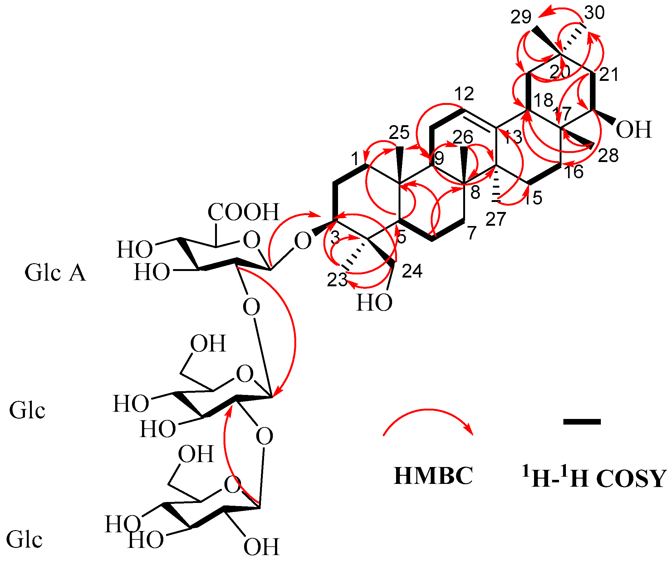
Compound 1 was obtained as a white amorphous powder with [α]25D = +54.8 (c 0.02, MeOH). HR-ESI-MS analysis revealed an [M–H] ion at m/z 957.5222 (calcd. for 957.5059), suggesting the molecular formula C48H78O19 with an index of hydrogen deficiency (IHD) of 10. The 1H-NMR spectrum (Figure S1) showed seven angular methyl singlets at δH 0.69, 0.92, 0.96, 1.20, 1.24, 1.27, and 1.36; one olefinic methine signal at δH 5.27 (br t-like); and three anomeric proton signals at δH 5.08 (1 H, J = 7 Hz), 5.24 (1 H, J = 7.4 Hz), and 5.58 (1 H, J = 7 Hz). The 13C-NMR spectrum (Figure S2), exhibited a carbonyl signal at δC 172.2 and two olefinic carbon signals at δC 122.5 and 144.6. These spectroscopic data suggested that compound 1 is an oleanane-type triterpenoid saponin. The signal at δC 90.2 for C-3 indicated glycosylation at this position. Three anomeric carbon signals at δC 103.3, 104.3, and 106.4 further suggested that 1 is a trisaccharide glycoside of an oleanane-type aglycone linked at C-3. The aglycone proton and carbon signals (Table 1) were fully assigned using distortionless enhancement by polarization transfer (DEPT) spectroscopy (Figure S3), homonuclear correlation spectroscopy (1H-1H COSY) (Figure S4), heteronuclear single-quantum coherence spectroscopy (HSQC) (Figure S5), heteronuclear multiple-bond correlation spectroscopy (HMBC) (Figure S6), nuclear Overhauser effect spectroscopy (NOESY) (Figure S8), and total correlation spectroscopy (TOCSY) (Figure S7). Consistent with the NMR data reported for Soyasaponin I/V and Azukisaponin II/V, the aglycone moiety was therefore assigned as Soyasapogenol B [20]. Acid hydrolysis followed by gas chromatographic analysis of the corresponding trimethylsilylated L-cysteine derivatives, compared with standard samples, identified the three sugar units as two D-glucoses and one D-glucuronic acid (Figure S13). The large anomeric proton coupling constants (J = 7–9 Hz) observed after hydrolysis indicated β-configurations for all sugars. The connectivity and sequence of the sugar chain were determined through analysis of 2D NMR data. Key evidence included the HMBC correlation between GlcA H-1 (δH 5.08) and the aglycone C-3 (δC 90.2), the HMBC correlation between Glc‴ H-1 (δH 5.24) and Glc″ C-2 (δC 85.5), and the 1H-1H COSY correlation between Glc″ H-1 (δH 5.58) and H-2 (δH 4.16) (Figure 2). Therefore, the structure of compound 1 was determined to be 3-O-[β-D-glucopyranosyl-(1 → 2)-β-D-glucopyranosyl-(1 → 2)-β-D-glucuronopyranosyl] 3β,22β,24-trihydroxyolean-12-ene (1).
The other compounds were identified as azukisaponin V(2) [21], Yunganoside B1 (3) [22], azukisaponin I (4) [20], azukisaponin II (5) [23], 3β-acetyl oleanolic acid (6) [24], β-amyrin (7) [25], (+)-catechin 7-O-β-D-glucopyranoside (8) [26], Rutin (9) [27], isoquercitrin (10) [28], kaempferol 3-o-rutinoside (11) [29], and (+)- catechin (12) [30]. The structures of all isolated compounds (112) are shown in Figure 1.

3.2. Inhibitory Activity of α-Glucosidase and Pancreatic Lipase

The inhibition activity tests against α-glucosidase and pancreatic lipase indicated that all tested compounds (13, 612) exhibited significant inhibitory effects on both enzymes, demonstrating clear structure–activity relationships (Table 2). This finding aligns with prior reports on the enzyme-inhibitory potential of adzuki bean extracts [8]. In the α-glucosidase inhibition assay, IC50 values for the isolated compounds ranged from 0.1 to 2.5 mM. The triterpenoid saponins (13) exhibited the strongest activity within the series, consistent with the reported potency of saponin-rich fractions from adzuki beans [8]. Their potent activity aligns with the established critical role of the C-3 glycosyl moiety in oleanane-type saponins for α-glucosidase inhibition, as evidenced by the strong activity (IC50 18.7–154.3 µM, superior to acarbose) reported for ligushicosides A–E from Ligulariopsis shichuana [31]. Notably, the flavonoid compounds 8, 9, and 12 also showed considerable inhibitory potency, with their activities statistically comparable to those of saponins 13 (Table 2, sharing superscript ‘a’). This is consistent with the reported structure–activity relationship that flavonols (e.g., compounds 8 and 9) generally exhibit stronger α-glucosidase inhibition than flavanones (e.g., 12) and that hydroxylation on the B-ring is a key activity-enhancing feature [32]. In contrast, the remaining flavonoids and triterpenoids (compounds 6, 7, 10, and 11) were significantly less active (p < 0.05). All tested compounds were, however, significantly less potent than the positive control acarbose (IC50 = 0.026 ± 0.05 mM) (p < 0.05), a trend previously noted for adzuki bean-derived flavonoids [33]. The strong efficacy of saponins is further supported by computational studies predicting high binding affinity for similar compounds, such as adzukisaponin VI and IV, towards α-glucosidase [34]. In the pancreatic lipase inhibition assay, a more pronounced effect was observed. Triterpenoid saponins (13) along with flavonoids 8 and 11 not only showed superior activity to triterpenoids but were also stronger than the positive control orlistat (p < 0.05, Table 2, group ‘b’ vs. ‘c’). This highlights their notable advantage against this target, and extends previous reports on the lipase-inhibitory activity of adzuki bean saponins [8]. The remaining compounds (6, 7, 9, 10, and 12) exhibited activity comparable to or slightly weaker than orlistat. Activity data for compounds 4 and 5 are not reported due to insufficient sample quantity and chemical instability, respectively.

3.3. Molecular Docking Results

Molecular docking analysis was performed to provide a structural rationale for the observed inhibitory activities. As shown in Table 3, the binding energies of compounds 13 and 612 with both α-glucosidase and pancreatic lipase were below −4 kcal/mol, supporting their potential for direct interaction. Notably, the docking results strongly corroborated the structure–activity relationships observed in vitro. The most potent inhibitors identified in the enzymatic assays—triterpenoid saponins 13 and flavonoid 8—consistently yielded the most favorable (most negative) docking scores for both targets. Specifically, saponin 1 demonstrated a predicted binding affinity substantially stronger than that of the drug acarbose (−8.2 vs. −6.2 kcal/mol) against α-glucosidase and superior to orlistat (−8.8 vs. −6.8 kcal/mol) against pancreatic lipase. This computational evidence directly aligns with its exceptional experimental potency. These findings are consistent with prior in silico studies on adzuki bean saponins, which also predicted strong binding to carbohydrate-digesting enzymes [34]. Our results extend this understanding, demonstrating that specific triterpenoid saponins and flavonoids can achieve predicted binding modes that outperform standard inhibitors, providing a plausible mechanistic basis for their significant in vitro efficacy.

4. Conclusions

This study systematically isolated and identified 12 compounds from the adzuki bean (Vigna angularis), including triterpenoid saponins, flavonoids, and triterpenes, with one being a novel compound and three reported from this source for the first time. Bioactivity evaluation revealed that the triterpenoid saponins (13) acted as potent dual inhibitors of α-glucosidase and pancreatic lipase. Notably, their inhibitory activity against pancreatic lipase surpassed that of the drug orlistat. Molecular docking further supported these findings, revealing strong binding interactions with the enzymes’ active sites, which correlates with the observed inhibitory potency.
These results underscore adzuki bean as a promising natural source of dual-enzyme inhibitors and provide a chemical and mechanistic basis for its potential use in functional foods aimed at metabolic health. The identified triterpenoid saponins represent compelling lead compounds for further development, pending future validation in cellular and in vivo models.

Supplementary Materials

The following supporting information can be downloaded at https://www.mdpi.com/article/10.3390/life16020235/s1: Figure S1. 1H NMR spectrum of compound 1 in C5D5N (400 MHz); Figure S2. 13C NMR spectrum of compound 1 in C5D5N (100 MHz); Figure S3. DEPT spectrum of compound 1 in C5D5N (400 MHz); Figure S4. COSY spectrum of compound 1 in C5D5N (400 MHz); Figure S5. HSQC spectrum of compound 1 in C5D5N (400 MHz); Figure S6. HMBC spectrum of compound 1 in C5D5N (400 MHz); Figure S7. TOCSY spectrum of compound 1 in C5D5N (400 MHz); Figure S8. NOESY spectrum of compound 1 in C5D5N (400 MHz); Figure S9. HRESIMS spectrum of compound 1; Figure S10. EIMS spectrum of Compound 1; Figure S11. UV spectrum of Compound 1; Figure S12. Comparison of the acid hydrolysis products of compound 1 with glucose and glucuronic acid by GC.

Author Contributions

Conceptualization, Data curation, Formal analysis, Funding acquisition, Investigation, Methodology, Visualization, Writing—original draft, Q.G.; Visualization, Writing—original draft, Investigation, Data curation, Formal analysis, Writing—review & editing, X.Z.; Investigation, Writing—review & editing. H.Z. (Hao Zhang), H.Z. (Haoran Zhou), M.W., M.C.; Formal analysis, Writing—review & editing, Z.Y., J.Z.; Supervision, Writing—review & editing, B.Y., L.W.; Conceptualization, Methodology, Project administration, Supervision, Writing—original draft, L.C. All authors have read and agreed to the published version of the manuscript.

Funding

This project was supported by the Tackling-plan Project of Henan Department of Science and Technology (242102311281, 242102311271); Key Scientific Research Projects of Colleges and Universities in Henan Province (24B360009, 24B360010); Innovation and Entrepreneurship training Program for College students in Henan Province (S202511834046, 2025xscxcy109); and 2022 Henan Province Private Education Professional Construction Funding Project—Pharmaceutical Preparation (ZLG202202), HenanProvince’s New Round Key Discipline (Pharmacy) (Jiao Yan (2023) No. 235).

Institutional Review Board Statement

Not applicable.

Informed Consent Statement

Not applicable.

Data Availability Statement

The original contributions presented in this study are included in the article/Supplementary Material. Further inquiries can be directed to the corresponding author.

Conflicts of Interest

The authors declare no conflicts of interest.

References

  1. Hajfathalian, M.; Ghelichi, S.; Jacobsen, C. Anti-obesity peptides from food: Production, evaluation, sources, and commercialization. Compr. Rev. Food Sci. Food Saf. 2025, 24, e70158. [Google Scholar] [CrossRef]
  2. Canning, K.L.; Brown, R.E.; Jamnik, V.K.; Kuk, J.L. Relationship between obesity and obesity-related morbidities weakens with aging. J. Gerontol. A Biol. Sci. Med. Sci. 2014, 69, 87–92. [Google Scholar] [CrossRef]
  3. Wen, X.; Zhang, B.; Wu, B.; Xiao, H.; Li, Z.; Li, R.; Xu, X.; Li, T. Signaling pathways in obesity: Mechanisms and therapeutic interventions. Signal Transduct. Target. Ther. 2022, 7, 298. [Google Scholar] [CrossRef]
  4. Fu, C.; Jiang, Y.; Guo, J.; Su, Z. Natural products with anti-obesity effects and different mechanisms of action. J. Agric. Food Chem. 2016, 64, 9571–9585. [Google Scholar] [CrossRef]
  5. Liu, T.T.; Liu, X.T.; Chen, Q.X.; Shi, Y. Lipase inhibitors for obesity: A review. Biomed. Pharmacother. 2020, 128, 110314. [Google Scholar] [CrossRef] [PubMed]
  6. Guo, Q.; Luo, J.; Zhang, X.; Zhi, J.; Yin, Z.; Zhang, J.; Zhang, W.; Xu, B.; Chen, L. A Comprehensive Review of the Chemical Constituents and Functional Properties of Adzuki Beans (Vigna angulariz). J. Agric. Food Chem. 2025, 73, 6361–6384. [Google Scholar] [CrossRef] [PubMed]
  7. Lee, P.S.; Teng, C.Y.; Hsieh, K.F.; Chiou, Y.S.; Wu, J.C.; Lu, T.J.; Pan, M.H. Adzuki bean water extract attenuates obesity by modulating M2/M1 macrophage polarization and gut microbiota composition. Mol. Nutr. Food Res. 2019, 63, 1900626. [Google Scholar] [CrossRef]
  8. Liu, R.; Zheng, Y.; Cai, Z.; Xu, B. Saponins and flavonoids from adzuki bean (Vigna angularis L.) ameliorate high-fat dietinduced obesity in ICR mice. Front. Pharmacol. 2017, 8, 687. [Google Scholar] [CrossRef]
  9. Itoh, T.; Kobayashi, M.; Horio, F.; Furuichi, Y. Hypoglycemic effect of hot-water extract of adzuki (Vigna angularis) in spontaneously diabetic KK-Ay mice. Nutrition 2009, 25, 134–141. [Google Scholar] [CrossRef] [PubMed]
  10. Han, K.H.; Fukushima, M.; Ohba, K.; Shimada, K.I.; Sekikawa, M.; Chiji, H.; Lee, C.H.; Nakano, M. Hepatoprotective effects of the water extract from adzuki bean hulls on acetaminophen-induced damage in rat liver. J. Nutr. Sci. Vitaminol. 2004, 50, 380–383. [Google Scholar] [CrossRef]
  11. Baracho, N.C.D.V.; Monteiro, N.F.; Borges, M.G.; Arguelho, R.R.D.M. Effect of aqueous extract of the Vigna angularis in rats subjected to an experimental model of moderate chronic kidney disease. Acta Cir. Bras. 2016, 31, 527–532. [Google Scholar] [CrossRef]
  12. Liu, R.; Cai, Z.; Xu, B. Characterization and quantification of flavonoids and saponins in adzuki bean (Vigna angularis L.) by HPLC–DAD–ESI–MSn analysis. Chem. Cent. J. 2017, 11, 93. [Google Scholar] [CrossRef]
  13. Jiang, Y.; Zeng, K.W.; David, B.; Massiot, G. Constituents of Vigna angularis and their in vitro anti-inflammatory activity. Phytochemistry 2014, 107, 111–118. [Google Scholar] [CrossRef]
  14. Takahama, U.; Yamauchi, R.; Hirota, S. Isolation and characterization of a cyanidin-catechin pigment from adzuki bean (Vigna angularis). Food Chem. 2013, 141, 282–288. [Google Scholar] [CrossRef]
  15. Li, L.; Gou, M.L.; He, Y.X. Mandshunosides C–E from the roots and rhizomes of Clematis mandshurica. Phytochem. Lett. 2013, 6, 570–574. [Google Scholar] [CrossRef]
  16. Wang, X.; Zhang, W.; Gao, K.; Lu, Y.; Tang, H.; Sun, X. Oleanane-type saponins from Anemone taipaiensis and their cytotoxic activities. Fitoterapia 2013, 89, 224–230. [Google Scholar] [CrossRef]
  17. Kang, W.Y.; Li, Y.Y.; Gu, X.Z.; Xu, Q.T.; Huang, X. Antioxidant activities, α-glucosidase inhibitory effect in vitro and antihyperglycemic of Trapa acornis shell in alloxan-induced diabetic rats. J. Med. Plant Res. 2011, 5, 6805–6812. [Google Scholar] [CrossRef]
  18. Kim, Y.S.; Lee, Y.M.; Kim, H.; Kim, J.; Jang, D.S.; Kim, J.H.; Kim, J.S. Anti-obesity effect of Morus bombycis root extract: Anti-lipase activity and lipolytic effect. J. Ethnopharmacol. 2010, 130, 621–624. [Google Scholar] [CrossRef] [PubMed]
  19. Ren, X.; Wen, Y.; Li, C.; Yuan, M.; Zhang, J.; Li, S.; Zhang, X.; Wang, L.; Wang, S. Integrated network pharmacology and comprehensive bioinformatics identifying the mechanisms and molecular targets of Jieyu Anshen Granules for treating comorbidity with Alzheimer’s disease and depression. Arab. J. Chem. 2024, 17, 105485. [Google Scholar] [CrossRef]
  20. Kinjo, J.; Hatakeyama, M.; Udayama, M.; Tsutanaga, Y.; Yamashita, M.; Nohara, T.; Okubo, K. HPLC profile analysis of oleanene-glucuronides in several edible beans. Biosci. Biotechnol. Biochem. 1998, 62, 429–433. [Google Scholar] [CrossRef][Green Version]
  21. Shaker, K.H.; Bernhardt, M.; Elgamal, M.H.A.; Seifert, K. Triterpenoid Saponins from Astragalus Trigonus. Z. Naturforsch. C 2001, 56, 699–702. [Google Scholar] [CrossRef]
  22. Ohtani, K.; Ogawa, K.; Kasai, R.; Yang, C.R.; Zhou, J.; Tanaka, O. Oleanane Glycosides from Glycyrrhiza yunnanensis roots. Phytochemistry 1992, 31, 1747–1752. [Google Scholar] [CrossRef]
  23. Kitagawa, I.; Wang, H.K.; Saito, M.; Yoshikawa, M. Saponin and Sapogenol. XXXII. Chemical Constituents of the Seeds of Vigna angularis (WILLD.) OHWI et OHASHI. (2). Azukisaponins I, II, III, and IV. Chem. Pharm. Bull. 1983, 31, 674–682. [Google Scholar] [CrossRef]
  24. Hue Ngan, D.; Hoai, H.T.C.; Mai Huong, L.; Hansen, P.E.; Vang, O. Bioactivities and chemical constituents of a Vietnamese medicinal plant Che Vang, Jasminum subtriplinerve Blume (Oleaceae). Nat. Prod. Res. 2008, 22, 942–949. [Google Scholar] [CrossRef] [PubMed]
  25. Afata, T.N.; Dekebo, A. Chemical composition and antimicrobial effect of western Ethiopian propolis. Chem. Biodivers. 2023, 20, e202200922. [Google Scholar] [CrossRef] [PubMed]
  26. Horii, Y.; Murasato, T.; Fukumura, M.; Toriizuka, K.; Iida, Y. Constituents and antioxidative activity of a hot-water extract of adzuki (Vigna angularis) beans. J. Jpn. Soc. Food Sci. Technol. 2009, 62, 3–11. [Google Scholar] [CrossRef]
  27. Fathiazad, F.; Delazar, A.; Amiri, R.; Sarker, S.D. Extraction of flavonoids and quantification of rutin from waste tobacco leaves. Iran. J. Pharm. Res. 2006, 5, 222–227. [Google Scholar] [CrossRef]
  28. Lee, J.H.; Ku, C.H.; Baek, N.L.; Kim, S.H.; Park, H.W.; Kim, D.K. Phytochemical constituents from Diodia teres. Arch. Pharm. Res. 2004, 27, 40–43. [Google Scholar] [CrossRef] [PubMed]
  29. Zhang, X.Q.; Min, J.G.; Lu, Z.B.; Shi, Y.H.; Zou, X.; Zhou, Y.L. Chemical constituents from Siegesbeckia pubescens. Chin. Tradit. Herbal Drugs 2025, 56, 3806–3818. [Google Scholar] [CrossRef]
  30. Feng, J.; Zhang, Y.; Hu, R.C.; Lai, K.D.; Yuan, J.T.; Chai, L. Flavonoids from Sauropus quadrangularis and their anti-oxidant activities. Chin. Tradit. Herbal Drugs 2025, 56, 3417–3425. [Google Scholar] [CrossRef]
  31. Wu, H.-B.; Liu, T.-T.; Wang, W.-S.; Feng, J.-C.; Tian, H.-M. Oleanane-Type Saponins from the Roots of Ligulariopsis shichuana and Their α-Glucosidase Inhibitory Activities. Molecules 2017, 22, 1981. [Google Scholar] [CrossRef] [PubMed]
  32. Jia, Y.; Ma, Y.; Cheng, G.; Zhang, Y.; Cai, S. Comparative Study of Dietary Flavonoids with Different Structures as α-Glucosidase Inhibitors and Insulin Sensitizers. J. Agric. Food Chem. 2019, 67, 10521–10533. [Google Scholar] [CrossRef] [PubMed]
  33. Yao, Y.; Cheng, X.; Wang, L.; Wang, S.; Ren, G. A determination of potential α-glucosidase inhibitors from azuki beans (Vigna angularis). Int. J. Mol. Sci. 2011, 12, 6445–6451. [Google Scholar] [CrossRef] [PubMed]
  34. Moussa, A.Y.; Alanzi, A.; Luo, J.; Chung, S.K.; Xu, B. Potential anti-obesity effect of saponin metabolites from adzuki beans: A computational approach. Food Sci. Nutr. 2024, 12, 3612–3627. [Google Scholar] [CrossRef]
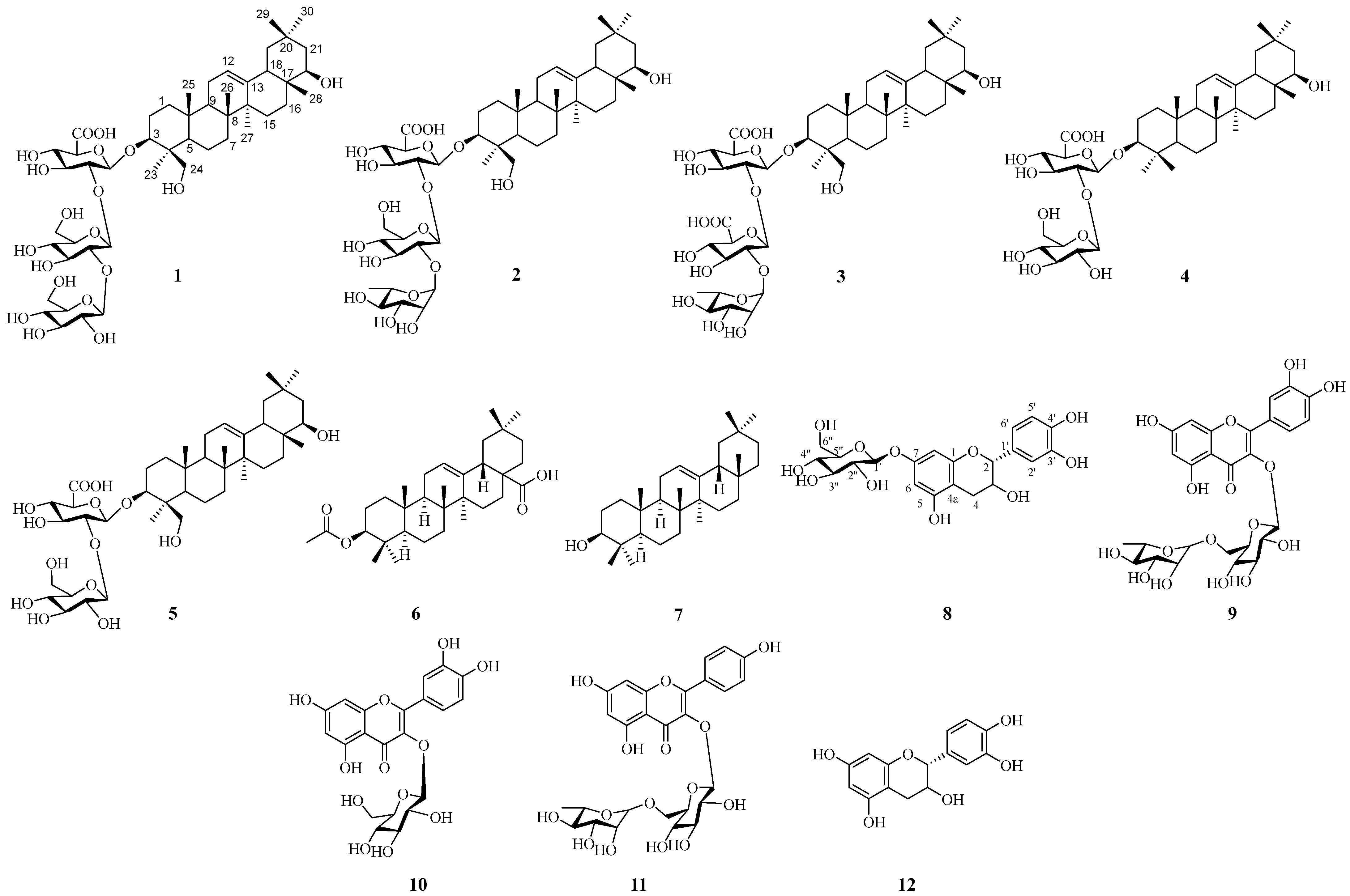
Figure 1. Structure of compounds 112 isolated from adzuki bean (V. angularis). The compounds are: 1 (adzukisaponin A), 2 (azukisaponin V), 3 (yunganoside B1), 4 (azukisaponin I), 5 (azukisaponin II), 6 (3β-acetyl oleanolic acid), 7 (β-amyrin), 8 ((+)-catechin 7-O-β-D-glucopyranoside), 9 (rutin), 10 (isoquercitrin), 11 (kaempferol 3-O-rutinoside), and 12 ((+)-catechin). The numbers and letters represent the standard atom/site numbering system for triterpenoid aglycones and flavonoid glycosides, respectively, following IUPAC nomenclature.
Life 16 00235 g001
Figure 2. 1H-1H COSY, selected HMBC correlations of compound 1.
Life 16 00235 g002
Table 1. 1H (400 MHz) and 13C (100 MHz) NMR data for compound 1 in C5D5N.
NOHCNOHC
11.31 (m), 0.74 (m)38.3250.69 (s)15.4
22.30 (d, J = 10.8), 1.93 (m)26.4260.9216.7
33.44 (d, J = 7.9)90.2271.24 (s)25.5
4-43.4281.20 (s)20.9
50.81 (m)55.8290.96 (s)33.1
61.52 (m), 1.21 (m)18.3301.27 (s)28.4
71.43 (m), 1.25 (m)32.91′5.08 (d, J = 7.0)104.3
8-39.62′4.10 (m)82.2
91.51 (o), 1.13(o)47.43′4.71 (m)77.2
10-36.24′4.56 (m)72.1
111.77 (m)23.75′4.67 (o)77.6
125.27 (t-like)122.16′ 172.2
13-144.61″5.58 (d, J = 7.0)103.3
14-42.12″4.16 (m)85.5
151.81 (m), 0.99 (m)26.23″4.27 (m)76.9
161.87 (m), 1.30 (m)28.44″4.4369.1
17-37.75″3.64 (o)77.8
182.37(d, J = 13.2)45.06″4.40 (m), 4.29 (m)60.8
191.89 (m), 1.12 (m)46.51‴5.24 (d, J = 7.4)106.4
20-30.72‴4.06 (o)76.7
211.72 (m), 1.62 (m)42.13‴4.1777.3
223.71 (m)75.34‴4.07 (o)71.1
231.36 (s)22.45‴3.96 (o)79.2
244.33 (d, J = 11.9), 3.35 (d, J = 11.1)63.26‴4.61(m), 4.26 (m)62.5
Table 2. Inhibition activity of isolated compounds 13 and 612 against α-glucosidase and pancreatic lipase 13 and 612.
CompoundsIC50 (mM) ± SEM/% Inhibition
α-GlucosidasePancreatic Lipase
10.14 ± 0.11 a0.40 ± 0.21 b
20.17 ± 0.21 a0.17 ± 0.21 b
30.23 ± 0.17 a0.11 ± 0.18 b
62.47 ± 0.25 c2.8 ± 0.27 c
71.19 ± 0.12 c1.35 ± 0.19 c
80.43 ± 0.06 a0.41 ± 0.07 b
90.35 ± 0.08 a 1.29 ± 0.11 c
101.20 ± 0.31 c0.77 ± 0.06 c
110.95 ± 0.12 c0.39 ± 0.04 b
120.56 ± 0.11 a1.25 ± 0.17 c
Acarbose0.026 ± 0.05 d/
Orlistat/1.02 ± 0.25 c
Note: Within each column, values with different lowercase superscript letters (a, b, c, d) are significantly different (p < 0.05)
Table 3. Binding affinities of compounds 13 and 612 against α-glucosidase and pancreatic lipase.
CompoundsBinding Affinities (kcal/mol)
α-GlucosidasePancreatic Lipase
1−8.2−8.8
2−7.4−7.4
3−7.2−7.5
6−5.6−5.2
7−4.7−4.0
8−7.0−6.9
9−6.5−5.5
10−5.7−6.4
11−6.2−5.1
12−5.9−6.5
Acarbose−6.2/
Orlistat/−6.8
Disclaimer/Publisher’s Note: The statements, opinions and data contained in all publications are solely those of the individual author(s) and contributor(s) and not of MDPI and/or the editor(s). MDPI and/or the editor(s) disclaim responsibility for any injury to people or property resulting from any ideas, methods, instructions or products referred to in the content.