Cytokines Adsorption During Ex Situ Machine Perfusion of Liver Grafts from Elderly Donors: A Pilot, Prospective, Randomized Study

 , , , , ,
  ,
,  and add
Show full author list
and add
Show full author list
Abstract
1. Introduction
2. Materials and Methods
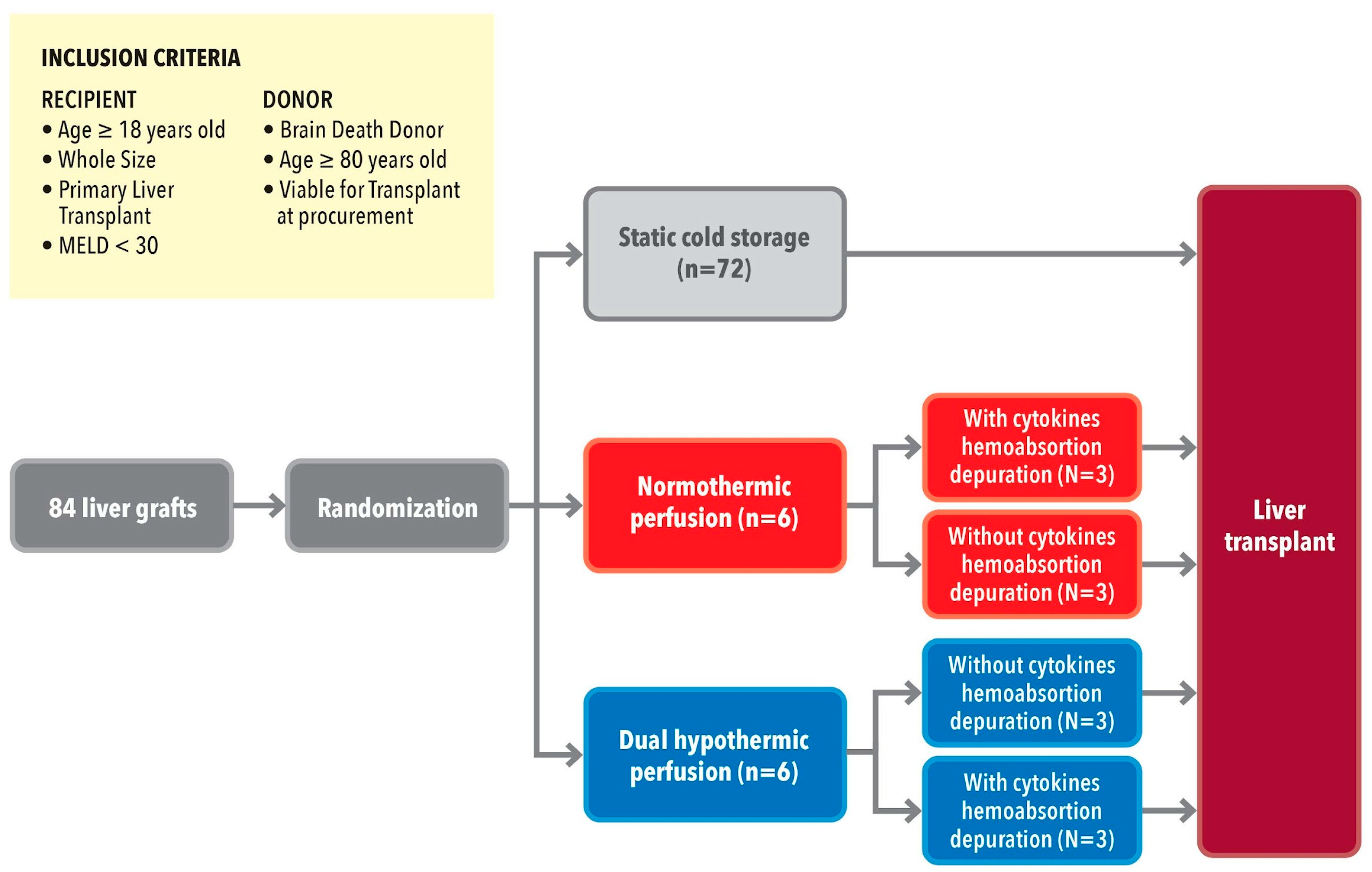
2.1. Study Design and Setting
2.2. Participants (Recipient and Donor Selection)
2.3. Allocation and Transplantation Criteria
2.4. Liver Procurement
2.5. Ex Situ Organ Preservation
2.5.1. SCS Procedures
2.5.2. MP Procedures
2.6. Purification of the Perfusate by Sorbent Device During MP
2.7. LT Surgery
2.8. Data Collection
2.8.1. Donor, Graft, and Recipient
2.8.2. MP Characteristics
2.8.3. Cytokine Levels Determination
2.8.4. Liver Biopsies
2.8.5. Variables, Clinical Outcomes, and Definitions
- (a)
- Perfusate cytokine levels and CA efficacy determination with the mass balance (MB) calculation are expressed as follows.
- (b)
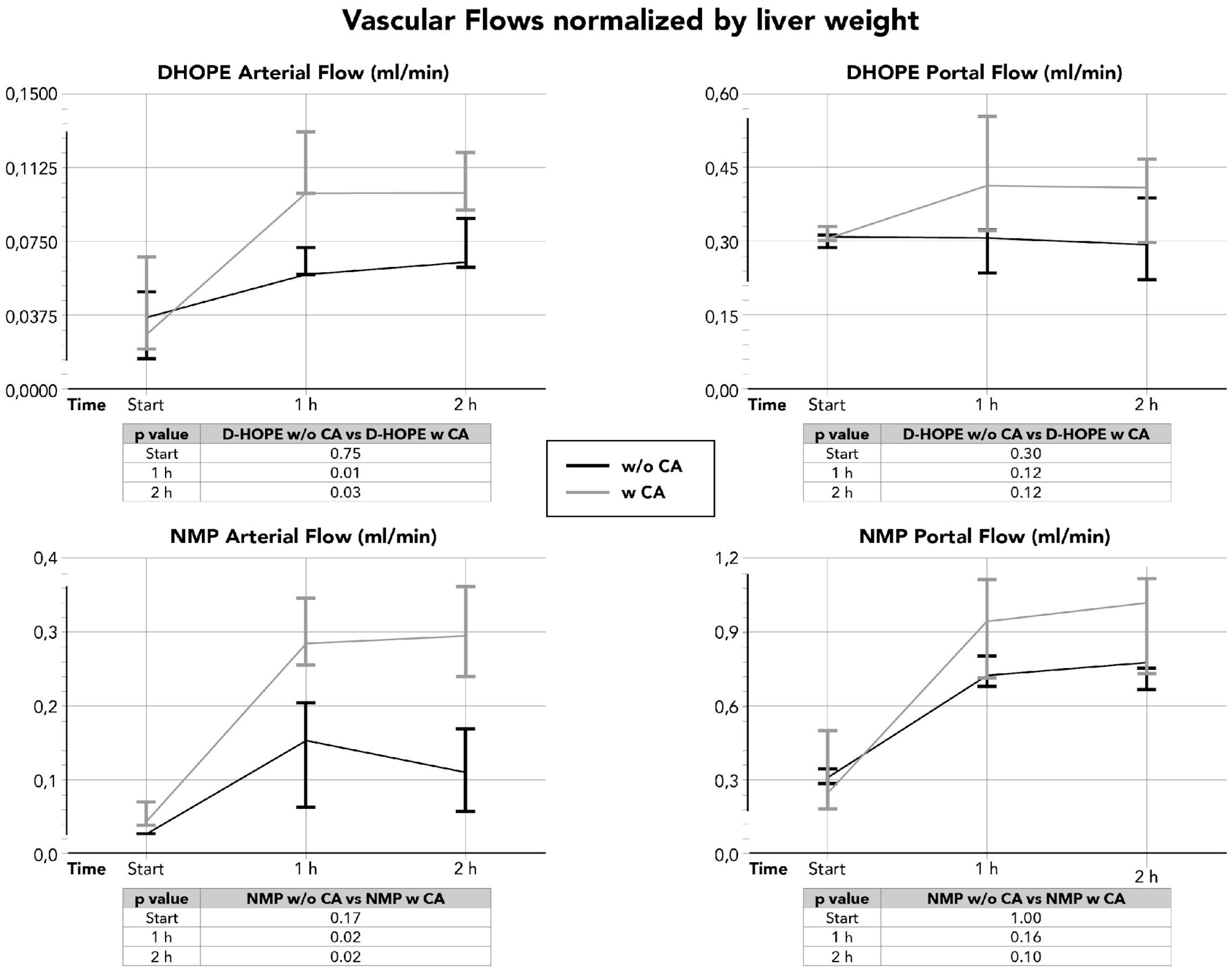
- The effect of CA on MP hemodynamics: Vascular flows were measured by the study group and normalized by liver weight as well.
- (c)
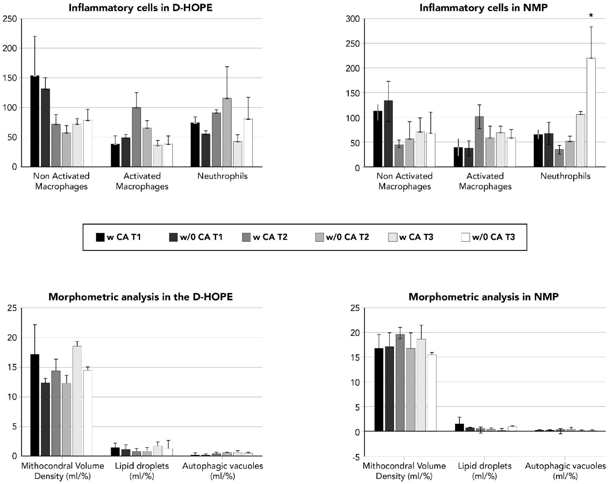
- Effect of CAon liver graft histology and inflammation.
2.9. Statistical Analysis
3. Results
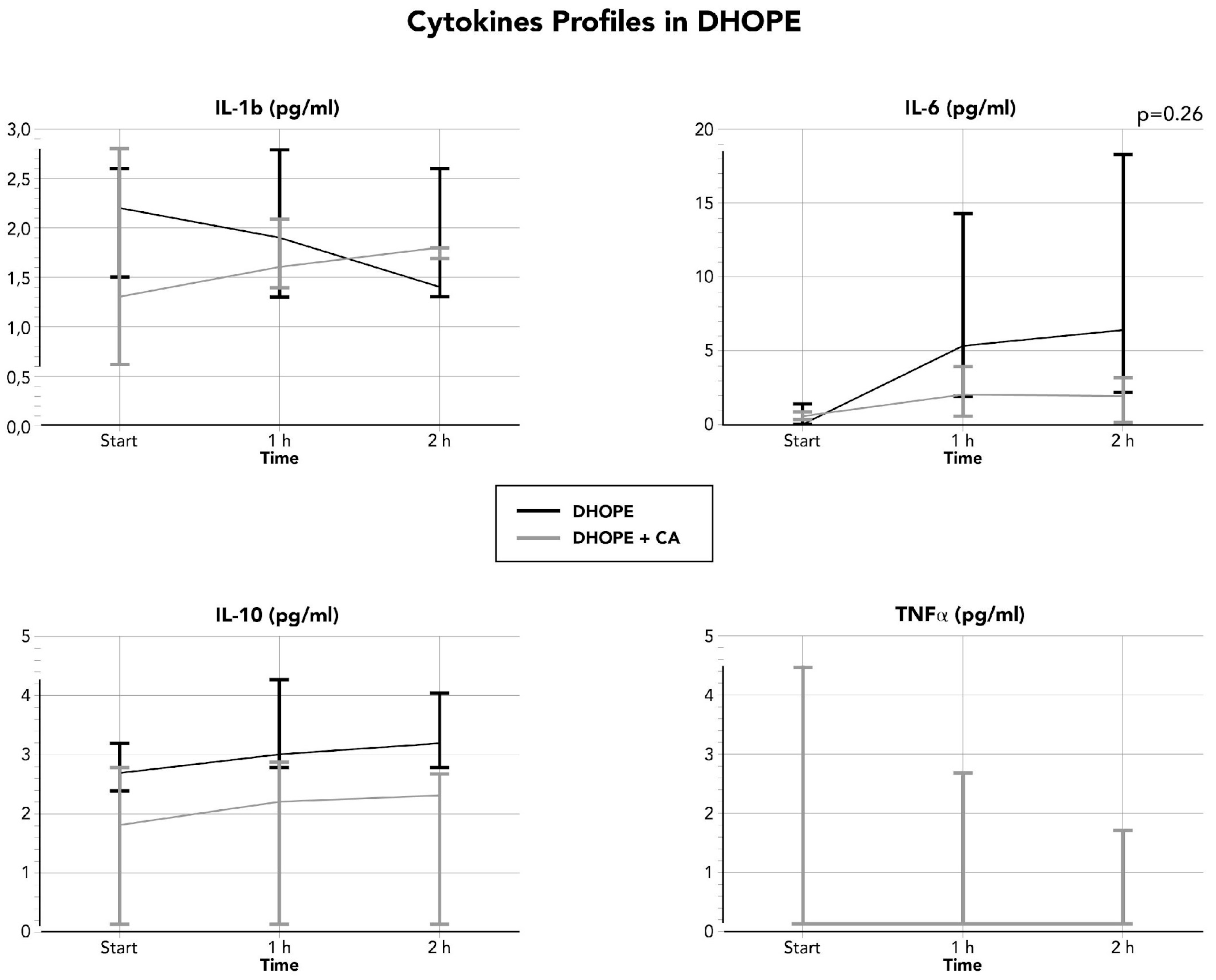
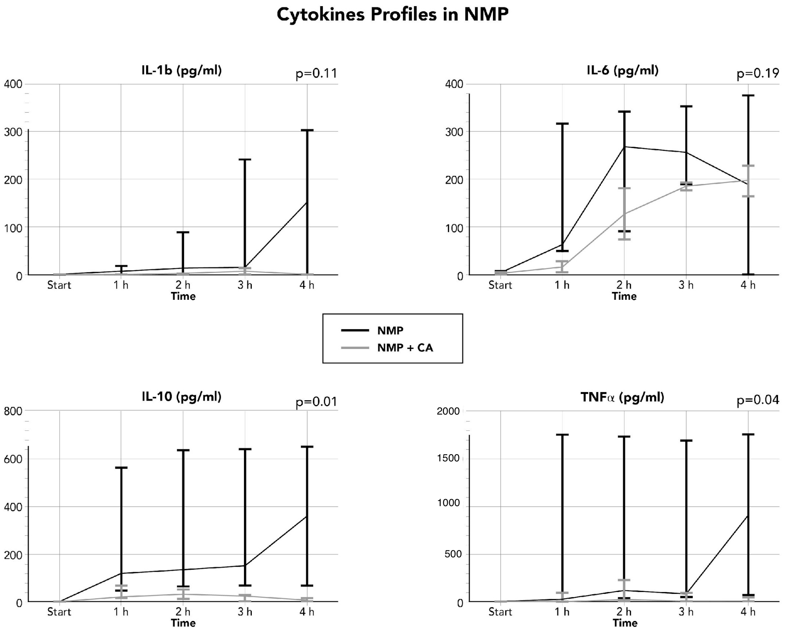
3.1. Cytokines Concentration During Ex Situ Perfusion and Efficacy of CA
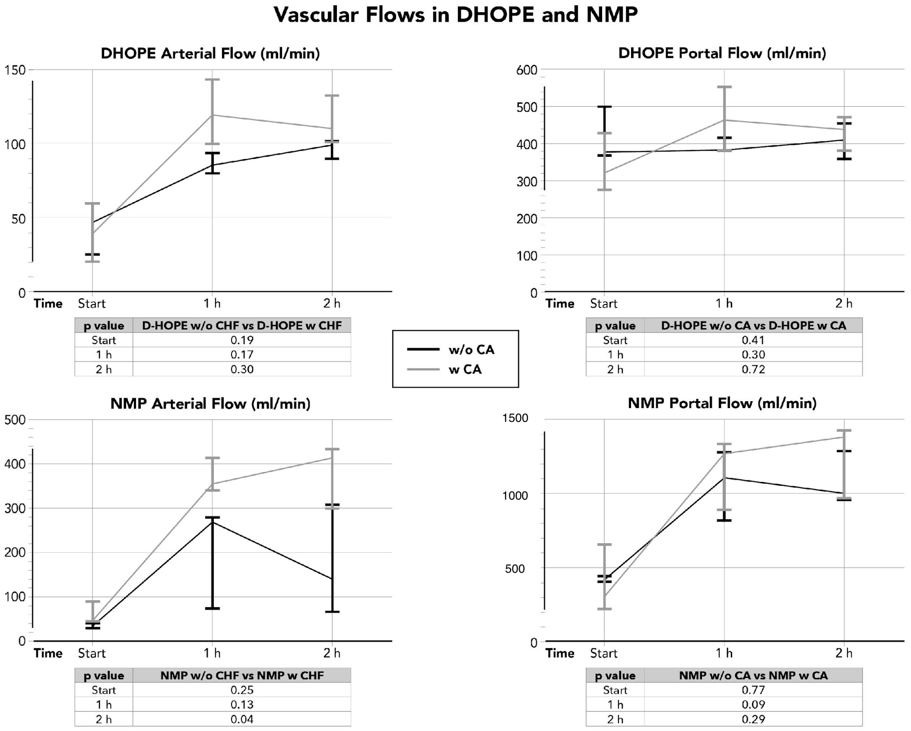
3.2. Effect of the Cytokines CA on the Hepatic Artery and Portal Vein Flows
3.3. Histology Evaluation of Liver Graft Biopsies
3.4. Electronic Microscopy Evaluation (EM) of Liver Graft Biopsies
3.5. Overall Clinical Outcomes
3.6. Clinical Outcomes (D-HOPE and NMP, with and Without CS)
4. Discussion
5. Conclusions
Author Contributions
Funding
Institutional Review Board Statement
Informed Consent Statement
Data Availability Statement
Conflicts of Interest
Abbreviations
| ALT | alanine amino transferase |
| AT | aspartate amino transferase |
| AKI | acute kidney injury |
| BMI | body mass index |
| CCI | comprehensive complication index |
| CA | cytokine adsorption |
| CNT | national transplant authority |
| DBD | donation after brain death |
| D-HOPE | dual hypothermic oxygenated machine perfusion |
| EAD | early allograft dysfunction |
| fWIT | functional warm ischemia time |
| tWIT | total warm ischemia time |
| GGT | gamma glutamyl transpeptidase |
| IC | ischemic cholangiopathy |
| ICU | intensive care unit |
| IL-1 | interleukin 1 |
| IL-10 | interleukin 10 |
| IL-6 | interleukin 6 |
| IQR | interquartile range |
| KDIGO | kidney disease improving global outcomes |
| LT | liver transplantation |
| WIT | warm ischemia time |
| MEAF | model for early allograft function |
| MELD | model for end-stage liver disease |
| MP | machine perfusion |
| NMP | normothermic machine perfusion |
| NRP | normothermic regional perfusion |
| PNF | primary non-function |
| PRS | post-reperfusion syndrome |
| TNF-a | tumor necrosis factor |
Appendix A

Appendix B

| DHOPE w CA | DHOPE w/o CA | p | NMP w CA | NMP w/o CA | p | |
|---|---|---|---|---|---|---|
| Liver Parenchyma | ||||||
| End of Procurement | ||||||
| -Fibrosis 1-2, n | 2 (66) | 3 (100) | 0.374 | 2 (66) | 2 (66) | 0.495 |
| -Necrosis > 10%, n | 0 | 0 | 0 | 0 | 0 | |
| -Macrosteatosi > 30% | 0 | 0 | 0 | 0 | 0 | |
| -Microsteatosis > 0% | 0 | 0 | 0 | 0 | 0 | |
| -PAS | 60 (45–75) | 90 (65–90) | 0.609 | 85 (82.5–87.5) | 80 (60–80) | 0.374 |
| -Suzuki’s score | 1 (0.5–1) | 1 (0.5–1) | 1.0 | 1 (0.5–1.5) | 1 (1–1.5) | 0.724 |
| 0 | 1 (33) | 1 (33) | 0 | 1 (33) | 0 (0) | |
| 1 | 2 (66) | 2 (66) | 0 | 1 (33) | 2 (66) | |
| 2 | 0 | 0 | 0 | 1 (33) | 1 (33) | |
| End of MP | ||||||
| -Fibrosis 1-2, y | 2 (66) | 3 (100) | 0.374 | 2 (66) | 1 (33) | 0.219 |
| -Necrosis > 10%, y | 0 | 0 | 0 | 1 (33) | 1 (33) | 0.789 |
| -Macrosteatosi > 30% | 0 | 0 | 0 | 0 | 0 | |
| -Microsteatosi > 10% | 0 | 0 | 0 | 0 | 0 | |
| -PAS | 80 (60–85) | 80 (75–80) | 0.692 | 75 (72.5–77.5) | 70 (55–75) | 0.518 |
| -Suzuki’s score | 1 (0.5–1) | 0 (0–0.5) | 0.519 | 1.5(1.25–1.75) | 1 (1–2) | 0.870 |
| 0 | 1 (33) | 2 (66) | 0 | 0 | 0 | |
| 1 | 2 (66) | 1 (33) | 0 | 1 (33) | 2 (66) | |
| 2 | 0 | 0 | 0 | 1 (33) | 0 | |
| 3 | 0 | 0 | 0 | 0 | 1 (33) | |
| End of LT | ||||||
| -Fibrosis 1-2, y | 2 (66) | 3 (100) | 0.374 | 0 | 2 (66) | 0.219 |
| -Necrosis > 10%, y | 0 | 0 | 0 | 1 (33) | 3 (100) | 0.272 |
| -Macrosteatosi > 30% | 0 | 0 | 0 | 0 | 1 (33) | 0.495 |
| -Microsteatosi > 10% | 0 | 1 (33) | 0.374 | 0 | 2 (66) | 0.219 |
| -PAS %, median (IQR) | 42.3 (23.5–60) | 50 (40–65) | 0.690 | 30 (15–45) | 70 (50–80) | 0.373 |
| -Suzuki’s score | 0 (0–0.5) | 1 (1–1) | 0.116 | 1 (0.5–1.5) | 3 (2.5–4.5) | 0.219 |
| 0 | 2 (66) | 0 | 0 | 1 (33) | 0 | |
| 1 | 1 (33) | 3 (100) | 0 | 0 | 0 | |
| 2 | 0 | 0 | 0 | 1 (33) | 1 (33) | |
| 3 | 0 | 0 | 0 | 0 | 1 (33) | |
| 6 | 0 | 0 | 0 | 0 | 1 (33) | |
| Bile duct | ||||||
| End of Procurement | ||||||
| -Epithelial damage | 3 (100) | 3 (100) | 2 (66) | 3 (100) | 0.374 | |
| -Necrosis | 0 | 0 | 0 | 0 | ||
| -Vascular plexus damage | 0 | 0 | 0 | 0 | ||
| -Intravascular thrombi | 0 | 0 | 0 | 0 | ||
| -Intramural bleeding | 0 | 0 | 0 | 0 | ||
| -Super peribiliary gland damage | 2 (66) | 3 (100) | 0.374 | 2 (66) | 3 (100) | |
| -Deep peribiliary gland injury | 1 (33) | 0 | 0.374 | 1 (33) | 0 | 0.272 |
| -Inflammation | 0 | 0 | 0 | 2 (66) | 0.219 | |
| End of MP | ||||||
| -Epithelial damage | 3 (100) | 3 (100) | 1.0 | 2 (66) | 3 (100) | 0.374 |
| -Necrosis | 0 | 0 | 1 (33) | 1 (33) | 0.789 | |
| -Vascular plexus damage | 0 | 0 | 0 | 0 | ||
| -Intravascular thrombi | 0 | 0 | 0 | 0 | ||
| -Intramural bleeding | 0 | 0 | 0 | 0 | ||
| -Super peribiliary gland damage | 3 (100) | 3 (100) | 1.0 | 2 (66) | 3 (100) | 0.374 |
| -Deep peribiliary gland injury | 1 (33) | 0 | 0.374 | 1 (33) | 0 | 0.272 |
| -Inflammation | 0 | 0 | 1 (33) | 3 (100) | 0.272 | |
| End of LT | ||||||
| -Epithelial damage | 2 (66) | 2 (66) | 1.0 | 2 (66) | 1 (33) | 0.219 |
| -Necrosis | 0 | 0 | 2 (66) | 1 (33) | 0.219 | |
| -Vascular plexus damage | 0 | 0 | 0 | 0 | ||
| -Intravascular thrombi | 0 | 0 | 0 | 0 | ||
| -Intramural bleeding | 0 | 0 | 0 | 0 | ||
| -Super peribiliary gland damage | 2 (66) | 2 (66) | 1.0 | 1 (33) | 2 (66) | 0.219 |
| -Deep peribiliary gland injury | 0 | 1 (33) | 0.423 | 0 | 0 | 0.667 |
| -Inflammation | 0 | 0 | 0 | 1 (33) | 0.495 | |
| D-HOPE w CA | D-HOPE w/o CA | NMP w CA | NMP w/o CA | |
|---|---|---|---|---|
| Day 1 | ||||
| -pH | 7.73 ± 0.2 | 7.9 ± 0.1 | 7.87 ± 0.2 | 7.85 ± 0.2 |
| -Lactate (mmol/L) | 0.7 ± 0.4 | 0.65 ± 0.4 | 0.5 ± 0.0 | 4.8 ± 7.0 |
| -HCO3 | 26.4 ± 2.6 | 28.5 ± 2.3 | 30.5 ± 1.2 | 22.7 ± 2.2 |
| -glucose (mg/dL) | 57 ± 59 | 12.5 ±5.4 | 15 ± 4.0 | 28 ± 23.5 |
| -Na, mEq/L | 140 ± 1.0 | 137 ± 2.1 | 140 ± 2.1 | 139 ± 6.5 |
| -K, mEq/L | 4.7 ± 0.6 | 4.7 ± 0.2 | 4.5 ± 0.3 | 4.4 ± 0.8 |
| -Cl, mEq/L | 114 ± 6.5 | 114 ± 5.0 | 114 ± 7.1 | 112 ± 2.9 |
| -pO2 | 152 ± 12.5 | 169 ± 1.4 | 159 ± 9.6 | 174 ± 6.6 |
| Day 3 | ||||
| -pH | 7.9 ± 0.1 | 8.0 ± 0.0 | 7.9 ± 0.2 | 8.0 ± 0.0 |
| -Lactate (mmol/L) | 0.6 ± 0.5 | 0.6 ± 0.1 | 0.5 ± 0.1 | 0.7 ± 0.6 |
| -HCO3 | 35.6 ± 3.0 | 37.0 ± 0.0 | 33.8 ± 0.0 | out of scale |
| -glucose (mg/dL) | 13.5 ± 11.8 | 7.5 ± 3.7 | 8.0 ± 1.4 | 9.0 ± 7.0 |
| -Na, mEq/L | 143 ± 3.0 | 138 ± 1.4 | 141.7 ± 2.5 | 137.5 ± 2.1 |
| -K, mEq/L | 4.2 ± 0.3 | 4.2 ± 0.3 | 4.0 ± 0.2 | 4.2 ± 0.7 |
| -Cl, mEq/L | 111.5 ± 1.5 | 109 ± 7.1 | 106 ± 5.3 | 103 ± 7.1 |
| -pO2 | 152.5 ± 15.5 | 165 ± 8.5 | 166.7 ± 28.4 | 176.0 ± 0.0 |
| Day 7 | ||||
| -pH | 7.7 ± 0.1 | 7.7 ± 0.0 | 7.9 ± 0.2 | 7.7 ± 0.1 |
| -Lactate (mmol/L) | 0.9 ± 0.5 | 1.1 ± 0.4 | 0.9 ± 0.6 | 0.6 ± 0.4 |
| -HCO3 | 26.3 ± 4.0 | 28.2 ± 8.0 | 29.8 ± 0.0 | 24.7 ± 1.4 |
| -glucose (mg/dL) | 10.3 ± 10.2 | 14.0 ± 14.1 | 10.0 ± 1.4 | 7.5 ± 4.9 |
| -Na, mEq/L | 138 ± 6.2 | 133 ± 4.2 | 135.5 ± 2.1 | 139 ± 9.9 |
| -K, mEq/L | 4.3 ± 0.2 | 4.7 ± 0.9 | 4.3 ± 0.0 | 4.1 ± 0.6 |
| -Cl, mEq/L | 105 ± 2.1 | 103 ± 1.4 | 106.5 ± 0.7 | 109 ± 3.5 |
| -pO2 | 150 ± 10.8 | 152 ± 43.8 | 177 ± 8.4 | 157 ± 16.9 |
References
- Dickson, K.M.; Martins, P.N. Implications of liver donor age on ischemia reperfusion injury and clinical outcomes. Transplant. Rev. 2020, 34, 100549. [Google Scholar] [CrossRef] [Scilit]
- Dasari, B.V.M.; Schlegel, A.; Mergental, H.; Perera, M.T.P.R. The use of old donors in liver transplantation. Best Pract. Res. Clin. Gastroenterol. 2017, 31, 211–217. [Google Scholar] [CrossRef] [Scilit]
- Dasari, B.V.; Mergental, H.; Isaac, J.R.; Muiesan, P.; Mirza, D.F.; Perera, T. Systematic review and meta-analysis of liver transplantation using grafts from deceased donors aged over 70 years. Clin. Transplant. 2017, 31, e13139. [Google Scholar] [CrossRef] [Scilit]
- Durand, F.; Levitsky, J.; Cauchy, F.; Gilgenkrantz, H.; Soubrane, O.; Francoz, C. Age and liver transplantation. J. Hepatol. 2019, 70, 745–758. [Google Scholar] [CrossRef] [Scilit] [PubMed]
- Guan, L.Y.; Fu, P.Y.; Li, P.D.; Li, Z.-N.; Liu, H.-Y.; Xin, M.-G.; Li, W. Mechanisms of hepatic ischemia-reperfusion injury and protective effects of nitric oxide. World J. Gastrointest. Surg. 2014, 6, 122–128. [Google Scholar] [CrossRef] [Scilit]
- Ghinolfi, D.; Melandro, F.; Torri, F.; Martinelli, C.; Cappello, V.; Babboni, S.; Silvestrini, B.; De Simone, P.; Basta, G.; Del Turco, S. Extended criteria grafts and emerging therapeutics strategy in liver transplantation. The unstable balance between damage and repair. Transplant. Rev. 2021, 35, 100639. [Google Scholar] [CrossRef] [Scilit] [PubMed]
- Ali, A.L.; Nailwal, N.P.; Doshi, G.M. Emerging Role of Interleukins for the Assessment and Treatment of Liver Diseases. Endocr. Metab. Immune Disord. Drug Targets 2022, 22, 371–382. [Google Scholar] [CrossRef] [Scilit]
- Ramos-Tovar, E.; Muriel, P. Molecular Mechanisms That Link Oxidative Stress, Inflammation, and Fibrosis in the Liver. Antioxidants 2020, 9, 1279. [Google Scholar] [CrossRef] [Scilit]
- Dery, K.J.; Kupiec-Weglinski, J.W. New insights into ischemia-reperfusion injury signaling pathways in organ transplantation. Curr. Opin. Organ Transplant. 2022, 27, 424–433. [Google Scholar] [CrossRef] [Scilit] [PubMed]
- Hautz, T.; Salcher, S.; Fodor, M.; Sturm, G.; Ebner, S.; Mair, A.; Trebo, M.; Untergasser, G.; Sopper, S.; Cardini, B.; et al. Immune cell dynamics deconvoluted by single-cell RNA sequencing in normothermic machine perfusion of the liver. Nat. Commun. 2023, 14, 2285. [Google Scholar] [CrossRef] [Scilit]
- Markmann, J.F.; Abouljoud, M.S.; Ghobrial, R.M.; Bhati, C.S.; Pelletier, S.J.; Lu, A.D.; Ottmann, S.; Klair, T.; Eymard, C.; Roll, G.R.; et al. Impact of Portable Normothermic Blood-Based Machine Perfusion on Outcomes of Liver Transplant: The OCS Liver PROTECT Randomized Clinical Trial. JAMA Surg. 2022, 157, 189–198. [Google Scholar] [CrossRef] [Scilit]
- Pezzati, D.; Ghinolfi, D.; Balzano, E.; De Simone, P.; Coletti, L.; Roffi, N.; Rreka, E.; Meacci, L.; Campani, D.; Mazzoni, A.; et al. Salvage of an Octogenarian Liver Graft Using Normothermic Perfusion: A Case Report. Transplant. Proc. 2017, 49, 726–728. [Google Scholar] [CrossRef] [Scilit]
- Parente, A.; Tirotta, F.; Pini, A.; Eden, J.; Dondossola, D.; Manzia, T.M.; Dutkowski, P.; Schlegel, A. Machine perfusion techniques for liver transplantation—A meta-analysis of the first seven randomized-controlled trials. J. Hepatol. 2023, 79, 1201–1213. [Google Scholar] [CrossRef] [Scilit]
- Scheuermann, U.; Zhu, M.; Song, M.; Yerxa, J.; Gao, Q.; Davis, R.P.; Zhang, M.; Parker, W.; Hartwig, M.G.; Kwun, J.; et al. Damage-Associated Molecular Patterns Induce Inflammatory Injury During Machine Preservation of the Liver: Potential Targets to Enhance a Promising Technology. Liver Transplant. 2019, 25, 610–626. [Google Scholar] [CrossRef] [Scilit]
- Obara, H.; Morito, N.; Matsuno, N.; Yoshikawa, R.; Nakajo, T.; Gochi, M.; Otani, M.; Shonaka, T.; Furukawa, H.; Hirano, T.; et al. Initial perfusate purification during subnormothermic machine perfusion for porcine liver donated after cardiac death. J. Artif. Organs 2020, 23, 62–69. [Google Scholar] [CrossRef] [Scilit] [PubMed]
- Jansen, A.; Waalders, N.J.B.; van Lier, D.P.T.; Kox, M.; Pickkers, P. CytoSorb hemoperfusion markedly attenuates circulating cytokine concentrations during systemic inflammation in humans in vivo. Crit. Care 2023, 27, 117. [Google Scholar] [CrossRef] [Scilit]
- Gruda, M.C.; Ruggeberg, K.G.; O’Sullivan, P.; Guliashvili, T.; Scheirer, A.R.; Golobish, T.D.; Capponi, V.J.; Chan, P.P. Broad adsorption of sepsis-related PAMP and DAMP molecules, mycotoxins, and cytokines from whole blood using CytoSorb® sorbent porous polymer beads. PLoS ONE 2018, 13, e0191676. [Google Scholar] [CrossRef] [Scilit] [PubMed]
- Saemann, L.; Pohl, S.; Wächter, K.; Georgevici, A.I.; Köhler, C.; Jünger, J.; Hoorn, F.; Gharpure, N.; Großkopf, A.; Korkmaz-Icöz, S.; et al. Cytokine Adsorption During Ex Vivo Blood Perfusion Improves Contractility of Donation After Circulatory Death Hearts. J. Am. Heart Assoc. 2024, 13, e036872. [Google Scholar] [CrossRef] [Scilit] [PubMed] [PubMed Central]
- Iskender, I.; Arni, S.; Maeyashiki, T.; Citak, N.; Sauer, M.; Rodriguez, J.M.; Frauenfelder, T.; Opitz, I.; Weder, W.; Inci, I. Perfusate adsorption during ex vivo lung perfusion improves early post-transplant lung function. J. Thorac. Cardiovasc. Surg. 2021, 161, e109–e121. [Google Scholar] [CrossRef] [Scilit]
- Ehrsam, J.P.; Arni, S.; Weisskopf, M.; Nowack, M.; Inci, I. Extracorporeal cytokine adsorption reduces systemic cytokine storm and improves graft function in lung transplantation. JTCVS Open 2023, 15, 497–507. [Google Scholar] [CrossRef] [Scilit] [PubMed]
- Ghinolfi, D.; Melandro, F.; Patrono, D.; Lai, Q.; De Carlis, R.; Camagni, S.; Gambella, A.; Ruberto, F.; De Simone, P. A new ex-situ machine perfusion device. A preliminary evaluation using a model of donors after circulatory death pig livers. Artif. Organs 2022, 12, 2493–2499. [Google Scholar] [CrossRef] [Scilit]
- Scalera, I.; Franzin, R.; Stasi, A.; Castellaneta, A.; Fischetti, E.; Morelli, G.; Raele, M.; Panetta, E.; Kurevija, A.; Pulga, W.; et al. Haemoadsorption cartridge connected to the machine perfusion for donation after circulatory death porcine liver marginal grafts. World J. Transplant. 2025, 15, 99287. [Google Scholar] [CrossRef] [Scilit] [PubMed]
- Ghinolfi, D.; Tincani, G.; Rreka, E.; Roffi, N.; Coletti, L.; Balzano, E.; Catalano, G.; Meli, S.; Carrai, P.; Petruccelli, S.; et al. Dual aortic and portal perfusion at procurement prevents ischaemic-type biliary lesions in liver transplantation when using octogenarian donors: A retrospective cohort study. Transpl. Int. 2019, 32, 193–205. [Google Scholar] [CrossRef] [Scilit]
- Ghinolfi, D.; Patrono, D.; De Carlis, R.; Melandro, F.; Buscemi, V.; Farnesi, F.; Torri, F.; Lauterio, A.; Di Salvo, M.; Cerchione, R.; et al. Liver transplantation with uncontrolled versus controlled DCD donors using normothermic regional perfusion and ex-situ machine perfusion. Liver Transplant. 2024, 30, 46–60. [Google Scholar] [CrossRef] [Scilit]
- Ghinolfi, D.; Pezzati, D.; Rreka, E.; Balzano, E.; Catalano, G.; Coletti, L.; Tincani, G.; Carrai, P.; Petruccelli, S.; Martinelli, C.; et al. Nonagenarian Grafts for Liver Transplantation. Liver Transplant. 2019, 25, 1439–1444. [Google Scholar] [CrossRef] [Scilit]
- Ghinolfi, D.; Rreka, E.; De Tata, V.; Franzini, M.; Pezzati, D.; Fierabracci, V.; Masini, M.; Cacciatoinsilla, A.; Bindi, M.L.; Marselli, L.; et al. Pilot, Open, Randomized, Prospective Trial for Normothermic Machine Perfusion Evaluation in Liver Transplantation From Older Donors. Liver Transplant. 2019, 25, 436–449. [Google Scholar] [CrossRef] [Scilit]
- Suzuki, S.; Toledo-Pereyra, L.H.; Rodriguez, F.J.; Cejalvo, D. Neutrophil infiltration as an important factor in liver ischemia and reperfusion injury. Modulating effects of FK506 and cyclosporine. Transplantation 1993, 55, 1265–1272. [Google Scholar] [CrossRef] [Scilit]
- Olthoff, K.M.; Kulik, L.; Samstein, B.; Kaminski, M.; Abecassis, M.; Emond, J.; Shaked, A.; Christie, J.D. Validation of a current definition of early allograft dysfunction in liver transplant recipients and analysis of risk factors. Liver Transplant. 2010, 16, 943–949. [Google Scholar] [CrossRef] [Scilit]
- Aggarwal, S.; Kang, Y.; Freeman, J.; Fortunato, F.; Pinsky, M. Postreperfusion syndrome: Cardiovascular collapse following hepatic reperfusion during liver transplantation. Transplant. Proc. 1987, 19, 54–55. [Google Scholar]
- Khwaja, A. KDIGO clinical practice guidelines for acute kidney injury. Nephron Clin. Pract. 2012, 120, c179–c184. [Google Scholar] [CrossRef] [Scilit]
- Dindo, D.; Demartines, N.; Clavien, P.A. Classification of surgical complications: A new proposal with evaluation in a cohort of 6336 patients and results of a survey. Ann. Surg. 2004, 240, 205–213. [Google Scholar] [CrossRef] [Scilit]
- Sliwinski, S.; Heil, J.; Franz, J.; El Youzouri, H.; Heise, M.; Bechstein, W.O.; Schnitzbauer, A.A. A critical appraisal of the ISGLS definition of biliary leakage after liver resection. Langenbeck’s Arch. Surg. 2023, 408, 77. [Google Scholar] [CrossRef] [Scilit]
- Pezzati, D.; Torri, F.; Franzini, M.; Balzano, E.; Catalano, G.; Tincani, G.; Bronzoni, J.; Martinelli, C.; Trizzino, A.; Petagna, L.; et al. Association of perfusate cytokine concentrations during liver graft ex situ normothermic perfusion to donor type and postoperative outcomes. Liver Transplant. 2025, 31, 877–889. [Google Scholar] [CrossRef] [Scilit] [PubMed]
- Ly, M.; Lau, N.S.; Dennis, C.; Chen, J.; Risbey, C.; Tan, S.; Chen, R.; Wang, C.; Gorrell, M.D.; McKenzie, C.; et al. Long-term ex situ normothermic machine perfusion allows regeneration of human livers with severe bile duct injury. Am. J. Transplant. 2025, 25, 60–71. [Google Scholar] [CrossRef] [Scilit]
- Boffini, M.; Marro, M.; Simonato, E.; Scalini, F.; Costamagna, A.; Fanelli, V.; Barbero, C.; Solidoro, P.; Brazzi, L.; Rinaldi, M. Cytokines Removal During Ex-Vivo Lung Perfusion: Initial Clinical Experience. Transpl. Int. 2023, 36, 10777. [Google Scholar] [CrossRef] [Scilit]
- Kleiner, G.; Marcuzzi, A.; Zanin, V.; Monasta, L.; Zauli, G. Cytokine levels in the serum of healthy subjects. Mediat. Inflamm. 2013, 2013, 434010. [Google Scholar] [CrossRef] [Scilit] [PubMed]
- Solhjou, Z.; Uehara, M.; Bahmani, B.; Maarouf, O.H.; Ichimura, T.; Brooks, C.R.; Xu, W.; Yilmaz, M.; Elkhal, A.; Tullius, S.G.; et al. Novel Application of Localized Nanodelivery of Anti-Interleukin-6 Protects Organ Transplant from Ischemia-Reperfusion Injuries. Am. J. Transplant. 2017, 17, 2326–2337. [Google Scholar] [CrossRef] [Scilit] [PubMed]
- Del Turco, S.; Cappello, V.; Tapeinos, C.; Moscardini, A.; Sabatino, L.; Battaglini, M.; Melandro, F.; Torri, F.; Martinelli, C.; Babboni, S.; et al. Cerium oxide nanoparticles administration during machine perfusion of discarded human livers: A pilot study. Liver Transplant. 2022, 28, 1173–1185. [Google Scholar] [CrossRef] [Scilit]

| Variable | SCS (n = 72) | NMP (n = 6) | D-HOPE (n = 6) | pSCS-NMP | pSCS-DHOPE | pNMP-HOPE |
|---|---|---|---|---|---|---|
| Donor Characteristics | ||||||
| Age, years | 84 (81.8–87) | 82.5 (81.3–84.5) | 82 (81.3–86.5) | 0.251 | 0.641 | 0.547 |
| Gender (M/F) | 44/28 (61/39) | 3/3 (50)/(50) | 2/4 (33)/(67) | 0.599 | 0.188 | 0.599 |
| Location—regional | 6 (100) | 6 (100) | 6 (100) | 1.0 | 1.0 | 1.0 |
| Weight, Kg | 70 (60–80) | 68.5 (65–74.3) | 75 (63.8–85.5) | 0.556 | 0.731 | 0.575 |
| Height, cm | 170 (164–175) | 174 (167–175) | 168 (162–173) | 0.341 | 0.748 | 0.384 |
| BMI | 24.5 (23–27) | 24 (21–24) | 25–5 (23.3–28.5) | 0.120 | 0.566 | 0.207 |
| Cause of death: | ||||||
| CVA | 60 (83) | 5 (83) | 5 (83) | 0.887 | 0.887 | 1 |
| Trauma | 10 (14) | 1 (17) | 1 (17) | - | - | - |
| Anoxia | 2 (3) | 0 | 0 | - | - | - |
| ICU stay (days) | 3 (2–5.25) | 2 (2–2) | 2 (2–2.75) | 0.153 | 0.151 | 1 |
| Comorbidities: | ||||||
| Hypertension | 58 (81) | 2 (33) | 1 (16) | 0.008 * | 0.239 | 0.01 * |
| Diabetes | 16 (22) | 1 (16) | 1 (16) | 0.755 | 0.755 | 1 |
| Cardiomyopathy | 44 (61) | 2 (33) | 3 (50) | 0.188 | 0.599 | 0.599 |
| Dislipidemy | 29 (70) | 3 (50) | 4 (67) | 0.647 | 0.214 | 0.599 |
| Nephropathy | 8 (11) | 1 (16) | 1 (16) | 0.687 | 0.687 | 1 |
| Virology: | ||||||
| Hbc/HBV DNA+ | 11/1 (15/1.3) | 1 (16) | 1 (16) | 0.929/0.775 | 0.929/0.775 | 1/- |
| HCVAb/HCV RNA | 1/0 (1.3/0) | 0/0 | 0/0 | 0.775 | 0.775 | - |
| Last lab values: | ||||||
| Na, mEq/L | 148 (145–153) | 147 (145–152) | 146 (141–149) | 0.963 | 0.204 | 0.191 |
| AST, UI/L | 24 (19–37.5) | 20.5 (15–36.5) | 19.5 (17.3–39) | 0.907 | 0.614 | 0.583 |
| ALT, UI/L | 17 (11–29) | 12 (10–23) | 10 (8–17.3) | 0.491 | 0.323 | 0.342 |
| Bilirubin, mg/dL | 0.6 (0.42–1.02) | 0.395 (0.32–0.7) | 0.68 (0.39–0.7) | 0.290 | 0.483 | 0.659 |
| Recipient Characteristics | ||||||
| Age, years | 58 (54–63) | 62 (58.5–64.8) | 54.5(53.3–58.8) | 0.758 | 0.76 | 0.66 |
| Gender (M/F) | 49/23 (68/32) | 4/2 (67/33) | 4/2 (67/33) | 0.945 | 0.945 | 1 |
| Weight, Kg | 72 (62–76.5) | 74 (72.3–84.8) | 75 (70.5–91.5) | 0.130 | 0.246 | 0.812 |
| Height, cm | 168 (160–173) | 173 (170–177) | 169 (168–175) | 0.309 | 0.331 | 0.295 |
| BMI | 25 (22–28) | 25 (22.8–28) | 26 (25–29.3) | 0.528 | 0.403 | 0.907 |
| MELD | 11.5 (9–15) | 12.5 (9–13) | 11.5 (8.5–13.8) | 0.960 | 0.632 | 0.717 |
| Cause of transplant: | ||||||
| HCC | 32 (44) | 2 (33) | 2 (33) | 0.525 | 0.953 | 0.563 |
| HCV | 4 (5.5) | 0 | 0 | - | - | - |
| HBV | 9 (12.5) | 0 | 1 (16) | - | - | - |
| NASH | 5 (7) | 1 (16) | 2 (33) | - | - | - |
| ETOH | 9 (12.5) | 2 (33) | 1 (16) | - | - | - |
| CSP/CBP | 4 (5.5) | 1 (16) | 0 | - | - | - |
| Other | 9 (12.5) | 0 | 0 | - | - | - |
| Procurement Characteristics | ||||||
| Extraction time, min | 23.5 (14–30) | 22 (12.5–31.5) | 10 (10–17.5) | 0.832 | 0.085 | 0.203 |
| Arterial reconstruction | 12 (16) | 0 | 1 (16) | 0.283 | 1 | 0.341 |
| Biopsy (Y/N) | 33 (45)/39 (55) | 2 (33)/4 (67) | 1 (16)/5 (84) | 0.560 | 0.171 | 0.549 |
| CIT, min | 450 (392–488) | 292 (274–306) | 248 (237–259) | <0.01 * | <0.01 * | 0.04 * |
| Re-cooling time, min | - | 28.5 (18.3–19.5) | 17.5 (17–19.5) | - | - | 0.113 |
| WIT, min | 45 (37–55) | 46 (33.3–61.8) | 49.5 (30–54) | 0.858 | 0.648 | 0.851 |
| Liver weight, gr | 1320 (1180–1408) | 1263 (1203–1491) | 1250 (1040–1393) | 0.908 | 0.436 | 0.432 |
| Liver weight post-MP, gr | - | 1236 (1196–1403) | 1298 (1055–1465) | - | - | 0.589 |
| Transplant Characteristics | ||||||
| Duration, min | 365 (320–420) | 362 (340–426) | 349 (324–372) | 0.591 | 0.386 | 0.308 |
| Cava replacement (yes) | 48 (66) | 3 (50) | 4 (67) | 0.416 | 1 | 0.599 |
| Biliary reconstruction: | ||||||
| c-c, N | 70 (97) | 6 (100) | 6 (100) | 0.351 | 0.351 | 1 |
| e-d, N | 2 (3) | 0 | 0 | 0.684 | 0.684 | - |
| Outcomes | ||||||
| PNF (yes) | 0 (0) | 1 (17) | 0 (0) | 1.0 | 1.0 | 1.0 |
| PRS (yes) | 15 (21) | 1 (17) | 0 (0) | 1.0 | 0.58 | 1.0 |
| EAD (yes) | 11 (15) | 1 (17) | 0 (0) | 0.67 | 0.61 | 1.0 |
| AKI (yes) | 19 (26) | 2 (34) | 0 (0) | 0.96 | 0.36 | 0.43 |
| RRT (yes) | 6 (8%) | 1 (17%) | 0 (0) | 0.46 | 0.95 | 1.0 |
| PRBC, units | 2 (0–4) | 2 (0–3.5) | 2 (1–4) | 0.70 | 0.53 | 1.0 |
| FFP, units | 3 (1–5) | 3.5 (2–5.5) | 2 (1–4) | 0.76 | 0.12 | 0.32 |
| AST peak, UI/L | 461 (294–1042) | 663 (477–1500) | 240 (208–327) | 0.55 | 0.23 | 0.06 * |
| ALT peak, UI/L | 433 (277–720) | 279 (243–1253) | 135 (124–199) | 0.44 | 0.11 | 0.07 * |
| Hospital stay, days | 13 (10–23) | 15 (10–20) | 13.5 (9–20) | 0.95 | 0.62 | 0.71 |
| CD > 3° | 8 (11) | 1 (17) | 0 (0) | 0.55 | 1.0 | 1.0 |
| CCI at discharge | 20.9 (8.7–31.5) | 21.1 (8.7–35.5) | 8.7 (8.7–17.8) | 0.97 | 0.77 | 0.70 |
| Vascular complication (y) | 5 (7) | 0 | 0 | 0.508 | 0.508 | - |
| Biliary complicat., tot | 26 (36) | 1 (16) | 2 (33) | 0.343 | 0.893 | 0.549 |
| -ischemic cholangiopathy | 8 (11) | 1 (16) | 1 (16) | 0.687 | 0.687 | - |
| -anastomotic stenosis | 13 (18) | 0 | 1 (16) | 0.260 | 0.933 | 0.734 |
| -fistula | 3 (4) | 0 | 0 | 0.616 | 0.616 | - |
| -stones | 2 (2.7) | 0 | 0 | 0.684 | 0.684 | - |
| 1-year graft survival, % | 78 | 84 | 100 | 0.755 | 0.200 | 0.341 |
| 1-year patient survival, % | 85 | 100 | 100 | 0.308 | 0.308 | - |
| Bil. at 1 year, mg/dL | 0.99 (0.6–1.52) | 1.15 (1–1.19) | 0.55 (0.49–0.62) | 0.7 | 0.630 | 0.086 |
| GGT at 1 year, UI/L | 49 (27–121) | 34 (23–58) | 22.5 (17–28) | 0.426 | 0.161 | 0.441 |
| Median follow-up (months) | 29 (24.8–35) | 32.5 (28.3–33) | 38 (37.3–38) | 0.473 | 0.031 | 0.006 |
| Variable | D-HOPE w CAD | D-HOPE w/o CAD | p | NMP w CAD | NMP w/o CAD | p |
|---|---|---|---|---|---|---|
| Donor Characteristics | ||||||
| Age, years | 88 (85–89) | 81 (81–82) | 0.09 | 83 (82–84) | 81 (80–84) | 0.65 |
| Gender (M/F) | 1/2 (33/66) | 1/2 (33/66) | 1.0 | 2/1 (66/33) | 1/2 (33/66/) | 1.0 |
| Weight, Kg | 60 (52–68) | 89 (82–90) | 0.07 | 65 (60–70) | 72 (68–74) | 0.43 |
| Height, cm | 160 (157–169) | 168 (167–172) | 0.45 | 165 (162–170) | 175 (173–177) | 0.15 |
| BMI | 23.4 (21.1–23.7) | 29.0 (27.1–30.6) | 0.04 | 24.5 (22.3–24.99) | 24 (22.0–24.2) | 0.80 |
| Cause of death (CVA) | 3 (100) | 2 (66) | 1.0 | 2 (66) | 3 (100) | 1.0 |
| ICU stay (days) | 2 (1.5–3.5) | 2 (2–2.5) | 0.80 | 2 (2–2) | 2 (1.5–4) | 0.55 |
| Comorbidities: | ||||||
| -diabetes | 3 (100) | 0 | 0.10 | 0 | 1 (33) | 1.0 |
| -hypertension | 3 (100) | 3 (100) | 1.0 | 1 (33) | 1 (33) | 1.0 |
| -cardiopathy | 1 (33) | 1 (33) | 1.0 | 1 (33) | 1 (33) | 1.0 |
| -dyslipidemia | 2 (66) | 1 (33) | 1.0 | 2 (66) | 1 (33) | 1.0 |
| -nephropathy | 1 (33) | 0 | 1.0 | 0 | 1 (33) | 1.0 |
| -anti HBc pos | 1 (33) | 0 | 1.0 | 0 | 1 (33) | 1.0 |
| Last lab values: | ||||||
| -AST, UI/L | 45 (29–46) | 18 (17–20) | 0.18 | 18 (15–30) | 23 (18–65) | 0.33 |
| -ALT, UI/L | 19 (12–19) | 8 (8–10) | 0.38 | 10 (8–18) | 14 (12–28) | 0.55 |
| -BIL T, mg/dL | 0.7 (0.5–0.8) | 0.7 (0.5–0.8) | 0.83 | 0.3 (0.3–0.4) | 0.8 (0.6–1.0) | 0.11 |
| Recipient characteristics | ||||||
| Age, years | 55 (54–60) | 54 (53–57) | 0.67 | 65 (51–67) | 60 (59–62) | 0.72 |
| Gender (M/F) | 1/2 (3/66) | 3/0 (100/0) | 1.0 | 1/2 (3/66) | 3/0 (100/0) | 1.0 |
| Weight, Kg | 72 (65–84) | 74 (71–87) | 0.70 | 72 (68–92) | 75 (74–81) | 0.80 |
| Height, cm | 168 (166–174) | 170 (169–173) | 0.87 | 170 (170–174) | 175 (172–180) | 0.47 |
| BMI, N | 27.0 (23.8–28.5) | 25.3 (25.1–29.1) | 0.66 | 25.3 (23.5–27.1) | 24.9 (23.5–30.3) | 0.65 |
| MELD | 13 (11–15) | 8 (7–11) | 0.29 | 13 (12–13) | 8 (7–15) | 0.95 |
| Procurement Characteristics | ||||||
| Arterial reconstruction N | 0 | 1 | 1.0 | 0 | 0 | 1.0 |
| CIT, min | 228 (218–251) | 231 (227–238) | 0.24 | 240 (238–260) | 269 (266–294) | 0.24 |
| WIT, min | 55 (39–62) | 48 (36–50) | 0.65 | 65 (48–70) | 40 (30–46) | 0.29 |
| Liver weight, gr | 980 (937–1205) | 1280 (1250–1442) | 0.26 | 1225 (1210–1262) | 1555 (1367–1585) | 0.21 |
| Liver weight post-MP, g | 1020 (978–1248) | 1435 (1297–1532) | 0.29 | 1260 (1225–1355) | 1215 (1200–1552) | 0.62 |
| MP duration, min | 128 (125–143) | 157 (145–162) | 0.34 | 279 (274–293) | 241 (220–244) | 0.04 |
| Transplant Characteristics | ||||||
| Cava replacement (yes) | 2 | 2 | 1.0 | 1 | 2 | 1.0 |
| Biliary reconstruction | ||||||
| -duct to duct w T-tube | 3 | 3 | 1.0 | 3 | 3 | 1.0 |
| Outcomes | ||||||
| PNF (yes) | 0 | 0 | 1.0 | 0 | 1 | 1.0 |
| PRS (yes) | 0 | 0 | 1.0 | 0 | 1 | 1.0 |
| EAD (yes) | 0 | 0 | 1.0 | 0 | 0 | 1.0 |
| AKI (yes) | 0 (0) | 0 (0) | 1.0 | 1 (33) | 1 (33) | 1.0 |
| RRT, (yes) | 0 (0) | 0 (0) | 1.0 | 0 (0) | 1 (33) | 1.0 |
| PRBC, N units | 4 (3–5) | 0 (0–1) | 0.07 | 1 (0.5–2) | 2 (1–9) | 0.45 |
| FFP, N units | 2 (2–3) | 1 (1–3) | 0.51 | 4 (3–5) | 3 (1–5) | 0.90 |
| AST peak, UI/L | 260 (232–452) | 220 (203–285) | 0.85 | 878 (678–1079) | 243 (236–871) | 0.75 |
| ALT peak, UI/L | 133 (110–218) | 137 (129–178) | 0.47 | 1214 (713–1714) | 663 (491–958) | 0.61 |
| Hospital stay, days | 18 (13–19) | 9 (8–19) | 0.34 | 12 (11–14) | 20 (14–25) | 0.44 |
| CD > 3° | 0 | 0 | 1.0 | 0 | 1 | 1.0 |
| CCI at discharge | 20.9 | 8.7 | 0.37 | 8.7 | 33.5 | 0.23 |
| Vascular complication | 0 (0) | 0 (0) | 1.0 | 0 (0) | 0 (0) | 1.0 |
| Biliary complications tot: | 0 (0) | 2 (67) | 0.68 | 1 (33) | 0 (0) | 0.37 |
| -ischemic cholangiopathy | 0 (0) | 1 (33) | 1 (33) | 0 (0) | ||
| -anastomotic stenosis | 0 (0) | 1 (33) | 0 (0) | 0 (0) | ||
| -fistula | 0 (0) | 0 (0) | 0 (0) | 0 (0) | ||
| -stones | 0 (0) | 0 (0) | 0 (0) | 0 (0) | ||
| Bil at 1 year, mg/dL | 0.490 (0.465–0.495) | 0.630 (0.615–1.1) | 0.22 | 1.15 (1.07–1.17) | 1.16 (0.99–1.33) | 0.87 |
| GGT at 1 year, UI/L | 25 (19.5–27.2) | 20 (18–52) | 0.48 | 58 (46–99.5) | 12 (6.5–17.5) | 0.22 |
| 1-year graft survival % | 100 | 100 | 1 | 100 | 67 | 0.37 |
| 1-year patient survival, % | 100 | 100 | 1 | 100 | 100 | 1 |
| Median follow-up, months | 38 (37.5–38) | 38 (37.5–38) | 1 | 32 (27.5–32.5) | 33 (30–35) | 0.52 |
| Cytokine | Start | 1 h | 2 h | 3 h | 4 h |
|---|---|---|---|---|---|
| Overall Concentration * | |||||
| D-HOPE | |||||
| IL-1 | 2.0 (1.55–2.0) | 2.05 (1.55–2.25) | 2.15 (1.57–2.2) | \ | \ |
| IL-6 | 1.65 (1.2–3.75) | 3.05 (2.02–5.05) | 2.65 (2.05–5.65) | \ | \ |
| IL-10 | 2.35 (2.2–2.88) | 3.1 (2.4–3.27) | 3.15 (2.5–3.2) | \ | \ |
| TNF-a | 0.3 (0.1–1.85) | 0.1 (0.1–0.1) | 0.1 (0.1–0.1) | \ | \ |
| NMP | |||||
| IL-1 | 0.1 (0.1–0.2) | 4.4 (2.6–11.0) | 10.8 (5.95–49.7) | 15.3 (8.3–129) | 153 (77.0–228) |
| IL-6 | 3.0 (1.55–4.4) | 62.0 (190–20.5) | 139 (389–154) | 158 (115–402) | 364 (219–509) |
| IL-10 | 0.1 (0.1–0.1) | 61.9 (30.2–110) | 48.3 (38.9–108) | 40.8 (29.1–77.2) | 17.9 (10.9–70.6) |
| TNF-a | 0.1 (0.1–0.1) | 23.1 (3.15–88) | 18.8 (9.15–183) | 19.1 (3.8–108) | 7.3 (2.4–11.8) |
| Mass Balance (pg) # | |||||
| D-HOPE | 0–60 min | 60–120 min | 120–180 min | 180–240 min | |
| IL-1 | 1.080 (0–1560) | 1.440 (864–2.400) | \ | \ | |
| IL-6 | 2.400 (660–7.380) | 4.320 (0–11.880) | \ | \ | |
| IL-10 | 0 (0–1.620) | 0 (0–6.480) | \ | \ | |
| TNF-a | 0 (0–2.640) | 0 (0–2.880) | \ | \ | |
| NMP | 0–60 min | 60–120 min | 120–180 min | 180–240 min | |
| IL-1 | 528 (0–801) | 4.596 (0–8.184) | 0 (0–108.768) | 3.951 (0–87.837) | |
| IL-6 | 2.112 (0–24.297) | 229.215 (0–591.294) | 289.677 (0–797.220) | 271.302 (0–718.428) | |
| IL-10 | 11.250 (8.316–63.279) | 166.026 (123.192–319.176) | 159.777 (61.893–160.680) | 99.636 (39.852–104.043) | |
| TNF-a | 1.056 (0–87.042) | 11.682 (0–706.893) | 30.498 (0–678.564) | 148.821 (22.356–517.845) | |
Disclaimer/Publisher’s Note: The statements, opinions and data contained in all publications are solely those of the individual author(s) and contributor(s) and not of MDPI and/or the editor(s). MDPI and/or the editor(s) disclaim responsibility for any injury to people or property resulting from any ideas, methods, instructions or products referred to in the content. |
© 2026 by the authors. Licensee MDPI, Basel, Switzerland. This article is an open access article distributed under the terms and conditions of the Creative Commons Attribution (CC BY) license.
Share and Cite
Cirillo, G.; Bernardi, L.; Pezzati, D.; Franzini, M.; Balzano, E.; Tincani, G.; Bronzoni, J.; Martinelli, C.; Trizzino, A.; Petagna, L.; et al. Cytokines Adsorption During Ex Situ Machine Perfusion of Liver Grafts from Elderly Donors: A Pilot, Prospective, Randomized Study. Life 2026, 16, 167. https://doi.org/10.3390/life16010167
Cirillo G, Bernardi L, Pezzati D, Franzini M, Balzano E, Tincani G, Bronzoni J, Martinelli C, Trizzino A, Petagna L, et al. Cytokines Adsorption During Ex Situ Machine Perfusion of Liver Grafts from Elderly Donors: A Pilot, Prospective, Randomized Study. Life. 2026; 16(1):167. https://doi.org/10.3390/life16010167
Chicago/Turabian StyleCirillo, Giulia, Lorenzo Bernardi, Daniele Pezzati, Maria Franzini, Emanuele Balzano, Giovanni Tincani, Jessica Bronzoni, Caterina Martinelli, Arianna Trizzino, Lorenzo Petagna, and et al. 2026. "Cytokines Adsorption During Ex Situ Machine Perfusion of Liver Grafts from Elderly Donors: A Pilot, Prospective, Randomized Study" Life 16, no. 1: 167. https://doi.org/10.3390/life16010167
APA StyleCirillo, G., Bernardi, L., Pezzati, D., Franzini, M., Balzano, E., Tincani, G., Bronzoni, J., Martinelli, C., Trizzino, A., Petagna, L., Carrai, P., Petruccelli, S., Vukotic, R., Uruci, E., Masini, M., Babboni, S., Del Turco, S., Morganti, R., De Tata, V., ... Ghinolfi, D. (2026). Cytokines Adsorption During Ex Situ Machine Perfusion of Liver Grafts from Elderly Donors: A Pilot, Prospective, Randomized Study. Life, 16(1), 167. https://doi.org/10.3390/life16010167