Objective and Non-Invasive Evaluation of Fascial Layers Related to Surgical or Post-Traumatic Scars: A Systematic Review of the Literature

 ,
,  ,
,  ,
,  ,
, 
Abstract
1. Introduction
2. Methods
2.1. Information Sources and Strategy
2.2. Eligibility Criteria
2.3. Definition of Wound
2.4. Study Selection
2.5. Data Charting Process and Synthesis of Results
2.6. Risk of Bias in Individual Studies
3. Results
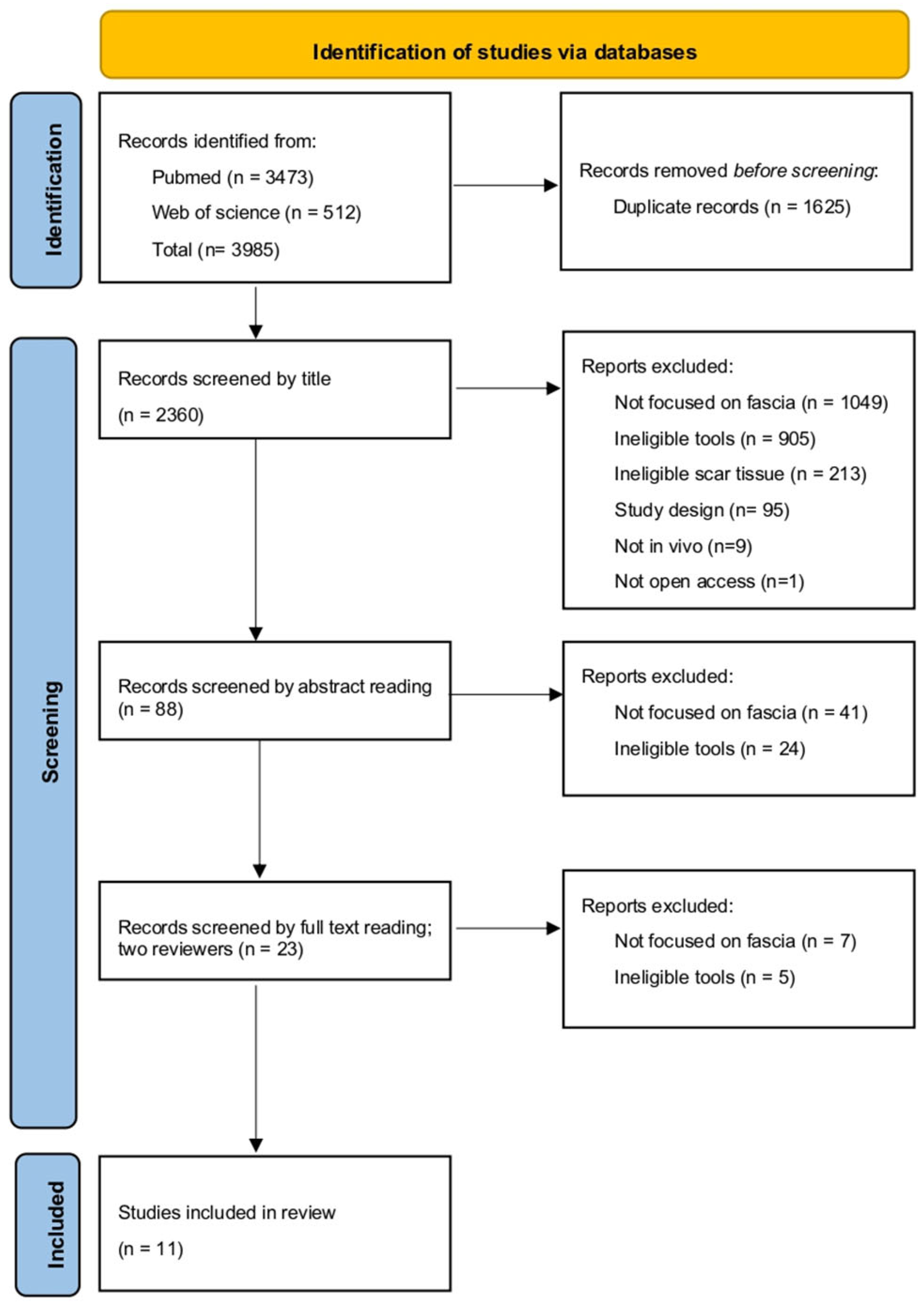
3.1. Study Selection
3.2. Study Characteristics
3.3. Etiology and Location of the Scars
3.4. Assessed Tissue
3.5. Included Tools
3.6. Outcomes of the Articles
3.7. Risk of Bias in Individual Studies
4. Discussion
4.1. Considered Tissue
4.2. Included Tools
4.3. Outcomes of the Articles
4.4. Limitations
5. Conclusions
Author Contributions
Funding
Institutional Review Board Statement
Informed Consent Statement
Data Availability Statement
Conflicts of Interest
References
- Martin, P.; Nunan, R. Cellular and molecular mechanisms of repair in acute and chronic wound healing. Br. J. Dermatol. 2015, 173, 370–378. [Google Scholar] [CrossRef] [PubMed]
- Bordoni, B.; Zanier, E. Skin, fascias, and scars: Symptoms and systemic connections. J. Multidiscip. Healthc. 2013, 7, 11–24. [Google Scholar] [CrossRef]
- Ferguson, M.W.J.; O’Kane, S. Scar–free healing: From embryonic mechanisms to adult therapeutic intervention. Philos. Trans. R. Soc. Lond. Ser. B Biol. Sci. 2004, 359, 839–850. [Google Scholar] [CrossRef]
- Niessen, F.; Spauwen, P.; Schalkwijk, J.; Kon, M. On the nature of hypertrophic scars and keloids: A systematic review. Plast. Reconstr. Surg. 1999, 104, 1435–1458. [Google Scholar] [CrossRef]
- Adib, Y.; Bensussan, A.; Michel, L. Cutaneous Wound Healing: A Review about Innate Immune Response and Current Therapeutic Applications. Mediat. Inflamm. 2022, 2022, 5344085. [Google Scholar] [CrossRef]
- Velnar, T.; Bailey, T.; Smrkolj, V. The Wound Healing Process: An Overview of the Cellular and Molecular Mechanisms. J. Int. Med. Res. 2009, 37, 1528–1542. [Google Scholar] [CrossRef]
- Broughton, G.; Janis, J.E.; Attinger, C.E. The Basic Science of Wound Healing. Plast. Reconstr. Surg. 2006, 117, 12S–34S. [Google Scholar] [CrossRef]
- Delaleu, J.; Charvet, E.; Petit, A. Keloid disease: Review with clinical atlas. Part I: Definitions, history, epidemiology, clinics and diagnosis. Ann. Dermatol. Vénéréologie 2023, 150, 3–15. [Google Scholar] [CrossRef]
- Finnerty, C.C.; Jeschke, M.G.; Branski, L.K.; Barret, J.P.; Dziewulski, P.; Herndon, D.N. Hypertrophic scarring: The greatest unmet challenge after burn injury. Lancet 2016, 388, 1427–1436. [Google Scholar] [CrossRef] [PubMed]
- Al-Attar, A.; Mess, S.; Thomassen, J.M.; Kauffman, C.L.; Davison, S.P. Keloid Pathogenesis and Treatment. Plast. Reconstr. Surg. 2006, 117, 286–300. [Google Scholar] [CrossRef] [PubMed]
- Slemp, A.E.; Kirschner, R.E. Keloids and scars: A review of keloids and scars, their pathogenesis, risk factors, and management. Curr. Opin. Pediatr. 2006, 18, 396–402. [Google Scholar] [CrossRef] [PubMed]
- Lin, T.R.; Chou, F.; Wang, H.; Wang, R. Effects of scar massage on burn scars: A systematic review and meta-analysis. J. Clin. Nurs. 2023, 32, 3144–3154. [Google Scholar] [CrossRef] [PubMed]
- Ogawa, R. The Most Current Algorithms for the Treatment and Prevention of Hypertrophic Scars and Keloids: A 2020 Update of the Algorithms Published 10 Years Ago. Plast. Reconstr. Surg. 2022, 149, 79e–94e. [Google Scholar] [CrossRef] [PubMed]
- Mustoe, T.; Cooter, R.; Gold, M.; Hobbs, R.; Ramelet, A.; Shakespeare, P.; Stella, M.; Téot, L.; Wood, F.M.; Ziegler, U.E.; et al. International clinical recommendations on scar management. Plast. Reconstr. Surg. 2002, 110, 560–571. [Google Scholar] [CrossRef]
- Lewit, K.; Olsanska, S. Clinical Importance of Active Scars: Abnormal Scars as a Cause of Myofascial Pain. J. Manip. Physiol. Ther. 2004, 27, 399–402. [Google Scholar] [CrossRef]
- Correa-Gallegos, D.; Ye, H.; Dasgupta, B.; Sardogan, A.; Kadri, S.; Kandi, R.; Dai, R.; Lin, Y.; Kopplin, R.; Shenai, D.S.; et al. CD201+ fascia progenitors choreograph injury repair. Nature 2023, 623, 792–802. [Google Scholar] [CrossRef]
- Stecco, C.; Pratt, R.; Nemetz, L.D.; Schleip, R.; Stecco, A.; Theise, N.D. Towards a comprehensive definition of the human fascial system. J. Anat. 2025, 246, 1084–1098. [Google Scholar] [CrossRef]
- Adstrum, S.; Hedley, G.; Schleip, R.; Stecco, C.; Yucesoy, C.A. Defining the fascial system. J. Bodyw. Mov. Ther. 2017, 21, 173–177. [Google Scholar] [CrossRef]
- Deflorin, C.; Hohenauer, E.; Stoop, R.; Van Daele, U.; Clijsen, R.; Taeymans, J. Physical Management of Scar Tissue: A Systematic Review and Meta-Analysis. J. Altern. Complement. Med. 2020, 26, 854–865. [Google Scholar] [CrossRef]
- Reinke, J.M.; Sorg, H. Wound Repair and Regeneration. Eur. Surg. Res. 2012, 49, 35–43. [Google Scholar] [CrossRef]
- Yin, J.; Zhang, S.; Yang, C.; Wang, Y.; Shi, B.; Zheng, Q.; Zeng, N.; Huang, H. Mechanotransduction in skin wound healing and scar formation: Potential therapeutic targets for controlling hypertrophic scarring. Front. Immunol. 2022, 13, 1028410. [Google Scholar] [CrossRef]
- Rippa, A.L.; Kalabusheva, E.P.; Vorotelyak, E.A. Regeneration of Dermis: Scarring and Cells Involved. Cells 2019, 8, 607. [Google Scholar] [CrossRef]
- Monstrey, S.; Middelkoop, E.; Vranckx, J.J.; Bassetto, F.; Ziegler, U.E.; Meaume, S.; Téot, L. Updated Scar Management Practical Guidelines: Non-invasive and invasive measures. J. Plast. Reconstr. Aesthetic Surg. 2014, 67, 1017–1025. [Google Scholar] [CrossRef]
- Baron, J.M.; Glatz, M.; Proksch, E. Optimal Support of Wound Healing: New Insights. Dermatology 2020, 236, 593–600. [Google Scholar] [CrossRef] [PubMed]
- Thulabandu, V.; Chen, D.; Atit, R.P. Dermal fibroblast in cutaneous development and healing. WIREs Dev. Biol. 2018, 7, e307. [Google Scholar] [CrossRef] [PubMed]
- Knoedler, S.; Broichhausen, S.; Guo, R.; Dai, R.; Knoedler, L.; Kauke-Navarro, M.; Diatta, F.; Pomahac, B.; Machens, H.G.; Jiang, D.; et al. Fibroblasts—The cellular choreographers of wound healing. Front. Immunol. 2023, 14, 1233800. [Google Scholar] [CrossRef] [PubMed]
- Luo, R.; Dai, J.; Zhang, J.; Li, Z. Accelerated Skin Wound Healing by Electrical Stimulation. Adv. Healthc. Mater. 2021, 10, 2100557. [Google Scholar] [CrossRef]
- Wang, Z.C.; Zhao, W.Y.; Cao, Y.; Liu, Y.Q.; Sun, Q.; Shi, P.; Cai, J.Q.; Shen, X.Z.; Tan, W.Q. The Roles of Inflammation in Keloid and Hypertrophic Scars. Front. Immunol. 2020, 11, 603187. [Google Scholar] [CrossRef]
- Martin, P. Wound Healing—Aiming for Perfect Skin Regeneration. Science 1997, 276, 75–81. [Google Scholar] [CrossRef]
- Zhang, T.; Wang, X.F.; Wang, Z.C.; Lou, D.; Fang, Q.Q.; Hu, Y.Y.; Zhao, W.Y.; Zhang, L.Y.; Wu, L.H.; Tan, W.Q. Current potential therapeutic strategies targeting the TGF-β/Smad signaling pathway to attenuate keloid and hypertrophic scar formation. Biomed. Pharmacother. 2020, 129, 110287. [Google Scholar] [CrossRef]
- Frech, F.S.; Hernandez, L.; Urbonas, R.; Zaken, G.A.; Dreyfuss, I.; Nouri, K. Hypertrophic Scars and Keloids: Advances in Treatment and Review of Established Therapies. Am. J. Clin. Dermatol. 2023, 24, 225–245. [Google Scholar] [CrossRef]
- Sitohang, I.B.S.; Sirait, S.A.P.; Suryanegara, J. Microneedling in the treatment of atrophic scars: A systematic review of randomised controlled trials. Int. Wound J. 2021, 18, 577–585. [Google Scholar] [CrossRef] [PubMed]
- Almadori, A.; Butler, P.E. Scarring and Skin Fibrosis Reversal with Regenerative Surgery and Stem Cell Therapy. Cells 2024, 13, 443. [Google Scholar] [CrossRef]
- Taheri, A.; Mansoori, P.; Al-Dabagh, A.; Feldman, S.R. Are corticosteroids effective for prevention of scar formation after second-degree skin burn? J. Dermatol. Treat. 2014, 25, 360–362. [Google Scholar] [CrossRef]
- Bayat, A. Skin scarring. BMJ 2003, 326, 88–92. [Google Scholar] [CrossRef]
- Waldorf, H.; Fewkes, J. Wound healing. Adv. Dermatol. 1995, 10, 77–96. [Google Scholar]
- Beanes, S.R.; Dang, C.; Soo, C.; Ting, K. Skin repair and scar formation: The central role of TGF-β. Expert Rev. Mol. Med. 2003, 5, 1–22. [Google Scholar] [CrossRef]
- Rhett, J.M.; Ghatnekar, G.S.; Palatinus, J.A.; O’Quinn, M.; Yost, M.J.; Gourdie, R.G. Novel therapies for scar reduction and regenerative healing of skin wounds. Trends Biotechnol. 2008, 26, 173–180. [Google Scholar] [CrossRef]
- Van Zuijlen, P.P.M.; Angeles, A.P.; Kreis, R.W.; Bos, K.E.; Middelkoop, E. Scar Assessment Tools: Implications for Current Research. Plast. Reconstr. Surg. 2002, 109, 1108–1122. [Google Scholar] [CrossRef]
- Browne, A.L.; Andrews, R.; Schug, S.A.; Wood, F. Persistent Pain Outcomes and Patient Satisfaction With Pain Management After Burn Injury. Clin. J. Pain 2011, 27, 136–145. [Google Scholar] [CrossRef]
- Awonuga, A.O.; Fletcher, N.M.; Saed, G.M.; Diamond, M.P. Postoperative Adhesion Development Following Cesarean and Open Intra-Abdominal Gynecological Operations: A Review. Reprod. Sci. 2011, 18, 1166–1185. [Google Scholar] [CrossRef] [PubMed]
- Kobesova, A.; Morris, C.E.; Lewit, K.; Safarova, M. Twenty-Year-Old Pathogenic “Active” Postsurgical Scar: A Case Study of a Patient With Persistent Right Lower Quadrant Pain. J. Manip. Physiol. Ther. 2007, 30, 234–238. [Google Scholar] [CrossRef]
- Hildebrand, K.A.; Frank, C.B. Scar formation and ligament healing. Can. J. Surg. J. Can. De Chir. 1998, 41, 425–429. [Google Scholar]
- Moher, D.; Liberati, A.; Tetzlaff, J.; Altman, D.G.; PRISMA Group. Preferred reporting items for systematic reviews and meta-analyses: The PRISMA statement. PLoS Med. 2009, 6, e1000097. [Google Scholar] [CrossRef]
- Frandsen, T.F.; Bruun Nielsen, M.F.; Lindhardt, C.L.; Eriksen, M.B. Using the full PICO model as a search tool for systematic reviews resulted in lower recall for some PICO elements. J. Clin. Epidemiol. 2020, 127, 69–75. [Google Scholar] [CrossRef] [PubMed]
- Crous, C.; Pretorius, J.; Petzer, A. A Review of the Current Trends in Chronic Wound and Scar Management. Wound Manag. Prev. 2024, 70. [Google Scholar] [CrossRef]
- Higgins, J.P.T.; Morgan, R.L.; Rooney, A.A.; Taylor, K.W.; Thayer, K.A.; Silva, R.A.; Lemeris, C.; Akl, E.A.; Bateson, T.F.; Berkman, N.D.; et al. A tool to assess risk of bias in non-randomized follow-up studies of exposure effects (ROBINS-E). Environ. Int. 2024, 186, 108602. [Google Scholar] [CrossRef]
- Gagnier, J.J.; Kienle, G.; Altman, D.G.; Moher, D.; Sox, H.; Riley, D.; CARE Group. The CARE Guidelines: Consensus-Based Clinical Case Reporting Guideline Development. Headache 2013, 53, 1541–1547. [Google Scholar] [CrossRef]
- Page, M.J.; McKenzie, J.E.; Bossuyt, P.M.; Boutron, I.; Hoffmann, T.C.; Mulrow, C.D.; Shamseer, L.; Tetzlaff, J.M.; Akl, E.A.; Brennan, S.E.; et al. The PRISMA 2020 statement: An updated guideline for reporting systematic reviews. BMJ 2021, 372, n71. [Google Scholar] [CrossRef]
- Baron, J.; Tirosh, D.; Mastrolia, S.A.; Ben-Haroush, Y.; Schwartz, S.; Kerner, Y.; Hershkovitz, R. Sliding sign in third-trimester sonographic evaluation of intra-abdominal adhesions in women undergoing repeat Cesarean section: A novel technique. Ultrasound Obstet. Gynecol. 2018, 52, 662–665. [Google Scholar] [CrossRef]
- Charernjiratragul, K.; Suntharasaj, T.; Pranpanus, S.; Chanwadi, S.; Kwankaew, N.; Petpichetchian, C. Preoperative sonographic sliding sign for prediction of intra-abdominal adhesions before repeat cesarean delivery. Intl. J. Gynecol. Obstet. 2023, 161, 250–254. [Google Scholar] [CrossRef]
- Cocco, G.; Ricci, V.; Boccatonda, A.; Abate, M.; Guagnano, M.T.; Schiavone, C. Ultrasound follow-up of spontaneous tears of the plantar fascia treated with conservative therapies: Two case reports. Medicine 2019, 98, e18428. [Google Scholar] [CrossRef]
- Drukker, L.; Sela, H.Y.; Reichman, O.; Rabinowitz, R.; Samueloff, A.; Shen, O. Sliding Sign for Intra-abdominal Adhesion Prediction Before Repeat Cesarean Delivery. Obstet. Gynecol. 2018, 131, 529–533. [Google Scholar] [CrossRef]
- Kawai, T.; Takahashi, M.; Takamoto, K.; Bito, I. Hamstring strains in professional rugby players result in increased fascial stiffness without muscle quality changes as assessed using shear wave elastography. J. Bodyw. Mov. Ther. 2021, 27, 34–41. [Google Scholar] [CrossRef]
- Lobos, N.; Wortsman, X.; Valenzuela, F.; Alonso, F. Color Doppler Ultrasound Assessment of Activity in Keloids. Dermatol. Surg. 2017, 43, 817–825. [Google Scholar] [CrossRef]
- Pirri, C.; Stecco, A.; Fede, C.; De Caro, R.; Stecco, C.; Özçakar, L. Ultrasound imaging of a scar on the knee: Sonopalpation for fascia and subcutaneous tissues. Eur. J. Transl. Myol. 2020, 30, 150–153. [Google Scholar] [CrossRef]
- Seven, B.; Yorgancı, A.; Alkan, M.; Gündüz, Ö.; Keskin, H.L.; Engin-Üstün, Y. Subcutaneous tissue stiffness of cesarean incision scar by elastography as a predictor of intra-abdominal adhesions. J. Obstet. Gynaecol. Res. 2020, 46, 2390–2396. [Google Scholar] [CrossRef] [PubMed]
- Sönmez, S.; Akselim, B.; Karaşin, S.S. The effectiveness of preoperative diagnostic methods in predicting intra-abdominal adhesions before repeat cesarean section delivery. Rev. Assoc. Med. Bras. 2023, 69, e20221455. [Google Scholar] [CrossRef] [PubMed]
- Veronese, S.; Picelli, A.; Zoccatelli, A.; Amuso, D.; Amore, R.; Smania, N.; Frisone, A.; Sbarbati, A.; Scarano, A. Morphological characterization of two dermal and hypodermal alterations in an adult man: Surgical scar vs. stretch mark. J. Ultrasound 2024, 27, 857–862. [Google Scholar] [CrossRef] [PubMed]
- Yu, J.S.; Smith, G.; Ashman, C.; Kaeding, C. The plantar fasciotomy: MR imaging findings in asymptomatic volunteers. Skelet. Radiol. 1999, 28, 447–452. [Google Scholar] [CrossRef]
- Veronese, S.; Picelli, A.; Smania, N.; Sbarbati, A. Hypodermis involvement in skin disorders: Imaging and functional imaging diagnostic tools. Ski. Res. Technol. 2021, 27, 641–643. [Google Scholar] [CrossRef]
- Pirri, C.; Pirri, N.; Macchi, V.; Porzionato, A.; De Caro, R.; Stecco, C. Ultrasound Imaging of Thoracolumbar Fascia: A Systematic Review. Medicina 2024, 60, 1090. [Google Scholar] [CrossRef]
- Brown, T.M.; Krishnamurthy, K. Histology, Dermis; StatPearls Publishing: Treasure Island, FL, USA, 2025. Available online: http://www.ncbi.nlm.nih.gov/books/NBK535346/ (accessed on 21 July 2025).
- Moroki, T. Morphological characteristics and notes of the skin in preclinical toxicity assessment. J. Toxicol. Pathol. 2023, 36, 85–94. [Google Scholar] [CrossRef] [PubMed]
- Stecco, C.; Macchi, V.; Porzionato, A.; Duparc, F.; De Caro, R. The fascia: The forgotten structure. Ital. J. Anat. Embryol. 2011, 116, 127–138. [Google Scholar]
- Corvino, A.; Varelli, C.; Cocco, G.; Corvino, F.; Catalano, O. Seeing the unseen with superb microvascular imaging: Ultrasound depiction of normal dermis vessels. J. Clin. Ultrasound 2022, 50, 121–127. [Google Scholar] [CrossRef] [PubMed]
- Mizukoshi, K.; Kuribayashi, M.; Hirayama, K.; Yabuzaki, J.; Kurosumi, M.; Hamanaka, Y. Examination of age-related changes of viscoelasticity in the dermis and subcutaneous fat layer using ultrasound elastography. Ski. Res. Technol. 2021, 27, 618–626. [Google Scholar] [CrossRef]
- Catalano, O.; Wortsman, X. Dermatology Ultrasound. Imaging Technique, Tips and Tricks, High-Resolution Anatomy. Ultrasound Q. 2020, 36, 321–327. [Google Scholar] [CrossRef]
- Shen, L.; Zhou, Y.; Gong, J.; Fan, H.; Liu, L. The role of macrophages in hypertrophic scarring: Molecular to therapeutic insights. Front. Immunol. 2025, 16, 1503985. [Google Scholar] [CrossRef]
- Berry, C.E.; Downer, M.; Morgan, A.G.; Griffin, M.; Liang, N.E.; Kameni, L.; Laufey Parker, J.B.; Guo, J.; Longaker, M.T.; Wan, D.C. The effects of mechanical force on fibroblast behavior in cutaneous injury. Front. Surg. 2023, 10, 1167067. [Google Scholar]
- Caprini, J.A.; Arcelus, J.A.; Swanson, J.; Coats, R.; Hoffman, K.; Brosnan, J.J.; Blattner, S. The ultrasonic localization of abdominal wall adhesions. Surg. Endosc. 1995, 9, 283–285. [Google Scholar] [CrossRef]


| Research Term | Database Entries |
|---|---|
| Fascia | “fascia” OR “fasciae” OR “fascias” |
| Scar | “cicatrix” OR “cicatrix” OR “scar” |
| Imaging | “image” OR “images” OR “imaging” OR “imagings” |
| Outcome | “outcome” OR “outcomes” |
| Assessment | “assess” OR “assessed” OR “assessing” OR “assessment” OR “assessments” |
| Skin | “skin” [MeSH Terms] OR “skin” [All Fields] |
| Ultrasonography | “diagnostic imaging” [MeSH Subheading] OR (“diagnostic” AND “imaging”) OR “diagnostic imaging” OR “ultrasonography” OR “ultrasonography” [MeSH Terms] |
| Evaluation | “evaluability” OR “evaluate” OR “evaluated” OR “evaluates” OR “evaluating” OR “evaluation” OR “evaluations” OR “evaluative” OR “evaluator” OR “evaluators” |
| Hypodermis | “subcutaneous tissue” [MeSH Terms] OR (“subcutaneous” AND “tissue”) OR “subcutaneous tissue” OR “hypodermis” |
| Ultrasound | “diagnostic imaging” [MeSH Subheading] OR (“diagnostic” AND “imaging”) OR “diagnostic imaging” [All Fields] OR “ultrasound” [All Fields] OR “ultrasonography” [MeSH Terms] OR “ultrasonography” OR “ultrasonics” [MeSH Terms] OR “ultrasonics” OR “ultrasounds” |
| Subcutis | “subcutaneous tissue” [MeSH Terms] OR (“subcutaneous” [All Fields] AND “tissue” [All Fields]) OR “subcutaneous tissue” [All Fields] OR “subcutis” [All Fields] OR “subcutaneous fat” [MeSH Terms] OR (“subcutaneous” [All Fields] AND “fat” [All Fields]) OR “subcutaneous fat” [All Fields]) |
| Characteristics | Inclusion | Exclusion |
|---|---|---|
| Participants | Human participants, in vivo only | Cadavers, animals, or other models; individuals affected by neoplasms. |
| Intervention | Objective diagnostic technique is applied to assess the fascia at the site where a scar was formed for trauma or surgery. | Invasive assessing methods; in vitro analysis. |
| Control/Comparator | No control/comparator was included | |
| Outcome measures | Studies that qualitatively assessed the fascia layers, including the gliding of the fascia at the site of a scar; Studies that quantitatively assessed fascia layers, including thickness, stiffness of a scar. | Subjective reports |
| Study design | Trials, prospective cohort studies, prospective observational studies, case reports, reliability and validity studies, prospective cross-sectional studies, retrospective studies. | Descriptive commentaries, reviews |
| Publication | English; full text available. | Conference abstracts without full data, non-peer-reviewed articles, and non-original papers |
| Study | Participants | Intervention | Control | Outcome Measures | Study Design | Publication |
|---|---|---|---|---|---|---|
| Baron et al. [50] | Humans, in vivo | Ultrasound | No | Sliding | Prospective observational double-blinded study | English language, full text available |
| Charernjiratragul et al. [51] | Humans, in vivo | Ultrasound | No | Sliding | Prospective cohort study | English language, full text available |
| Cocco et al. [52] | Humans, in vivo | Ultrasound | No | Qualitative analysis | Case report | English language, full text available |
| Drukker et al. [53] | Humans, in vivo | Ultrasound | No | Sliding | Prospective blind observational study | English language, full text available |
| Kawai et al. [54] | Humans, in vivo | Shear wave elastography | No | Stiffness | Observational study | English language, full text available |
| Lobos et al. [55] | Humans, in vivo | Ultrasound | No | Qualitative analysis | Retrospective study | English language, full text available |
| Pirri et al. [56] | Humans, in vivo | Ultrasound | No | Thickness, qualitative analysis | Case report | English language, full text available |
| Seven et al. [57] | Humans, in vivo | Shear wave elastography | No | Stiffness | prospective cross-sectional study | English language, full text available |
| Sönmez et al. [58] | Humans, in vivo | Ultrasound | No | Sliding | prospective cohort study | English language, full text available |
| Veronese et al. [59] | Humans, in vivo | Ultrasound, Strain elastography | No | Stiffness, thickness, qualitative analysis | Case report | English language, full text available |
| Yu et al. [60] | Humans, in vivo | Magnetic resonance imaging | No | Thickness | Observational study | English language, full text available |
| Etiology | Location | |||||||
|---|---|---|---|---|---|---|---|---|
| C-Section | Orthopedic Surgery | General Surgery | Trauma | Keloids | Abdomen and Pelvis | Limbs | Face and Trunk | |
| Baron et al. [50] | X | X | ||||||
| Charernjiratragul et al. [51] | X | X | ||||||
| Cocco et al. [52] | X | X | ||||||
| Drukker et al. [53] | X | X | ||||||
| Kawai et al. [54] | X | X | ||||||
| Lobos et al. [55] | X | X | ||||||
| Pirri et al. [56] | X | X | ||||||
| Seven et al. [57] | X | X | ||||||
| Sönmez et al. [58] | X | X | ||||||
| Veronese et al. [59] | X | X | ||||||
| Yu et al. [60] | X | X | ||||||
| Visceral Fascia | Musculoskeletal Fascia | Hypodermis | Superficial Fascia | |
|---|---|---|---|---|
| Baron et al. [50] | X | |||
| Charernjiratragul et al. [51] | X | |||
| Cocco et al. [52] | X | |||
| Drukker et al. [53] | X | |||
| Kawai et al. [54] | X | |||
| Lobos et al. [55] | X | |||
| Pirri et al. [56] | X | X | ||
| Seven et al. [57] | X | |||
| Sönmez et al. [58] | X | |||
| Veronese et al. [59] | X | |||
| Yu et al. [60] | X |
| Thickness | Sliding | Stiffness | Qualitative Assessment | |
|---|---|---|---|---|
| Baron et al. [50] | X | |||
| Charernjiratragul et al. [51] | X | |||
| Cocco et al. [52] | X | |||
| Drukker et al. [53] | X | |||
| Kawai et al. [54] | X | |||
| Lobos et al. [55] | X | |||
| Pirri et al. [56] | X | X | ||
| Seven et al. [57] | X | |||
| Sönmez et al. [58] | X | |||
| Veronese et al. [59] | X | X | X | |
| Yu et al. [60] | X |
| Sensibility | Specificity | Positive Likelihood Ratio | Negative Likelihood Ratio | |
|---|---|---|---|---|
| Baron et al. [50] | 76.5% | 92.1% | ||
| Charernjiratragul et al. [51] | 39.1% | 95.2% | ||
| Drukker et al. [53] | 56.0% | 95.0% | 12.00 | 0.46 |
| Sönmez et al. [58] | 4.198 |
| Mean Shear Modulus (kPa) | p Value | ||
|---|---|---|---|
| Kawai et al. [54] | Injured Uninjured | 17.37 12.70 | 0.48 |
| Seven et al. [57] | Moderate and severe adhesions Mild adhesions | 51.50 36.80 | 0.003 |
| Stiffness Scar (%) | Stiffness Stretchmark (%) | Stiffness Intact Skin (%) | ||
|---|---|---|---|---|
| Veronese et al. [59] | Longitudinal Transversal | 59.30 58.23 | 59.44 47.20 | 44.04 60.79 |
| Pirri et al. [56] | Cocco et al. [52] | Veronese et al. [59] | |
|---|---|---|---|
| Were the patient’s demographic characteristics clearly described? (yes/no/N.A.) | Yes | Yes | Yes |
| Was the patient’s history clearly described and presented as a timeline? (yes/no/N.A.) | Yes | Yes | No |
| Was the current clinical condition of the patient on presentation clearly described? (yes/no/N.A.) | Yes | Yes | Yes |
| Were diagnostic tests or assessment methods and the results clearly described? (yes/no/N.A.) | Yes | Yes | Yes |
| Were the intervention(s) or treatment procedure(s) clearly described? (yes/no/N.A.) | N.A. | N.A. | N.A. |
| Was the post-intervention clinical condition clearly described? (yes/no/N.A.) | N.A. | N.A. | N.A. |
| Were adverse events (harms) or unanticipated events identified and described? (yes/no/N.A.) | N.A. | N.A. | N.A. |
| Does the case report provide takeaway lessons? (yes/no/N.A.) | Yes | Yes | Yes |
| Domain 1 Var A | Domain 2 Var A | Domain 3 | Domain 4 | Domain 5 | Domain 6 | Domain 7 | Overall Risk of Bias | |
|---|---|---|---|---|---|---|---|---|
| Baron et al. [50] | ● | ● | ● | ● | ● | ● | ● | ● |
| Charernjiratragul et al. [51] | ● | ● | ● | ● | ● | ● | ● | ● |
| Drukker et al. [53] | ● | ● | ● | ● | ● | ● | ● | ● |
| Kawai et al. [54] | ● | ● | ● | ● | ● | ● | ● | ● |
| Lobos et al. [55] | ● | ● | ● | ● | ● | ● | ● | ● |
| Seven et al. [57] | ● | ● | ● | ● | ● | ● | ● | ● |
| Sönmez et al. [58] | ● | ● | ● | ● | ● | ● | ● | ● |
| Yu et al. [60] | ● | ● | ● | ● | ● | ● | ● | ● |
Disclaimer/Publisher’s Note: The statements, opinions and data contained in all publications are solely those of the individual author(s) and contributor(s) and not of MDPI and/or the editor(s). MDPI and/or the editor(s) disclaim responsibility for any injury to people or property resulting from any ideas, methods, instructions or products referred to in the content. |
© 2026 by the authors. Licensee MDPI, Basel, Switzerland. This article is an open access article distributed under the terms and conditions of the Creative Commons Attribution (CC BY) license.
Share and Cite
De Luca, C.; Sun, Y.; Stecco, A.; Fede, C.; Clair, C.; Pirri, C.; Trovarelli, G.; Stecco, C. Objective and Non-Invasive Evaluation of Fascial Layers Related to Surgical or Post-Traumatic Scars: A Systematic Review of the Literature. Life 2026, 16, 133. https://doi.org/10.3390/life16010133
De Luca C, Sun Y, Stecco A, Fede C, Clair C, Pirri C, Trovarelli G, Stecco C. Objective and Non-Invasive Evaluation of Fascial Layers Related to Surgical or Post-Traumatic Scars: A Systematic Review of the Literature. Life. 2026; 16(1):133. https://doi.org/10.3390/life16010133
Chicago/Turabian StyleDe Luca, Clara, Yunfeng Sun, Antonio Stecco, Caterina Fede, Claudia Clair, Carmelo Pirri, Giulia Trovarelli, and Carla Stecco. 2026. "Objective and Non-Invasive Evaluation of Fascial Layers Related to Surgical or Post-Traumatic Scars: A Systematic Review of the Literature" Life 16, no. 1: 133. https://doi.org/10.3390/life16010133
APA StyleDe Luca, C., Sun, Y., Stecco, A., Fede, C., Clair, C., Pirri, C., Trovarelli, G., & Stecco, C. (2026). Objective and Non-Invasive Evaluation of Fascial Layers Related to Surgical or Post-Traumatic Scars: A Systematic Review of the Literature. Life, 16(1), 133. https://doi.org/10.3390/life16010133